Category: Knee fractures

  • What you need to know so they don’t hold you back – ACL Strong

    What you need to know so they don’t hold you back – ACL Strong


    Do you know how many people walk around with a torn meniscus without knowing they have a tear?

    💥About 5% of young people and up to 67% of older people have asymptomatic (non-painful) meniscus tears! 💥

    How is that possible?

    Depending on the type of injury and how it is treated, the symptoms of a meniscus tear may resolve within days to weeks or months; and you may not even know the tear exists!

    The initial injury may not even have been severe enough to warrant a visit to an orthopedist or ordering an X-ray or MRI, especially if the knee only hurt for a few days before the pain went away.

    Here’s what you need to know about meniscus injuries so they don’t hinder your favorite activities…

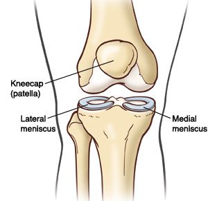
    Where is the meniscus?

    R6hmleXC6W0ny8ioxXQ1WJJ5 gBBz5pid7oXKzkdZjLCm j1pOMJVgGzlhelt2i2WWk4oBI10 viL69qw0Df1BHeXuhdytd k68eVv e9FM5ZebGSOte1IlG Z2oKmfmp4NUqpmdwZtgUbOInWzTjs

    The meniscus is a “C”-shaped disk made of cartilage that is located in the knee, between the shin bone and the femur. It sits on the weight-bearing surface of the tibia, or tibia, in the joint capsule.

    There are two menisci, one on the lateral (outside) of the knee and one on the medial (inside). A healthy meniscus provides space, cushion and lubrication to the joint so that the knee can bend, straighten and bear weight comfortably.

    The meniscus is located deep in the knee, along with the anterior cruciate ligament (ACL) and the posterior cruciate ligament (PCL). Due to their deep location, the menisci do not have a very good blood supply, so certain areas are not likely to heal from an injury.

    💥 🤯 However, the fact that the meniscus does not heal properly on its own does not mean that a tear always needs to be repaired!

    What does the meniscus do and why is it so important?

    Shock absorption: The meniscus acts as a shock absorber for shocks that occur during daily activities, such as walking, running, jumping and sports. As we move, force travels through the foot and through the shin bone to the knee, where the meniscus absorbs and disperses that force through the knee, so the impact is spread out and not concentrated in one area.

    Stability: The meniscus also acts as a wedge that supports the front and back of your knee, keeping the knee from going beyond a certain range of motion, such as hyperextension, which puts you at risk for other injuries.

    How does the meniscus become injured?

    The meniscus is most commonly injured during twisting of the knee when the foot is planted on the ground. Rotational stress or “shearing” can cause the femur and tibia to rotate beyond what the meniscus can handle, resulting in a tear in the cartilage.

    Meniscal tears are common in athletes who exert a large amount of force on one leg and twist their body in a different direction, such as football 🏈, soccer ⚽️, lacrosse 🥍, tennis 🎾, or basketball 🏀. Field and field athletes often put their feet down to stop and change direction; and in a split second the knee rotates with enough force to cause a meniscus tear.

    Snow sports, such as skiing ⛷ and snowboarding 🏂, also carry a high risk of meniscus injuries. The edge of a ski or board can unexpectedly hit the snow, causing a sudden rotation of the leg. The long lever arm can quickly generate a high-speed rotational force, strong enough to cause meniscus injury.

    Meniscal tears occur in people of all ages, including athletes and non-athletes. In middle-aged and older adults, meniscus tears can occur due to wear and tear on the knee joint. As the body ages, degeneration of the cartilage tissue occurs as the joints are exposed to force for years.

    Osteoarthritis, a condition in which cartilage wears away over time, is common in older adults and is often accompanied by a torn meniscus. A meniscus tear earlier in life can also make the knee more susceptible to earlier-onset osteoarthritis. That’s why it’s so important to learn the right moves and exercises to protect your knees!

    I have a meniscus tear. What now?

    image

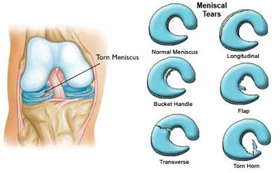
    Depending on the type of meniscus tear, you may experience symptoms such as pain, swelling, locking, or locking of the knee. Although a meniscal injury cannot be detected on X-rays, it can be diagnosed through a careful examination or an MRI.

    Most meniscal tears are initially treated conservatively (without surgery); although an orthopedic doctor may recommend surgery if symptoms persist and daily activities are limited.

    Meniscectomy: The most common surgery for a meniscus tear is called a menisectomy, in which the surgeon removes the torn piece of cartilage and cleans up the surrounding ragged tissue. Although it is a surgical procedure, recovery after a meniscectomy is relatively short. Most people can resume daily activities within a few weeks and exercise within a few months.

    It is important to add that everyone responds to surgery differently; and a ‘minor’ surgery for one person may be a more stressful experience and longer rehabilitation for someone else.

    Meniscus repair: An alternative surgery is a meniscus repair, which is performed when the orthopedic surgeon believes the torn cartilage structure can be sewn together, saving cartilage to prevent osteoarthritis in the future. This procedure is typically performed more in younger people to preserve the cartilage rather than remove it. Recovery from a meniscus repair takes longer than a menisectomy and involves close monitoring of range of motion, weight bearing, and activity progression during rehabilitation.

    Non-surgical treatment A meniscus tear is very common and results in great results in most cases! A diagnosis of a meniscus tear does NOT mean that surgery is always necessary!

    Although a meniscus tear will not heal itself, you can strengthen the knee and improve movement patterns to disperse forces and reduce stress at the site of the tear, which will help resolve symptoms.

    💪 With the right exercises and learning to move properly, most meniscus tears can be treated conservatively WITHOUT surgical intervention.

    💥It’s important to remember that you are not your MRI result; and a meniscus tear should not determine the rest of your life.

    How can you treat a meniscus injury without surgery?

    A meniscus injury must be properly guided through the healing phases. Pain and inflammation are likely to be early symptoms, while stiffness and weakness may be the main complaints after a few weeks. Dealing with injuries with “PEACE & LOVE” is a good reference for dealing with acute injuries.

    To return to normal activities, follow the guidance of your physical therapist, who will develop a care plan for strengthening, balance training, improving mobility, and returning to exercise.

    👉Doing the right exercises with good form and consistency will significantly increase your ability to recover from a meniscus tear and prevent future injuries.

    If you’re ready to start strengthening at home, ACL Strong has courses that include all the necessary components of exercise and movement to train your body and brain to protect your knees now and for the long term.

    Whether you have a torn meniscus or not, doing the right exercises the right way will keep your knees strong and stable, so you’ll be less prone to further injury and better equipped to maintain an active lifestyle for as long as you want. to lead!

    You can LIVE with a torn meniscus….

    And you can also PREVENT them!

    Source link

  • Why does my knee feel numb and tingly after ACL surgery?

    Why does my knee feel numb and tingly after ACL surgery?

    It is usually normal for your knee to feel numb and tingly after ACL surgery. This is because some small nerves are cut during the surgical procedure. Don’t worry, we’re not talking about the really important nerves that allow you to bend and straighten your knee, but the nerves that allow you to feel things around your knee. This video below explains all the reasons why your knee may feel numb and tingly after ACL surgery. For more information, read this blog about 5 tips to help you recover after ACL surgery.

    Lauren Youssef, a physiotherapy student at the University of Toronto, explains why your knee may feel numb and tingly after ACL surgery.

    Read Lauren’s blog: Why is my knee numb and tingling after ACL surgery? | Curovat

    There are also 5 great blogs written by Lauren on our site where you can learn more about surgery and injury recovery.

    If you have had an ACL injury or surgery, download Curovate from the links below. Curovate is a physical therapy app that offers daily video-guided exercises, progress tracking, in-app chat with a physical therapist to answer your questions about surgery or injury, and the ability to measure your knee’s range of motion using just your phone.

    If you need more tailored help during your surgery or recovery from your injury, check out our Virtual Physiotherapy page to book your 1-on-1 video session with a physiotherapist.

    acl knee physical therapy 1080x1080 2
    Download it on Google Play

    Other blogs related to ACL injuries:

    References

    1. Horteur, C., Cavalié, G., Gaulin, B., Cohen Bacry, M., Morin, V., Cavaignac, E., & Pailhé, R. (2020). Saphenous nerve damage after anterior cruciate ligament reconstruction: Decreased area of ​​numbness after ligamentoplasty using the quadriceps tendon compared to the hamstring tendon. The Knee, 27(4), 1151–1157.

    2. Inderhaug, E., Strand, T., and Solheim, E. (2015). The impact of sensory deficits after hamstring autograft harvesting for ACL reconstruction. Knee surgery, sports traumatology, arthroscopy: Official Journal of the ESSKA, 23(4), 1060–1064.

    3. Kjaergaard, J., Faunø, L. Z., & Faunø, P. (2008). Loss of sensation after ACL reconstruction with hamstring graft. International Journal of Sports Medicine, 29(6), 507–511.

    4. Nerve Blocks for Surgery – Yale Medicine. (2020, July 02). Retrieved from https://www.yalemedicine.org/conditions/nerve-blocks-for-surgery

    5. Ochiai, S., Hagino, T., Senga, S., Yamashita, T., Oda, K., & Haro, H. (2017). Damage to the infrapatellar branch of the saphenous nerve in anterior cruciate ligament reconstruction using vertical skin incision for hamstring harvesting: risk factors and influence on treatment outcome. Journal of Orthopedic Surgery and Research, 12(1), 101.

    6. Sanders, B., Rolf, R., McClelland, W., and Xerogeanes, J. (2007). Prevalence of saphenous nerve damage after autogenous hamstring harvest: an anatomical and clinical study of tailor branch injury. Arthroscopy: The Journal of Arthroscopic and Related Surgery: Official Publication of the Arthroscopy Association of North America and the International Arthroscopy Association, 23(9), 956–963.

    7. Wang, H. D., Zhang, H., Wang, T. R., Zhang, W. F., Wang, F. S., & Zhang, Y. Z. (2018). Comparison of clinical outcomes after anterior cruciate ligament reconstruction with hamstring tendon autograft versus soft tissue allograft: a meta-analysis of randomized controlled trials. International Journal of Surgery (London, England), 56, 174–183.

    Source link

  • Which graft is better: patellar tendon or hamstring tendon for an ACL

    Which graft is better: patellar tendon or hamstring tendon for an ACL

    The two most common areas where a surgeon can take a graft to replace your ACL are the patellar tendon and the hamstring tendon. If this is new information to you, please refer to our previous blog on where the most common areas for an ACL graft are. Also keep in mind that the patellar tendon and hamstring tendon are the most common but not the only sources for an ACL graft.

    As you read the differences below, it is important to remember that your surgeon will choose the graft site for your ACL surgery. In some cases, due to a previous injury or surgery, one or more of the graft sites may not be feasible. The good news is that our ACL rehabilitation app, Curovate, has a protocol for hamstring tendon, patellar tendon, quadriceps tendon, allograft, and non-surgical ACL rehabilitation.

    Overall, the patellar tendon is the more stable ACL graft and has a lower re-injury rate compared to the hamstring tendon graft. Read the advantages and disadvantages of each graft site in the table below.

    Hamstring Graft Surgery (HT) Patellar tendon surgery, also called bone-patellar-bone surgery (BTB).
    1. The surgery takes longer and the tourniquette, a device used during surgery to restrict blood flow in the leg, stays on longer2 1. The knee is stiffer after surgery2
    2. The knee moves more freely forward and backward when tested with a KT-1000 Arthrometer machine2a device that measures the forward and backward movement of your lower leg compared to your thigh. 2. Increased knee pain when kneeling on the ground3
    3. During a period of 15 years after surgery, more reinjury was reported.3 3. Over a period of 15 years after surgery, increased stiffness and difficulty straightening the knee were reported3.
    4. The need to perform another surgery to address problems with the first surgery is called revision surgery. The revision rate for hamstring operations increases by 0.65% within 1 year and by 4.45% within 5 years4. 4. The 1-year revision rate is 0.16% and the 5-year revision rate is 3.03% for patellar tendon graft surgery4.
    5. Hamstring muscle strength reduced by 15%1. Muscle strength is the maximum force you can exert in the shortest possible time.
    6. Muscle strength decreases by 11% when stretching the knee2. Muscle strength is the amount of force a muscle can produce during maximum effort.
    7. There is increased instability in the knee when force is applied to it2. Instability is unwanted movement in a joint.

    Conclusion

    Based on the research reviewed in this blog, the patellar tendon has a lower re-injury rate and is more stable at 1 and 5 years after ACL surgery. However, the location of the graft should be discussed with your surgeon and ultimately the surgeon will decide the most suitable area for the ACL graft.

    If you have had ACL surgery and want clear daily, weekly and monthly guidance for your knee extension and daily exercises for your recovery, try our Cuorvate app. Curovate offers video-guided daily exercises, weekly range of motion goals and exercises, progress tracking, the ability to measure your knee and hip range of motion, and in-app chat with a physical therapist.

    If you need more tailored help during your ACL recovery, check out our Virtual Physiotherapy page to book your 1-on-1 video session with a physiotherapist.

    acl knee physical therapy 1080x1080 2
    Download it on Google Play

    Other recommended blogs

    References

    1. Ageberg, E., Roos, HP, Silbernagel, KG, Thomeé, R., Roos, EM, akademin, S., Gothenburg University. (2009). Knee extension and flexion muscle strength after anterior cruciate ligament reconstruction with patellar tendon graft or hamstring tendon graft: a cross-sectional comparison 3 years after surgery. Knee Surgery, Sports Traumatology, Arthroscopy, 17(2), 162-169.
    2. Beynnon, B.D., Johnson, R.J., Fleming, B.C., Kannus, P., Kaplan, M., Samani, J., and Per Renstrom. (2002). Anterior cruciate ligament replacement: comparison of bone-patellar tendon-bone grafts with two-strand hamstring grafts A prospective, randomized study. The Journal of Bone & Joint Surgery, 84(9), 1503-1513.
    3. Leys, T., Salmon, L., Waller, A., Linklater, J., and Pinczewski, L. (2012). Clinical outcomes and risk factors for re-injury 15 years after anterior cruciate ligament reconstruction: a prospective study of hamstring and patellar tendon grafts. The American Journal of Sports Medicine, 40(3), 595-605.
    4. Rahr-Wagner, L., Thillemann, T.M., Pedersen, A.B., and Lind, M. (2014). Comparison of hamstring tendon and patellar tendon grafts in anterior cruciate ligament reconstruction in a nationwide population-based cohort study: results from the Danish Knee Ligament Reconstruction Register. The American Journal of Sports Medicine, 42(2), 278-284.

    Source link

  • Save Our Bones Bulletin: Newly Discovered Genetic Pathway for Bone Formation;  3D scans can predict the risk of fractures;  Evolutionary biology and bone regeneration

    Save Our Bones Bulletin: Newly Discovered Genetic Pathway for Bone Formation; 3D scans can predict the risk of fractures; Evolutionary biology and bone regeneration

    New research enriches our understanding of bone function and provides insight into maintaining its quality and strength. This bulletin delves into three new studies that point the way to the future of bone health.

    First we’ll look at a report on the genetics of bone loss. Scientists have identified a protein that plays an important role in regulating the genes that control bone regeneration.

    Next, you’ll learn about a group of Finnish scientists who are using 3D modeling to revolutionize the way we assess bone health and predict fracture risk.

    Finally, we look at a report from the PhyloBone project. The project’s leaders are using techniques from evolutionary biology to identify and study proteins in the bone matrix that may have the power to regulate bone regeneration.

    Study focuses on genes for another new osteoporosis drug

    A new study published in The FASEB Journal linked the expression of a certain gene to reduced bone loss in postmenopausal women.

    Researchers have identified a gene that regulates the high mobility group AT-hook 1 (Hmga1) protein. This protein regulates the expression of genes that convert bone marrow-derived mesenchymal stem cells (BMSCs) into cells that build new bone.

    Relevant excerpt

    “Tests on rats showed an increase in Hmga1 expression during bone formation, but a decrease when the rats underwent ovariectomy, which simulated the conditions of menopause. Introducing more Hmga1 to these rats led to a remarkable recovery in bone resorption.

    Yihe Hu, PhD, from Zhejiang University in China, the lead author, noted: “Our study showed that Hmga1 prevents bone loss by promoting the osteogenic differentiation of BMSCs in osteoporosis rats, suggesting that Hmga1 could be an important therapeutic target for osteoporosis. are.”1

    Unfortunately, when Dr. Yihe Hu calls “therapeutic target,” he is referring to a potential new osteoporosis drug that would be developed, marketed and sold by Big Pharma. Nevertheless, new information about the genetic pathways that lead to bone formation is welcome, as it could also prove to have non-pharmaceutical applications.

    Short content

    A new study identified a genetic pathway that increases bone formation. Researchers discovered that a protein called Hmga1 regulates the expression of genes that instruct bone marrow-derived mesenchymal stem cells (BMSCs) to become bone-building cells. Increasing Hmga1 in rats with induced bone loss led to restoration of bone loss.

    3D modeling can provide more accurate assessments of bone health

    Researchers in Sweden have developed a method to create a 3D model of a patient’s bones based on information from 2D X-ray images.

    The report, published in the Journal of Bone and Mineral Research, concluded that they were able to identify thousands of patients at risk of fractures who would be overlooked by current assessment methods.

    Relevant excerpt

    Lorenzo Grassi, associate professor of biomedical engineering at Lund University, was responsible for evaluating the method. He explains that the new method uses 2D X-ray images of bone density measurements to produce 3D models of the femur.

    “The shift from 2D to 3D is performed using a computer-simulated template that describes how bone geometry and density varies in the population.”

    The 3D model of the femur can be used to simulate various situations and scenarios that could have an influence, for example in the event of a fall. Information that makes it easier to estimate the risk of fractures.”2

    Grassi developed 3D simulations using data from 400 study participants who had previously undergone X-ray-based bone mineral density assessments. When he compared the accuracy of each method in predicting which patients would break their hip over the next decade, the 3D simulation provided a more accurate prediction.

    This new method could provide patients with a more comprehensive understanding of their bone health compared to DXA scans. However, because the modeling uses the information from the 2D X-ray, it would complement rather than replace DXA technology.

    Short content

    Researchers in Sweden developed a method for creating a 3D model of a patient’s bone from 2D DXA scans. Their 3D model proved to be more effective than DXA scans in predicting fractures.

    Evolutionary biology provides new data on bone regeneration

    Scientists in Finland have discovered hundreds of non-collagenous proteins in the bone matrix that may play a role in bone formation and regeneration.

    Their research, conducted as part of the PhyloBone project, uses the principles of evolutionary biology to identify molecular mechanisms that maintain bone health.

    Relevant excerpt

    “Since the bone matrix, which constitutes the majority of bone mass, plays both structural and regulatory roles, non-collagenous organic components play a key function in bone regulation. For example, it is known that few non-collagenous proteins, such as osteopontin, play an important role in bone formation. However, the bone matrix consists of hundreds of proteins that are still poorly understood and that may play an important regulatory role in bone regeneration and osteoporosis.

    “Our project has identified 255 proteins in 30 species of vertebrates. The aim of the project is to serve as a valuable resource for further research in bone regeneration, osteoporosis and related areas,” says Dr. Puigbò, co-principal investigator of the PhyloBone project.”3

    The project will continue to conduct studies to determine the regulatory role of bone proteins. They have shared their data on these understudied proteins with the scientific community, with the aim of encouraging further research and new discoveries in the field of bone matrix proteins.

    Hopefully, this research will lead to new knowledge on how bone regeneration can be supported and enhanced through behavioral and lifestyle changes. However, it may also stimulate the development of new drugs against osteoporosis.

    Short content

    Finnish scientists have identified hundreds of proteins in the bone matrix that may play a role in bone formation and regeneration. They have made their data on these underdeveloped proteins available to the scientific community, which will hopefully lead to more research and new discoveries.

    What this means for you

    New research offers new insights and information. We can apply this knowledge in our pursuit of stronger, healthier bones.

    The connection between the latest scientific research and an informed, drug-free approach to bone health is at the heart of the Osteoporosis Reversal Program. The Save Institute uses scientific research published in mainstream journals to ensure its drug-free approach is the safest and most effective method for reversing and preventing osteoporosis.

    As science advances, it increasingly emphasizes the importance of diet, exercise and lifestyle as the key tools for maintaining healthy bones and ensuring a robust, long and independent life.

    References

    1 https://www.revyuh.com/news/lifestyle/health-and-fitness/how-to-avoid-the-risk-of-osteoporosis-study-suggests-a-new-way-to-fight-brittle- bone disease/

    2 https://www.news-medical.net/news/20230913/New-method-could-improve-prediction-of-osteoporotic-fracture-risk.aspx

    3 https://medicalxpress.com/news/2023-08-bone-regeneration-osteoporosis-evolution.html


    Source link

  • 8 exercises in the Accelerate ACL Prehab protocol

    8 exercises in the Accelerate ACL Prehab protocol


    Previous posts have discussed the importance of ACL prehab, the goals you should aim for in prehab, and different types of exercises you can expect in a quality ACL prehab program.

    This message will dive one step further and reveal the exact exercises used in the Accelerate ACL prehab protocol. Please note that within the Accelerate ACL protocol, the effects of these exercises are magnified by the Neubie and coaching by Accelerate ACL trainers.

    The exercises used by Accelerate ACL are chosen to train the muscles so that they are properly activated through a full range of motion. When muscles are activated properly, they create and absorb force efficiently, meaning minimal force goes to the joint or injury site.

    As the body realizes that the muscles are functioning properly and it is safe for the knee to move, pain decreases, range of motion increases, normal walking is restored, and strength is developed.

    With the help of our trainers, these exercises can easily be adapted to suit any stage of the prehab or ACL recovery process. As a general rule of thumb, we recommend staying within the guidelines of your doctor and/or physical therapist and never using a range of motion that causes >2/10 pain.


    Source link