Knee composants and fonctions
As a physical therapist, I have seen many patients with knee injuries and conditions. Understanding the anatomy and function of the knee joint is essential to prevent injuries, manage pain, and recover from surgery. In this article, I will explain the basic structure of the knee, the role of ligaments and tendons, and the muscles responsible for movement.

The knee joint is the largest joint in the body and connects the thigh bone (femur) to the shin bone (tibia). The kneecap (patella) is a small bone that sits in front of the knee joint and protects the joint. The knee joint is a hinge joint that allows for flexion and extension of the leg. It also has some rotational movement, which is important for activities such as walking and running.
The knee joint is supported by several ligaments and tendons. Ligaments are tough bands of tissue that connect bones to bones, while tendons connect muscles to bones. The ligaments and tendons around the knee joint work together to provide stability and support to the joint. In the next section, I will discuss the role of these structures in more detail.
As the largest joint in the body, the knee is a complex structure that allows us to walk, run, jump, and perform other physical activities. It is a synovial joint, meaning it contains a fluid-filled capsule that lubricates the joint and reduces friction during movement.
The knee joint is formed by the articulation of three bones: the femur, tibia, and patella. The femur, or thigh bone, is the longest bone in the body and forms the upper part of the knee joint. The tibia, or shin bone, is the larger of the two bones in the lower leg and forms the lower part of the knee joint. The fibula is the smaller bone in the lower leg and is not directly involved in the knee joint.
The knee joint is actually two joints in one: the tibiofemoral joint and the patellofemoral joint. The tibiofemoral joint is the main joint between the femur and tibia, while the patellofemoral joint is the joint between the patella and the femur.
The patella, or kneecap, is a sesamoid bone that sits in front of the knee joint and helps to protect the knee and improve the leverage of the quadriceps muscle. The patella is unique in that it is not directly attached to any other bone in the body. Instead, it is connected to the quadriceps tendon and the patellar ligament.
In summary, the knee joint is a complex structure that is formed by the articulation of three bones: the femur, tibia, and patella. The knee joint is actually two joints in one: the tibiofemoral joint and the patellofemoral joint. The patella, or kneecap, is a sesamoid bone that sits in front of the knee joint and helps to protect the knee and improve the leverage of the quadriceps muscle.
The knee joint is stabilized and supported by a network of ligaments and tendons. These structures work together to provide strength and stability to the knee joint, allowing us to perform various activities such as walking, running, and jumping.
The collateral ligaments are located on the sides of the knee joint. The medial collateral ligament (MCL) is located on the inner side of the knee, while the lateral collateral ligament (LCL) is found on the outer side. These ligaments help to prevent excessive side-to-side movement of the knee joint.
The cruciate ligaments are located inside the knee joint and cross each other to form an “X” shape. The anterior cruciate ligament (ACL) is located in the front of the knee, while the posterior cruciate ligament (PCL) is located at the back. These ligaments help to prevent excessive forward and backward movement of the knee joint.
The patellar tendon and quadriceps tendon are two important tendons that are located in the knee joint. The patellar tendon connects the patella (kneecap) to the tibia (shinbone), while the quadriceps tendon connects the quadriceps muscle to the patella. These tendons help to provide stability to the knee joint and allow us to perform various movements such as jumping and climbing stairs.
In summary, the knee joint is stabilized and supported by a network of ligaments and tendons. The collateral ligaments help to prevent excessive side-to-side movement of the knee joint, while the cruciate ligaments help to prevent excessive forward and backward movement. The patellar and quadriceps tendons provide stability to the knee joint and allow us to perform various movements.
The knee joint is a hinge joint that allows for flexion and extension of the lower leg. The movement of the knee is controlled by a complex system of muscles, tendons, and ligaments that work together to stabilize and move the joint.
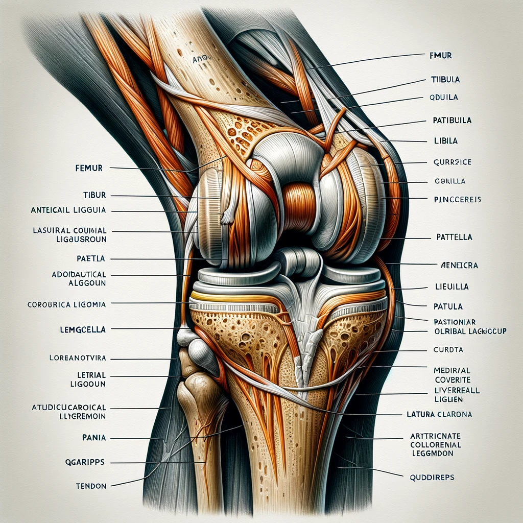
Anatomical illustration of the human knee joint, showing all the components along with their names. This includes the bones, ligaments, cartilage, and tendons, with clear labels for each part such as the femur, tibia, fibula, patella, meniscus, and various ligaments and tendons.
The quadriceps muscles are a group of four muscles located on the front of the thigh that work together to extend the knee joint. These muscles include the rectus femoris, vastus lateralis, vastus intermedius, and vastus medialis. The rectus femoris also works to flex the hip joint.
The hamstring muscles are a group of three muscles located on the back of the thigh that work together to flex the knee joint. These muscles include the biceps femoris, semitendinosus, and semimembranosus. The gracilis muscle also works to flex the knee joint.
The knee joint allows for a variety of movements, including flexion, extension, lateral rotation, and medial rotation. Flexion is the movement that brings the heel towards the buttocks, while extension is the movement that straightens the leg. Lateral rotation is the movement that turns the lower leg outward, while medial rotation is the movement that turns the lower leg inward.
The popliteus muscle is a small muscle located at the back of the knee joint that works to unlock the knee joint during flexion. The flexors and extensors of the knee joint work together to stabilize the joint during movement.
The tibiofemoral joint is the main joint of the knee, while the patellofemoral joint is the joint between the kneecap and the femur. The articularis genus muscle is a small muscle located at the front of the knee joint that works to pull the synovial membrane of the joint upward during extension.
The gastrocnemius and plantaris muscles are located at the back of the knee joint and work to plantarflex the ankle joint. Instability of the knee joint can lead to pain and difficulty with activities such as running and walking.
The knee is a complex joint that is susceptible to a variety of injuries. Some common knee injuries include:
| Section | Description |
|---|---|
| 1. | Maintain a healthy weight to reduce stress on the knee joint. |
| 2. | Wear appropriate shoes for the activity you are doing. |
| 3. | Warm up before exercising to increase blood flow to the muscles. |
| 4. | Use proper technique when exercising to avoid unnecessary stress on the knee joint. |
| 5. | Incorporate exercises that strengthen the muscles around the knee joint. |
| 6. | Avoid activities that put excessive stress on the knee joint, such as jumping or running on hard surfaces. |
| 7. | Take breaks during activities to rest and stretch the knee joint. |
| 8. | Use knee pads or braces for added support during high-impact activities. |
| 9. | Stay hydrated to help keep the joints lubricated. |
| 10. | Consult with a healthcare professional before starting a new exercise program. |
Treatment for knee injuries varies depending on the severity and type of injury. Some common treatments include:
In some cases, surgery may be necessary to repair a knee injury. Rehabilitation after surgery may include physical therapy and exercises to help regain strength and range of motion.
Overall, it is important to take steps to prevent knee injuries and to seek treatment promptly if an injury does occur. With proper care and treatment, many knee injuries can be successfully treated, allowing individuals to return to their normal activities.
The knee joint is a synovial joint that connects three bones: the femur, tibia, and patella. It is a complex hinge joint composed of two articulations: the tibiofemoral joint and patellofemoral joint.
Yes, the knee is a hinge joint. It allows for flexion and extension, as well as a small degree of medial and lateral rotation.
The three most commonly injured knee structures are the anterior cruciate ligament (ACL), medial collateral ligament (MCL), and meniscus.
The major anatomical features of the knee include bones (femur, tibia, and patella), cartilage, ligaments, tendons, and muscles.
The knee joint is responsible for weight-bearing and movement, allowing for flexion and extension, as well as a small degree of medial and lateral rotation. The major muscles involved in knee movement include the quadriceps, hamstrings, and calf muscles.
Symptoms of a torn ligament in your knee include pain, swelling, instability, and difficulty bearing weight. Depending on the severity of the tear, surgery may be necessary to repair the ligament.
Introduction If you're an athlete or someone who leads an active lifestyle, you've likely heard…
Key Takeaways Regular exercise and maintaining a healthy weight are crucial for knee health Proper…
Ever finished a run and felt your knee throbbing? You're not alone. Many athletes face…
https://youtu.be/YRrppL1WuCw Introduction As a weight lifter, you're no stranger to pushing your body to its…
Ever wondered why some hikers seem to move easily on tough trails while others find…
Are you an active person dealing with knee pain that stops you from doing what…