Did you know that knee pain affects millions of people worldwide?
Knee pain is a common problem that can significantly impact daily activities and quality of life. Whether it’s due to an injury, degenerative condition, or overuse, finding the right specialist for knee pain relief is essential to achieve optimal outcomes.
At the Noyes Knee Institute, we understand the challenges of living with knee pain, and our goal is to provide comprehensive treatment, diagnosis, and rehabilitation for various knee injuries and conditions. Our team of orthopedic knee surgeons and healthcare professionals has extensive experience in effectively managing knee pain while addressing the underlying causes.
When it comes to knee pain, knowing which doctor to see can make all the difference. While general practitioners may offer initial care, specialized experts like orthopedic knee surgeons are specifically trained in diagnosing and treating musculoskeletal issues related to the knee. Whether you need non-surgical interventions or surgical procedures, our orthopedic knee surgeons will provide personalized treatment options tailored to your needs.
Don’t let knee pain hold you back. Contact the Noyes Knee Institute today and discover the best specialist for your knee pain relief.
What Is Knee Pain and Who to Consult?
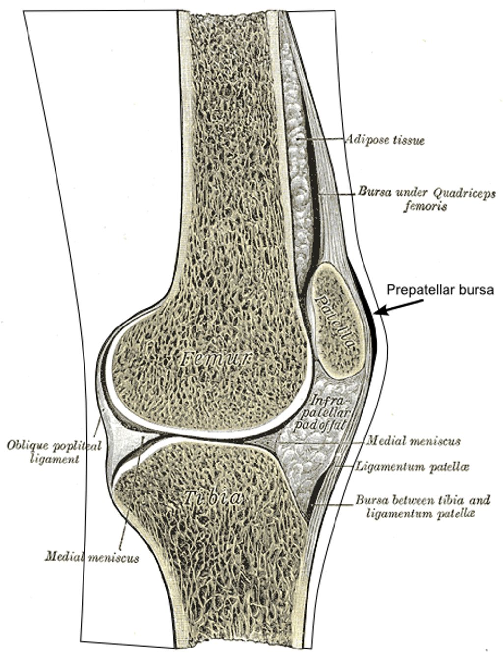
Knee pain refers to any discomfort or pain experienced in or around the knee joint, which is a complex structure composed of cartilage, tendons, bones, and muscles. It accounts for a significant number of visits to healthcare providers, particularly among adults over the age of 50.
Knee pain can be caused by various factors, including injuries, degenerative diseases like osteoarthritis, and overuse. Depending on the underlying cause and severity of the knee pain, different specialists may be consulted.
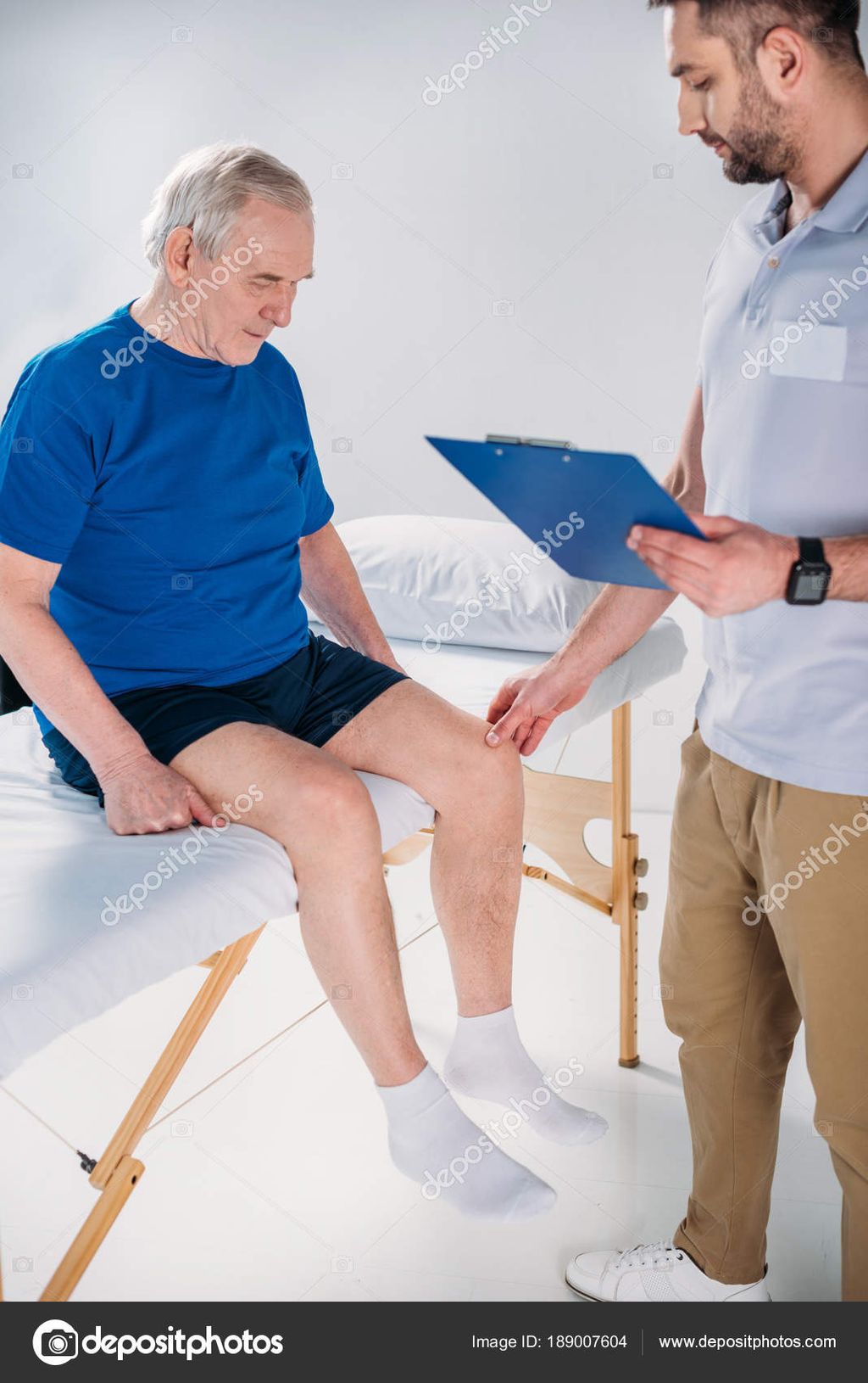
General practitioners, also known as family doctors or primary care physicians, are often the first point of contact for knee pain. They conduct medical history assessments, physical examinations, and may order diagnostic tests like X-rays or MRIs. General practitioners usually recommend conservative treatments such as rest, medication, physical therapy exercises, or lifestyle modifications.
If the knee pain is severe, non-responsive to conservative treatments, or requires specialized care, referral to a specialist like an orthopedic knee surgeon at a reputed clinic like the Noyes Knee Institute may be necessary.

| Specialist | Role |
|---|---|
| General Practitioner | – Conducts medical history assessments and physical examinations. – Orders diagnostic tests like X-rays or MRIs. – Recommends conservative treatments. |
| Orthopedic Knee Surgeon | – Specializes in diagnosing and treating knee conditions. – Develops tailored treatment plans. – Performs non-surgical interventions and surgeries. |
Sports Medicine and Physical Therapy for Knee Pain
Sports medicine specialists and physical therapists play vital roles in the treatment and prevention of knee pain. They possess specialized knowledge and expertise in addressing sports-related injuries and supporting patients in their recovery journey. At the Noyes Knee Institute, we offer comprehensive programs and customized treatment plans to address knee pain, prioritize injury prevention, improve knee function and mobility, and provide effective knee pain management.
Sportmetrics™ Program: Tailored Treatment Plans for Efficient Recovery
Our Sportmetrics™ program is designed to meet the unique needs of individual patients. By assessing the underlying causes and severity of knee injuries, our certified trainers develop personalized treatment plans for efficient recovery. We prioritize safe return to sports and activities by combining evidence-based rehabilitation techniques with cutting-edge technologies. With meticulous care and attention to detail, our trainers guide athletes through targeted exercises, focusing on strengthening and conditioning the knee joint and surrounding structures.
- Customized training programs to prevent future knee injuries
- Guidance on proper nutrition, rest, and recovery techniques for optimal performance
- Minimization of knee-related issues through comprehensive injury prevention strategies
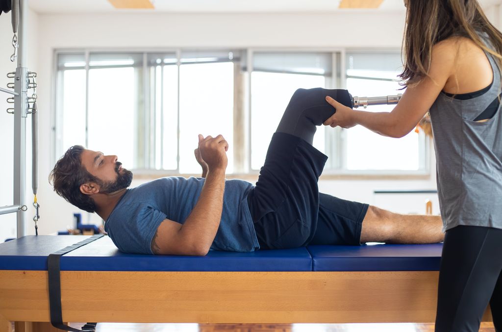
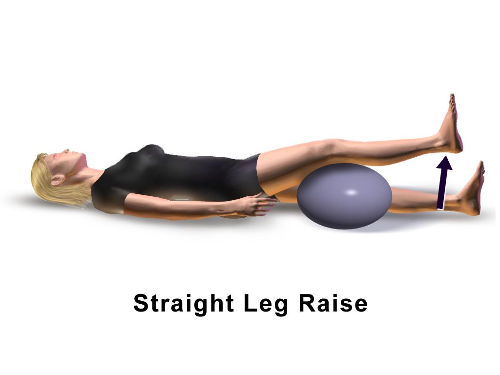
Physical Therapy: Restoring Function and Mobility
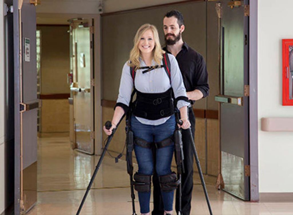
Physical therapists at the Noyes Knee Institute specialize in improving, maintaining, and restoring knee function and mobility. Through comprehensive evaluations, our skilled therapists develop tailored treatment plans that address the unique needs and goals of each patient. These plans often include a combination of exercise, manual therapy, and other modalities to reduce pain, swelling, and inflammation.
Our collaborative approach with orthopedic knee surgeons allows us to provide seamless post-surgery rehabilitation support, ensuring patients achieve optimal recovery and regain full knee function.
Our physical therapists educate patients on proper techniques for activities like walking and running, enabling them to prevent further knee injuries or aggravation of existing pain. Additionally, they provide guidance on lifestyle modifications to minimize knee stress and discomfort, promoting long-term knee health.
By combining the expertise of sports medicine specialists and physical therapists, the Noyes Knee Institute is dedicated to delivering comprehensive care that optimizes knee function, enhances mobility, and offers effective knee pain management strategies for our patients.
Importance of Orthopedic Knee Surgeons for Knee Pain Treatment
Orthopedic knee surgeons are highly skilled specialists in the diagnosis and treatment of various musculoskeletal conditions that affect the knee joint. At the Noyes Knee Institute, our team of orthopedic knee surgeons possesses extensive training and expertise in evaluating knee problems and formulating tailored treatment plans.
Working collaboratively with physical therapists and pain management specialists, our orthopedic knee surgeons offer comprehensive care to patients. After conducting a thorough evaluation, including X-rays or MRIs, they develop individualized treatment plans based on the specific needs of each patient.
Non-surgical interventions, such as physical therapy, medications, or injections, may be recommended to manage pain and facilitate healing. In cases where surgery is necessary, our orthopedic knee surgeons are proficient in a range of procedures to repair or reconstruct damaged knee structures.
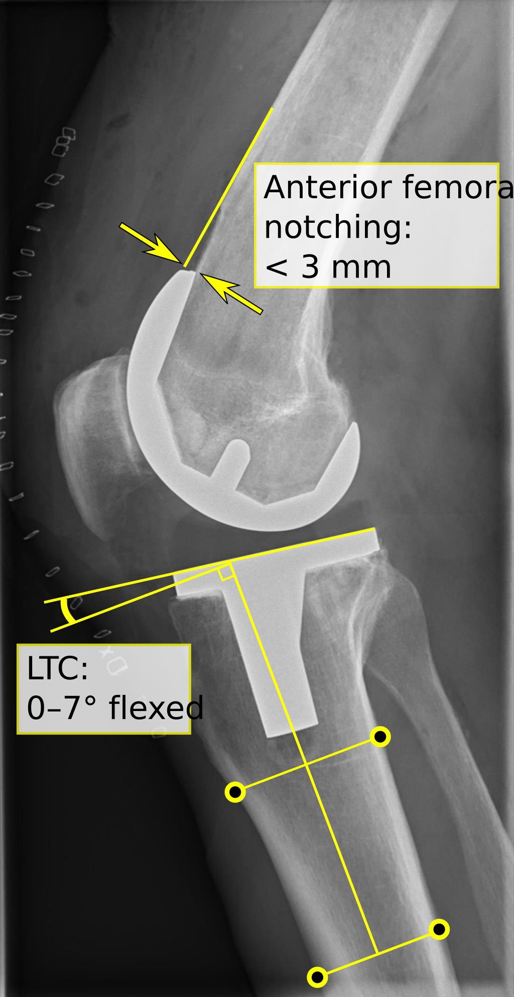
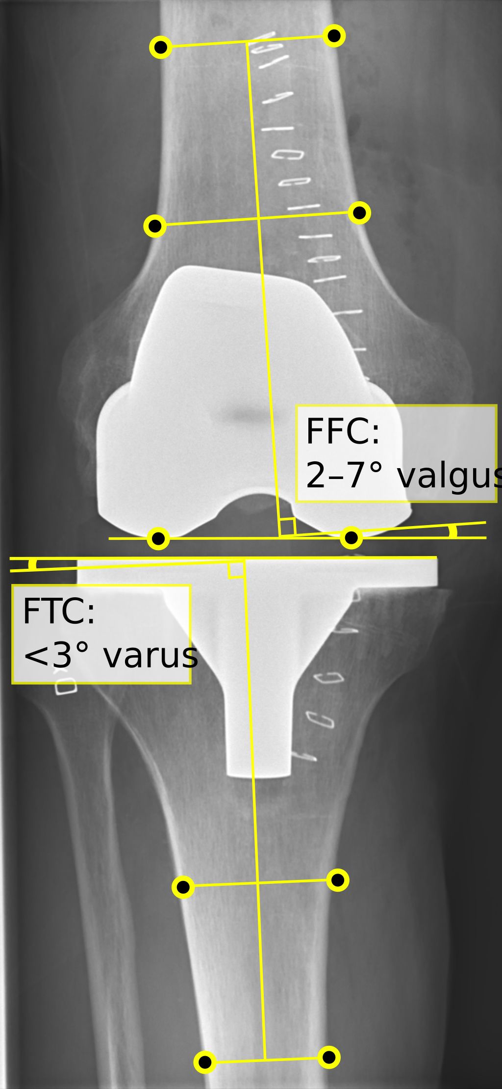
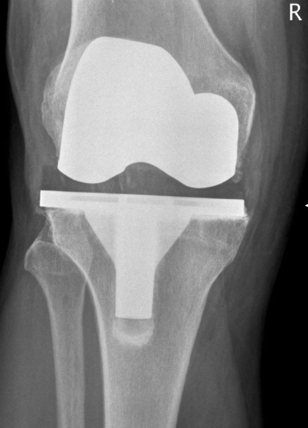
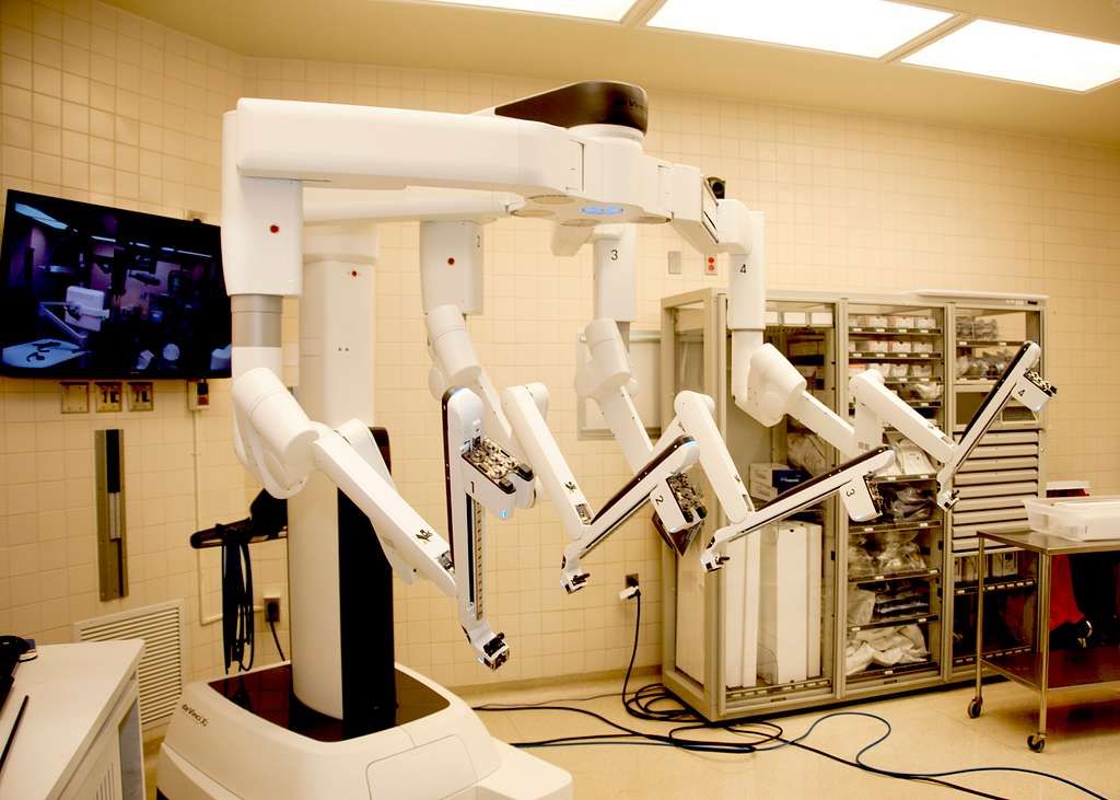
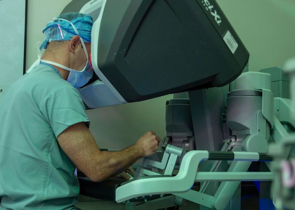
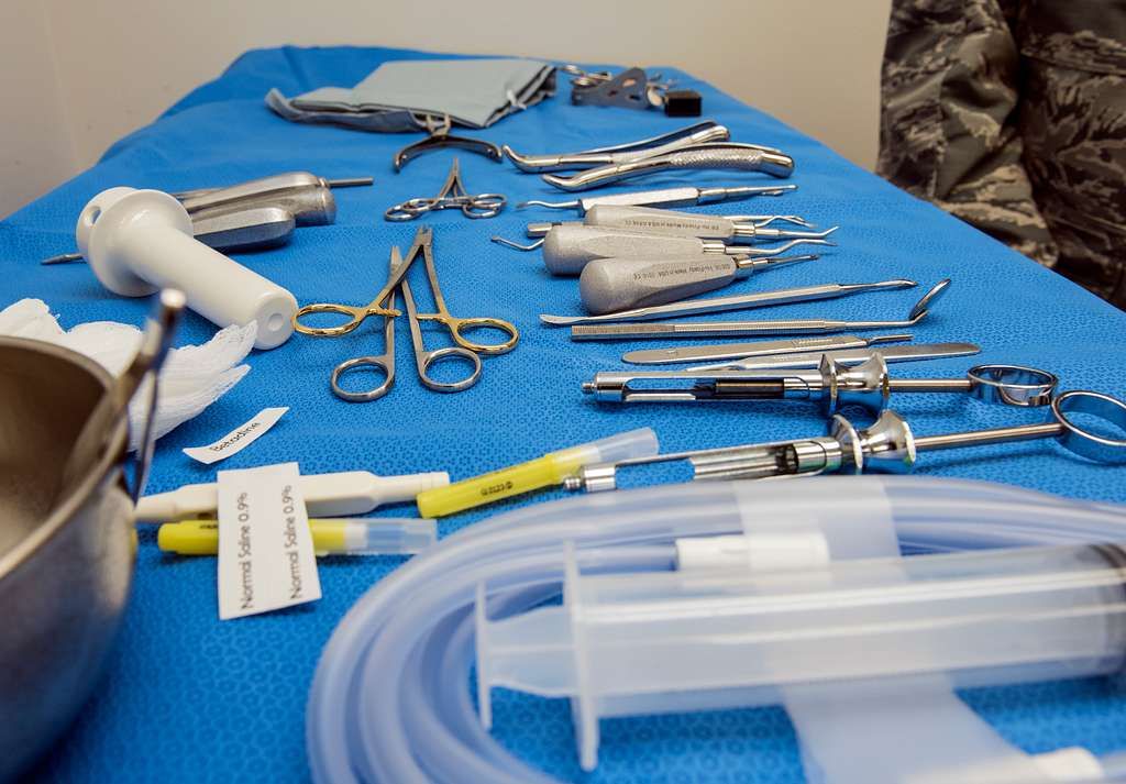
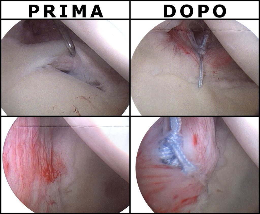
These procedures can involve arthroscopic surgery, which utilizes a miniature camera to guide the surgeon during repairs, or joint replacement surgery, where a damaged knee joint is replaced with an artificial joint. With their expertise, our orthopedic knee surgeons address complex knee conditions, delivering effective treatment options to relieve pain and restore function. At the Noyes Knee Institute, we remain at the forefront of utilizing advanced technology and techniques to achieve optimal outcomes for patients with knee pain.
FAQ
What types of doctors should I see for knee pain?
Depending on the severity and underlying cause of your knee pain, you may need to consult with different specialists. General practitioners are usually the first point of contact and can recommend conservative treatments. However, if your knee pain is severe or requires specialized care, you may be referred to a sports medicine specialist, physical therapist, or an orthopedic knee surgeon.
When should I see a knee pain specialist?
If your knee pain is persistent, worsening, or affecting your daily activities, it’s advisable to consult a knee pain specialist. They can provide a comprehensive evaluation, diagnosis, and treatment options tailored to your specific condition. Orthopedic knee surgeons, in particular, are experts in diagnosing and treating knee conditions, and they can offer both non-surgical and surgical interventions as necessary.
How can a sports medicine specialist help with knee pain?
Sports medicine specialists have specialized knowledge in treating and preventing sports-related injuries, including knee injuries. They can develop customized treatment plans, recommend injury prevention strategies, and provide guidance on safe and effective return-to-sports protocols. They understand the unique demands that sports activities place on the knees and can help athletes optimize their performance while minimizing the risk of knee-related issues.
What can physical therapy do for knee pain?
Physical therapists play a crucial role in managing knee pain and improving knee function and mobility. They conduct comprehensive evaluations to develop personalized treatment plans, which may include exercises, manual therapy, and other modalities. Physical therapists work closely with orthopedic knee surgeons to assist in post-surgery rehabilitation, educate patients on proper techniques for activities, and provide guidance on lifestyle modifications to minimize knee stress.
Why are orthopedic knee surgeons important for knee pain treatment?
Orthopedic knee surgeons specialize in diagnosing and treating various knee conditions. They have extensive training and expertise in assessing knee problems and formulating appropriate treatment plans. They can utilize non-surgical interventions like physical therapy, medication, or injections to manage pain and promote healing. If surgery is necessary, orthopedic knee surgeons can perform procedures such as arthroscopy or joint replacement to repair or reconstruct damaged knee structures, helping to relieve pain and restore function.