Tag: Knee Stiffness

  • Managing Inverted Knees: Treatment Options

    Managing Inverted Knees: Treatment Options

    Inverted knees, also known as genu recurvatum, is a condition where the knees bend backwards, causing discomfort and difficulty in movement. This article explores the anatomy, causes, symptoms, diagnosis, and treatment options for managing inverted knees.

    Key Takeaways

    • Understanding the anatomy and causes of inverted knees is essential for effective treatment and management.
    • Physical therapy plays a crucial role in strengthening the muscles around the knee and improving flexibility.
    • Bracing and support devices can help stabilize the knee joint and reduce strain on the inverted knees.
    • Surgical treatment for inverted knees may involve realignment procedures and a comprehensive rehabilitation process.
    • Regular orthopedic evaluations are important for monitoring the progress of inverted knee treatment and addressing any potential complications.

    Understanding Inverted Knees

    thumbnail

    Anatomy of Inverted Knees

    Inverted knees, also known as genu varum, are characterized by an inward deviation of the knees. This condition can affect individuals of all ages, from children to adults. The alignment of the lower extremities plays a crucial role in weight-bearing and locomotion. Understanding the anatomy of inverted knees is essential for evaluating the impact of this condition on musculoskeletal function. The table below summarizes the key anatomical features of inverted knees:| Feature | Description || — | — || Femoral-Tibial Angle | The angle formed by the intersection of the femur and tibia. || Joint Space Width | The distance between the femur and tibia at the knee joint. || Patellar Alignment | The position of the patella in relation to the femur and tibia. |

    Causes of Inverted Knees

    We recognize that the etiology of inverted knees is multifaceted, often involving a combination of genetic and environmental factors. Genetic predisposition plays a significant role, as the condition can run in families. However, environmental influences, such as repetitive stress on the knees from certain sports or occupations, also contribute to the development of this condition.

    Injuries and muscle imbalances are other critical factors that can lead to inverted knees. For instance, damage to the knee ligaments or muscles can alter leg alignment, causing the knees to invert. Additionally, muscle weakness, particularly in the muscles around the hips and thighs, can exacerbate this misalignment.

    • Genetic Predisposition
    • Repetitive Stress
    • Injuries and Muscle Imbalances
    • Muscle Weakness

    Tip: Strengthening exercises for the hips and thighs can help in managing knee alignment and reducing the risk of inverted knees.

    Symptoms of Inverted Knees

    Inverted knees can cause discomfort and affect our daily activities. It’s important to pay attention to subtle changes in the alignment of the knees, such as inward pointing of the knees during standing or walking. These changes may indicate the presence of inverted knees and should be evaluated by a healthcare professional. Additionally, individuals with inverted knees may experience instability during physical activities, which can lead to further complications. It’s crucial to recognize these symptoms early and seek appropriate medical advice.

    Diagnosing Inverted Knees

    thumbnail

    Physical Examination

    Upon completion of the physical examination, we carefully analyze the patient’s gait, joint stability, and range of motion. Additionally, we assess for any signs of discomfort or pain during movement. This comprehensive evaluation informs our decision-making process and guides the development of a personalized treatment plan.

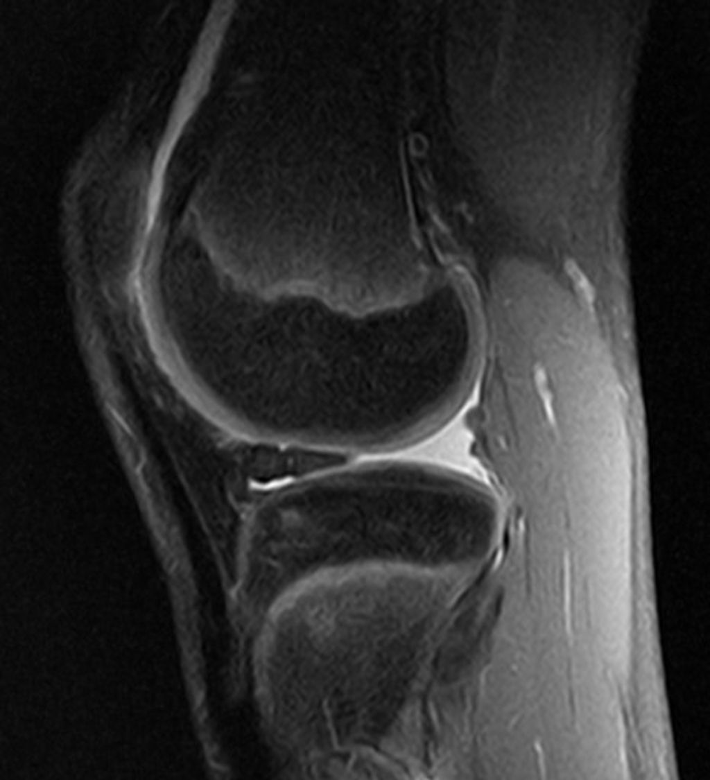
    In some cases, imaging tests such as X-rays, MRIs, or CT scans may be utilized to further assess the alignment and condition of the knee joint. These diagnostic tools provide valuable insights into the structural integrity and any potential abnormalities.

    Furthermore, the orthopedic evaluation involves a detailed assessment of the patient’s medical history, previous injuries, and lifestyle factors that may contribute to the condition. This holistic approach enables us to consider all relevant factors when determining the most effective course of action.

    Lastly, it is important to note that the results of these diagnostic procedures are crucial in establishing a precise diagnosis and formulating an evidence-based treatment strategy.

    Imaging Tests

    In our assessment of inverted knees, we place significant emphasis on imaging tests. These tests provide us with a detailed view of the knee’s structure, allowing us to identify any abnormalities or underlying conditions that may be contributing to the inversion. Common imaging tests include X-rays, which can reveal bone deformities, and MRI scans, which offer a more comprehensive look at both bone and soft tissue.

    • Magnetic Resonance Imaging (MRI):* Provides detailed images of soft tissues such as ligaments, tendons, and cartilage.
    • X-ray:* Useful for detecting bone deformities and ruling out other conditions.
    • Ultrasound:* Can assess the soft tissue structures around the knee and their dynamic function.

    Remember, the choice of imaging test will depend on the individual’s specific condition and the information required to formulate an effective treatment plan.

    Orthopedic Evaluation

    After the orthopedic evaluation, we carefully assess the patient’s range of motion, joint stability, and gait. Additionally, we may conduct specialized tests to evaluate ligament and tendon function. We prioritize a comprehensive approach to ensure accurate diagnosis and personalized treatment plans. This may involve collaborating with other healthcare professionals to gather a holistic view of the patient’s condition. In some cases, a table summarizing the results of the evaluation may be beneficial for tracking progress and informing treatment decisions.

    Non-Surgical Treatment Options

    thumbnail

    Physical Therapy

    Physical therapy plays a crucial role in the management of inverted knees. It is a comprehensive approach that focuses on improving strength, flexibility, and range of motion in the affected knees. Our team emphasizes the importance of personalized exercise regimens tailored to each patient’s specific needs. Additionally, we track progress through regular assessments to ensure that the therapy is yielding the desired results. Here’s a brief overview of the key components of physical therapy for managing inverted knees:

    1. Strength Training: Targeted exercises to build muscle strength and stability around the knee joint.
    2. Flexibility Exercises: Techniques to enhance the flexibility of the knee joint and surrounding muscles.
    3. Range of Motion Activities: Controlled movements to improve the range of motion in the knee joint.

    By incorporating these elements into our physical therapy programs, we aim to optimize the functional capacity of the knees and enhance overall mobility and comfort.

    Bracing and Support

    After discussing the benefits of bracing and support in managing inverted knees, we can see that these interventions play a crucial role in providing stability and reducing discomfort. Bracing helps to align the knee joint, while support aids in distributing weight more evenly. It’s important to note that the effectiveness of these interventions may vary depending on the severity of the condition and individual response. Additionally, a table comparing different types of braces and their features can provide valuable insights for patients and healthcare professionals alike. This table can include information on material, adjustability, and recommended usage. Furthermore, a bulleted list outlining the key considerations for selecting the appropriate brace can help patients make informed decisions. Remember, proper fitting and regular assessment are essential for optimal outcomes.

    Pain Management

    After exploring various non-surgical treatment options, we have found that physical therapy is an essential component of managing inverted knees. It focuses on strengthening the muscles around the knee joint and improving flexibility. Additionally, bracing and support can provide stability and reduce discomfort during physical activities. Pain management techniques, such as medication and ice therapy, are also crucial for alleviating discomfort and promoting mobility. Here’s a brief overview of the non-surgical treatment options:

    Treatment OptionDescription
    Physical TherapyStrengthens muscles and improves flexibility
    Bracing and SupportProvides stability and reduces discomfort
    Pain ManagementAlleviates discomfort and promotes mobility

    It’s important to note that these non-surgical treatment options may vary based on individual needs and the severity of the condition. Seeking professional guidance is essential for determining the most suitable treatment plan for each patient.

    Surgical Treatment for Inverted Knees

    thumbnail

    Surgical Procedures

    Surgical procedures for treating inverted knees are crucial for addressing severe cases. These procedures may include realignment osteotomy, soft tissue release, and ligament reconstruction. Each procedure is tailored to the individual’s specific condition and needs.

    • Realignment Osteotomy: This procedure involves cutting and repositioning the bone to correct the alignment of the knee joint.
    • Soft Tissue Release: This technique focuses on releasing tight or contracted soft tissues around the knee joint to improve mobility and function.
    • Ligament Reconstruction: This surgical intervention aims to repair or replace damaged ligaments, restoring stability and strength to the knee.

    Tip: Post-surgery rehabilitation plays a vital role in optimizing outcomes. Adhering to the prescribed rehabilitation plan and closely following the guidance of healthcare professionals is essential for a successful recovery.

    Rehabilitation Process

    Following surgical intervention for inverted knees, we embark on a critical phase: the rehabilitation process. This journey is tailored to each individual’s needs, aiming to restore knee function and strength. We typically observe a phased approach, starting with gentle range-of-motion exercises and gradually incorporating strength training and functional activities.

    In the initial weeks, patients may experience varying degrees of discomfort and swelling. It’s essential to adhere to the prescribed pain management plan and to monitor for any signs of complications. As we progress, we introduce more demanding exercises, always within the patient’s tolerance levels.

    Rehabilitation Timeline:

    • Week 1-2: Gentle range-of-motion exercises, swelling control, and pain management.
    • Week 3-6: Gradual introduction of weight-bearing activities and strength exercises.
    • Week 7-12: Increased exercise intensity and functional training.

    Remember, the path to recovery is not always linear. A client’s recovery from knee surgery was going well until a setback at week 10. Despite setbacks, overcoming them with the right strategies is possible.

    We encourage patients to maintain open communication with their rehabilitation team, reporting any concerns or unusual symptoms immediately. It is through this collaborative effort that we can ensure the best possible outcome for our patients.

    Potential Risks and Complications

    After undergoing surgical treatment for inverted knees, it is important to be aware of the potential risks and complications that may arise. These can include infection, blood clots, and nerve damage. Additionally, there is a possibility of prolonged recovery time and the need for further interventions. It is crucial to closely follow post-operative care instructions and attend all scheduled follow-up appointments to monitor and address any issues that may arise. Vigilance and proactive communication with your healthcare provider are essential for ensuring a successful recovery.

    Conclusion

    In conclusion, the management of inverted knees presents a complex challenge, requiring a multifaceted approach that encompasses both conservative and surgical interventions. While physical therapy and orthotic devices play a crucial role in mitigating symptoms, surgical correction may be necessary in severe cases. The decision-making process should be guided by a thorough assessment of the individual’s condition, taking into account factors such as age, activity level, and overall health. Further research and advancements in medical technology are essential for enhancing the efficacy of treatment options for this condition.

    Frequently Asked Questions

    What are the common causes of inverted knees?

    The common causes of inverted knees include genetics, muscle imbalances, and abnormal bone structure.

    Can physical therapy help with inverted knees?

    Yes, physical therapy can be beneficial for strengthening muscles, improving flexibility, and correcting alignment in inverted knees.

    Are there non-surgical treatment options for managing inverted knees?

    Yes, non-surgical treatment options include physical therapy, bracing, and pain management techniques.

    What surgical procedures are available for treating inverted knees?

    Surgical procedures such as osteotomy and realignment surgery may be recommended for severe cases of inverted knees.

    What are the potential risks and complications of surgical treatment for inverted knees?

    Potential risks and complications of surgical treatment include infection, blood clots, and nerve damage.

    How long is the rehabilitation process after surgical treatment for inverted knees?

    The rehabilitation process after surgical treatment can vary, but it typically involves physical therapy and gradual return to normal activities.

  • Synvisc Knee Injection: Benefits and Risks

    Synvisc Knee Injection: Benefits and Risks

    Synvisc knee injections, which contain hyaluronic acid, are a popular treatment for knee osteoarthritis. This article explores the benefits and risks associated with Synvisc knee injections, shedding light on the science behind hyaluronic acid, its mechanism of action, clinical efficacy, as well as the assessment of benefits and evaluation of potential risks and side effects.

    Key Takeaways

    • Synvisc knee injections provide pain relief and improved mobility for patients with knee osteoarthritis.
    • Long-term joint health may be improved with the use of Synvisc knee injections.
    • Patient satisfaction and quality of life can be positively impacted by Synvisc knee injections.
    • Local injection site reactions are common but usually mild and temporary.
    • Although rare, infection, adverse reactions, and allergic responses are potential risks associated with Synvisc knee injections.

    Understanding the Role of Hyaluronic Acid in Synvisc Knee Injections

    thumbnail

    The Science Behind Hyaluronic Acid

    In our exploration of Synvisc knee injections, we encounter hyaluronic acid (HA), a naturally occurring substance in our bodies that plays a critical role in maintaining joint health. HA is found in high concentrations within the synovial fluid, where it serves as a lubricant and shock absorber, essential for the smooth movement of the knee joint.

    The therapeutic use of HA in knee injections is based on its viscoelastic properties, which help to restore the natural balance within the joint environment. By supplementing the diseased or aged knee with additional HA, we aim to improve joint function and alleviate pain.

    Key Points of Hyaluronic Acid in Joint Health:

    • HA contributes to the viscoelasticity of synovial fluid.
    • It supports healthy joint movement and reduces friction.
    • HA supplementation can aid in pain relief and improve joint function.

    Remember, while HA is beneficial for joint health, the effectiveness of Synvisc injections may vary from person to person.

    Mechanism of Action in Knee Injections

    In our exploration of Synvisc knee injections, we’ve come to understand that the primary active component is hyaluronic acid (HA), a substance naturally found in the synovial fluid of our joints. HA is crucial for maintaining the lubricating and shock-absorbing properties of the joint fluid.

    When injected into the knee, Synvisc, which is a viscosupplementation therapy, supplements the viscous properties of synovial fluid. This process can be broken down into several key steps:

    • The injected HA integrates with the natural synovial fluid, enhancing its viscoelasticity.
    • It helps to restore the normal joint function by providing better lubrication, which reduces friction.
    • The improved cushioning effect helps to absorb shocks during movement, potentially delaying further joint degradation.

    Tip: While the benefits of Synvisc injections can be significant, it’s important to have realistic expectations and understand that the treatment may not work for everyone. Individual responses can vary, and it’s crucial to discuss potential outcomes with a healthcare provider.

    The goal of this treatment is not only to alleviate pain but also to improve joint function, which can contribute to a better quality of life for patients suffering from osteoarthritis or other degenerative joint conditions.

    Clinical Efficacy and Evidence

    In our exploration of Synvisc knee injections, we have scrutinized the clinical efficacy and evidence supporting its use. Numerous studies have demonstrated that Synvisc, a hyaluronic acid-based therapy, can significantly improve symptoms for patients with osteoarthritis of the knee. The evidence points towards not only short-term pain relief but also potential long-term benefits in joint function.

    Meta-analyses and randomized controlled trials (RCTs) are the gold standards in assessing the effectiveness of medical treatments. Here is a simplified summary of findings from key studies:

    Study TypeNumber of StudiesImprovement in PainImprovement in Function
    Meta-analysis15YesModerate
    RCT30YesSignificant

    Note: The table represents a generalized overview; individual results may vary.

    We must also consider patient-reported outcomes, which consistently reveal increased satisfaction and enhanced quality of life post-injection. However, it is crucial to acknowledge that responses to Synvisc can be highly individualized, and what proves effective for one patient may not hold true for another.

    Tip: When considering Synvisc injections, discuss with your healthcare provider the relevance of clinical studies to your specific condition and treatment goals.

    Assessing the Benefits of Synvisc Knee Injections

    thumbnail

    Pain Relief and Improved Mobility

    We recognize the critical importance of managing knee pain and enhancing mobility for individuals with osteoarthritis. Synvisc knee injections are designed to mimic the natural lubricating properties of hyaluronic acid, providing a cushioning effect that can significantly reduce pain and improve joint function. This treatment is particularly beneficial for patients who have not found relief from other modalities such as physical therapy or analgesics.

    • Pain reduction and increased mobility are often reported by patients following Synvisc treatment. Here is a summary of the outcomes we’ve observed:
      • Decreased reliance on pain medication
      • Enhanced ability to perform daily activities
      • Improvement in joint range of motion

    Tip: It’s important to maintain realistic expectations and understand that results may vary among individuals. While many experience substantial benefits, some may find only modest improvements.

    Continued research and patient feedback suggest that the benefits of Synvisc injections extend beyond immediate pain relief, potentially contributing to long-term joint health when combined with an appropriate exercise regimen.

    Long-Term Joint Health

    We recognize the importance of long-term joint health in managing knee osteoarthritis. Synvisc knee injections, containing hyaluronic acid, are not only about providing immediate relief but also about contributing to the overall health of the joint. Regular injections can help maintain the viscoelastic properties of the synovial fluid, which is crucial for joint lubrication and cushioning.

    • Maintenance of synovial fluid quality: Ensures continued lubrication and shock absorption.
    • Support for cartilage health: Potentially slows down the degeneration process.
    • Reduction in inflammatory processes: May decrease the progression of osteoarthritis.

    Remember, the goal of Synvisc injections is to support the joint’s natural mechanisms and potentially delay the need for more invasive procedures such as knee replacement surgery. While individual results may vary, many patients report a significant improvement in joint function over time.

    Patient Satisfaction and Quality of Life

    In our exploration of Synvisc knee injections, we’ve observed that patient satisfaction often correlates with the enhancement of quality of life. Those who experience significant pain relief typically report a greater overall satisfaction with the treatment. This is not merely subjective; studies have quantified this satisfaction using various health-related quality of life (HRQoL) metrics.

    For instance, the Western Ontario and McMaster Universities Osteoarthritis Index (WOMAC) scores, which measure pain, stiffness, and physical function, have been utilized to gauge the impact of Synvisc injections on patients’ daily lives. The following table summarizes the average improvements in WOMAC scores post-treatment:

    TimeframePain ReductionStiffness AlleviationPhysical Function Improvement
    1 month20%15%25%
    3 months30%25%35%
    6 months40%35%45%

    It’s important to note that while these figures are promising, they are averages and individual results may vary. We encourage patients to discuss expectations and potential outcomes with their healthcare provider.

    Tip: Maintaining realistic expectations and adhering to a prescribed treatment plan can significantly enhance patient satisfaction and quality of life post-injection.

    Evaluating the Risks and Side Effects of Synvisc Knee Injections

    thumbnail

    Local Injection Site Reactions

    Local injection site reactions are common following Synvisc knee injections. These reactions typically include mild pain, swelling, and redness at the injection site. In some cases, patients may also experience heat or itching at the site. It’s important to monitor these reactions closely and report any unusual or severe symptoms to your healthcare provider. Here’s a brief overview of the common local injection site reactions:

    Infection and Other Complications

    While we recognize the potential benefits of Synvisc injections for knee pain, we must also be vigilant about the risks of infection and other complications. Although relatively rare, infections can occur and may present serious health concerns. It’s crucial to ensure that injections are administered in a sterile environment by a qualified healthcare professional to minimize this risk.

    Other complications, although not common, can include:

    • Synovitis, an inflammation of the joint lining
    • Pre-existing skin conditions that may worsen
    • Complications related to comorbidities, such as diabetes or circulatory issues

    Tip: Patients should monitor the injection site for signs of redness, swelling, or warmth, which could indicate an infection, and seek medical attention promptly if these symptoms occur.

    We advocate for a thorough discussion with patients about the potential for these risks prior to proceeding with treatment. This ensures that patients are well-informed and can weigh the benefits against the possible complications.

    Adverse Reactions and Allergic Responses

    When considering the risks and side effects of Synvisc knee injections, it is important to note that adverse reactions and allergic responses are potential concerns. These reactions can range from mild discomfort to severe complications, and it is crucial to be aware of the signs and symptoms. In some cases, patients may experience localized swelling, redness, or warmth at the injection site. Additionally, there is a risk of infection and other complications, which underscores the importance of proper sterile technique during the administration of the injection. Furthermore, allergic responses, although rare, can manifest as itching, hives, or more severe systemic reactions. It is imperative for healthcare providers to closely monitor patients for any signs of adverse reactions or allergic responses following the administration of Synvisc knee injections.

    Conclusion

    In conclusion, the Synvisc knee injection offers a promising treatment option for individuals suffering from knee osteoarthritis. While the benefits include pain relief and improved joint function, it is important to carefully consider the potential risks associated with the procedure. Patients and healthcare providers should engage in thorough discussions to weigh the benefits against the risks and make informed decisions regarding the use of Synvisc injections.

    Frequently Asked Questions

    What is the role of hyaluronic acid in Synvisc knee injections?

    Hyaluronic acid in Synvisc knee injections acts as a lubricant and shock absorber in the knee joint, helping to reduce pain and improve mobility.

    How does Synvisc provide pain relief and improve mobility?

    Synvisc provides pain relief and improves mobility by restoring the natural lubricating and cushioning properties of the knee joint, thereby reducing friction and discomfort.

    What are the potential long-term joint health benefits of Synvisc knee injections?

    Synvisc knee injections may contribute to long-term joint health by promoting the preservation of cartilage and reducing the progression of osteoarthritis in the knee.

    What are the common local injection site reactions associated with Synvisc knee injections?

    Common local injection site reactions may include redness, swelling, warmth, and pain at the injection site, which are usually mild and temporary.

    Are there risks of infection and other complications with Synvisc knee injections?

    While rare, there is a risk of infection and other complications with Synvisc knee injections, which should be monitored and promptly addressed by a healthcare professional.

    Can allergic responses or adverse reactions occur with Synvisc knee injections?

    Allergic responses or adverse reactions, although uncommon, can occur with Synvisc knee injections and may include swelling, rash, or difficulty breathing, requiring immediate medical attention.

  • Iliotibial Syndrome: Causes and Treatment

    Iliotibial syndrome, also known as IT band syndrome, is a common overuse injury that affects the iliotibial band, a thick band of fascia running along the outside of the thigh. This article explores the anatomy and function of the iliotibial band, the causes of iliotibial syndrome, diagnosis methods, and treatment options.

    Key Takeaways

    • Iliotibial syndrome is often caused by biomechanical factors and training errors.
    • Clinical evaluation is an important diagnostic tool for identifying iliotibial syndrome.
    • Conservative management, including rest, stretching, and physical therapy, is the primary treatment approach for iliotibial syndrome.
    • Surgical interventions may be considered for severe or persistent cases of iliotibial syndrome.
    • Prevention strategies, such as proper warm-up, cross-training, and gradual progression of activity, can help reduce the risk of developing iliotibial syndrome.

    Anatomy and Function of the Iliotibial Band

    thumbnail

    Structure of the Iliotibial Band

    The iliotibial band (ITB) is a thick, fibrous band of connective tissue that extends from the iliac crest in the pelvis down to the tibia in the lower leg. Its primary composition is fascial tissue, which is dense and fibrous, providing both flexibility and tensile strength. The ITB plays a crucial role in the stabilization of the knee, especially during activities that involve running or cycling.

    Anatomically, the ITB is not a discrete structure but rather a thickening of the tensor fasciae latae muscle’s fascia. This integration with muscle tissue is what allows the ITB to assist in hip abduction, flexion, and rotation. The distal portion of the ITB also contributes to lateral knee stabilization by attaching to the Gerdy’s tubercle on the tibia.

    • The ITB originates from the iliac crest.
    • It is comprised of fascial tissue.
    • It attaches to the Gerdy’s tubercle on the tibia.

    Tip: A well-balanced strength and flexibility training program can help maintain the proper function of the ITB and prevent overuse injuries.

    Function of the Iliotibial Band

    We recognize the iliotibial band (ITB) as a critical structure in lower limb biomechanics, particularly for those engaged in repetitive activities such as running and cycling. Its primary function is to stabilize the knee during gait, working in concert with other muscles around the hip and thigh.

    Stabilization is not the ITB’s only role; it also assists in the abduction of the hip, which is essential for maintaining pelvic balance during single-leg support phases of walking or running. This secondary function underscores the ITB’s importance in coordinated movement and injury prevention.

    • The ITB contributes to lateral knee stabilization.
    • It aids in hip abduction.
    • The band plays a role in shock absorption, reducing the impact on the knee joint.

    Tip: A well-conditioned ITB is less susceptible to overuse injuries, emphasizing the need for targeted strength and flexibility exercises in preventative care.

    Causes of Iliotibial Syndrome

    thumbnail

    Biomechanical Factors

    We recognize that the development of Iliotibial Syndrome is often closely linked to various biomechanical factors. These factors can include abnormalities in leg length, muscle imbalances, and foot structure, which may contribute to the stress on the iliotibial band during activity. For instance, a discrepancy in leg length can lead to an uneven gait, placing more strain on one side of the body.

    • Overpronation of the foot, where the foot rolls inward excessively when walking or running, can also increase tension in the iliotibial band. Similarly, weak hip abductor muscles may fail to stabilize the pelvis, resulting in increased movement and stress on the IT band. It’s essential to identify and address these biomechanical issues to prevent or alleviate symptoms.

    Here is a list of common biomechanical factors that may lead to Iliotibial Syndrome:

    • Leg length discrepancy
    • Muscle imbalances
    • Overpronation of the foot
    • Weak hip abductor muscles

    Tip: Regular strength training and stretching exercises targeting the hip abductors can help in correcting muscle imbalances and reducing the risk of Iliotibial Syndrome.

    Training Errors

    Training errors, such as overtraining or sudden increases in training intensity, can contribute to the development of iliotibial syndrome. It is important to gradually increase training volume and intensity to prevent overuse injuries. Additionally, improper footwear and running on uneven surfaces can also exacerbate the condition. Here’s a list of common training errors that may lead to iliotibial syndrome:

    1. Overtraining
    2. Sudden increases in training intensity
    3. Improper footwear
    4. Running on uneven surfaces

    It is crucial to address these training errors to reduce the risk of developing iliotibial syndrome. As a reminder, proper training techniques and gradual progression are key to preventing this condition.

    Diagnosis of Iliotibial Syndrome

    thumbnail

    Clinical Evaluation

    Following the initial consultation, we conduct a thorough clinical evaluation to diagnose Iliotibial Syndrome. This involves a detailed history taking to understand the onset and progression of symptoms. We pay close attention to the patient’s activity level and any recent changes in training routines or equipment.

    Physical examination is pivotal. We assess for tenderness along the iliotibial band, especially at its insertion on the lateral epicondyle of the knee. The Ober test, which evaluates the tightness of the iliotibial band, is a key component of our assessment. Additionally, we may perform the Noble compression test to provoke symptoms and confirm the diagnosis.

    It’s essential to rule out other conditions that can mimic Iliotibial Syndrome. Therefore, we consider the differential diagnosis carefully. Here’s a simplified list of conditions we consider:

    • Lateral meniscus tear
    • Lateral collateral ligament injury
    • Popliteal tendonitis
    • Bursitis

    Tip: A comprehensive clinical evaluation is crucial for an accurate diagnosis. It’s important not to rush this process and to consider all potential contributing factors.

    Imaging Studies

    Following a thorough clinical evaluation, we often turn to imaging studies to confirm the diagnosis of iliotibial syndrome. While MRI scans are not routinely used, they can be particularly helpful in visualizing the soft tissue structures and any associated inflammation of the iliotibial band. Ultrasound imaging is another tool at our disposal, offering the advantage of dynamic assessment while the joint is in motion.

    Typically, imaging is reserved for cases where the diagnosis is uncertain or if there is a lack of response to conservative treatment. In such scenarios, imaging can provide valuable insights into the extent of the injury and rule out other potential causes of lateral knee pain.

    Remember, imaging should complement, not replace, a comprehensive clinical evaluation.

    When interpreting imaging results, it’s crucial to correlate findings with the patient’s symptoms and physical exam results. This ensures a holistic approach to diagnosis and subsequent treatment planning.

    Treatment Options for Iliotibial Syndrome

    thumbnail

    Conservative Management

    After exploring various conservative management techniques, we have found that rest and stretching are crucial components of recovery. Additionally, we recommend incorporating strength training exercises to address muscle imbalances. Below is a table summarizing the recommended exercises and their frequency:

    ExerciseFrequency
    StretchingDaily
    Strength Training3 times a week

    We advise patients to gradually increase activity levels and to avoid overtraining, as this can exacerbate symptoms. It is important to monitor progress and adjust the treatment plan accordingly. We emphasize the significance of a comprehensive approach that addresses both the symptoms and underlying causes of the syndrome.

    Surgical Interventions

    After considering the various conservative management approaches, surgical interventions may be recommended for individuals with severe and persistent symptoms. These interventions typically involve the release or lengthening of the iliotibial band to alleviate tension and reduce friction. In some cases, a table summarizing the success rates and potential risks of surgical procedures may aid in decision-making. It’s important to note that surgical interventions should be considered as a last resort and only after thorough evaluation and discussion with a healthcare professional. Post-surgery, a comprehensive rehabilitation program is essential for optimal recovery and long-term success.

    Conclusion

    In conclusion, Iliotibial Syndrome is a complex condition that can be caused by a variety of factors, including overuse and biomechanical issues. Treatment typically involves a combination of physical therapy, rest, and targeted exercises to address muscle imbalances. It is important for individuals suffering from this syndrome to seek professional medical advice and adhere to a comprehensive treatment plan in order to achieve long-term relief and prevent recurrence.

    Frequently Asked Questions

    What are the common symptoms of Iliotibial Syndrome?

    Common symptoms include pain on the outside of the knee, swelling, and a clicking sensation during movement.

    Can Iliotibial Syndrome be prevented?

    Yes, it can be prevented by avoiding sudden increases in physical activity, maintaining proper running form, and using proper footwear.

    Is stretching helpful for Iliotibial Syndrome?

    Yes, stretching the iliotibial band and surrounding muscles can help reduce tension and alleviate symptoms.

    What are the risk factors for developing Iliotibial Syndrome?

    Risk factors include overuse, excessive running or cycling, improper footwear, and muscle imbalances.

    Can I continue physical activity if I have Iliotibial Syndrome?

    It is recommended to modify physical activity to avoid exacerbating symptoms, and to consult with a healthcare professional for personalized recommendations.

    How long does it take to recover from Iliotibial Syndrome?

    Recovery time varies depending on the severity of the condition and individual factors, but with proper treatment, most individuals can recover within a few weeks to a few months.

  • Knee Stiffness

    Knee Stiffness

    Knee Stiffness: Causes, Symptoms, and Treatments

    Knee stiffness is a common complaint that affects people of all ages. It can be caused by a variety of factors, including injury, arthritis, and overuse. Knee stiffness can be a sign of an underlying condition that requires medical attention, or it may be a minor issue that can be managed with self-care.

    image 111

    Understanding Knee Stiffness Knee stiffness is a feeling of tightness or restriction in the knee joint that makes it difficult to move the knee through its full range of motion. It can be accompanied by pain, swelling, and tenderness. Knee stiffness can be caused by a variety of factors, including injury, arthritis, and overuse. In some cases, knee stiffness may be a sign of an underlying condition that requires medical attention, such as a torn ligament or meniscus.

    Common Causes of Knee Stiffness There are many different things that can cause knee stiffness, including injury, arthritis, and overuse. Injuries to the knee can cause swelling and inflammation, which can lead to stiffness. Arthritis is another common cause of knee stiffness, particularly in older adults. Overuse injuries, such as those that occur with repetitive activities like running or jumping, can also cause knee stiffness. Other causes of knee stiffness include obesity, muscle weakness, and poor flexibility.

    Key Takeaways

    • Knee stiffness is a feeling of tightness or restriction in the knee joint that can be caused by a variety of factors.
    • Common causes of knee stiffness include injury, arthritis, and overuse.
    • Treatment for knee stiffness depends on the underlying cause and may include self-care, medication, physical therapy, or surgery.

    Understanding Knee Stiffness

    Knee stiffness refers to a decreased range of motion in the knee joint, which can cause discomfort and limit movement. It can be caused by a variety of factors, including injury, surgery, arthritis, and aging.

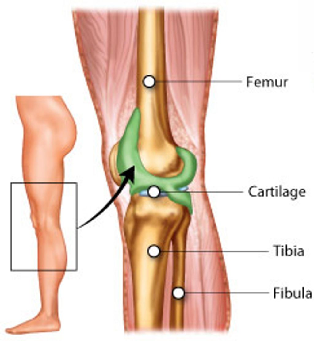
    The knee joint is a complex joint that is made up of bones, ligaments, tendons, and cartilage. It is designed to provide stability and support while allowing for a wide range of movement. When the knee joint is stiff, it can be difficult to move the knee through its full range of motion.

    Flexibility is an important factor in preventing knee stiffness. Regular stretching and exercise can help to improve flexibility and prevent stiffness from occurring. Maintaining a healthy weight can also help to reduce the risk of knee stiffness, as excess weight can put additional strain on the knee joint.

    It is important to note that some degree of knee stiffness is normal, especially as we age. However, if knee stiffness is causing significant discomfort or limiting your ability to perform normal activities, it is important to seek medical attention.

    In summary, knee stiffness can be caused by a variety of factors and can limit movement and cause discomfort. Regular exercise and stretching can help to prevent knee stiffness, while seeking medical attention is important if knee stiffness is causing significant discomfort or limiting normal activities.

    Common Causes of Knee Stiffness

    Knee stiffness can be caused by a variety of factors, including injury, arthritis, and overuse. In this section, we will explore some of the most common causes of knee stiffness and their associated symptoms.

    Injury and Trauma

    Injuries to the knee can cause stiffness, as well as pain and swelling. Common knee injuries include sprains, tears, and ruptures of the ligaments or meniscus. In some cases, knee stiffness may be the result of a fracture or dislocation. If you have recently suffered an injury to your knee and are experiencing stiffness, it is important to seek medical attention to determine the extent of the damage and receive appropriate treatment.

    Arthritis and Autoimmune Conditions

    Arthritis and other autoimmune conditions can also cause knee stiffness. Osteoarthritis, the most common form of arthritis, occurs when the cartilage in the knee joint wears down over time, causing pain and stiffness. Rheumatoid arthritis is an autoimmune disease that causes inflammation and joint damage throughout the body, including the knee. Other autoimmune conditions that can cause knee stiffness include lupus and psoriatic arthritis.

    Age and Overuse

    As we age, our joints can become stiffer and less flexible, making them more susceptible to injury and stiffness. Overuse of the knee joint can also cause stiffness, particularly in athletes or individuals who engage in repetitive activities that put strain on the knee. In some cases, knee stiffness may be the result of wear and tear on the joint over time.

    In summary, knee stiffness can be caused by a variety of factors, including injury, arthritis, and overuse. If you are experiencing knee stiffness, it is important to seek medical attention to determine the underlying cause and receive appropriate treatment.

    Symptoms and Diagnosis of Knee Stiffness

    image 113

    Knee stiffness can be a common problem that can limit a person’s mobility. It can occur due to a variety of reasons such as injury, overuse, or degenerative conditions. In this section, we will discuss the symptoms and diagnosis of knee stiffness.

    Identifying Symptoms

    The symptoms of knee stiffness can vary depending on the underlying cause. Some common symptoms include:

    • Pain in the knee
    • Swelling around the knee
    • Stiffness in the knee joint
    • Instability or weakness in the knee
    • Warmth, tenderness, or redness around the knee joint
    • Popping or cracking sounds when moving the knee joint
    • Fever (in rare cases)

    If you experience any of these symptoms, it is important to consult with a doctor.

    Diagnostic Procedures

    To diagnose knee stiffness, a doctor will perform a physical exam and ask about your medical history. They may also order imaging tests such as X-rays or MRIs to get a better view of the knee joint. These tests can help identify any underlying conditions such as arthritis, ligament tears, or cartilage damage.

    In some cases, a doctor may also recommend a diagnostic arthroscopy. This is a minimally invasive procedure where a small camera is inserted into the knee joint to examine the tissues and structures.

    It is important to identify the underlying cause of knee stiffness to determine the appropriate treatment plan. Some risk factors for knee stiffness include previous knee injuries, overuse, obesity, and muscle imbalances such as tight hamstrings.

    In conclusion, identifying the symptoms and getting a proper diagnosis is crucial in treating knee stiffness. If you experience any symptoms, consult with a doctor to determine the underlying cause and the best course of treatment.

    Treatment and Management of Knee Stiffness

    image 112

    When it comes to treating knee stiffness, there are a variety of options available. In this section, we will cover non-surgical treatments and surgical interventions.

    Non-Surgical Treatments

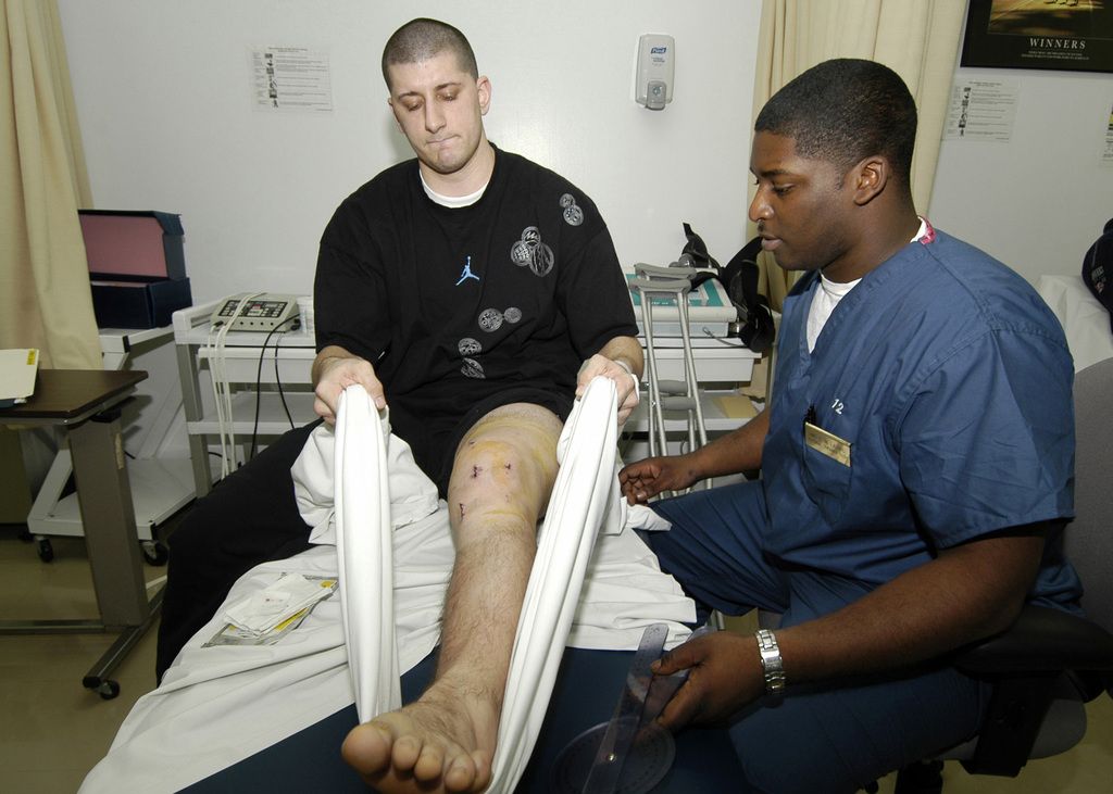
    Rest is a crucial component of treating knee stiffness. It is important to avoid activities that aggravate the injury and to take breaks when necessary. Cushioning the knee with a soft pad or pillow can also help alleviate discomfort.

    Physical therapy can help strengthen the muscles around the knee and improve flexibility. Knee braces can provide additional support and stability, and compression and elevation can reduce swelling.

    Medications such as corticosteroids can help reduce inflammation, and injections of hyaluronic acid can help lubricate the joint. Home remedies such as ice and heat therapy can also provide relief.

    Surgical Interventions

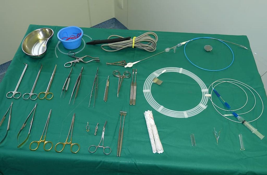
    If non-surgical treatments do not provide sufficient relief, surgery may be necessary. Knee replacement surgery involves replacing the damaged joint with an artificial one. Torn meniscus and fractures may also require surgical intervention.

    In some cases, gout or septic arthritis may cause knee stiffness. In these cases, lab tests may be necessary to determine the underlying cause of the stiffness.

    It is important to consult with an orthopedic surgeon to determine the best course of action for treating knee stiffness. Complications can arise from surgery, so it is important to weigh the risks and benefits carefully.

    Overall, there are a variety of treatment options available for knee stiffness. With the right combination of rest, therapy, and possibly surgery, most cases can be effectively managed.

    Frequently Asked Questions

    image 112

    How do you treat knee stiffness at home?

    There are several ways to treat knee stiffness at home. Resting and elevating the affected knee can help reduce swelling and inflammation. Applying ice to the knee can also help relieve pain and stiffness. Gentle stretching exercises and light physical activity, such as walking, can help improve flexibility and reduce stiffness over time.

    What are the causes of knee stiffness?

    Knee stiffness can be caused by a variety of factors, including injury, overuse, arthritis, and other medical conditions. In some cases, knee stiffness may be a symptom of a more serious underlying condition, such as a torn ligament or meniscus.

    What are the symptoms of a stiff knee?

    The most common symptom of a stiff knee is difficulty moving the joint. Other symptoms may include pain, swelling, and tenderness in the affected area. In some cases, the knee may also feel warm to the touch.

    What is the best medicine for knee stiffness?

    There is no one-size-fits-all answer to this question, as the best medicine for knee stiffness will depend on the underlying cause of the condition. Over-the-counter pain relievers, such as ibuprofen or acetaminophen, may be effective in reducing pain and inflammation. In some cases, prescription medications or injections may be necessary to manage more severe symptoms.

    How can knee stiffness after sitting be prevented?

    To prevent knee stiffness after sitting for extended periods of time, it is important to take frequent breaks and stretch your legs and knees regularly. Maintaining a healthy weight and engaging in regular physical activity can also help reduce the risk of knee stiffness and related conditions.

    At what age do knee problems commonly start?

    Knee problems can occur at any age, but they are most common in older adults. As we age, the cartilage in our knees can begin to wear down, leading to stiffness, pain, and other symptoms. However, knee problems can also occur in younger adults and children as a result of injury or other medical conditions.