Tag: Ligament

  • Iliotibial Syndrome: Causes and Treatment

    Iliotibial syndrome, also known as IT band syndrome, is a common overuse injury that affects the iliotibial band, a thick band of fascia running along the outside of the thigh. This article explores the anatomy and function of the iliotibial band, the causes of iliotibial syndrome, diagnosis methods, and treatment options.

    Key Takeaways

    • Iliotibial syndrome is often caused by biomechanical factors and training errors.
    • Clinical evaluation is an important diagnostic tool for identifying iliotibial syndrome.
    • Conservative management, including rest, stretching, and physical therapy, is the primary treatment approach for iliotibial syndrome.
    • Surgical interventions may be considered for severe or persistent cases of iliotibial syndrome.
    • Prevention strategies, such as proper warm-up, cross-training, and gradual progression of activity, can help reduce the risk of developing iliotibial syndrome.

    Anatomy and Function of the Iliotibial Band

    thumbnail

    Structure of the Iliotibial Band

    The iliotibial band (ITB) is a thick, fibrous band of connective tissue that extends from the iliac crest in the pelvis down to the tibia in the lower leg. Its primary composition is fascial tissue, which is dense and fibrous, providing both flexibility and tensile strength. The ITB plays a crucial role in the stabilization of the knee, especially during activities that involve running or cycling.

    Anatomically, the ITB is not a discrete structure but rather a thickening of the tensor fasciae latae muscle’s fascia. This integration with muscle tissue is what allows the ITB to assist in hip abduction, flexion, and rotation. The distal portion of the ITB also contributes to lateral knee stabilization by attaching to the Gerdy’s tubercle on the tibia.

    • The ITB originates from the iliac crest.
    • It is comprised of fascial tissue.
    • It attaches to the Gerdy’s tubercle on the tibia.

    Tip: A well-balanced strength and flexibility training program can help maintain the proper function of the ITB and prevent overuse injuries.

    Function of the Iliotibial Band

    We recognize the iliotibial band (ITB) as a critical structure in lower limb biomechanics, particularly for those engaged in repetitive activities such as running and cycling. Its primary function is to stabilize the knee during gait, working in concert with other muscles around the hip and thigh.

    Stabilization is not the ITB’s only role; it also assists in the abduction of the hip, which is essential for maintaining pelvic balance during single-leg support phases of walking or running. This secondary function underscores the ITB’s importance in coordinated movement and injury prevention.

    • The ITB contributes to lateral knee stabilization.
    • It aids in hip abduction.
    • The band plays a role in shock absorption, reducing the impact on the knee joint.

    Tip: A well-conditioned ITB is less susceptible to overuse injuries, emphasizing the need for targeted strength and flexibility exercises in preventative care.

    Causes of Iliotibial Syndrome

    thumbnail

    Biomechanical Factors

    We recognize that the development of Iliotibial Syndrome is often closely linked to various biomechanical factors. These factors can include abnormalities in leg length, muscle imbalances, and foot structure, which may contribute to the stress on the iliotibial band during activity. For instance, a discrepancy in leg length can lead to an uneven gait, placing more strain on one side of the body.

    • Overpronation of the foot, where the foot rolls inward excessively when walking or running, can also increase tension in the iliotibial band. Similarly, weak hip abductor muscles may fail to stabilize the pelvis, resulting in increased movement and stress on the IT band. It’s essential to identify and address these biomechanical issues to prevent or alleviate symptoms.

    Here is a list of common biomechanical factors that may lead to Iliotibial Syndrome:

    • Leg length discrepancy
    • Muscle imbalances
    • Overpronation of the foot
    • Weak hip abductor muscles

    Tip: Regular strength training and stretching exercises targeting the hip abductors can help in correcting muscle imbalances and reducing the risk of Iliotibial Syndrome.

    Training Errors

    Training errors, such as overtraining or sudden increases in training intensity, can contribute to the development of iliotibial syndrome. It is important to gradually increase training volume and intensity to prevent overuse injuries. Additionally, improper footwear and running on uneven surfaces can also exacerbate the condition. Here’s a list of common training errors that may lead to iliotibial syndrome:

    1. Overtraining
    2. Sudden increases in training intensity
    3. Improper footwear
    4. Running on uneven surfaces

    It is crucial to address these training errors to reduce the risk of developing iliotibial syndrome. As a reminder, proper training techniques and gradual progression are key to preventing this condition.

    Diagnosis of Iliotibial Syndrome

    thumbnail

    Clinical Evaluation

    Following the initial consultation, we conduct a thorough clinical evaluation to diagnose Iliotibial Syndrome. This involves a detailed history taking to understand the onset and progression of symptoms. We pay close attention to the patient’s activity level and any recent changes in training routines or equipment.

    Physical examination is pivotal. We assess for tenderness along the iliotibial band, especially at its insertion on the lateral epicondyle of the knee. The Ober test, which evaluates the tightness of the iliotibial band, is a key component of our assessment. Additionally, we may perform the Noble compression test to provoke symptoms and confirm the diagnosis.

    It’s essential to rule out other conditions that can mimic Iliotibial Syndrome. Therefore, we consider the differential diagnosis carefully. Here’s a simplified list of conditions we consider:

    • Lateral meniscus tear
    • Lateral collateral ligament injury
    • Popliteal tendonitis
    • Bursitis

    Tip: A comprehensive clinical evaluation is crucial for an accurate diagnosis. It’s important not to rush this process and to consider all potential contributing factors.
    Imaging Studies

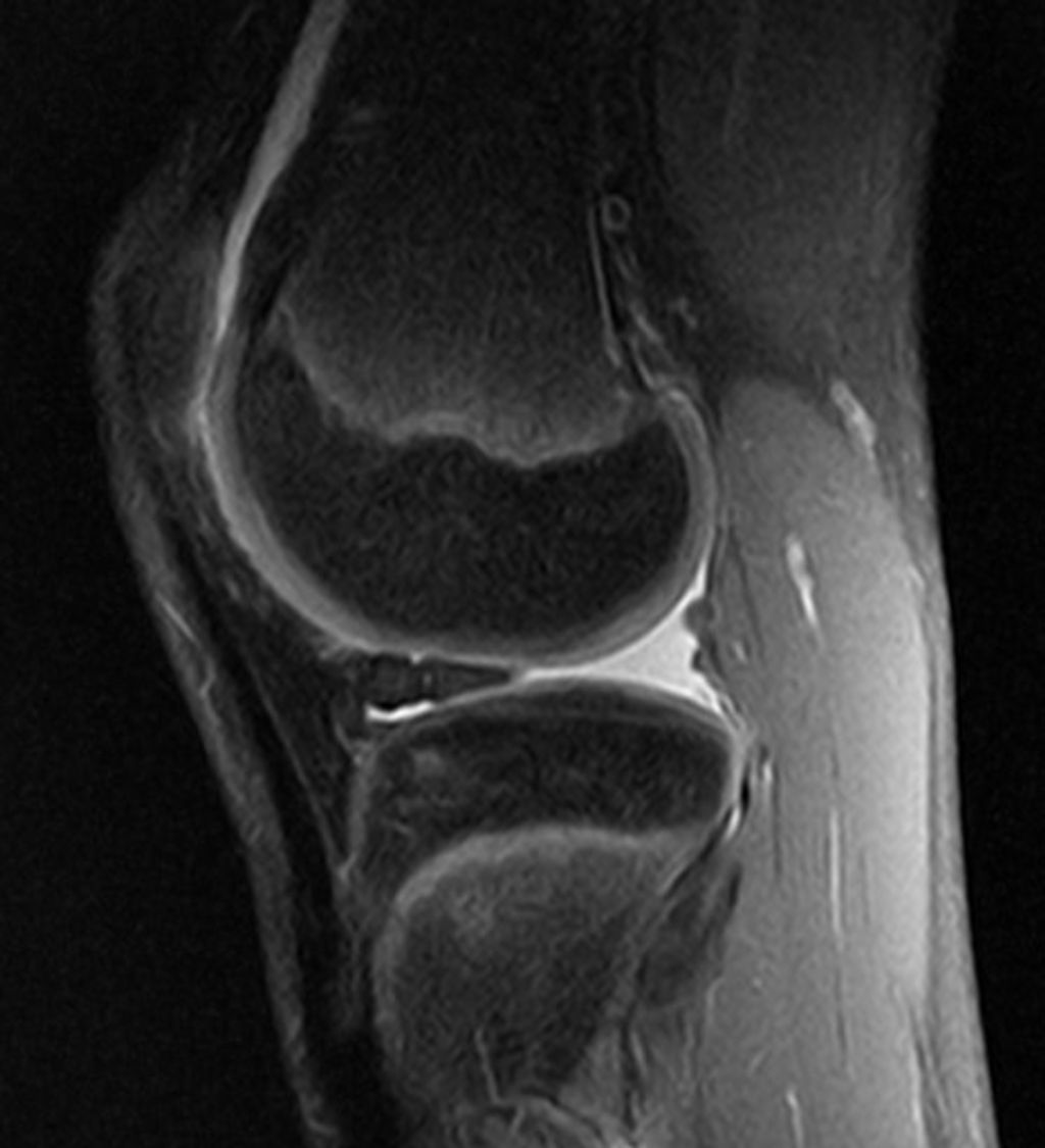
    Following a thorough clinical evaluation, we often turn to imaging studies to confirm the diagnosis of iliotibial syndrome. While MRI scans are not routinely used, they can be particularly helpful in visualizing the soft tissue structures and any associated inflammation of the iliotibial band. Ultrasound imaging is another tool at our disposal, offering the advantage of dynamic assessment while the joint is in motion.

    Typically, imaging is reserved for cases where the diagnosis is uncertain or if there is a lack of response to conservative treatment. In such scenarios, imaging can provide valuable insights into the extent of the injury and rule out other potential causes of lateral knee pain.

    Remember, imaging should complement, not replace, a comprehensive clinical evaluation.

    When interpreting imaging results, it’s crucial to correlate findings with the patient’s symptoms and physical exam results. This ensures a holistic approach to diagnosis and subsequent treatment planning.

    Treatment Options for Iliotibial Syndrome

    thumbnail

    Conservative Management

    After exploring various conservative management techniques, we have found that rest and stretching are crucial components of recovery. Additionally, we recommend incorporating strength training exercises to address muscle imbalances. Below is a table summarizing the recommended exercises and their frequency:

    ExerciseFrequency
    StretchingDaily
    Strength Training3 times a week

    We advise patients to gradually increase activity levels and to avoid overtraining, as this can exacerbate symptoms. It is important to monitor progress and adjust the treatment plan accordingly. We emphasize the significance of a comprehensive approach that addresses both the symptoms and underlying causes of the syndrome.

    Surgical Interventions

    After considering the various conservative management approaches, surgical interventions may be recommended for individuals with severe and persistent symptoms. These interventions typically involve the release or lengthening of the iliotibial band to alleviate tension and reduce friction. In some cases, a table summarizing the success rates and potential risks of surgical procedures may aid in decision-making. It’s important to note that surgical interventions should be considered as a last resort and only after thorough evaluation and discussion with a healthcare professional. Post-surgery, a comprehensive rehabilitation program is essential for optimal recovery and long-term success.

    Conclusion

    In conclusion, Iliotibial Syndrome is a complex condition that can be caused by a variety of factors, including overuse and biomechanical issues. Treatment typically involves a combination of physical therapy, rest, and targeted exercises to address muscle imbalances. It is important for individuals suffering from this syndrome to seek professional medical advice and adhere to a comprehensive treatment plan in order to achieve long-term relief and prevent recurrence.

    Frequently Asked Questions

    What are the common symptoms of Iliotibial Syndrome?

    Common symptoms include pain on the outside of the knee, swelling, and a clicking sensation during movement.

    Can Iliotibial Syndrome be prevented?

    Yes, it can be prevented by avoiding sudden increases in physical activity, maintaining proper running form, and using proper footwear.

    Is stretching helpful for Iliotibial Syndrome?

    Yes, stretching the iliotibial band and surrounding muscles can help reduce tension and alleviate symptoms.

    What are the risk factors for developing Iliotibial Syndrome?

    Risk factors include overuse, excessive running or cycling, improper footwear, and muscle imbalances.

    Can I continue physical activity if I have Iliotibial Syndrome?

    It is recommended to modify physical activity to avoid exacerbating symptoms, and to consult with a healthcare professional for personalized recommendations.

    How long does it take to recover from Iliotibial Syndrome?

    Recovery time varies depending on the severity of the condition and individual factors, but with proper treatment, most individuals can recover within a few weeks to a few months.

  • Prevention Strategies for Knee Ligament Injuries

    Prevention Strategies for Knee Ligament Injuries

    Prevention Strategies for Knee Ligament Injuries: Professional Tips

    Knee ligament injuries are common among athletes and can be a frustrating setback for anyone who enjoys an active lifestyle. The knee joint is one of the largest and most complex joints in the body, making it vulnerable to various types of injuries. Understanding and identifying knee ligament injuries is crucial to prevent further damage and promote quick recovery.

    v2 32d9h 7v8oi

    The most common knee ligament injuries are the anterior cruciate ligament (ACL), medial collateral ligament (MCL), posterior cruciate ligament (PCL), and lateral collateral ligament (LCL). These injuries can occur due to direct trauma, such as contusions and strains, or from indirect trauma, such as sudden changes in direction. Symptoms of knee ligament injuries include pain, swelling, inflammation, and joint laxity. In this article, we will discuss five prevention strategies for knee ligament injuries to help maintain knee stability and prevent knee instability.

    professional tips for prevention strategies to help avoid knee ligament injuries:

    CategoryTips
    Strength & Flexibility Training– Emphasize hamstring, glute, quad, hip & core exercises<br>- Ensure muscle imbalances are addressed<br>- Use proper form/technique to prevent overstress
    Movement Control & Balance– Practice proper landing mechanics when jumping <br>- Enhance proprioception & body control<br>- Integrate deceleration drills
    Equipment & Environment– Wear supportive & appropriate footwear <br>- Tape or brace vulnerable knees if needed <br>- Ensure safe, even playing surfaces
    Loading & Recovery– Follow loading guidelines for ramping up intensity<br>- Avoid aggressive pivoting until tissues adapted<br>- Schedule adequate rest periods between sessions
    Body Composition & Nutrition– Prevent muscle loss during injury layoffs<br>- Maintain healthy BMI year-round<br>- Support exercise recovery nutritionally
    Mindset & Motivation– Foster positive, confident mentality<br>- Set smaller achievable goal steps <br>- Enlist social support structure

    The most effective ACL/knee ligament injury prevention programs take a comprehensive approach. Optimizing strength, movement mechanics, loading, and nutrition helps create resilience. Confidence in knee stability paired with caution towards unsafe mechanics allows for safer progression. Expert guidance tailoring programs is key for best injury deterrence.

    9c0fad6b da19 4bad 92cb dac1d9705c7d

    Key Takeaways

    • Understanding the anatomy of the knee joint and identifying knee ligament injuries can promote quick recovery.
    • Direct and indirect trauma can cause knee ligament injuries, leading to pain, swelling, inflammation, and joint laxity.
    • Five prevention strategies can help maintain knee stability and prevent knee instability, reducing the risk of knee ligament injuries.

    Understanding and Identifying Knee Ligament Injuries

    Knee ligament injuries are common among athletes and individuals who engage in physical activities that require jumping, pivoting, or sudden changes in direction. In this section, we will discuss the common types and causes of knee ligament injuries, how to identify symptoms and diagnose the injury, treatment and rehabilitation options, and the role of sex and age in knee ligament injuries.

    Common Types and Causes

    The most common types of knee ligament injuries are the anterior cruciate ligament (ACL) and medial collateral ligament (MCL) injuries. The ACL is usually injured during non-contact activities such as landing after a jump or sudden stops and changes in direction. On the other hand, the MCL is usually injured from a direct blow to the outer side of the knee, such as when playing football or hockey.

    Symptoms and Diagnosis

    The symptoms of knee ligament injuries include pain, swelling, bruising, and instability. The diagnosis of knee ligament injuries is usually made through a physical exam and imaging tests such as MRI or X-rays. It is important to seek medical attention if you experience any of these symptoms.

    Treatment and Rehabilitation

    The treatment and rehabilitation of knee ligament injuries depend on the severity of the injury. Treatment options include rest, ice, compression, and elevation (RICE), physical therapy, and surgery. Rehabilitation includes exercises to improve range of motion, strength, and mobility. It is important to follow the guidance of a physical therapist or orthopedic surgeon to ensure proper recovery.

    Role of Sex and Age

    Research shows that females are more prone to knee ligament injuries than males due to differences in anatomy, form, and hormones such as estrogen and relaxin. Additionally, age plays a role in knee ligament injuries, with individuals over 50 years old being at a higher risk due to decreased muscle strength and flexibility.

    Sports and Injuries

    Knee ligament injuries are common in sports that require sudden stops and changes in direction such as soccer, basketball, and football (soccer). Injury prevention programs that include warm-up, neuromuscular training, and proprioception exercises can reduce the frequency of knee ligament injuries by up to 50%. Coaches and athletes should also prioritize proper technique, posture, and fatigue management to prevent knee ligament injuries.

    In conclusion, knee ligament injuries are common among athletes and individuals who engage in physical activities. Understanding the common types and causes of knee ligament injuries, identifying symptoms and diagnosing the injury, and following proper treatment and rehabilitation options can help prevent long-term damage. Coaches, athletes, and individuals should prioritize injury prevention programs and proper technique to reduce the frequency of knee ligament injuries.

    Prevention Strategies for Knee Ligament Injuries

    When it comes to knee ligament injuries, prevention is key. Here are some effective strategies that we recommend:

    Strengthening and Conditioning

    Strengthening and conditioning exercises can help improve muscle strength and endurance, which can help reduce the risk of knee ligament injuries. Exercises that target the hips, ankles, and core can be particularly effective in improving stability and reducing the risk of injury.

    Injury Prevention Programs

    Injury prevention programs can be an effective way to reduce the risk of knee ligament injuries. These programs typically include a combination of warm-up exercises, neuromuscular training, and strength and conditioning exercises. They can be particularly effective for athletes who participate in high-risk sports, such as soccer, basketball, and football.

    Importance of Compliance and Frequency

    Injury prevention strategies are only effective if they are followed consistently and frequently. Compliance with injury prevention programs is essential for reducing the risk of knee ligament injuries. It is important to follow the program as prescribed and to make sure that exercises are performed correctly and with good form.

    In conclusion, preventing knee ligament injuries requires a combination of strategies that target strength, stability, and flexibility. Injury prevention programs that incorporate warm-up exercises, neuromuscular training, and strength and conditioning exercises can be particularly effective. However, compliance with these programs is essential for success. By following these strategies, we can help reduce the risk of knee ligament injuries and keep athletes healthy and active.

  • Knee Ligament Injury Symptoms and Treatment

    Knee Ligament Injury Symptoms and Treatment

    As we all know, knee ligament injuries are common among athletes and people with an active lifestyle. These injuries can be caused by various factors such as sudden twisting or pivoting movements, direct impact, or overuse. Knee ligament injuries can be debilitating and may require immediate medical attention. In this article, we will discuss the symptoms and treatment of knee ligament injuries.

    v2 329cj 3ezlj

    Understanding Knee Ligament Injuries Knee ligament injuries are usually caused by a sudden twist or a direct impact on the knee joint. The most common types of knee ligament injuries are ACL (anterior cruciate ligament) and MCL (medial collateral ligament) injuries. Symptoms of knee ligament injuries include pain, swelling, instability, and difficulty walking. If you experience any of these symptoms, it is important to seek medical attention immediately.

    Diagnosis and Treatment The diagnosis of knee ligament injuries usually involves a physical examination, imaging tests such as X-rays and MRI, and arthroscopy. Treatment options for knee ligament injuries depend on the severity of the injury. Mild injuries can be treated with rest, ice, compression, and elevation (RICE) therapy, while more severe injuries may require surgery. Rehabilitation is also an important part of the treatment process, as it helps to restore strength and flexibility to the knee joint.

    knee ligament injury symptoms and treatment options:

    Ligament InjuredCommon SymptomsNonsurgical Treatment OptionsSurgical Treatment Options
    Anterior Cruciate Ligament (ACL)Instability of the knee, knee “giving way”, loss of full range of motion, swelling, pain with pivoting or decelerationRest, ice, compression, elevation, anti-inflammatory medication, bracing, physical therapy focusing on range of motion and strengtheningACL reconstruction surgery with autograft or allograft tissue
    Posterior Cruciate Ligament (PCL)Instability of the knee, swelling, pain with decelerationBracing, physical therapy, anti-inflammatory medicationPCL repair or reconstruction if symptoms do not improve with conservative treatment
    Medial Collateral Ligament (MCL)Pain along the inner knee, instability with valgus stress, swellingBracing, anti-inflammatory medication, physical therapySurgical repair or reconstruction for complete ruptures or severe instability
    Lateral Collateral Ligament (LCL)Pain along the outer knee, instability with varus stress, swellingBracing, anti-inflammatory medication, physical therapySurgical repair or reconstruction for complete ruptures or instability
    MeniscusCatching or locking of the knee, swelling, limited range of motionAnti-inflammatory medication, physical therapy, steroid injectionsMeniscectomy (removal of torn meniscus) or meniscal repair surgery

    The chosen treatment path depends greatly on injury severity, patient symptoms and functional limitations, and patient expectations. Many knee ligament injuries, especially mild sprains, can heal successfully without surgery. More severe instability often requires surgical reconstruction and extensive rehabilitation.

    Key Takeaways

    • Knee ligament injuries can be caused by sudden twisting or direct impact on the knee joint.
    • Symptoms of knee ligament injuries include pain, swelling, instability, and difficulty walking.
    • Treatment options for knee ligament injuries depend on the severity of the injury and may include RICE therapy, surgery, and rehabilitation.

    Understanding Knee Ligament Injuries

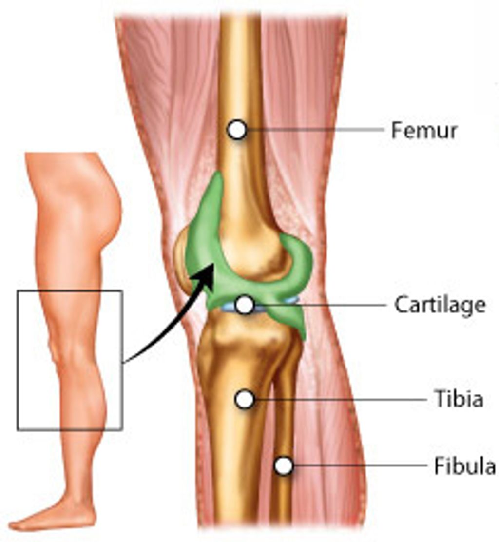
    Knee ligament injuries can be caused by trauma, such as a car accident or sports injuries. The knee has four major ligaments: the anterior cruciate ligament (ACL), the posterior cruciate ligament (PCL), the medial collateral ligament (MCL), and the lateral collateral ligament (LCL). These ligaments are elastic bands of tissue that connect the femur (thigh bone) to the tibia (shin bone) and provide stability and strength to the joint.

    Types of Knee Ligaments

    The ACL is located in the center of the knee and controls rotation and forward movement of the tibia. The PCL is located in the back of the knee and controls backward movement of the tibia. The MCL is located on the inside of the knee and helps to prevent the knee from bending inward. The LCL is located on the outside of the knee and helps to prevent the knee from bending outward.

    Causes and Risk Factors

    Knee ligament injuries can occur due to a blow to the knee, a sudden twisting motion, or a direct impact to the knee. Sports injuries, such as those that occur in basketball, football, skiing, and hockey, are common causes of knee ligament injuries. Female athletes are also at a higher risk of experiencing knee ligament injuries due to differences in anatomy and function.

    Injuries to the knee ligaments can cause pain, swelling, and deformity of the knee joint. A torn ligament can result in the knee feeling unstable and may require surgery to repair. Treatment for knee ligament injuries may include rest, ice, compression, and elevation (RICE), physical therapy, and surgery in severe cases.

    In conclusion, knee ligament injuries can be caused by trauma or sports injuries and can result in pain, swelling, and deformity of the knee joint. Treatment for knee ligament injuries may include RICE, physical therapy, and surgery in severe cases. It is important to seek medical attention if you suspect you have a knee ligament injury to prevent further damage to the joint.

    Diagnosis and Treatment

    When it comes to knee ligament injuries, prompt diagnosis and treatment are crucial to prevent further damage and ensure a full recovery. In this section, we will discuss the identification of symptoms, diagnostic procedures, treatment options, and prevention and rehabilitation methods.

    Identifying Symptoms

    The most common symptoms of knee ligament injuries include pain, swelling, and instability in the joint. Patients may also experience a popping or buckling sensation, stiffness, and clicking sounds when moving the knee. These signs and symptoms can vary depending on the type and severity of the injury.

    Diagnostic Procedures

    To diagnose a knee ligament injury, a physical exam is usually the first step. During the exam, a doctor will assess the range of motion, stability, and overall function of the joint. Imaging tests such as X-rays or magnetic resonance imaging (MRI) may also be necessary to confirm the diagnosis and determine the extent of the damage.

    Treatment Options

    The treatment for knee ligament injuries depends on the severity of the injury. In some cases, rest, ice, compression, and elevation (RICE) may be sufficient to reduce pain and swelling. Other treatments may include medication, physical therapy, and rehabilitation exercises to strengthen the muscles around the knee.

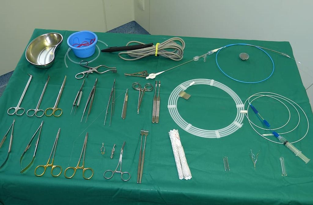
    For more severe injuries, knee surgery may be necessary. Arthroscopy is a minimally invasive surgical procedure that uses a small camera to view the inside of the knee joint and repair any damage. Knee surgery may also involve the use of a protective knee brace and grafts to replace damaged ligaments.

    Prevention and Rehabilitation

    Preventing knee ligament injuries involves taking precautions during physical activities and sports, such as wearing appropriate footwear and protective gear. Rehabilitation exercises and muscle-strengthening exercises can also help prevent future injuries and improve overall knee function.

    In conclusion, knee ligament injuries can be debilitating and require prompt medical attention. If you experience any signs or symptoms of a knee ligament injury, seek medical care from a qualified healthcare professional such as Dr. Andrew Cosgarea or Dr. Jay Lee. With proper diagnosis and treatment, most patients can recover fully and return to their normal activities.