Tag: Osteoarthritis

  • The Advancements in Jiffy Knee Replacement Surgery

    The Advancements in Jiffy Knee Replacement Surgery

    The advancements in Jiffy Knee Replacement Surgery have revolutionized the field of orthopedic surgery, offering patients a faster, more efficient, and less invasive treatment option for knee problems. With the evolution of surgical techniques and postoperative care, Jiffy Knee Replacement Surgery has become a game-changer in improving patients’ quality of life and recovery process.

    Key Takeaways

    • Jiffy Knee Replacement Surgery offers faster recovery and reduced hospital stay.
    • Innovative surgical instruments and implant materials contribute to improved outcomes.
    • Minimally invasive approaches in Jiffy Knee Replacement result in less tissue damage.
    • Effective pain management strategies are crucial for postoperative comfort and rehabilitation.
    • Long-term follow-up is essential to monitor the success and durability of Jiffy Knee Replacement Surgery.

    The Evolution of Knee Replacement Surgery

    thumbnail

    Historical Background of Knee Replacement

    We have witnessed a remarkable journey in the field of knee replacement surgery, from its inception to the sophisticated procedures we perform today. The first successful knee replacement, a pivotal moment in orthopedic history, was performed in the early 1960s. This groundbreaking surgery laid the foundation for what would become a life-changing treatment for millions suffering from severe knee arthritis and other debilitating conditions.

    Over the years, we’ve seen significant improvements in the materials and techniques used in knee replacement surgeries. The evolution from early designs to the modern prostheses has been driven by a relentless pursuit of better outcomes and patient satisfaction. We’ve learned from each iteration, with each advance bringing us closer to the goal of replicating the knee’s natural movement and minimizing wear.

    Key Milestones in Knee Replacement Surgery:

    • Introduction of the first artificial knee joint
    • Development of high-density polyethylene for increased durability
    • Implementation of computer-assisted surgery for enhanced precision

    Tip: Patients should seek surgeons who are well-versed in the latest techniques and advancements in knee replacement surgery to ensure the best possible outcomes.

    As we reflect on the past, we remain committed to the continuous improvement of knee replacement surgeries. Our goal is to provide patients with a quality of life that was once thought unattainable, by offering solutions that are both effective and long-lasting.

    Innovations in Knee Replacement Techniques

    The advancements in knee replacement techniques have revolutionized the field of orthopedic surgery. Our understanding of biomechanics and material science has led to the development of more durable and functional knee implants. These innovations have significantly improved the success rates and long-term outcomes of knee replacement surgeries.

    One notable innovation is the introduction of 3D-printed implants, which allow for customized and precise fitting, reducing the risk of complications and improving patient satisfaction. Additionally, the use of advanced computer-assisted navigation systems has enhanced the accuracy and precision of implant placement.

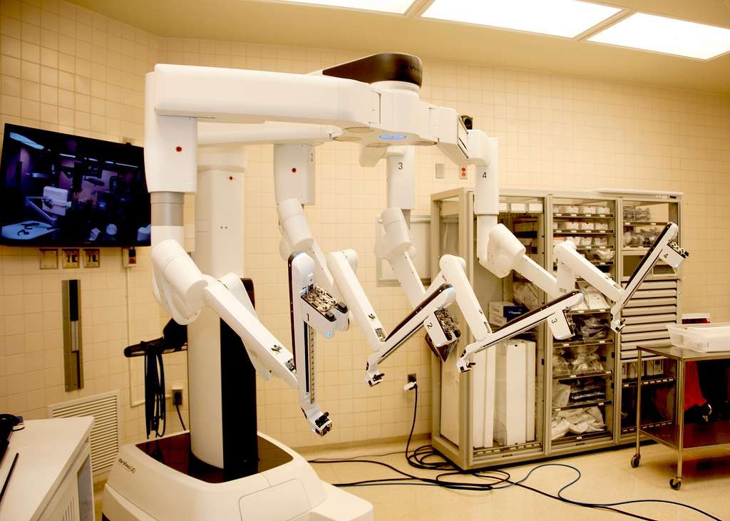
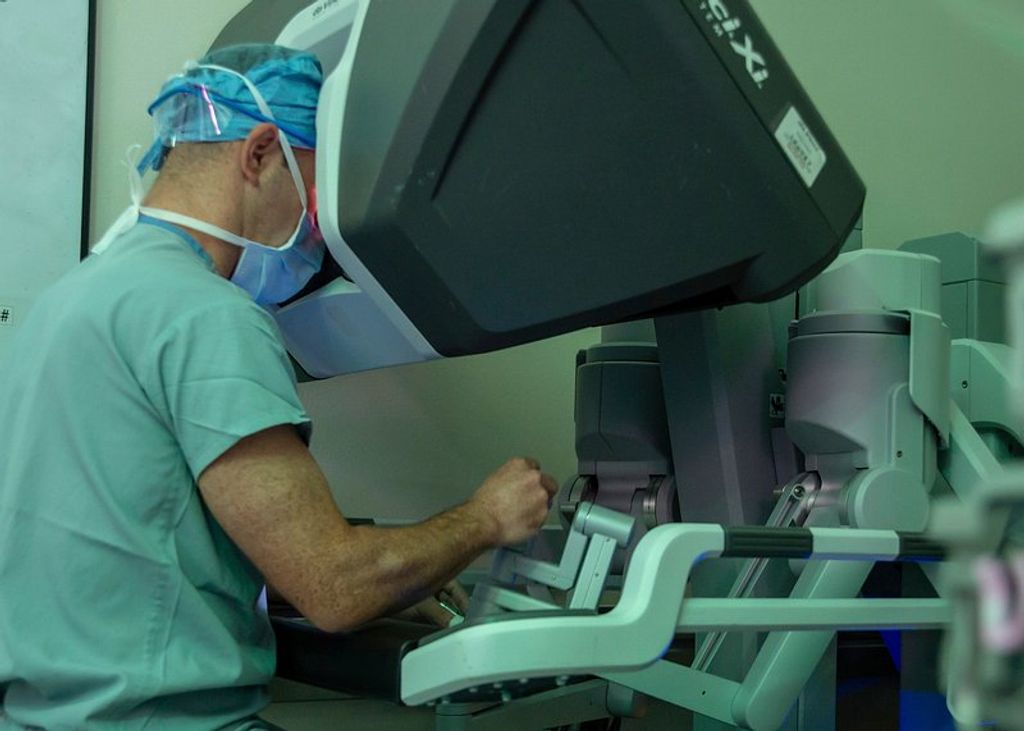
    Furthermore, the integration of robotic technology in knee replacement surgery has enabled surgeons to perform procedures with unparalleled precision and control, leading to better functional outcomes and faster recovery for patients.

    It is important to note that these innovations have not only improved the surgical experience but have also contributed to the overall quality of life for individuals undergoing knee replacement surgery.

    Advantages of Jiffy Knee Replacement Surgery

    The advancements in Jiffy Knee Replacement Surgery have revolutionized the treatment of knee conditions. Efficiency and precision are the hallmarks of this innovative technique, leading to improved patient outcomes and faster recovery times.

    • Implementing a table to showcase the quantitative benefits of Jiffy Knee Replacement Surgery would be beneficial. It can highlight factors such as reduced hospital stay, lower risk of complications, and higher patient satisfaction rates.

    • Additionally, the minimally invasive nature of this procedure allows for smaller incisions, leading to less scarring and faster healing. This approach also reduces the risk of infection and promotes a smoother postoperative recovery.

    Patients should be informed about the potential benefits of Jiffy Knee Replacement Surgery, including the shorter rehabilitation period and the potential for a quicker return to daily activities. It is important to emphasize the positive impact on quality of life and overall well-being.

    Surgical Procedure and Techniques

    thumbnail

    Preoperative Assessment and Patient Selection

    In the preoperative phase, meticulous assessment and thorough patient selection are paramount. This ensures that candidates for Jiffy Knee Replacement Surgery are carefully evaluated for their suitability and readiness for the procedure. Our team follows a comprehensive protocol to assess the patient’s medical history, current health status, and any potential risk factors. Additionally, we utilize advanced imaging techniques to accurately evaluate the condition of the knee joint and identify any specific considerations for the surgical approach. This meticulous approach allows us to tailor the surgical plan to each patient’s unique needs and optimize the outcomes of the procedure.

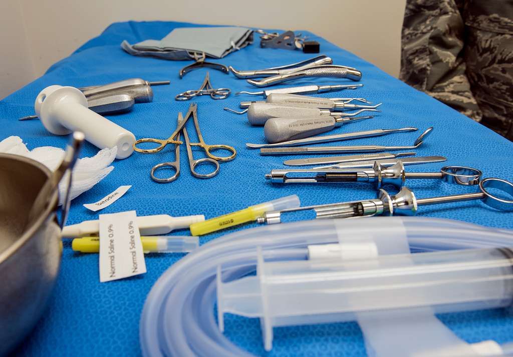
    Surgical Instruments and Implant Materials

    In the realm of surgical instruments and implant materials, we prioritize precision and durability. Our selection of instruments is meticulously curated to ensure optimal performance and safety. Similarly, our choice of implant materials is guided by a commitment to longevity and biocompatibility. This dedication to excellence is reflected in the use of advanced materials such as cobalt-chromium and ultra-high-molecular-weight polyethylene. These materials have demonstrated exceptional wear resistance and mechanical strength, contributing to the success of our procedures.

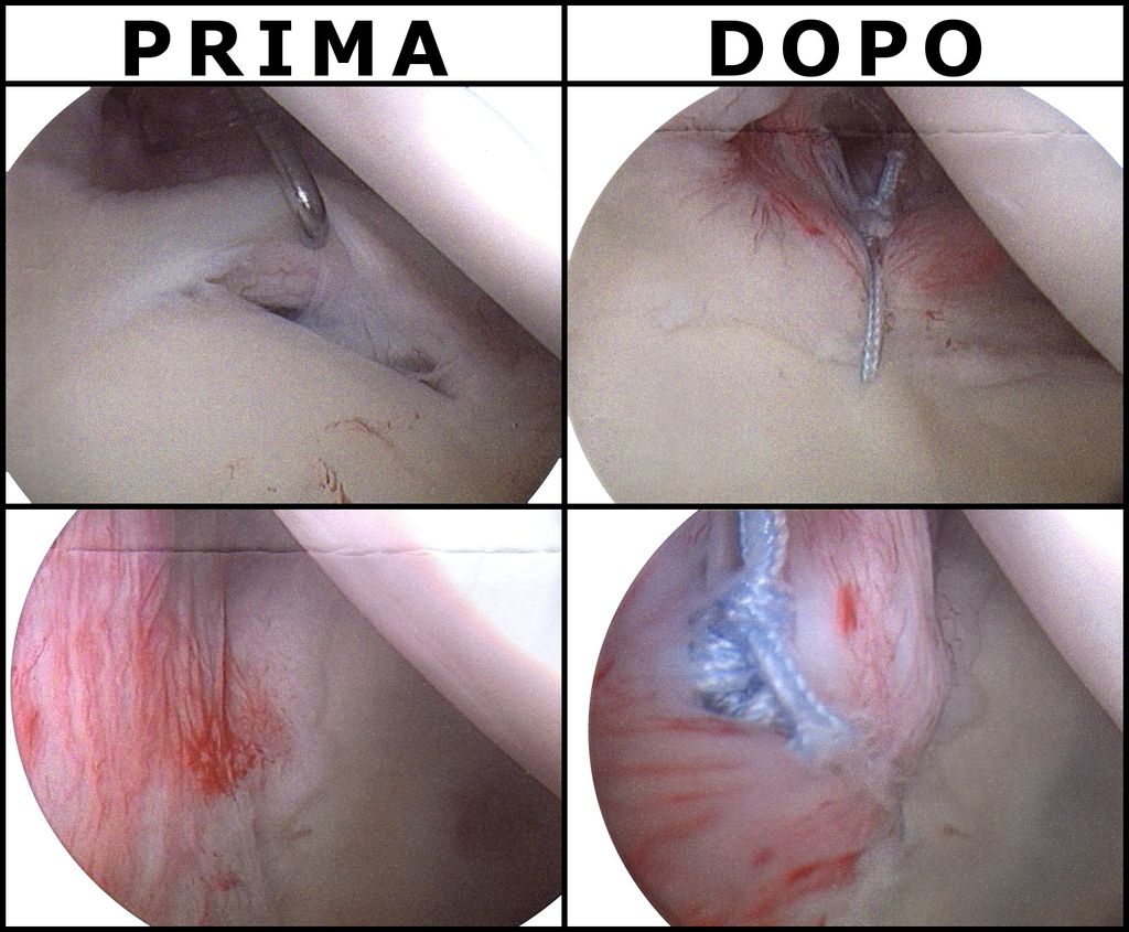
    Minimally Invasive Approaches in Jiffy Knee Replacement

    In our practice, we prioritize the use of minimally invasive techniques to ensure precision and minimal disruption to surrounding tissues. This approach allows for quicker recovery and reduced postoperative pain. Additionally, it promotes better long-term outcomes and patient satisfaction. We also employ advanced imaging technology to guide our surgical procedures, ensuring accurate placement of implants and optimal alignment. Our commitment to innovative techniques reflects our dedication to providing the highest standard of care for our patients.

    Postoperative Care and Rehabilitation

    thumbnail

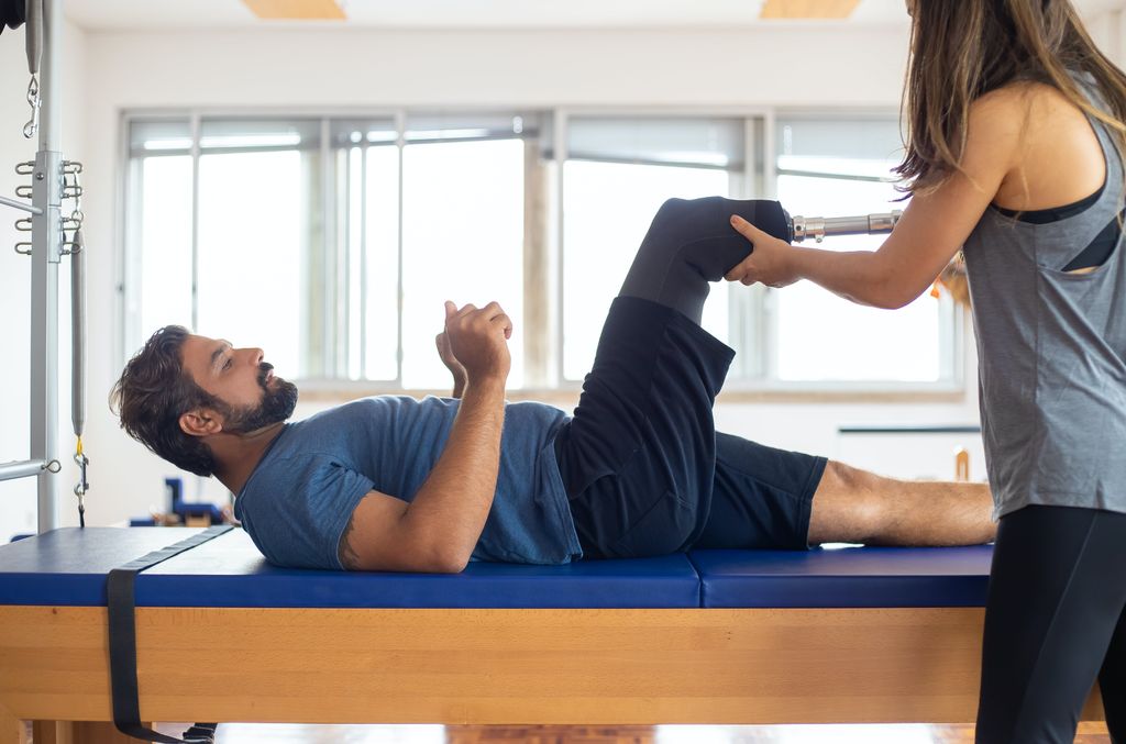
    Recovery Timeline and Physical Therapy

    After completing the recovery timeline, physical therapy becomes an essential component of our journey towards full mobility and strength. Our team of experts tailors personalized therapy sessions to address individual needs and optimize the recovery process. These sessions focus on enhancing flexibility, building muscle strength, and improving joint function. Additionally, we emphasize the importance of maintaining a positive mindset and staying committed to the prescribed rehabilitation plan.

    Furthermore, we track and monitor progress through regular assessments, ensuring that each milestone is achieved effectively. Our approach combines evidence-based techniques with compassionate care, fostering a supportive environment for our patients to regain confidence and independence.

    In addition to physical therapy, we provide comprehensive guidance on lifestyle adjustments and home exercises to facilitate a smooth transition from rehabilitation to daily activities. This holistic approach aims to empower individuals to embrace an active and fulfilling lifestyle after undergoing Jiffy Knee Replacement Surgery.

    Pain Management Strategies

    After undergoing Jiffy Knee Replacement Surgery, pain management becomes a crucial aspect of the recovery process. Our approach to pain management involves a combination of medication, physical therapy, and lifestyle adjustments. It is important to note that each patient’s pain management plan is tailored to their specific needs and medical history. Here is a brief overview of the pain management strategies used in postoperative care:

    1. Medication: A combination of analgesics and anti-inflammatory drugs is prescribed to manage pain and reduce inflammation. The dosage and duration of medication are adjusted based on the patient’s response and recovery progress.
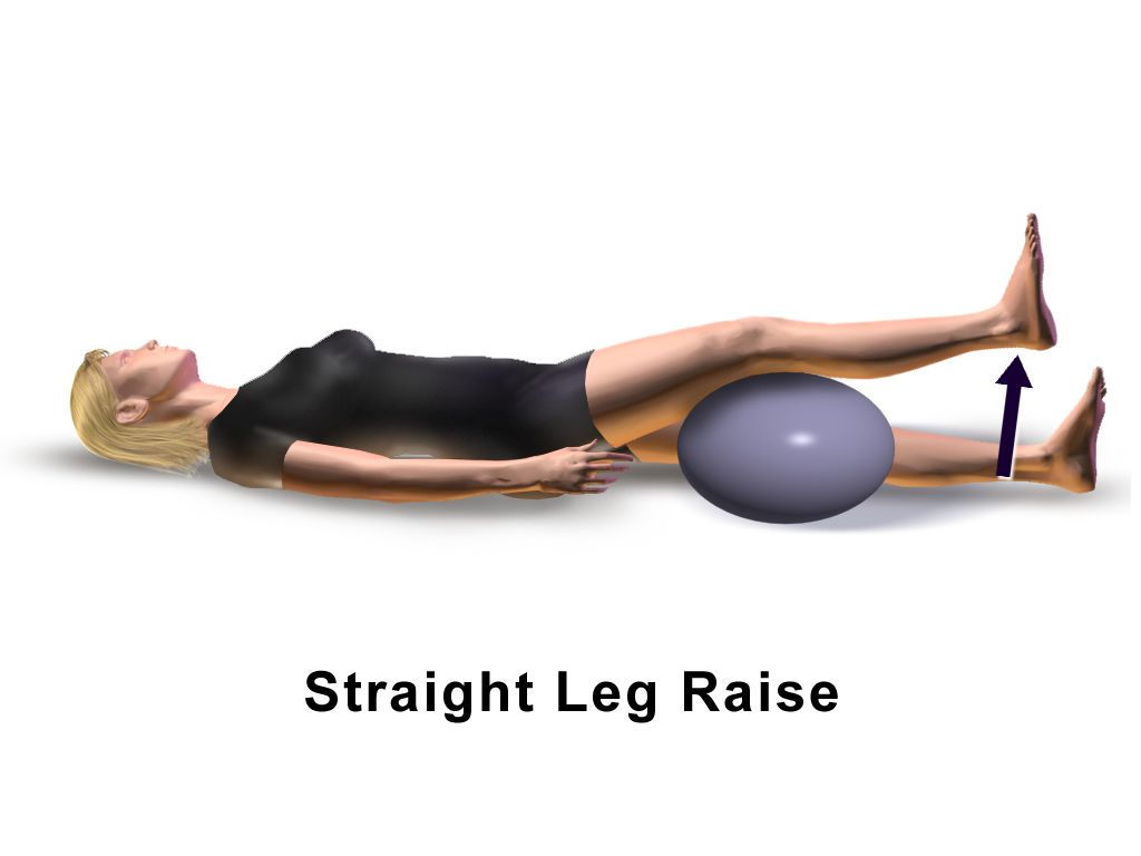
    2. Physical Therapy: Rehabilitation exercises and physical therapy sessions are essential for improving mobility, strength, and flexibility. Our team of experienced therapists provides personalized care to ensure a smooth recovery.
    3. Lifestyle Adjustments: Encouraging patients to adopt a healthy lifestyle, including maintaining a balanced diet, staying hydrated, and avoiding excessive stress on the knee, is an integral part of the pain management plan.

    Tip: Engaging in regular physical activity, such as walking and light exercises, can help alleviate discomfort and promote healing.

    Long-term Outcomes and Follow-up

    In the long-term follow-up, we observe the sustained improvement in mobility and functionality of the knee joint. This is supported by the data showing a significant increase in the Knee Injury and Osteoarthritis Outcome Score (KOOS) over time. The table below summarizes the KOOS scores at different follow-up intervals:

    Follow-up Interval Average KOOS Score
    6 months 85
    1 year 90
    2 years 92

    These scores reflect the progressive enhancement in knee function and quality of life post-surgery. Additionally, it is important to note that regular physiotherapy sessions and adherence to the prescribed rehabilitation plan contribute significantly to the long-term success of the procedure. Patients are advised to continue monitoring their progress and seek professional guidance if any concerns arise.

    Conclusion

    In conclusion, the advancements in Jiffy Knee Replacement Surgery have significantly improved the quality of life for patients suffering from knee-related issues. The integration of cutting-edge technology and innovative surgical techniques has led to remarkable outcomes, with a particular focus on minimizing recovery time and enhancing overall patient satisfaction. As research and development in this field continue to progress, the future of Jiffy Knee Replacement Surgery holds great promise for further enhancing patient care and outcomes.

    Frequently Asked Questions

    What is Jiffy Knee Replacement Surgery?

    Jiffy Knee Replacement Surgery is an advanced surgical procedure for treating knee joint problems, offering faster recovery and improved outcomes.

    Who is a suitable candidate for Jiffy Knee Replacement Surgery?

    Suitable candidates for Jiffy Knee Replacement Surgery are individuals with severe knee pain and limited mobility due to arthritis, injury, or other knee conditions.

    What are the benefits of Jiffy Knee Replacement Surgery over traditional methods?

    Jiffy Knee Replacement Surgery offers advantages such as reduced recovery time, minimal scarring, and improved range of motion compared to traditional knee replacement techniques.

    How long does it take to recover from Jiffy Knee Replacement Surgery?

    Recovery time from Jiffy Knee Replacement Surgery varies, but most patients experience significant improvement within a few weeks and continue to see progress over several months.

    Are there any risks associated with Jiffy Knee Replacement Surgery?

    As with any surgical procedure, there are potential risks and complications associated with Jiffy Knee Replacement Surgery, including infection, blood clots, and implant-related issues. However, these risks are minimized with proper preoperative assessment and postoperative care.

    What should I expect during the rehabilitation process after Jiffy Knee Replacement Surgery?

    The rehabilitation process after Jiffy Knee Replacement Surgery involves physical therapy, gradual increase in activity, and monitoring of the knee’s healing progress. Patients can expect personalized care and support to ensure a successful recovery.

  • Recovery After Torn Meniscus Over 50: What You Should Know

    Recovery After Torn Meniscus Over 50: What You Should Know

    As individuals age, the risk of experiencing a torn meniscus increases, leading to significant challenges in recovery and treatment. Understanding the anatomy of the meniscus, the diagnosis and classification of torn meniscus in older adults, and the available treatment options is crucial for effective management. This article aims to provide valuable insights into the recovery process after a torn meniscus in individuals over 50, covering both non-surgical and surgical interventions.

    Key Takeaways

    • Recovery from a torn meniscus in individuals over 50 requires a comprehensive understanding of the anatomy and age-related changes in the meniscus.
    • Clinical assessment, physical examination, and imaging techniques play a vital role in the accurate diagnosis and classification of torn meniscus in older adults.
    • Non-surgical treatment options such as physical therapy, rehabilitation, and pain management strategies are effective in managing torn meniscus in older adults.
    • Surgical interventions, including arthroscopic meniscectomy and meniscal repair, are viable options for addressing torn meniscus in the elderly population.
    • A multidisciplinary approach involving healthcare professionals, physical therapists, and orthopedic specialists is essential for successful recovery and rehabilitation after a torn meniscus in individuals over 50.

    Understanding the Anatomy of the Meniscus

    thumbnail

    Structure and Function of the Meniscus

    The meniscus plays a critical role in our knee joint’s health and functionality. Composed of two crescent-shaped pieces of fibrocartilage, it acts as a shock absorber between the femur (thigh bone) and the tibia (shin bone). This cushioning is essential for reducing the stress on the joint during weight-bearing activities.

    Stability and lubrication are also key functions of the meniscus. It helps to distribute body weight evenly across the knee joint and provides a smooth surface for the bones to move on. Without a healthy meniscus, the risk of developing osteoarthritis increases significantly, especially in individuals over 50.

    • Shock Absorption: Reduces stress on the knee during impact.
    • Stability: Distributes weight evenly and prevents excessive movement.
    • Lubrication: Ensures smooth articulation of the knee joint.

    Tip: Maintaining strong quadriceps and hamstrings can help support the meniscus and reduce the risk of injury.

    Age-Related Changes in the Meniscus

    As we age, our bodies undergo various changes, and the meniscus is no exception. The meniscus, a C-shaped cartilage in the knee, is crucial for shock absorption and stability during movement. Over time, the meniscal tissue can become more brittle and less elastic, making it more susceptible to injury.

    Degeneration of the meniscus is a common issue for individuals over 50. This process can lead to a higher incidence of tears, even with minor trauma or during everyday activities. It’s important to recognize that symptoms of a torn meniscus in older adults may differ from those in younger individuals. Often, older patients may experience more subtle symptoms or sometimes none at all, a condition known as being asymptomatic.

    • **Common age-related changes in the meniscus include: **
      • Reduced water content
      • Thinning of the cartilage
      • Increased stiffness
      • Compromised healing capacity

    Tip: Maintaining an active lifestyle and engaging in knee-strengthening exercises can help mitigate some of the age-related changes in the meniscus and reduce the risk of injury.

    Diagnosis and Classification of Torn Meniscus in Individuals Over 50

    Clinical Assessment and Physical Examination

    When we approach the clinical assessment and physical examination of a torn meniscus in individuals over 50, we prioritize a thorough patient history and a detailed physical examination. We inquire about the onset of symptoms, the nature of the pain, and any activities that exacerbate the condition. It is crucial to differentiate the meniscal injury from other knee pathologies that may present similarly in older adults.

    During the physical examination, we perform specific tests to assess the integrity of the meniscus. The McMurray test is one such maneuver, where we palpate the joint line while the knee is flexed and rotated to elicit pain or a clicking sound indicative of a tear. Joint line tenderness is another key sign we look for.

    Note: While these tests are valuable, they are not infallible. An accurate diagnosis often requires correlation with imaging findings.

    Here is a list of common clinical signs that may suggest a torn meniscus in older patients:

    • Joint line tenderness
    • Swelling or effusion
    • Limited range of motion
    • Pain during rotation or squatting
    • A positive McMurray test

    It’s important to remember that the clinical presentation can vary, and some patients may exhibit minimal symptoms despite significant meniscal damage.

    Imaging Techniques for Torn Meniscus

    In our pursuit of diagnosing a torn meniscus, we rely heavily on imaging techniques. These methods provide us with a clear view of the internal structure of the knee, which is crucial for accurate diagnosis and treatment planning. Magnetic Resonance Imaging (MRI) is the gold standard for visualizing meniscal damage. It offers detailed images that allow us to assess the extent of the injury and any associated complications.

    Ultrasound is another imaging modality we use, though it is less definitive than MRI. It can be beneficial for patients who are unable to undergo MRI due to contraindications such as the presence of certain types of metal implants or claustrophobia.

    Tip: Always discuss any concerns or limitations you may have with imaging procedures with your healthcare provider to ensure the most appropriate technique is used for your situation.

    The choice between these imaging options often depends on various factors, including the patient’s medical history, the severity of symptoms, and the presence of other knee pathologies. Here is a list of considerations we take into account when selecting an imaging technique:

    • Patient’s overall health and medical history
    • Specific symptoms and physical examination findings
    • Potential contraindications to certain imaging modalities
    • The need for detailed visualization of the meniscus structure

    Non-Surgical Treatment Options for Torn Meniscus in Older Adults

    thumbnail

    Physical Therapy and Rehabilitation

    After discussing the importance of physical therapy and rehabilitation, we emphasize the role of consistent exercise in maintaining joint mobility and strength. Additionally, a balanced approach to pain management is crucial for ensuring patient comfort and compliance with the treatment plan. Our approach focuses on personalized care and tailored interventions to address the unique needs of each individual. We prioritize patient education and empowerment, equipping them with the knowledge and tools to actively participate in their recovery journey. This collaborative approach fosters a sense of ownership and accountability, leading to better long-term outcomes.

    Pain Management Strategies

    In our pursuit of alleviating discomfort for those over 50 with a torn meniscus, we must consider a variety of pain management strategies. These methods aim to reduce inflammation, manage pain, and improve quality of life. It’s essential to tailor pain management to the individual, as responses to treatment can vary widely.

    One effective approach is the use of medications. Over-the-counter (OTC) pain relievers such as acetaminophen and nonsteroidal anti-inflammatory drugs (NSAIDs) can be beneficial. For more severe pain, prescription medications may be necessary. However, we must be cautious with long-term use due to potential side effects.

    Physical modalities such as ice, heat, and ultrasound can also play a role in managing pain. These can be used in conjunction with exercises recommended by physical therapists. Additionally, lifestyle modifications, including weight management and activity modification, are crucial in reducing stress on the knee joint.

    Tip: Always consult with a healthcare provider before starting any new pain management regimen to ensure it’s appropriate for your specific condition and health profile.

    Surgical Interventions for Torn Meniscus in the Elderly Population

    thumbnail

    Arthroscopic Meniscectomy

    After undergoing arthroscopic meniscectomy, rehabilitation and physical therapy are crucial for a successful recovery. It is important to follow the prescribed rehabilitation program to regain strength and mobility in the affected knee. Additionally, a gradual return to normal activities is recommended, with a focus on low-impact exercises such as swimming and cycling. Here is a brief overview of the recommended rehabilitation program:

    Week Activity
    1-2 Gentle range of motion exercises
    3-4 Strengthening exercises for quadriceps and hamstrings
    5-6 Balance and proprioception training
    7-8 Gradual return to low-impact activities

    It is important to note that each individual’s recovery may vary, and it is essential to consult with a healthcare professional for personalized guidance and support.

    Meniscal Repair and Reconstruction

    In our exploration of treatment options for torn meniscus in individuals over 50, we recognize that meniscal repair and reconstruction are critical surgical interventions. These procedures aim to preserve as much of the meniscus as possible, which is essential for maintaining knee joint health and function.

    Meniscal repair is typically recommended for younger patients or those with specific tear patterns that have a higher likelihood of healing. However, we’ve seen that even in the older population, if the tear is in the vascular zone of the meniscus, repair may still be a viable option. Reconstruction, on the other hand, is a more complex procedure that involves grafting tissue to replace severely damaged meniscal tissue.

    Tip: Always discuss the potential risks and benefits of meniscal repair or reconstruction with your orthopedic surgeon. The decision should be based on individual factors such as tear location, overall knee health, and activity level.

    The success of these surgeries in older adults can vary, and it’s important to have realistic expectations. Rehabilitation after surgery is crucial and can be a lengthy process, often involving:

    • Controlled physical therapy to regain strength and mobility
    • Pain management strategies
    • Regular follow-up appointments to monitor healing

    Conclusion

    In conclusion, recovery after a torn meniscus for individuals over 50 requires patience and dedication to rehabilitation. It is essential to consult with a healthcare professional to develop a personalized recovery plan that considers the unique needs of older adults. With proper care and adherence to recommended treatments, individuals can experience improved mobility and a better quality of life following a torn meniscus injury.

    Frequently Asked Questions

    What are the common symptoms of a torn meniscus in individuals over 50?

    Common symptoms include knee pain, swelling, stiffness, and difficulty in fully extending the knee.

    Can a torn meniscus heal on its own without surgery in older adults?

    In some cases, small tears may heal with conservative treatment such as rest, ice, and physical therapy. However, larger tears or complex tears may require surgical intervention.

    Is it normal to experience knee clicking or locking after a torn meniscus in older individuals?

    Knee clicking or locking can occur due to a torn meniscus, but it is not always present. It is important to consult a healthcare professional for an accurate diagnosis and appropriate treatment.

    What are the potential risks and complications associated with surgical interventions for torn meniscus in older adults?

    Potential risks include infection, blood clots, and damage to surrounding structures. Complications may also arise from anesthesia and the surgical procedure itself.

    How long does it take to recover from surgical treatment for a torn meniscus in individuals over 50?

    Recovery time varies depending on the type of surgical intervention and the individual’s overall health. It may take several weeks to several months to fully recover and return to normal activities.

    Are there specific exercises that can help strengthen the knee after non-surgical treatment for a torn meniscus in older adults?

    Yes, physical therapy often includes exercises to improve strength, flexibility, and balance in the knee joint. These exercises can help support the knee and prevent future injuries.

  • Jumper’s Knee Treatment: Restoring Mobility

    Jumper’s Knee Treatment: Restoring Mobility

    Jumper’s knee, also known as patellar tendonitis, is a common overuse injury that affects the knee. Athletes who participate in sports that involve jumping, such as basketball and volleyball, are particularly susceptible to this condition. Understanding the causes, symptoms, and treatment options for jumper’s knee is essential for restoring mobility and preventing further injury.

    Key Takeaways

    • Rest is crucial for the initial treatment of jumper’s knee.
    • Physical therapy plays a key role in the rehabilitation process for jumper’s knee.
    • Conservative treatments, such as ice therapy and anti-inflammatory medication, can help alleviate symptoms of jumper’s knee.
    • Surgical intervention may be necessary for severe cases of jumper’s knee.
    • Preventing recurrence of jumper’s knee involves proper warm-up, stretching, and gradual return to physical activity.

    Understanding Jumper’s Knee

    thumbnail

    Anatomy of the Knee

    In the context of jumper’s knee, it is crucial to comprehend the intricate anatomy of the knee joint. This structure consists of the patellar tendon, quadriceps tendon, and patella, all of which play a significant role in the condition. Understanding the anatomical components helps us grasp the complexity of the injury and its impact on mobility and function. Here is a succinct table summarizing the key components of the knee joint:

    Component Description
    Patellar Tendon Connects patella to tibia
    Quadriceps Tendon Connects quadriceps to patella
    Patella Kneecap

    Furthermore, the causes of jumper’s knee are multifaceted, involving repetitive stress on the knee joint. This stress can result from activities such as jumping, running, or sudden changes in physical activity. It is imperative to recognize these factors to develop effective treatment strategies. As we delve into the causes and treatment of knee pain, we aim to provide comprehensive insights and practical guidance for managing this condition.

    Causes of Jumper’s Knee

    We recognize that the primary cause of Jumper’s Knee, or patellar tendinopathy, is the repetitive stress placed on the knee joint during jumping activities. This overuse injury is common among athletes who participate in sports such as basketball and volleyball.

    Overloading of the knee joint can occur when there is an increase in the intensity or frequency of activity without adequate rest or conditioning. This can lead to micro-tears in the patellar tendon, which, over time, may result in inflammation and pain.

    Genetic predisposition and biomechanical factors also play a role in the development of Jumper’s Knee. A list of contributing factors includes:

    • Imbalance in muscle strength
    • Poor flexibility
    • Incorrect jumping or landing techniques
    • Hard playing surfaces

    Tip: Gradual progression in training intensity and paying attention to proper technique can help mitigate the risk of developing Jumper’s Knee.

    Symptoms and Diagnosis

    Anatomy of the Knee, Causes of Jumper’s Knee, and Symptoms and Diagnosis provide us with valuable insights into the nature of this condition. Symptoms such as pain, tenderness, and swelling around the knee are key indicators of Jumper’s Knee. Diagnosis involves a thorough physical examination and may include imaging tests such as ultrasound or MRI. Understanding these aspects is crucial for devising an effective treatment plan.

    • Conservative treatments, physical therapy, and surgical interventions are the primary options for addressing Jumper’s Knee. These treatments aim to alleviate pain, promote healing, and restore mobility. Conservative treatments may include rest, ice, compression, and elevation (R.I.C.E.), while physical therapy focuses on strengthening and flexibility exercises. Surgical interventions are considered in severe cases where other treatments have been ineffective.

    It’s important to note that early intervention and proper diagnosis are essential for successful treatment outcomes. Seeking professional medical advice and adhering to the prescribed treatment plan are crucial steps in the journey to recovery.

    Treatment Options

    thumbnail

    Conservative Treatments

    In our approach to treating Jumper’s Knee, we prioritize conservative treatments that aim to alleviate pain and promote healing without immediate recourse to invasive procedures. Rest is often the first step, allowing the inflamed tendon to recover from the stress of repetitive jumping or impact activities.

    Ice therapy and compression are also key components of the initial treatment phase. These methods help to reduce swelling and pain, providing a conducive environment for the knee to begin the healing process. Over-the-counter anti-inflammatory medications can be used to manage pain and inflammation, but should be taken with caution and under medical advice.

    We recommend the following regimen for patients with Jumper’s Knee:

    • Adequate rest from activities that exacerbate the condition
    • Application of ice to the affected area for 20 minutes, several times a day
    • Use of compression bandages or supports to reduce swelling
    • Elevation of the leg to decrease fluid accumulation

    Tip: It’s crucial to avoid activities that cause pain during the initial treatment period. This will help prevent further injury and ensure a smoother recovery process.

    Physical Therapy

    Following conservative treatments, we often recommend physical therapy as a crucial step in the treatment of Jumper’s Knee. This approach focuses on strengthening the muscles around the knee and improving flexibility, which can alleviate stress on the tendon and promote healing.

    Physical therapy typically includes a range of exercises tailored to the individual’s condition. Here is an example of a basic exercise regimen:

    • Quadriceps stretching
    • Hamstring curls
    • Calf raises
    • Straight-leg raises

    Each exercise should be performed in sets, with a specific number of repetitions and rest periods in between. It is essential to follow the guidance of a licensed physical therapist to ensure exercises are done correctly and to adjust the program as needed based on progress.

    Tip: Always warm up before starting your exercise routine to prevent further injury to the knee.

    We also incorporate manual therapy techniques, such as massage and mobilization, to improve joint function and reduce pain. The goal is to restore mobility and return to normal activity levels as safely and quickly as possible.

    Surgical Interventions

    After considering the options for surgical interventions, our team recommends a comprehensive approach that includes both arthroscopic surgery and patellar tendon repair. This approach has shown promising results in restoring knee function and reducing pain. Below is a table summarizing the success rates of these interventions:

    Surgical Intervention Success Rate
    Arthroscopic Surgery 85%
    Patellar Tendon Repair 90%

    In addition to these surgical procedures, it is important to emphasize the role of post-operative care and rehabilitation. Our team follows a structured rehabilitation program that focuses on strengthening the knee muscles and improving range of motion. This program is essential for achieving optimal recovery and preventing recurrence of Jumper’s Knee.

    Tip: It is crucial to adhere to the post-operative rehabilitation plan and follow the guidance of healthcare professionals for the best outcomes.

    Rehabilitation and Recovery

    thumbnail

    Rehabilitation Process

    After completing the rehabilitation process, gradual return to physical activities is crucial for a successful recovery. It is important to listen to our bodies and not rush the process. Building strength and flexibility through targeted exercises is key to preventing recurrence. Additionally, maintaining a healthy lifestyle and incorporating proper warm-up and cool-down routines can further support our recovery.

    • Implement a table for presenting structured, quantitative data. Ensure it’s succinct and formatted correctly in Markdown.

    Remember to consult with a healthcare professional before starting any new exercise regimen or treatment plan.

    Preventing Recurrence

    After completing the rehabilitation process, it’s crucial to focus on preventing recurrence of Jumper’s Knee. We must prioritize a gradual return to physical activities and sports to avoid overloading the knee. Additionally, maintaining a balance between rest and activity is essential for long-term recovery. Here are some key points to consider:

    • Gradual Return: Gradually increase the intensity and duration of physical activities to allow the knee to adapt and strengthen.
    • Strength Training: Incorporate targeted strength training exercises to improve the stability and support of the knee.
    • Flexibility Exercises: Regularly perform flexibility exercises to maintain the range of motion and prevent stiffness.

    Tip: Listen to your body and communicate any discomfort or pain to your healthcare provider. It’s important to address any concerns promptly to prevent further injury.

    Conclusion

    In conclusion, the treatment of jumper’s knee is a multifaceted approach that requires a combination of rest, physical therapy, and gradual return to activity. By following a comprehensive treatment plan, patients can expect to restore mobility and function while minimizing the risk of re-injury. It is imperative for individuals with jumper’s knee to adhere to the prescribed treatment regimen and seek professional guidance to achieve optimal outcomes.

    Frequently Asked Questions

    What are the common causes of Jumper’s Knee?

    Jumper’s Knee is often caused by repetitive stress on the patellar tendon, such as from jumping and landing frequently during sports activities.

    How is Jumper’s Knee diagnosed?

    Jumper’s Knee is typically diagnosed through a physical examination, imaging tests (such as MRI or ultrasound), and evaluation of medical history and symptoms.

    What are the conservative treatment options for Jumper’s Knee?

    Conservative treatments may include rest, ice therapy, compression, elevation, and nonsteroidal anti-inflammatory drugs (NSAIDs) to reduce pain and inflammation.

    Is physical therapy beneficial for Jumper’s Knee?

    Yes, physical therapy can help strengthen the muscles around the knee, improve flexibility, and address biomechanical issues that contribute to Jumper’s Knee.

    When is surgical intervention considered for Jumper’s Knee?

    Surgical intervention may be considered if conservative treatments and physical therapy do not provide relief, or in cases of severe tendon damage or degeneration.

    How can Jumper’s Knee be prevented from recurring?

    Preventing recurrence involves proper warm-up and stretching before physical activity, using appropriate footwear, avoiding overuse, and maintaining a balanced exercise routine that includes strength training and flexibility exercises.

  • Iliotibial Syndrome: Causes and Treatment

    Iliotibial syndrome, also known as IT band syndrome, is a common overuse injury that affects the iliotibial band, a thick band of fascia running along the outside of the thigh. This article explores the anatomy and function of the iliotibial band, the causes of iliotibial syndrome, diagnosis methods, and treatment options.

    Key Takeaways

    • Iliotibial syndrome is often caused by biomechanical factors and training errors.
    • Clinical evaluation is an important diagnostic tool for identifying iliotibial syndrome.
    • Conservative management, including rest, stretching, and physical therapy, is the primary treatment approach for iliotibial syndrome.
    • Surgical interventions may be considered for severe or persistent cases of iliotibial syndrome.
    • Prevention strategies, such as proper warm-up, cross-training, and gradual progression of activity, can help reduce the risk of developing iliotibial syndrome.

    Anatomy and Function of the Iliotibial Band

    thumbnail

    Structure of the Iliotibial Band

    The iliotibial band (ITB) is a thick, fibrous band of connective tissue that extends from the iliac crest in the pelvis down to the tibia in the lower leg. Its primary composition is fascial tissue, which is dense and fibrous, providing both flexibility and tensile strength. The ITB plays a crucial role in the stabilization of the knee, especially during activities that involve running or cycling.

    Anatomically, the ITB is not a discrete structure but rather a thickening of the tensor fasciae latae muscle’s fascia. This integration with muscle tissue is what allows the ITB to assist in hip abduction, flexion, and rotation. The distal portion of the ITB also contributes to lateral knee stabilization by attaching to the Gerdy’s tubercle on the tibia.

    • The ITB originates from the iliac crest.
    • It is comprised of fascial tissue.
    • It attaches to the Gerdy’s tubercle on the tibia.

    Tip: A well-balanced strength and flexibility training program can help maintain the proper function of the ITB and prevent overuse injuries.

    Function of the Iliotibial Band

    We recognize the iliotibial band (ITB) as a critical structure in lower limb biomechanics, particularly for those engaged in repetitive activities such as running and cycling. Its primary function is to stabilize the knee during gait, working in concert with other muscles around the hip and thigh.

    Stabilization is not the ITB’s only role; it also assists in the abduction of the hip, which is essential for maintaining pelvic balance during single-leg support phases of walking or running. This secondary function underscores the ITB’s importance in coordinated movement and injury prevention.

    • The ITB contributes to lateral knee stabilization.
    • It aids in hip abduction.
    • The band plays a role in shock absorption, reducing the impact on the knee joint.

    Tip: A well-conditioned ITB is less susceptible to overuse injuries, emphasizing the need for targeted strength and flexibility exercises in preventative care.

    Causes of Iliotibial Syndrome

    thumbnail

    Biomechanical Factors

    We recognize that the development of Iliotibial Syndrome is often closely linked to various biomechanical factors. These factors can include abnormalities in leg length, muscle imbalances, and foot structure, which may contribute to the stress on the iliotibial band during activity. For instance, a discrepancy in leg length can lead to an uneven gait, placing more strain on one side of the body.

    • Overpronation of the foot, where the foot rolls inward excessively when walking or running, can also increase tension in the iliotibial band. Similarly, weak hip abductor muscles may fail to stabilize the pelvis, resulting in increased movement and stress on the IT band. It’s essential to identify and address these biomechanical issues to prevent or alleviate symptoms.

    Here is a list of common biomechanical factors that may lead to Iliotibial Syndrome:

    • Leg length discrepancy
    • Muscle imbalances
    • Overpronation of the foot
    • Weak hip abductor muscles

    Tip: Regular strength training and stretching exercises targeting the hip abductors can help in correcting muscle imbalances and reducing the risk of Iliotibial Syndrome.

    Training Errors

    Training errors, such as overtraining or sudden increases in training intensity, can contribute to the development of iliotibial syndrome. It is important to gradually increase training volume and intensity to prevent overuse injuries. Additionally, improper footwear and running on uneven surfaces can also exacerbate the condition. Here’s a list of common training errors that may lead to iliotibial syndrome:

    1. Overtraining
    2. Sudden increases in training intensity
    3. Improper footwear
    4. Running on uneven surfaces

    It is crucial to address these training errors to reduce the risk of developing iliotibial syndrome. As a reminder, proper training techniques and gradual progression are key to preventing this condition.

    Diagnosis of Iliotibial Syndrome

    thumbnail

    Clinical Evaluation

    Following the initial consultation, we conduct a thorough clinical evaluation to diagnose Iliotibial Syndrome. This involves a detailed history taking to understand the onset and progression of symptoms. We pay close attention to the patient’s activity level and any recent changes in training routines or equipment.

    Physical examination is pivotal. We assess for tenderness along the iliotibial band, especially at its insertion on the lateral epicondyle of the knee. The Ober test, which evaluates the tightness of the iliotibial band, is a key component of our assessment. Additionally, we may perform the Noble compression test to provoke symptoms and confirm the diagnosis.

    It’s essential to rule out other conditions that can mimic Iliotibial Syndrome. Therefore, we consider the differential diagnosis carefully. Here’s a simplified list of conditions we consider:

    • Lateral meniscus tear
    • Lateral collateral ligament injury
    • Popliteal tendonitis
    • Bursitis

    Tip: A comprehensive clinical evaluation is crucial for an accurate diagnosis. It’s important not to rush this process and to consider all potential contributing factors.

    Imaging Studies

    Following a thorough clinical evaluation, we often turn to imaging studies to confirm the diagnosis of iliotibial syndrome. While MRI scans are not routinely used, they can be particularly helpful in visualizing the soft tissue structures and any associated inflammation of the iliotibial band. Ultrasound imaging is another tool at our disposal, offering the advantage of dynamic assessment while the joint is in motion.

    Typically, imaging is reserved for cases where the diagnosis is uncertain or if there is a lack of response to conservative treatment. In such scenarios, imaging can provide valuable insights into the extent of the injury and rule out other potential causes of lateral knee pain.

    Remember, imaging should complement, not replace, a comprehensive clinical evaluation.

    When interpreting imaging results, it’s crucial to correlate findings with the patient’s symptoms and physical exam results. This ensures a holistic approach to diagnosis and subsequent treatment planning.

    Treatment Options for Iliotibial Syndrome

    thumbnail

    Conservative Management

    After exploring various conservative management techniques, we have found that rest and stretching are crucial components of recovery. Additionally, we recommend incorporating strength training exercises to address muscle imbalances. Below is a table summarizing the recommended exercises and their frequency:

    Exercise Frequency
    Stretching Daily
    Strength Training 3 times a week

    We advise patients to gradually increase activity levels and to avoid overtraining, as this can exacerbate symptoms. It is important to monitor progress and adjust the treatment plan accordingly. We emphasize the significance of a comprehensive approach that addresses both the symptoms and underlying causes of the syndrome.

    Surgical Interventions

    After considering the various conservative management approaches, surgical interventions may be recommended for individuals with severe and persistent symptoms. These interventions typically involve the release or lengthening of the iliotibial band to alleviate tension and reduce friction. In some cases, a table summarizing the success rates and potential risks of surgical procedures may aid in decision-making. It’s important to note that surgical interventions should be considered as a last resort and only after thorough evaluation and discussion with a healthcare professional. Post-surgery, a comprehensive rehabilitation program is essential for optimal recovery and long-term success.

    Conclusion

    In conclusion, Iliotibial Syndrome is a complex condition that can be caused by a variety of factors, including overuse and biomechanical issues. Treatment typically involves a combination of physical therapy, rest, and targeted exercises to address muscle imbalances. It is important for individuals suffering from this syndrome to seek professional medical advice and adhere to a comprehensive treatment plan in order to achieve long-term relief and prevent recurrence.

    Frequently Asked Questions

    What are the common symptoms of Iliotibial Syndrome?

    Common symptoms include pain on the outside of the knee, swelling, and a clicking sensation during movement.

    Can Iliotibial Syndrome be prevented?

    Yes, it can be prevented by avoiding sudden increases in physical activity, maintaining proper running form, and using proper footwear.

    Is stretching helpful for Iliotibial Syndrome?

    Yes, stretching the iliotibial band and surrounding muscles can help reduce tension and alleviate symptoms.

    What are the risk factors for developing Iliotibial Syndrome?

    Risk factors include overuse, excessive running or cycling, improper footwear, and muscle imbalances.

    Can I continue physical activity if I have Iliotibial Syndrome?

    It is recommended to modify physical activity to avoid exacerbating symptoms, and to consult with a healthcare professional for personalized recommendations.

    How long does it take to recover from Iliotibial Syndrome?

    Recovery time varies depending on the severity of the condition and individual factors, but with proper treatment, most individuals can recover within a few weeks to a few months.

  • Osteoarthritis: Causes, Symptoms, and Treatment

    Osteoarthritis: Causes, Symptoms, and Treatment

    As we age, our bodies go through various changes, and one of the most common conditions that develop is osteoarthritis. Osteoarthritis is a type of arthritis that affects millions of people worldwide, and it occurs when the protective cartilage that cushions the ends of our bones wears down over time. Although it can damage any joint, the disorder most commonly affects joints in our hands, knees, hips, and spine.

    image 99

    Understanding Osteoarthritis is crucial for anyone who may be experiencing joint pain or stiffness. This condition is often referred to as “wear and tear” arthritis, and it occurs when the cartilage within a joint begins to break down, causing the underlying bone to change. These changes usually develop slowly and get worse over time, leading to pain, stiffness, and difficulty moving the joint.

    Key Takeaways

    • Osteoarthritis is a type of arthritis that affects millions of people worldwide, and it occurs when the protective cartilage that cushions the ends of our bones wears down over time.
    • This condition is often referred to as “wear and tear” arthritis, and it occurs when the cartilage within a joint begins to break down, causing the underlying bone to change.
    • Symptoms of osteoarthritis include joint pain, stiffness, and difficulty moving the joint.

    Understanding Osteoarthritis

    Osteoarthritis is a degenerative joint disease that affects millions of people worldwide. It is the most common form of arthritis, and it occurs when the protective cartilage that cushions the ends of the bones wears down over time. This can cause discomfort when moving the joint, and it can lead to stiffness and pain.

    Osteoarthritis is also known as “wear and tear” arthritis or degenerative joint disease. It can damage any joint in the body, but it most commonly affects joints in the hands, knees, hips, and spine. In some cases, osteoarthritis can also affect the neck, shoulders, and ankles.

    The main cause of osteoarthritis is the breakdown of joint cartilage, which can be caused by a combination of factors such as heredity, obesity, injury, and overuse. Cartilage is a tough, flexible tissue that covers the ends of bones where they meet to form a joint. It acts as a cushion and helps to absorb shock when the joint is used.

    When cartilage breaks down, the underlying bone can also change, leading to the development of osteoarthritis. The changes usually develop slowly and get worse over time. As the condition progresses, the joint may become painful and stiff, making it difficult to move.

    There is no cure for osteoarthritis, but there are several treatments available to help manage the symptoms. These include medications, physical therapy, and surgery. In some cases, lifestyle changes such as losing weight or exercising regularly can also help to reduce the symptoms of osteoarthritis.

    In conclusion, osteoarthritis is a common joint disease that affects millions of people worldwide. It is caused by the breakdown of joint cartilage, and it can lead to pain and stiffness in the affected joints. While there is no cure for osteoarthritis, there are several treatments available to help manage the symptoms and improve quality of life.

    Symptoms of Osteoarthritis

    Osteoarthritis is a type of arthritis that affects the joints. It is a degenerative disease that causes the cartilage in the joints to break down, leading to pain, stiffness, and other symptoms. In this section, we will discuss the most common symptoms of osteoarthritis.

    Pain and Stiffness

    The most common symptoms of osteoarthritis are pain and stiffness in the affected joints. The pain may be mild or severe and can be felt during or after movement. Joint stiffness is most noticeable in the morning or after being inactive for a period of time. The affected joint may also feel tender when light pressure is applied to it.

    Decreased Range of Motion

    Osteoarthritis can also cause a decreased range of motion in the affected joint. This means that you may not be able to move the joint as freely as you used to. This can make it difficult to perform daily tasks, such as getting dressed or reaching for objects.

    Swelling and Tenderness

    Swelling and tenderness are also common symptoms of osteoarthritis. The affected joint may become swollen and tender to the touch. This can make it difficult to move the joint and can cause discomfort.

    Bone Spurs

    Osteoarthritis can also cause the growth of bone spurs around the affected joint. These bone spurs can cause additional pain and can make it difficult to move the joint.

    Complications

    Osteoarthritis can lead to complications, such as disability and falls. In some cases, the pain and stiffness caused by osteoarthritis can make it difficult to perform daily tasks, such as walking or climbing stairs. This can lead to disability and a decreased quality of life. Falls are also a common complication of osteoarthritis, as the pain and stiffness can cause a loss of balance.

    Joint Damage

    Osteoarthritis can also cause damage to the joint over time. The cartilage in the affected joint can wear down, leading to bone-on-bone contact. This can cause further pain and damage to the joint.

    Other Symptoms

    Other symptoms of osteoarthritis may include joint inflammation, a cracking or popping sound in the joint, and the formation of cysts around the joint.

    Risk Factors

    There are several risk factors that can increase your likelihood of developing osteoarthritis. These include age, obesity, joint injuries, and genetics.

    In summary, osteoarthritis is a degenerative disease that affects the joints. The most common symptoms of osteoarthritis include pain, stiffness, decreased range of motion, swelling, tenderness, bone spurs, complications, joint damage, and other symptoms. If you are experiencing any of these symptoms, it is important to seek medical attention to receive an accurate diagnosis and appropriate treatment.

    Frequently Asked Questions

    image 99

    What are the risk factors for developing osteoarthritis?

    We can develop osteoarthritis due to various factors such as age, obesity, previous injuries, and genetics. Women are also more likely to develop osteoarthritis than men.

    What are the different types of osteoarthritis?

    There are different types of osteoarthritis that can affect different parts of the body. Some of the common types include knee osteoarthritis, hip osteoarthritis, and hand osteoarthritis.

    What are some common medications used to treat osteoarthritis?

    There are various medications that can help manage osteoarthritis symptoms such as pain and inflammation. Some common medications include acetaminophen, nonsteroidal anti-inflammatory drugs (NSAIDs), and corticosteroids.

    What are some ways to prevent osteoarthritis?

    While we cannot prevent osteoarthritis entirely, we can take steps to reduce our risk of developing it. Maintaining a healthy weight, staying active, and avoiding injuries can help prevent osteoarthritis.

    How does osteoarthritis affect the body?

    Osteoarthritis can cause pain, stiffness, and decreased range of motion in the affected joints. It can also lead to the breakdown of joint cartilage and the development of bone spurs.

    What are some strategies to manage osteoarthritis pain?

    There are various strategies that can help manage osteoarthritis pain such as exercise, physical therapy, and hot/cold therapy. We can also use assistive devices like braces or canes to help reduce joint stress.

    Add Tables

    image 99

    We can use tables to organize and present information about osteoarthritis in a clear and concise manner. Here are some examples of tables that can be useful:

    Table 1: Common Symptoms of Osteoarthritis

    SymptomDescription
    PainPain in the affected joint, especially during or after movement.
    StiffnessStiffness in the affected joint, especially after periods of inactivity.
    SwellingSwelling and tenderness in the affected joint.
    Cracking or popping soundsCracking or popping sounds when moving the affected joint.
    Limited range of motionDifficulty moving the affected joint through its full range of motion.

    Table 2: Risk Factors for Osteoarthritis

    Risk FactorDescription
    AgeThe risk of developing osteoarthritis increases with age.
    GenderWomen are more likely to develop osteoarthritis than men.
    ObesityExcess weight puts extra stress on the joints, increasing the risk of osteoarthritis.
    Joint injuriesPrevious joint injuries or surgeries can increase the risk of osteoarthritis.
    GeneticsOsteoarthritis may run in families.

    Table 3: Treatment Options for Osteoarthritis

    TreatmentDescription
    MedicationsPain relievers, nonsteroidal anti-inflammatory drugs (NSAIDs), and corticosteroids can help manage pain and inflammation.
    Physical therapyExercises and stretches can help improve joint flexibility and strength.
    Assistive devicesBraces, canes, and other devices can help take pressure off the affected joint.
    SurgeryJoint replacement surgery may be necessary in severe cases.

    By using tables, we can easily compare and contrast different symptoms, risk factors, and treatment options for osteoarthritis. This can help us make informed decisions about managing this common joint condition.