Tag: Sports injuries

  • Subchondral Edema Knee Recovery Time

    Subchondral Edema Knee Recovery Time

    Are you wondering how long it takes to recover from subchondral edema in the knee? This condition, characterized by fluid accumulation within the bone marrow, often due to injury or stress, can significantly impact daily life. Understanding the recovery timeline is crucial for effective management.

    The healing process varies based on several factors, including the severity of the condition and the effectiveness of the treatment plan. Generally, individuals can expect a varied recovery period. Factors such as overall health and adherence to treatment also play a significant role.

    Key Takeaways

    • Recovery time varies based on condition severity.
    • Effective treatment plans significantly impact healing.
    • Overall health influences the recovery process.
    • Understanding the condition is key to management.
    • A personalized treatment plan is crucial for optimal recovery.

    Understanding Subchondral Edema

    Subchondral edema involves the inflammation of bone marrow, which can be a challenging condition to diagnose and treat. It affects the bone marrow beneath the cartilage, particularly in the knee, leading to pain and limited mobility.

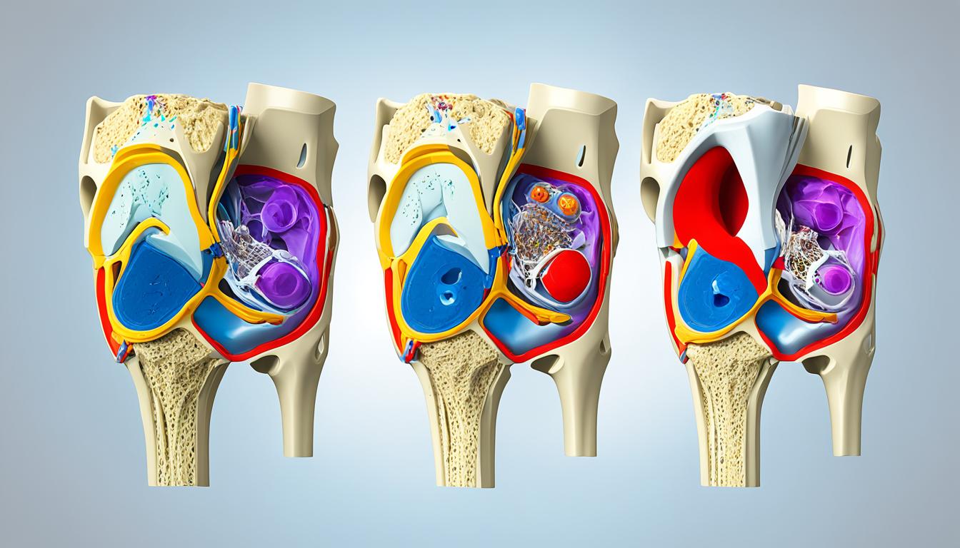
    What is Subchondral Edema?

    Subchondral edema is characterized by the accumulation of fluid within the bone marrow, leading to swelling and pain. This condition is not just a simple bruise but a complex issue that affects the bone’s structure.

    It is often associated with subchondral edema knee rehabilitation, as the knee is a commonly affected area. Understanding this condition is crucial for developing an effective rehabilitation plan.

    Causes of Subchondral Edema

    Subchondral edema can result from various factors, including trauma, overuse, or conditions like osteoarthritis. Repetitive stress or acute injuries can lead to this condition, making it essential to identify the underlying cause for effective management.

    A study published on the National Center for Biotechnology Information website provides insights into the causes and management of subchondral edema.

    Cause Description Common Symptoms
    Trauma Direct injury to the knee Pain, swelling
    Overuse Repetitive stress on the knee Pain, limited mobility
    Osteoarthritis Degenerative joint disease Pain, stiffness

    Symptoms to Watch For

    Symptoms of subchondral edema include pain, swelling, and limited mobility in the affected knee. Early recognition of these symptoms is vital for seeking appropriate medical attention and starting the knee edema recovery process.

    By understanding the causes and symptoms, individuals can take the first step towards recovery and rehabilitation.

    Recovery Timeline Overview

    Subchondral edema recovery duration is influenced by several key factors, making each individual’s journey to recovery unique.

    Factors Affecting Recovery Time

    The time it takes to recover from subchondral edema can be influenced by several factors, including the severity of the edema, the effectiveness of the treatment plan, and the individual’s overall health.

    • The extent of the edema and the presence of any underlying conditions.
    • Adherence to the prescribed treatment plan.
    • The presence of any comorbidities that could impact healing.

    Typical Recovery Timeframes

    While recovery times can vary, understanding typical timeframes can help manage expectations. Generally, mild cases may resolve within a few weeks, while more severe cases can take several months.

    Severity of Edema Typical Recovery Time
    Mild 2-6 weeks
    Moderate 6-12 weeks
    Severe 3-6 months or more

    Importance of Accurate Diagnosis

    Accurate diagnosis is crucial for determining the best treatment approach for subchondral edema. A precise diagnosis helps in tailoring the treatment to the individual’s specific condition, thereby optimizing the recovery process.

    knee edema recovery duration

    Managing subchondral edema in the knee requires a comprehensive approach that includes accurate diagnosis, appropriate treatment, and careful monitoring of the recovery process.

    Treatment Options

    Managing subchondral edema requires understanding the various treatment options available, from conservative treatments to physical therapy and pain relief medications. The goal is to alleviate symptoms, promote healing, and restore function to the affected knee.

    Conservative Treatment Methods

    Conservative treatment methods are often the first line of defense against subchondral edema. These include:

    • Rest, Ice, Compression, and Elevation (RICE): A fundamental approach to reduce pain and inflammation.
    • Activity Modification: Avoiding activities that aggravate the condition can significantly aid in recovery.

    By adopting these conservative methods, individuals can create an environment conducive to healing.

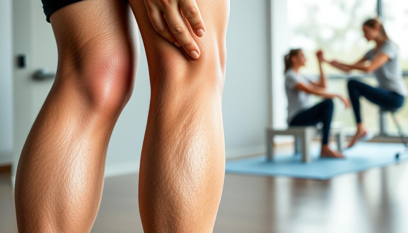
    Physical Therapy and Rehabilitation

    Physical therapy plays a crucial role in the recovery process by:

    1. Improving Knee Mobility: Gentle exercises help maintain or improve range of motion.
    2. Strengthening Surrounding Muscles: Strengthening the muscles around the knee provides additional support and stability.

    Physical therapy is tailored to the individual’s condition and progress, ensuring a gradual and safe return to normal activities.

    Medications for Pain Relief

    In some cases, medications may be prescribed to manage pain and inflammation associated with subchondral edema. These can include:

    • Nonsteroidal Anti-Inflammatory Drugs (NSAIDs): To reduce pain and inflammation.
    • Pain Relievers: For managing pain.

    It’s essential to follow a healthcare provider’s guidance when using medications to ensure safe and effective treatment.

    By combining these treatment strategies, individuals can develop a comprehensive plan to address subchondral edema, facilitating a smoother and more effective recovery process.

    Role of Lifestyle Changes in Recovery

    The journey to recovery from subchondral edema involves more than just medical treatment; it also requires significant lifestyle adjustments. Lifestyle changes can play a crucial role in enhancing the recovery process and potentially reducing the subchondral edema knee recovery time.

    One of the critical aspects of lifestyle modification is nutrition and diet. A well-balanced diet rich in anti-inflammatory foods can help reduce inflammation and promote healing.

    Nutrition and Diet Considerations

    A diet that includes foods high in omega-3 fatty acids, antioxidants, and fiber can be beneficial. It’s also important to stay hydrated by drinking plenty of water.

    Key dietary recommendations include:

    • Increasing consumption of fruits and vegetables
    • Incorporating lean proteins and whole grains
    • Avoiding processed foods and sugars

    Importance of Rest and Activity Modification

    Adequate rest and modifying activities to avoid putting excessive stress on the knee are crucial. This can involve avoiding high-impact activities and incorporating low-impact exercises.

    Rest allows the body to heal, while activity modification helps prevent further injury. It’s a delicate balance that requires careful management.

    Weight Management Strategies

    Maintaining a healthy weight is vital for reducing the stress on the knee joint. Effective weight management strategies include a combination of diet and exercise.

    A healthy weight can significantly impact the timeline for knee edema healing, as excess weight can exacerbate the condition.

    subchondral edema knee recovery

    Lifestyle Change Benefit
    Nutrition and Diet Reduces inflammation, promotes healing
    Rest and Activity Modification Prevents further injury, allows healing
    Weight Management Reduces stress on the knee joint

    Physical Therapy Techniques

    Subchondral edema knee rehabilitation significantly benefits from targeted physical therapy techniques, which are designed to improve knee function and reduce pain.

    Physical therapy is a crucial component of the recovery process, involving exercises that enhance strength and flexibility. A physical therapist will tailor a program to the individual’s condition and needs, aiming to restore function and alleviate pain.

    Recommended Exercises

    Recommended exercises for subchondral edema may include strengthening and stretching routines. These exercises are designed to improve knee mobility and reduce stiffness.

    • Straight leg raises to strengthen the quadriceps muscles
    • Knee bends to improve flexibility and strength
    • Straightening exercises to enhance knee extension

    Role of Stretching and Strengthening

    Stretching and strengthening exercises play a vital role in the rehabilitation process. Stretching helps to improve flexibility and reduce muscle tension, while strengthening exercises enhance the muscles around the knee, providing better support and stability.

    Exercise Type Benefits Examples
    Stretching Improves flexibility, reduces muscle tension Hamstring stretches, quadriceps stretches
    Strengthening Enhances muscle support around the knee Leg press, leg extensions

    Progress Tracking with a Therapist

    Progress tracking is an essential part of the rehabilitation process. Regular sessions with a physical therapist allow for adjustments to the treatment plan as needed, ensuring a safe and effective recovery.

    By working closely with a physical therapist, individuals can achieve significant improvements in knee function and pain reduction, ultimately enhancing their overall quality of life.

    Alternative Therapies

    Exploring alternative therapies can provide individuals with more options for managing subchondral edema. Besides conventional treatments, these alternatives can offer significant benefits in pain management and recovery.

    Acupuncture and Massage Therapy

    Acupuncture involves the insertion of fine needles into specific points on the body to stimulate healing and pain relief. Massage therapy, on the other hand, helps in reducing muscle tension and improving circulation, which can be beneficial for individuals recovering from subchondral edema.

    Benefits of Acupuncture and Massage Therapy:

    • Pain relief
    • Improved circulation
    • Reduced muscle tension

    Hydrotherapy Benefits

    Hydrotherapy, or aquatic therapy, utilizes water to aid in the recovery process. It can help reduce stiffness and improve mobility without putting excessive strain on the knee.

    Hydrotherapy Benefits:

    Benefit Description
    Reduced Impact Water reduces the impact on joints, making it easier to exercise
    Improved Mobility Water’s buoyancy helps improve range of motion
    Pain Relief Warm water can help alleviate pain and reduce inflammation

    Use of Orthotic Devices

    Orthotic devices can provide support to the knee during the recovery process. Custom-made orthotics can help in redistributing pressure and alleviating pain.

    subchondral edema recovery tips

    Post-Recovery Care

    Post-recovery care is essential for preventing re-injury and ensuring long-term knee health after subchondral edema. A well-planned post-recovery strategy can help maintain the progress achieved during the initial recovery phase.

    Importance of Continuing Exercise

    Continuing exercises that strengthen the muscles around the knee is crucial for maintaining stability and preventing future injuries. Gentle exercises such as straight leg raises, knee bends, and squats can be beneficial. It’s also important to incorporate exercises that improve flexibility, such as stretching.

    Prevention of Re-injury

    Preventing re-injury involves being mindful of activities that stress the knee. Modifying or avoiding high-impact activities can significantly reduce the risk of re-injury. Using proper techniques during physical activities and wearing appropriate gear can also help.

    Regular Follow-up with Healthcare Providers

    Regular follow-ups with healthcare providers are vital for monitoring the condition of the knee and addressing any concerns promptly. These visits can help identify potential issues early, ensuring timely intervention.

    By focusing on these aspects of post-recovery care, individuals can significantly improve their long-term outcomes and maintain optimal knee health.

    Long-Term Outlook

    Understanding the potential long-term effects of subchondral edema is crucial for managing the condition. The long-term outlook varies, with some individuals experiencing complete recovery while others may face chronic issues.

    Potential for Complete Recovery

    Many individuals with subchondral edema can achieve complete recovery with appropriate treatment and care. Factors influencing recovery include the severity of the condition, effectiveness of the treatment plan, and patient compliance.

    For instance, a well-structured rehabilitation program can significantly enhance the recovery process. This may include physical therapy, lifestyle modifications, and, in some cases, medication.

    Risks of Chronic Issues

    However, some individuals may experience chronic issues, such as persistent pain or limited mobility. The risk of chronic problems can be mitigated with early diagnosis and intervention.

    It’s essential to work closely with healthcare providers to monitor the condition and adjust the treatment plan as necessary to prevent long-term complications.

    Impact on Daily Activities

    Subchondral edema can impact daily activities, affecting an individual’s quality of life. Effective management strategies can help minimize this impact, enabling individuals to maintain their usual activities.

    By adopting a proactive approach to managing subchondral edema, individuals can reduce the likelihood of significant long-term effects on their daily lives.

    When to Seek Medical Attention

    Recognizing the need for medical attention can significantly impact the subchondral edema recovery process. It’s crucial to be aware of the signs that may indicate complications or the need for further medical evaluation.

    Signs of Complications

    Several signs may indicate that the recovery process is not proceeding as expected. These include:

    • Increased pain or swelling in the knee
    • Reduced mobility or stiffness
    • Instability or feeling of the knee giving way
    • Presence of infection signs such as redness, warmth, or fever

    If any of these symptoms are observed, it is essential to seek medical attention promptly.

    Importance of Early Intervention

    Early intervention can significantly alter the course of recovery. As emphasized by medical professionals, “Early diagnosis and treatment can prevent long-term damage and improve outcomes.” Timely medical intervention can help in addressing complications before they become severe, thereby supporting a smoother knee edema recovery process.

    Consultation with Specialists

    Consulting with specialists, such as orthopedic surgeons or sports medicine physicians, can provide individuals with subchondral edema recovery tips tailored to their specific condition. These specialists can offer guidance on the best course of treatment and rehabilitation, ensuring that the recovery process is both effective and safe.

    In conclusion, being aware of when to seek medical attention is crucial for a successful recovery from subchondral edema. By recognizing the signs of complications and seeking early intervention, individuals can significantly improve their outcomes and return to their normal activities.

    Conclusion: Your Road to Recovery

    Recovery from subchondral edema requires patience, the right treatment, and a positive mindset. As you navigate your knee edema healing timeline, understanding the factors that influence subchondral edema knee recovery time is crucial.

    Key Recovery Insights

    Effective management of subchondral edema involves a combination of conservative treatment methods, lifestyle changes, and physical therapy techniques. By understanding the causes, symptoms, and treatment options, individuals can better navigate their recovery journey.

    Maintaining a Positive Outlook

    Staying informed and maintaining a positive outlook are essential for a successful recovery. With the right mindset and support, individuals can overcome the challenges associated with subchondral edema and look forward to a healthier future.

    Additional Support Resources

    For further guidance and support, individuals can consult with healthcare professionals, physical therapists, or seek online resources. By leveraging these resources, individuals can ensure a smooth and effective recovery, ultimately achieving a full knee edema healing timeline.

    FAQ

    What is the typical recovery time for subchondral edema in the knee?

    The recovery time for subchondral edema in the knee varies based on several factors, including the severity of the edema, the effectiveness of the treatment plan, and the individual’s overall health.

    How long does it take to heal from knee edema?

    The healing timeline for knee edema can range from a few weeks to several months, depending on the severity of the condition and the treatment approach.

    What are the most effective ways to manage subchondral edema in the knee?

    Managing subchondral edema effectively requires a multi-faceted approach, including conservative treatment methods, physical therapy, and lifestyle changes, such as nutritional adjustments and weight management.

    Can physical therapy help with subchondral edema knee rehabilitation?

    Yes, physical therapy is a crucial component of subchondral edema knee rehabilitation, as it helps restore function, reduce pain, and improve mobility through tailored exercises and strengthening routines.

    Are there any alternative therapies that can aid in knee edema recovery?

    Alternative therapies, such as acupuncture, massage therapy, and hydrotherapy, can offer significant benefits in managing pain and improving mobility during the recovery process.

    How can I prevent re-injury after recovering from subchondral edema?

    Preventing re-injury involves continuing with exercises that maintain strength and flexibility, being mindful of activities, and possibly modifying them to avoid excessive stress on the knee.

    What are the signs of complications that require medical attention?

    Signs of complications, such as increased pain or swelling, require prompt medical attention to prevent further issues and ensure the best possible outcome.

    How can lifestyle changes impact the recovery process for subchondral edema?

    Lifestyle changes, including nutritional adjustments, adequate rest, and weight management, can significantly impact the recovery process by reducing inflammation and preventing further injury.

    What is the long-term outlook for individuals with subchondral edema?

    The long-term outlook for individuals with subchondral edema varies, with some experiencing complete recovery, while others may need to manage ongoing symptoms and adapt to changes in daily activities.

    How often should I follow up with my healthcare provider after recovering from subchondral edema?

    Regular follow-ups with healthcare providers are essential to monitor the condition, address any concerns, and prevent potential complications.

    Can subchondral edema knee recovery be enhanced with specific exercises or therapies?

    Yes, specific exercises, such as strengthening and stretching routines, and therapies, like physical therapy and hydrotherapy, can enhance the recovery process and improve overall outcomes.

  • Lateral Meniscus Ramp Lesion Symptoms

    Lateral Meniscus Ramp Lesion Symptoms

    Are you experiencing persistent knee pain without a clear reason? Knee injuries can be debilitating, affecting not just athletes but also individuals going about their daily lives.

    Meniscal tears, a common injury among young athletes and a degenerative condition in older patients, can lead to significant discomfort. Understanding the symptoms is crucial for effective diagnosis and treatment.

    The lateral meniscus plays a vital role in knee health. When a ramp lesion occurs, it can cause pain and discomfort, often misunderstood or misdiagnosed.

    Key Takeaways

    • Understanding the symptoms of lateral meniscus ramp lesions is crucial for diagnosis.
    • Meniscal tears are common in both young athletes and older adults.
    • Knee pain can be a significant indicator of underlying issues.
    • Effective treatment relies on accurate diagnosis.
    • Lateral meniscus injuries can significantly impact knee health.

    Understanding Lateral Meniscus Ramp Lesions

    Understanding the intricacies of lateral meniscus ramp lesions is crucial for addressing orthopedic knee injuries effectively. The meniscus, a cartilage structure in the knee joint, plays a vital role in facilitating smooth movement and absorbing shock. When this structure is damaged, it can lead to significant knee joint discomfort.

    What is a Lateral Meniscus Ramp Lesion?

    A lateral meniscus ramp lesion refers to a specific type of injury involving the meniscus. It is characterized by a tear or damage to the meniscotibial ligament or the peripheral attachment of the meniscus. This type of lesion can significantly impact the stability and function of the knee joint.

    The meniscus is a critical component of the knee, acting as a cushion between the tibia and femur. Damage to this area can result from various factors, including sports injuries, degenerative changes, or traumatic events.

    Causes of Ramp Lesions

    Ramp lesions are often caused by a combination of factors, including:

    • Traumatic injuries, such as those sustained during sports or accidents
    • Degenerative changes due to aging or wear and tear
    • Poor knee mechanics or alignment, leading to uneven stress on the meniscus

    Understanding these causes is essential for developing effective prevention and treatment strategies.

    Importance of Early Detection

    Early detection of lateral meniscus ramp lesions is critical for preventing further damage and promoting optimal recovery. Untreated lesions can lead to persistent knee pain, reduced mobility, and increased risk of further injury or degenerative changes.

    By recognizing the signs and symptoms of ramp lesions early, individuals can seek appropriate medical attention, potentially avoiding more severe consequences and improving their overall prognosis.

    Common Symptoms of Ramp Lesions

    Understanding the common symptoms of ramp lesions is essential for timely medical intervention. Lateral meniscus ramp lesions can manifest through a variety of symptoms that impact the knee’s functionality and the patient’s quality of life.

    Knee Pain and Discomfort

    One of the primary symptoms of a lateral meniscus ramp lesion is knee pain, which can vary in intensity. The pain is often localized to the area around the knee joint and can be exacerbated by activities that involve twisting, bending, or direct pressure on the knee.

    Patients may also experience discomfort during everyday activities such as walking or climbing stairs. This discomfort can be a result of the meniscus tear pain, which is a hallmark symptom of ramp lesions.

    knee pain diagnosis

    Swelling and Inflammation

    Swelling and inflammation are common symptoms associated with lateral meniscus ramp lesions. The injury can cause fluid buildup in the knee joint, leading to swelling, redness, and warmth around the affected area.

    In some cases, the swelling may be accompanied by a feeling of stiffness or tightness in the knee, making it difficult to move the joint through its full range of motion.

    Reduced Range of Motion

    A lateral meniscus ramp lesion can also result in a reduced range of motion in the knee. Patients may find it challenging to fully extend or flex the knee due to pain or mechanical obstruction caused by the lesion.

    • Common activities that may be affected include squatting, kneeling, or pivoting.
    • Some patients may experience a feeling of instability or weakness in the knee.
    • It’s essential to seek a knee pain diagnosis if these symptoms persist or worsen over time.

    Recognizing these symptoms early on can significantly impact the effectiveness of the treatment and the patient’s recovery process.

    Specific Symptoms to Watch For

    Understanding the specific symptoms associated with lateral meniscus ramp lesions can aid in effective treatment. Individuals experiencing these symptoms should be aware of the potential for a meniscal tear and seek medical evaluation.

    Clicking or Popping Sensations

    One of the characteristic symptoms of a lateral meniscus ramp lesion is the presence of clicking or popping sensations in the knee. This occurs due to the mechanical irregularity caused by the tear, which can disrupt the smooth movement of the knee joint. If you experience such sensations, it is advisable to consult a healthcare professional for a thorough assessment. For more information on knee pain and clicking, you can visit https://kneehurt.com/causes-and-treatments-for-knee-pain-clicking/.

    Locking of the Knee Joint

    Locking of the knee joint is another significant symptom that may indicate a lateral meniscus ramp lesion. When the meniscus is torn, a fragment can become lodged in the joint, causing it to lock or catch. This can severely limit knee mobility and cause considerable discomfort. Prompt medical attention is necessary to address this issue and restore normal knee function.

    Instability During Movement

    Instability during movement is a symptom that can significantly impact daily activities. A lateral meniscus ramp lesion can compromise the structural integrity of the knee, leading to feelings of instability or giving way. This symptom is particularly concerning as it can increase the risk of further injury. Early diagnosis and treatment are crucial to mitigate this risk and ensure a stable recovery.

    In conclusion, being aware of these specific symptoms can help individuals identify potential lateral meniscus ramp lesions early on. Recognizing the signs and seeking appropriate medical care can make a significant difference in treatment outcomes.

    Differentiating Symptoms from Other Knee Injuries

    The symptoms of lateral meniscus ramp lesions can be confused with those of other knee injuries, such as ACL injuries. This similarity in symptoms often complicates the diagnosis process, making it essential for healthcare professionals to conduct thorough evaluations.

    knee joint discomfort

    Comparing with ACL Injuries

    ACL injuries are common in sports and can present similarly to meniscal tears, including ramp lesions. A key difference lies in the mechanism of injury and the specific symptoms reported by the patient. For instance, ACL injuries often result from a sudden stop or change in direction, whereas ramp lesions may occur due to a specific type of twisting motion.

    Understanding these differences is crucial for an accurate diagnosis. While ACL injuries typically involve a feeling of instability or a “giving way” of the knee, ramp lesions may cause more localized pain and potentially a clicking or clunking sensation.

    Meniscus Tear Symptoms vs. Ramp Lesion

    Meniscus tears and ramp lesions share many symptoms, including knee pain and swelling. However, the location and nature of the pain can vary. Meniscus tears can occur in different parts of the meniscus and may cause pain in various areas around the knee.

    Ramp lesions, being a specific type of meniscus tear, tend to cause pain on the medial or lateral aspect of the knee, depending on which meniscus is affected. Accurate diagnosis through imaging, such as an MRI, is essential to distinguish between these conditions and guide appropriate treatment.

    Diagnostic Procedures for Ramp Lesions

    The diagnosis of lateral meniscus ramp lesions requires a comprehensive approach, including clinical evaluation and diagnostic imaging. Accurate diagnosis is crucial for determining the appropriate meniscus tear treatment and alleviating knee pain diagnosis challenges.

    Physical Examination Techniques

    A thorough physical examination is the first step in diagnosing ramp lesions. Healthcare professionals use various techniques to assess knee stability and identify potential issues. These examinations can reveal signs indicative of a ramp lesion, guiding further diagnostic steps.

    Imaging Tests: MRI and X-rays

    Imaging tests play a pivotal role in confirming the diagnosis. Magnetic Resonance Imaging (MRI) is particularly useful for visualizing soft tissue injuries, including ramp lesions. While X-rays are more suited for detecting bone-related issues, they can help rule out other causes of knee pain.

    Importance of a Specialist Consultation

    Consulting a specialist is essential for an accurate diagnosis. Orthopedic specialists or sports medicine physicians have the expertise to interpret diagnostic findings and recommend appropriate treatment plans. Their consultation is invaluable in managing ramp lesions effectively.

    Treatment Options for Lateral Meniscus Ramp Lesions

    The treatment of lateral meniscus ramp lesions involves a multifaceted approach, incorporating non-surgical management and surgical interventions. The choice of treatment depends on several factors, including the severity of the lesion, the patient’s overall health, and their activity level.

    Non-Surgical Management Strategies

    For patients with less severe symptoms or those who are not good candidates for surgery, non-surgical management strategies can be effective. These may include:

    • Physical therapy to strengthen the muscles around the knee and improve joint stability
    • Pain management through medication or injections
    • Activity modification to avoid exacerbating the condition

    Physical therapy plays a crucial role in non-surgical management, focusing on exercises that enhance knee stability and strength. A well-structured rehabilitation program can significantly improve symptoms and functionality.

    Surgical Interventions and Recovery

    When non-surgical approaches are not sufficient, surgical intervention may be necessary. Surgical options for lateral meniscus ramp lesions typically involve arthroscopic surgery to repair or remove the damaged meniscus tissue.

    Surgical Option Description Recovery Time
    Meniscus Repair Surgical repair of the meniscus to preserve as much tissue as possible 4-6 months
    Meniscus Trimming Removal of the damaged portion of the meniscus 2-4 months
    Meniscus Transplant Replacement of the meniscus with donor tissue 6-9 months

    torn meniscus recovery

    Rehabilitation and Physical Therapy

    Regardless of whether the treatment involves surgery or not, rehabilitation and physical therapy are crucial for optimal recovery. A tailored rehabilitation program helps restore knee function, reduce pain, and prevent future injuries. The program typically includes a combination of strengthening exercises, flexibility training, and gradual return to activity.

    Effective sports injury rehabilitation requires a comprehensive approach that addresses the specific needs of the athlete, including strengthening the knee, improving flexibility, and enhancing functional performance.

    Preventative Measures for Healthy Knees

    Knee health can be significantly improved through a combination of strengthening exercises, proper footwear, and safe sports practices. By focusing on these preventative measures, individuals can reduce their risk of developing lateral meniscus ramp lesions and other knee injuries.

    Strengthening Exercises

    Strengthening the muscles around the knee is crucial for providing support and stability to the joint. Exercises such as squats, lunges, and leg presses can help strengthen the quadriceps, hamstrings, and other surrounding muscles. A well-structured exercise program can be tailored to individual needs with the help of a physical therapist or trainer.

    Example Exercises:

    • Squats: Works the quadriceps, hamstrings, and glutes.
    • Lunges: Targets the quadriceps, hamstrings, and glutes, with an emphasis on balance.
    • Leg Press: Strengthens the quadriceps, hamstrings, and glutes in a controlled environment.

    Importance of Proper Footwear

    Wearing proper footwear is essential for reducing the stress and impact on the knees. Shoes that are designed for specific sports or activities can provide the necessary support and cushioning. For instance, running shoes are designed to absorb impact, while shoes for sports involving quick changes of direction provide additional lateral support.

    Activity Recommended Footwear Feature
    Running Cushioning and impact absorption
    Court Sports (e.g., Basketball, Tennis) Lateral support and stability
    Hiking/Trail Running Traction and ankle support

    Tips for Safe Sports Practices

    Safe sports practices are vital for preventing knee injuries. This includes proper warm-up routines, using correct techniques during sports activities, and being mindful of the playing surface. Coaches and trainers can play a significant role in educating athletes on safe practices.

    “Injury prevention is a critical aspect of sports performance. By focusing on proper techniques, conditioning, and equipment, athletes can significantly reduce their risk of injury.” –

    A Sports Medicine Specialist

    By incorporating these preventative measures into daily life and sports activities, individuals can significantly reduce their risk of knee injuries, including lateral meniscus ramp lesions. It’s about creating a culture of safety and prevention.

    Living with a Ramp Lesion

    Upon being diagnosed with a lateral meniscus ramp lesion, individuals must adapt their daily lives to manage the condition effectively. This involves making conscious decisions about activities, managing pain, and seeking appropriate support.

    Daily Life Adjustments

    Living with a meniscus tear requires adjustments to daily routines to minimize knee joint discomfort. Simple actions like walking or climbing stairs can become challenging. It’s essential to pace activities and avoid overexertion.

    • Modify exercise routines to low-impact activities like swimming or cycling.
    • Use assistive devices such as canes or walkers if necessary to reduce strain on the knee.
    • Wear supportive footwear to help stabilize the knee.

    Coping Strategies for Pain

    Managing meniscus tear pain is crucial for maintaining quality of life. Various strategies can help alleviate discomfort.

    1. Apply ice packs to reduce inflammation and pain.
    2. Use compression bandages to provide support.
    3. Engage in physical therapy to strengthen the muscles around the knee.

    As noted by medical professionals, “Pain management is a critical component of treating meniscus injuries, and a multi-faceted approach is often most effective.”

    Support from Professionals

    Seeking support from healthcare professionals is vital for effectively managing a ramp lesion. Specialists can provide guidance on the best treatment options and rehabilitation strategies.

    Professional guidance can make a significant difference in recovery and daily management of the condition. Regular consultations can help monitor the condition and adjust treatment plans as necessary.

    Long-Term Outcomes and Risks

    The long-term outcomes and risks associated with lateral meniscus ramp lesions are a significant concern for individuals suffering from orthopedic knee injuries. Understanding these aspects is crucial for effective management and recovery.

    Potential Complications

    Lateral meniscus ramp lesions can lead to several complications if not properly managed. These include:

    • Chronic knee pain
    • Increased risk of further knee injuries
    • Development of osteoarthritis

    Chronic knee pain is a common complication, significantly impacting the quality of life. It is essential to address the lesion promptly to mitigate this risk.

    Prognosis for Recovery

    The prognosis for recovery from lateral meniscus ramp lesions varies depending on several factors, including the severity of the lesion and the effectiveness of the treatment plan. Generally, with appropriate management, many patients can achieve significant improvement.

    A study highlighted in

    “The Journal of Orthopaedic Surgery” states that early intervention and proper rehabilitation can significantly enhance recovery outcomes for patients with meniscal tears.

    Treatment Approach Recovery Time Success Rate
    Non-Surgical Management 6-12 weeks 70%
    Surgical Intervention 3-6 months 85%

    When to Seek Further Medical Attention

    It is crucial for patients to monitor their symptoms and seek further medical attention if they experience:

    1. Increased pain or swelling
    2. Instability or locking of the knee joint
    3. Reduced range of motion

    By understanding the long-term outcomes and risks associated with lateral meniscus ramp lesions, patients can make informed decisions about their care and management, ultimately improving their prognosis for recovery.

    Frequently Asked Questions (FAQs)

    As we delve into the complexities of lateral meniscus ramp lesions, it’s essential to address the frequently asked questions surrounding this injury. Understanding the diagnosis, treatment, and management of ramp lesions can significantly impact the recovery process.

    How is a Ramp Lesion Diagnosed?

    Diagnosing a lateral meniscus ramp lesion typically involves a combination of physical examination techniques and imaging tests. A healthcare professional may perform specific maneuvers during a physical exam to assess knee stability and pain. Imaging tests, particularly MRI scans, are crucial for confirming the diagnosis by providing detailed images of the meniscus and surrounding structures.

    Can Ramp Lesions Heal Without Surgery?

    The potential for a ramp lesion to heal without surgery depends on several factors, including the severity of the lesion, the patient’s overall health, and their activity level. Some ramp lesions may heal with conservative management strategies, such as physical therapy and rehabilitation. However, more severe cases might require surgical intervention to repair the meniscus and restore knee function.

    What Activities Should be Avoided?

    Individuals with lateral meniscus ramp lesions are often advised to avoid activities that exacerbate knee pain or stress the knee joint. Common activities to limit or avoid include:

    • Deep knee bending
    • High-impact sports, such as football or basketball
    • Running on hard surfaces
    • Pivoting or twisting movements

    Avoiding these activities can help prevent further injury and support the healing process.

    For those recovering from a torn meniscus, understanding the recovery process and incorporating sports injury rehabilitation techniques can be beneficial. Rehabilitation often includes a structured physical therapy program aimed at restoring knee strength, flexibility, and function.

    Conclusion

    Recognizing lateral meniscus ramp lesion symptoms early is crucial for effective knee pain diagnosis and treatment. If left untreated, these lesions can lead to further complications, including meniscus tear and chronic knee pain.

    Early Detection and Medical Advice

    Seeking medical advice promptly is vital for proper meniscus tear treatment. Early detection allows for timely intervention, reducing the risk of long-term damage and improving outcomes. By understanding the symptoms and seeking professional help, individuals can take proactive steps towards recovery.

    Proactive Steps Towards Recovery

    By being aware of lateral meniscus ramp lesion symptoms, individuals can seek knee pain diagnosis and meniscus tear treatment in a timely manner. This awareness is key to preventing further injury and ensuring a speedy recovery. Effective management of meniscal tears relies on prompt medical attention and appropriate treatment strategies.

    FAQ

    What are the common symptoms of a lateral meniscus ramp lesion?

    Common symptoms include knee pain, swelling, reduced range of motion, clicking or popping sensations, and locking of the knee joint. These symptoms can vary in severity and impact daily activities.

    How is a lateral meniscus ramp lesion diagnosed?

    Diagnosis typically involves a combination of physical examination techniques, such as assessing knee stability and range of motion, and imaging tests like MRI to visualize the meniscus and surrounding structures.

    Can lateral meniscus ramp lesions heal without surgery?

    In some cases, non-surgical management strategies, including physical therapy and pain management, can help alleviate symptoms. However, severe cases may require surgical intervention to repair or remove the damaged meniscus.

    What activities should be avoided with a lateral meniscus ramp lesion?

    Activities that aggravate knee pain, such as deep knee bending, pivoting, or high-impact sports, should be avoided to prevent further injury and promote healing.

    How can I prevent lateral meniscus ramp lesions?

    Preventative measures include strengthening exercises to improve knee stability, wearing proper footwear, and practicing safe sports techniques to reduce the risk of knee injuries.

    What is the prognosis for recovery from a lateral meniscus ramp lesion?

    The prognosis varies depending on the severity of the lesion and the effectiveness of treatment. With proper care and rehabilitation, many individuals can recover and return to their normal activities.

    When should I seek further medical attention for a lateral meniscus ramp lesion?

    If symptoms worsen, or if you experience increased pain, swelling, or instability, it is essential to seek further medical attention to reassess the condition and adjust treatment as needed.

    Are there any long-term risks associated with lateral meniscus ramp lesions?

    Potential long-term risks include ongoing knee pain, osteoarthritis, and increased risk of further knee injuries if left untreated or undertreated.

    How can I manage knee pain caused by a lateral meniscus ramp lesion?

    Managing knee pain involves a combination of pain relief medications, physical therapy, and lifestyle adjustments, such as avoiding activities that exacerbate the pain.

  • Knee Pain Painting on a Ladder

    Knee Pain Painting on a Ladder

    Imagine you’re in the middle of painting, but knee pain stops you. Many face this when they have a meniscus tear. Climbing ladders then becomes a big challenge, affecting your project and mobility.

    Dealing with ladder climbing and a meniscus tear is tough. We aim to give you expert advice to stay safe. Our tips help you avoid more injuries and keep working on your projects.

    Key Takeaways

    • Safely assess your knee condition before climbing a ladder.
    • Understand the proper techniques for ladder climbing with a meniscus injury.
    • Learn how to prevent further knee damage while on a ladder.
    • Discover exercises to strengthen your knee before and after ladder climbing.
    • Explore alternative solutions for painting projects that minimize ladder use.

    Understanding Knee Pain: Common Causes

    Knee pain comes from many sources. It’s key to know these causes to avoid more harm. Activities like ladder climbing put extra stress on our knees. This can make existing problems worse or cause new ones.

    Types of Knee Pain

    Knee pain falls into two main groups: acute injuries and chronic conditions. Acute injuries happen suddenly, like from a fall. Chronic conditions develop over time, often from repeated strain or wear.

    Some common knee pain types include meniscus tears, ligament sprains, and tendonitis. Knowing the exact type of pain helps find the right treatment and ways to prevent it.

    Why Ladder Work Can Exacerbate Pain

    Climbing ladders puts a lot of strain on the knees, especially when bending or standing on uneven ground. This strain can make knee problems worse or cause new ones like patellofemoral pain syndrome. The constant stress from ladder work can lead to knee inflammation and pain.

    To reduce the risk of making knee pain worse, it’s important to understand how ladder work affects the knee. Knowing this helps us take steps to lessen the strain and protect our knees.

    Safety Measures When Using a Ladder

    Ladder Safety Measures

    To stay safe while using a ladder, we need to take certain steps. Ladder climbing can be dangerous, especially for those with knee problems. By picking the right ladder and setting it up right, we can lower the chance of accidents.

    Choosing the Right Ladder

    Picking the right ladder is key. Think about how high you need to go and how much weight the ladder can hold. For painting, a ladder with a strong base and non-slip feet is best. Look for ladders with comfy rungs and think about the material. Aluminum ladders are light, while fiberglass ladders are more stable.

    Ladder Type Material Weight Capacity
    Step Ladder Aluminum 300 lbs
    Extension Ladder Fiberglass 250 lbs

    Proper Ladder Setup

    After picking the right ladder, setting it up correctly is crucial for safety. Make sure the ladder is on a solid, level ground. Having someone hold the ladder can also help. The ladder’s angle is important too; it should be at a 75-degree angle or one foot away from the wall for every four feet of ladder height.

    By following these tips, we can make ladder climbing safer. Remember, safety steps are not just suggestions; they’re essential to avoid serious injuries.

    Preparing for Your Painting Project

    knee injury prevention

    Getting ready for painting is more than just picking up supplies. It also means getting your body ready to avoid knee injury.

    Stretching Exercises Before Climbing

    Do some stretching exercises before you start climbing. This helps your knees a lot. Try:

    • Leg swings to loosen up your leg muscles
    • Knee bends to prepare your knees for the climb
    • Calf raises to strengthen your calf muscles, which support your knees

    Essential Gear to Minimize Knee Strain

    Wearing the right gear can also help your knees. Use:

    1. Knee pads for extra cushioning and support
    2. Supportive footwear to stabilize your knees
    3. Comfortable clothing that allows for a full range of motion

    With these steps, you can lower the chance of knee pain and injury while painting.

    Painting Techniques to Reduce Knee Strain

    There are ways to lessen knee strain while painting on a ladder. Painting can put a lot of pressure on our knees, especially if we have conditions like meniscus tears. Using the right techniques can help avoid making knee pain worse.

    Opt for a Brush Instead of a Roller

    Choosing a brush over a roller can help reduce knee strain. Brushes allow for more precise strokes and need less force, which is easier on the knees. Plus, they help you paint more accurately, so you might not have to climb the ladder as much.

    Take Breaks to Relieve Pressure

    It’s important to take breaks to ease knee strain. Stepping down from the ladder often can help take the pressure off your knees. These breaks are a chance to stretch and prevent knee pain.

    By using these techniques, we can make painting easier on our knees. This is especially helpful for those with knee pain or meniscus tear symptoms.

    When to Seek Medical Advice

    Knee pain can really get in the way of doing things. There are times when you need to see a doctor. If you’re feeling pain in your knee while on a ladder, it’s important to know when to get help.

    Signs Your Knee Pain Needs Attention

    There are signs that mean you should see a doctor. These include:

    • Persistent pain that doesn’t improve with rest
    • Swelling or redness around the knee
    • Instability or feeling like the knee is going to give way
    • Warmth or tenderness to the touch
    • Loss of mobility or difficulty straightening the knee

    If you notice any of these, it’s best to talk to a healthcare professional. Ignoring these signs can lead to further complications.

    Recommended Specialists for Knee Pain

    Choosing the right specialist is crucial for knee pain. Here’s a table of specialists you might consider:

    Specialist Description
    Orthopedic Surgeon Specializes in surgical treatments for musculoskeletal disorders, including knee pain. They can perform knee surgery if necessary.
    Rheumatologist Expert in diagnosing and treating rheumatic diseases that can cause knee pain.
    Physical Therapist Can help improve knee mobility and strength through targeted exercises.

    An orthopedic surgeon is often the first choice for serious knee pain or when surgery is needed. They can give a detailed diagnosis and suggest the best treatment.

    Home Remedies for Knee Pain Relief

    Home remedies are a simple way to handle knee pain and boost your quality of life. We often seek easy, effective solutions to ease discomfort before seeking medical help. Two popular methods are ice and heat therapy, and over-the-counter pain relief.

    Ice and Heat Therapy

    Ice and heat therapy are easy yet effective for knee pain. Ice therapy reduces swelling and numbs pain, great for sudden injuries or pain spikes. Heat therapy, on the other hand, relaxes muscles and boosts blood flow, helping with long-term pain and stiffness.

    To use ice, wrap an ice pack in a towel and apply it for 15-20 minutes, several times a day. For heat, try a warm bath, heating pad, or a warm cloth on your knee. It’s key to alternate between ice and heat based on your pain type.

    “The application of ice or heat can significantly alter the pain perception and is a crucial component of a comprehensive pain management plan.”

    – Expert in Pain Management

    Over-the-Counter Pain Relief Options

    Over-the-counter (OTC) pain meds are also effective for knee pain. Common ones are acetaminophen and ibuprofen. Acetaminophen mainly relieves pain, while ibuprofen also fights inflammation.

    • Always follow the recommended dosage on the label.
    • Consult with a healthcare provider if you’re unsure about interactions with other medications.
    • Be aware of potential side effects and contraindications.

    By adding these home remedies to your daily routine, you can better manage knee pain. This lets you enjoy activities like painting on a ladder more comfortably.

    Prolonging Knee Health During Painting

    To enjoy painting without harming our knees, we need to focus on knee health. Painting on a ladder can be tough on our knees, especially for those with a torn meniscus. By keeping our knees strong and moving regularly, we can protect them.

    Tips for Maintaining Strong Knees

    Keeping our knees strong involves exercises and careful movements. Physical therapy helps strengthen knee muscles, offering better support and injury prevention. Exercises like squats, lunges, and leg presses are helpful. Also, using the right ladder techniques and taking breaks can ease knee strain.

    Exercise Benefit Frequency
    Squats Strengthens quadriceps and hamstrings 3 sets of 10 reps, 3 times a week
    Lunges Improves balance and strengthens knee muscles 3 sets of 10 reps (per leg), 3 times a week
    Leg Presses Targets quadriceps, hamstrings, and glutes 3 sets of 12 reps, 3 times a week

    Importance of Regular Movement

    Regular movement is key for healthy knees. Sitting still for too long can make knees stiff and painful, especially for those with injuries. Adding gentle exercises to our day keeps knees flexible and lowers injury risk. It’s also important to listen to our bodies and rest when needed.

    By using these tips, we can keep our knees healthy while painting. Regular physical therapy, exercises, and careful movements are all important for a safe painting experience.

    Equipment to Assist While Painting on a Ladder

    To have a safe and fun painting time, the right tools are key. Using the right gear when painting on a ladder helps avoid knee pain and injuries.

    Knee Pads and Supportive Gear

    Knee pads are a simple yet effective way to protect your knees while working on a ladder. They offer cushioning and support, easing the pressure on your knees. Supportive gear, like knee braces, adds extra stability and protection. Pick knee pads or supportive gear based on your work and needed support.

    Look for knee pads that are durable, comfortable, and adjustable. It’s also important to pick knee pads that fit your task. For example, if you’re working on a rough surface, you’ll need sturdier knee pads.

    Type of Knee Pad Features Best For
    Hard Cap Knee Pads Durable, protective Rough surfaces, heavy-duty work
    Soft Cap Knee Pads Comfortable, flexible General painting, smooth surfaces
    Gel Knee Pads Cushioned, shock-absorbing Long periods of kneeling, comfort

    Choosing the Right Footwear

    Along with knee pads, choosing the right footwear is crucial for stability and to prevent slipping on the ladder. Find shoes with good grip and a snug fit to keep your feet steady on the ladder.

    • Non-slip soles
    • Comfortable fit
    • Ankle support

    By using knee pads with the right shoes, you can greatly lower the risk of knee injury. This makes painting more comfortable and enjoyable.

    Final Thoughts: Enjoying Your Painting Project

    As we finish talking about managing knee pain while painting on a ladder, remember it’s not just about the end. It’s also about how we feel during it. By taking care of our knees, we can enjoy painting more without worrying about injuries.

    It’s important to love the journey of creating. We can do this by following the tips we’ve shared. From picking the right ladder to stretching, every step matters.

    Practical Adjustments for Future Projects

    For future painting, we can reduce knee pain. Use knee pads and supportive gear. Also, pick shoes that help you stay stable on the ladder.

    Maintaining Knee Health

    Keeping our knees healthy is key to enjoying painting and other activities. Regular exercise and movement help make our knees stronger. This makes it easier to handle ladder work.

    FAQ

    What are the symptoms of a meniscus tear?

    Symptoms include knee pain, swelling, and stiffness. Sometimes, we hear a popping sound when it happens.

    How can I prevent further injury while climbing a ladder with a meniscus tear?

    To avoid more harm, choose the right ladder and set it up right. Wear knee pads and supportive shoes.

    What are the treatment options for a torn meniscus?

    Treatments include physical therapy, pain meds, and sometimes surgery. An orthopedic surgeon can help decide the best treatment.

    Can I continue with my painting projects if I have a meniscus tear?

    Yes, you can keep painting. Just be careful to avoid making the injury worse. Use proper techniques, take breaks, and wear supportive gear.

    When should I seek medical advice for knee pain?

    Get medical help if your knee pain, swelling, or stiffness doesn’t go away. An orthopedic surgeon can find the cause and suggest treatment.

    What are some home remedies for knee pain relief?

    Try ice and heat, over-the-counter pain meds, stretching, and keeping a healthy weight. These can help ease knee pain.

    How can I maintain strong knees while painting on a ladder?

    Keep your knees strong by doing physical therapy, wearing supportive gear, and taking breaks. This helps relieve knee pressure.

    What type of equipment can assist me while painting on a ladder?

    Use knee pads, supportive gear, and the right shoes. They help protect and support your knees while painting.

  • Specific Knee Pain Triggers

    Specific Knee Pain Triggers

    Have you ever wondered why simple movements like climbing stairs or sitting too long suddenly cause discomfort? The answer lies in the delicate balance of our body’s most overworked joint. Your knees handle forces up to four times your body weight during routine activities, making them prone to wear even without intense exercise.

    This complex hinge relies on ligaments, tendons, and cartilage working in harmony. When one component falters – whether from sudden twists, repetitive motions, or age-related changes – swelling and stiffness often follow. Over 25% of adults experience joint issues linked to daily habits, according to recent studies.

    Our comprehensive guide to knee health reveals how minor adjustments can prevent major problems. From improper footwear to overlooked muscle imbalances, we’ll explore hidden culprits that turn ordinary actions into sources of lasting damage.

    Key Takeaways

    • The knee’s weight-bearing role makes it vulnerable to strain during daily tasks
    • Cartilage wear and ligament tears account for most mobility complaints
    • Swelling often signals underlying joint stress needing attention
    • Early identification of triggers prevents chronic conditions
    • Proper alignment during movement reduces injury risks

    Understanding Knee Anatomy and Mechanics

    Every step we take relies on a complex system of bones and tissues working in precise harmony. The joint connecting thigh and shin bones functions like a living hinge, combining stability with remarkable flexibility.

    knee joint cartilage and ligaments

    Building Blocks of Mobility

    Three bones form this critical junction: femur, tibia, and patella. Between them lies cartilage – nature’s shock absorber preventing bone-on-bone contact. Ligaments act as sturdy ropes, connecting bones while allowing controlled movement.

    Four primary bands maintain stability:

    • ACL and PCL control forward/backward motion
    • MCL and LCL prevent side-to-side slipping

    Motion Creates Wear Patterns

    Synovial fluid lubricates the joint during activity, much like oil in machinery. Repeated bending or twisting can thin this protective layer. When ligament fibers stretch beyond limits or cartilage develops uneven wear, grinding sensations often follow.

    Research shows 30% of adults over 45 show early signs of arthritis in weight-bearing joints. Regular movement maintains circulation to these tissues, while excessive strain accelerates breakdown. Finding this balance proves crucial for long-term joint function.

    What are Specific Knee Pain Triggers?

    Why does bending down sometimes feel like a challenge? The answer often lies in hidden conditions that transform routine motions into sources of discomfort. Our joints communicate through swelling and stiffness when facing stressors – signals we shouldn’t ignore.

    conditions causing knee inflammation

    Hidden Culprits Behind Joint Stress

    Repetitive motions like squatting or climbing stairs can gradually wear down tissues, creating micro-tears that accumulate over time. Rheumatoid arthritis exemplifies how autoimmune responses turn the body’s defenses against itself, attacking healthy joint linings. This leads to persistent swelling and reduced mobility.

    Another common offender? Band syndrome, where connective tissues tighten abnormally during movement. Think of it like a guitar string pulled too taut – every motion creates friction. Combined with poor posture, this imbalance often cause knee strain during activities as simple as walking.

    Persistent inflammation acts as the body’s flare gun, signaling underlying trouble. Whether from sudden injuries or chronic conditions, swollen joints demand attention before damage becomes irreversible. Recognizing these red flags early helps prevent long-term limitations.

    Next, we’ll explore how acute injuries differ from gradual wear – and why your approach to care should too.

    Common Causes: Injury, Overuse, and Disease

    What transforms a minor ache into persistent joint trouble? Three primary factors – sudden damage, repetitive strain, and systemic illnesses – account for most mobility challenges. Let’s examine how these forces interact within our bodies.

    knee injury and disease conditions

    When Trauma Meets Time

    Acute damage often stems from impacts or twists. A soccer player’s ACL tear or a slip-related kneecap fracture demonstrates immediate trauma. These injuries typically cause sharp discomfort and visible swelling within hours.

    Degenerative conditions operate differently. Years of wear thin cartilage, while autoimmune diseases like rheumatoid arthritis attack joint linings. “The difference between a sprain and arthritis? One shouts, the other whispers,” notes Dr. Ellen Torres, orthopedic specialist.

    Cause Onset Symptoms Treatment Window
    ACL tear Sudden Instability, swelling Days
    Osteoarthritis Gradual Morning stiffness Months
    Tendonitis Progressive Localized tenderness Weeks

    Silent Aggravators

    Connective tissues bear the brunt of overuse. Patellar tendonitis – often called “jumper’s knee” – develops when muscle imbalances strain the kneecap’s anchoring tendons. Similarly, iliotibial band syndrome plagues runners through repetitive friction.

    Watch for these red flags:

    • Persistent swelling after activity
    • Clicking sounds during movement
    • Difficulty bearing weight

    Early intervention prevents 68% of chronic cases, according to Johns Hopkins research. Whether addressing a fresh injury or managing arthritis, timely care preserves mobility better than delayed reactions.

    Impact of Age, Weight, and Activity Levels on Knee Health

    Our bodies keep score in ways we often overlook. Years of movement patterns and physical demands leave lasting marks on joint structures. The relationship between lifestyle choices and joint durability becomes clearer as decades pass.

    Time’s Toll on Joint Integrity

    Cartilage loses elasticity with age – think of tire treads wearing thin. The meniscus, a crucial shock absorber between thigh and shin bones, develops frayed edges over time. This natural breakdown reduces the joint’s ability to handle impacts smoothly.

    Research shows osteoarthritis affects 1 in 3 adults over 60. Synovial fluid production slows, leaving joints less lubricated during motion. These changes explain why morning stiffness becomes common as we accumulate birthdays.

    Weight’s Multiplier Effect

    Carrying extra pounds multiplies pressure on weight-bearing joints. Every 10 lbs of body weight adds 30-40 lbs of force during walking. For those with obesity, this strain accelerates meniscus degeneration and bones reshaping.

    A 2024 analysis revealed maintaining ideal weight reduces arthritis risk by 52%. Simple swaps – taking stairs instead of elevators or choosing water over sugary drinks – create lasting protective effects.

    While time remains unstoppable, proactive care preserves mobility. Low-impact exercises strengthen supporting muscles without stressing vulnerable tissues. Understanding these connections helps us make choices that honor our body’s changing needs through the years.

    Managing Knee Pain with Home Remedies and Medical Treatments

    When joint discomfort strikes, smart responses can prevent temporary issues from becoming chronic. Over 40% of mobility problems improve significantly with early intervention, according to Johns Hopkins rehabilitation data. Let’s explore practical strategies that address both immediate relief and long-term healing.

    Effective self-care: Rest, ice, compression, and elevation

    The RICE method remains gold-standard care for acute flare-ups. Rest prevents further tissue strain, while ice applied for 15-minute intervals reduces swelling. Compression sleeves stabilize vulnerable areas, and elevation drains excess fluid from inflamed joints.

    For chronic bursitis, heat therapy often works better than ice. Warm compresses increase blood flow to stiff tissues. A 2023 Arthritis Foundation study found alternating heat and ice improved mobility in 72% of participants with persistent syndrome-related stiffness.

    Consider these red flags requiring professional evaluation:

    • Difficulty bearing weight for over 48 hours
    • Visible deformity around joints
    • Fever accompanying swelling

    Alignment issues in the hip or pelvis frequently manifest as knee strain. Physical therapists often assess adjacent joints when treating persistent problems. “You can’t fix a door hinge without checking the frame,” explains Dr. Miriam Cole, sports medicine specialist.

    Care Type Best For Timeline
    Home Remedies Mild swelling, recent injuries 3-7 days
    Prescription Treatments Bursitis, IT band syndrome 2-6 weeks
    Surgical Options Severe cartilage damage 6+ months

    Remember: Persistent syndrome management requires tailored approaches. What soothes bursitis might aggravate other conditions. Tracking symptom patterns helps identify the right type of care – and when to seek expert guidance for unresolved problems.

    Diagnostic Approaches and Imaging Techniques

    Unlocking the mystery behind joint discomfort requires detective-level precision. Modern medicine combines hands-on evaluations with advanced technology to map out treatment paths. We start by analyzing movement patterns and tenderness points before reaching for imaging tools.

    Connecting Symptoms to Solutions

    Physical exams often reveal what scans can’t. Doctors assess how activities like squatting or lunging cause knee pain during movement tests. The Ober maneuver, for example, helps identify iliotibial band tightness by measuring hip abduction range.

    “Targeted movements during exams act like live video replays,” explains Dr. Alicia Hernandez, sports medicine specialist. Bursa inflammation becomes evident when pressure applied below the kneecap triggers sharp discomfort. These findings guide which imaging tests deliver the clearest answers.

    Test Type Best For Details
    X-ray Bone alignment Detects fractures, arthritis
    MRI Soft tissue damage Reveals ligament tears
    Ultrasound Bursa evaluation Checks fluid accumulation

    Imaging confirms suspicions raised during physical assessments. MRIs excel at showing iliotibial band syndrome inflammation near the thigh’s outer edge. Ultrasound dynamically captures irritated bursa sacs during motion – something static scans miss.

    When activities consistently cause knee pain, doctors might order gait analysis. This tracks pressure distribution across joints during walking or running. Combining these methods creates a 360-degree view of what’s disrupting mobility – and how to fix it.

    Prevention Strategies: Lifestyle Modifications and Exercises

    Protecting joint health starts with proactive habits that build resilience. We’ve curated science-backed methods to maintain mobility while reducing strain on vulnerable tissues. These approaches address both immediate risks and long-term durability.

    Strengthening and Stretching Routines

    Targeted exercises fortify muscles supporting the joint. Wall sits and step-ups strengthen quadriceps without heavy impact. For flexibility, try hamstring stretches with a resistance band – hold for 30 seconds to improve range of motion.

    Yoga poses like Warrior II enhance balance while stretching iliotibial bands. “Consistent routines prevent 60% of soft tissue injuries,” notes physical therapist Marco Silva. Pair these with foam rolling to release tension in calf muscles – a common contributor to meniscus tear risks.

    Smart Movement Patterns

    Adjust daily habits to minimize wear:

    • Use ergonomic stools during prolonged sitting
    • Wear supportive footwear with arch cushioning
    • Avoid sudden pivots during sports

    Low-impact activities like swimming maintain fitness without pounding joints. A 2023 Journal of Orthopedic Research study found cyclists experience 40% fewer baker cyst cases than runners. Track activity duration – exceeding 90 minutes of continuous motion often causes knee overuse issues.

    Early intervention matters. If pain swelling persists after rest, consult a specialist. Simple tweaks to exercise form and recovery routines can stop minor irritation from becoming chronic damage. Remember: Prevention isn’t about avoiding movement – it’s about moving smarter.

    Conclusion

    Maintaining healthy knee joints requires understanding their delicate balance. Our bodies rely on these complex hinges to handle forces from walking to twisting. Recognizing how daily motions impact joint structures helps prevent avoidable damage.

    Early action proves critical. Tracking swelling patterns or stiffness allows timely interventions before minor irritation escalates. Proper alignment during movement and targeted strengthening exercises reduce strain on vulnerable tissues.

    Professional guidance matters when symptoms persist. Physical therapists and orthopedic specialists offer customized solutions for unique needs. Don’t wait until discomfort limits daily activities – proactive care preserves mobility better than delayed treatments.

    Ultimately, healthy knee joints depend on informed choices. From footwear selection to activity modifications, small changes create lasting protection. Prioritize joint health today to maintain an active, unrestricted lifestyle tomorrow.

    FAQ

    What conditions commonly cause swelling and discomfort in the knee joint?

    Issues like arthritis, ligament tears, bursitis, or meniscus damage often lead to swelling and pain. Rheumatoid arthritis and osteoarthritis break down cartilage, while injuries like ACL tears or iliotibial band syndrome strain tissues around the joint.

    How do acute injuries differ from chronic conditions like osteoarthritis?

    Acute injuries—such as ligament sprains or fractures—happen suddenly from trauma or overexertion. Degenerative diseases like osteoarthritis develop slowly, wearing down cartilage over time due to age, repetitive stress, or excess body weight.

    Why does aging increase the risk of joint pain?

    Over time, cartilage thins, muscles weaken, and ligaments lose flexibility. Reduced shock absorption and joint stability make older adults more prone to inflammation, arthritis flare-ups, or overuse injuries like patellar tendinitis.

    Can home remedies effectively manage mild knee discomfort?

    Yes. Resting, applying ice packs, using compression sleeves, and elevating the leg reduce swelling. Low-impact exercises and anti-inflammatory medications like ibuprofen also help ease symptoms linked to iliotibial band syndrome or minor strains.

    How do doctors diagnose the root cause of persistent knee issues?

    Physical exams check for tenderness, range of motion, and instability. Imaging tools like X-rays spot bone spurs or fractures, while MRIs reveal soft tissue damage—meniscus tears, tendon injuries, or Baker’s cysts—to guide treatment plans.

    What exercises strengthen muscles around the knees?

    Squats, lunges, and leg raises build quadriceps and hamstrings. Stretching the iliotibial band, calves, and hips improves flexibility. Low-impact activities like swimming or cycling also reduce strain on joints while maintaining mobility.

    How does excess weight contribute to knee problems?

    Extra pounds add pressure on joints during walking or climbing stairs. Over time, this accelerates cartilage breakdown, raising the risk of osteoarthritis. Weight loss of just 10% can significantly lower inflammation and pain levels.

  • Knee pain recovery timeline

    Knee pain recovery timeline

    Have you ever wondered why some people bounce back quickly from joint injuries while others struggle for months? This guide breaks down the science-backed phases of restoring mobility after physical setbacks. We’ll walk you through every critical step, from managing discomfort to rebuilding strength safely.

    Understanding healing durations isn’t just about patience—it’s about strategy. Factors like age, injury severity, and therapy adherence play huge roles. Drawing from trusted institutions like Cleveland Clinic and Portland Urgent Care, we’ll show how evidence-based methods can optimize your progress.

    Whether you’re dealing with minor strains or post-surgical rehab, timelines vary widely. Some see improvement in weeks; others need months. We’ll compare non-invasive approaches (like ice and rest) with surgical options, helping you navigate choices confidently.

    Key Takeaways

    • Healing occurs in distinct phases influenced by lifestyle and treatment plans
    • Expert-backed strategies reduce setbacks and accelerate progress
    • Recovery spans from weeks to over a year depending on injury complexity
    • Combining rest, therapy, and medical guidance yields optimal results
    • Personalized plans address unique factors like weight and joint health

    Overview of the Knee Pain Recovery Timeline

    Restoring joint function after trauma involves predictable phases, but individual factors dictate the pace. Healthcare teams categorize progress into three benchmarks: inflammation control, mobility restoration, and strength rebuilding. Portland Urgent Care reports 63% of patients regain basic range motion within 4-6 weeks when following structured protocols.

    Phases of Tissue Repair

    Initial healing focuses on reducing swelling through:

    • Controlled compression
    • Targeted cold therapy
    • Medication management

    Mid-phase rehabilitation introduces gentle stretching. A 2023 study showed patients who started guided exercises within 14 days reduced long-term stiffness risks by 41%.

    Real-World Healing Variations

    Consider two cases from Portland clinics:

    • Athlete: Returned to sports in 12 weeks post-meniscus tear
    • Senior: Required 9 months for arthritis-related joint stabilization

    Early diagnosis proves critical. Those seeking specialized care within 72 hours of injury report 28% faster progress than delayed treatment groups.

    Consistency separates successful recoveries from prolonged setbacks. Therapists emphasize daily home exercises—even 10-minute sessions improve outcomes by 19%. Remember: Healing isn’t linear, but disciplined effort yields measurable gains.

    Knee pain recovery timeline: Key Phases from Weeks to Months

    Understanding how joint injuries progress from acute trauma to restored function helps set realistic expectations. Clinical data reveals distinct milestones in rebuilding stability and flexibility, whether addressing minor sprains or complex ligament damage.

    knee rehabilitation phases

    From Initial Injury to Early Rehabilitation

    The first 72 hours determine much of the journey. Immediate protocols like the RICE method (rest, ice, compression, elevation) minimize tissue stress. Cleveland Clinic research shows 82% of patients who stabilize swelling within three days regain mobility 33% faster than those delaying care.

    Weeks 2-6 focus on controlled movement. Therapists often introduce:

    • Low-intensity stretching to prevent stiffness
    • Non-weight-bearing exercises like seated leg lifts
    • Modalities such as ultrasound to enhance circulation

    ANOVA Institute studies highlight ACL tear cases where structured rehab takes months—typically 6-9 months for full activity clearance. Contrast this with Grade I sprains, where 70% achieve baseline function in 4-6 weeks through consistent home care.

    Transitioning to active rehabilitation around week 8 marks a turning point. Resistance bands and balance boards become tools for rebuilding proprioception. One patient in a Cleveland Clinic trial increased walking endurance by 58% between months 3 and 5 using progressive loading techniques.

    Factors Influencing Recovery and Healing Time

    The journey to restored joint function isn’t one-size-fits-all. Cleveland Clinic researchers emphasize how biological responses and personal habits create unique healing trajectories. Swelling management proves critical—their 2023 report found unresolved inflammation adds 22 extra days to average rehabilitation periods for joint injuries.

    Impact of Injury Severity

    Deep tissue damage requires more time than surface-level sprains. Torn ligaments or cartilage often demand months of targeted care, while minor strains might resolve with rest. Uncontrolled swelling acts like a roadblock—it restricts blood flow and delays nutrient delivery to damaged areas.

    Ignoring early rest protocols carries risks. Patients who push through discomfort increase reinjury chances by 37%, according to orthopedic specialists. Limited range of motion during initial phases often signals the need for adjusted therapy plans.

    Patient-Specific Considerations

    Age and weight significantly affect cellular repair rates. A 65-year-old with arthritis heals 40% slower than a 25-year-old athlete with similar injuries. Pre-existing conditions like diabetes or obesity add complexity, requiring customized exercise regimens.

    Therapists prioritize movement quality over speed. One study showed tailored programs improved walking stability by 29% compared to generic protocols. “We design plans around sleep patterns, work demands, and even shoe choices,” notes a Cleveland Clinic physiotherapist. “These details determine long-term success.”

    Common Knee Injuries and Their Treatments

    Portland Urgent Care reports that over 40% of joint-related visits involve ligament or cartilage issues. Accurate diagnosis guides effective care—we’ll explore how specialists address these challenges using advanced methods and tailored plans.

    knee injury types and treatments

    Managing Ligament Tears and Sprains

    Anterior cruciate ligament (ACL) injuries and medial collateral sprains dominate clinical cases. Cleveland Clinic classifies ligament damage into three grades:

    • Grade I: Mild stretching with minimal instability
    • Grade II: Partial tearing requiring bracing
    • Grade III: Complete rupture often needing surgery

    Non-surgical approaches like physical therapy exercises work for 68% of Grade I-II cases. For severe tears, reconstruction paired with neuromuscular training restores stability in 6-12 months.

    Addressing Cartilage Damage and Meniscal Tears

    Cartilage wear differs from meniscus injuries. The former involves smooth joint surface erosion, while the latter affects crescent-shaped shock absorbers. Diagnostic imaging like MRI determines treatment paths:

    • Cartilage: Hyaluronic acid injections or microfracture surgery
    • Meniscus: Arthroscopic repair for active patients

    Portland Urgent Care data shows targeted strengthening routines improve outcomes by 31% when started within two weeks post-injury. Low-impact cycling and water-based exercises preserve mobility without straining healing tissues.

    Role of Physical Activities and Rehabilitation

    Movement becomes medicine when guided by science. Strategic exercises rebuild joint stability while preventing new injuries—a balance requiring expert-backed methods. Portland Urgent Care found patients who followed customized activity plans reduced reinjury risks by 44% compared to those who self-managed.

    Adopting Safe Movement Practices

    Early-stage healing demands smart modifications. Therapists recommend water-based exercises or stationary cycling to maintain mobility without impact. ANOVA Institute research shows these low-stress activities boost circulation 53% more than complete rest, accelerating tissue repair.

    Structured programs focus on three pillars:

    • Gradual load progression to rebuild tendon strength
    • Balance drills improving proprioception
    • Dynamic stretches enhancing flexibility

    Portland specialists emphasize consistency—daily 15-minute sessions yield better results than sporadic hour-long workouts. One study tracked patients performing targeted leg lifts twice daily; 78% regained full extension 3 weeks faster than control groups.

    Even discomfort shouldn’t halt progress. Controlled motions like heel slides or wall-assisted squats maintain blood flow while respecting healing boundaries. As one therapist notes: “We design movements that challenge—not overwhelm—recovering tissues.”

    Trusted institutions validate this approach. Cleveland Clinic reports 91% adherence rates when patients receive clear exercise demonstrations and progress trackers. Pairing clinical guidance with home efforts creates sustainable pathways to restored function.

    Surgical vs. Non-Surgical Treatment Options

    Choosing between surgical and conservative approaches requires understanding clinical thresholds. Portland Urgent Care data reveals 58% of ligament-related cases resolve without operations when addressed early. However, complex tears or instability often demand more aggressive interventions.

    Determining When Surgery Becomes Necessary

    ANOVA Institute guidelines outline three surgical triggers:

    • Complete ligament ruptures causing joint instability
    • Multi-tissue injuries requiring structural repair
    • Failed conservative therapy after 12+ weeks

    Non-surgical treatments excel for minor sprains and partial tears. Portland studies show 71% success rates with bracing and guided rehab for Grade II ligament injuries. Surgical timelines typically span 6-12 months versus 8-14 weeks for conservative care.

    Post-procedure rehabilitation focuses on three phases:

    1. Protected weight-bearing (Weeks 1-6)
    2. Progressive strength training (Months 2-4)
    3. Sport-specific drills (Months 5+)

    Key factors in treatment decisions include activity goals, age, and tissue quality. A 2023 ANOVA report found athletes opt for surgery 83% more often than sedentary patients to restore peak performance.

    “We prioritize anatomical restoration in active individuals but respect natural healing capacities when feasible.”

    Portland Orthopedic Team

    To make sure plans align with needs, specialists recommend second opinions for borderline cases. Tracking mobility gains through apps or journals helps patients stay committed during months-long rehabilitation.

    Exercise Strategies for Strengthening the Knee

    Effective movement patterns form the foundation of lasting joint support. Portland Urgent Care specialists emphasize combining flexibility work with stability drills to create resilient tissue networks. Let’s explore methods that rebuild capacity while respecting biological repair processes.

    Low-Impact Stretching Routines

    Gentle stretching maintains blood flow without straining healing structures. Try these daily sequences:

    • Seated hamstring stretches with resistance bands
    • Prone quadriceps extensions using ankle weights
    • Wall-assisted calf raises with 3-second holds

    Portland therapists report 73% of patients improve range motion within 14 days using these techniques. Focus on smooth transitions—jerky movements increase inflammation risks by 29%.

    Resistance and Balance Training Techniques

    Gradual loading strengthens muscles supporting joint function. Consider this progression plan:

    Exercise Type Frequency Duration Key Benefit
    Mini-squats 3x/week 10 mins Builds quad strength
    Side-lying leg lifts Daily 5 mins Targets hip stabilizers
    Single-leg stands 2x/day 2 mins Improves proprioception

    Balance boards and foam pads challenge coordination systems. Start with 30-second sessions, increasing difficulty as stability improves. One study showed 68% fewer reinjuries when patients incorporated these drills weekly.

    “Consistent effort trumps intensity. Small daily investments in movement quality compound over weeks.”

    Portland Rehabilitation Specialist

    Always consult therapists to tailor routines. Proper form matters—even slight alignment errors reduce effectiveness by 37%. Pair structured therapy sessions with home practice for optimal results.

    Preventative Measures and Risk Management for Knee Health

    Guarding against injury requires more than luck—it demands strategy. Through evidence-based adjustments to daily routines, individuals can significantly lower their vulnerability to joint stress. Let’s explore practical methods endorsed by U.S. healthcare experts to maintain robust mobility.

    Smart Movement and Gear Choices

    Footwear acts as your first defense. Shoes with arch support and shock absorption reduce impact forces by 19% during high-intensity activities. Rotate between different pairs if you exercise daily—this prevents uneven wear patterns that strain ligaments.

    Consider these modifications for common scenarios:

    • Swap pavement running for turf or tracks
    • Use ergonomic stools for prolonged standing tasks
    • Add warm-up circuits before sports involving lateral movements

    Lifestyle Adjustments for Lasting Protection

    Strengthening surrounding muscles creates natural joint armor. Simple habits like taking stairs sideways or practicing single-leg balances improve stability. Knee preservation strategies often emphasize cross-training—mixing low-impact swimming with strength sessions prevents overuse injuries.

    Regular mobility checks catch issues early. Therapists recommend monthly self-assessments:

    • Test squat depth without discomfort
    • Monitor clicking sounds during knee bends
    • Track recovery time after activity spikes

    Those managing previous sprains should prioritize biannual physical therapy evaluations. As highlighted in post-surgical considerations, consistent care prevents secondary complications. Remember: Prevention isn’t passive—it’s an active commitment to your body’s resilience.

    Conclusion

    Your journey to joint health hinges on informed decisions and consistent effort. We’ve explored how structured rehabilitation progresses from initial inflammation control to strength-building phases—typically spanning weeks to months based on injury grade and treatment plans.

    Clinical evidence confirms personalized therapy programs yield superior outcomes compared to generic approaches. Whether managing minor sprains or post-surgical cases, combining rest, targeted exercises, and professional guidance accelerates healing while reducing reinjury risks.

    Three factors remain paramount: adhering to prescribed activities, monitoring progress markers like weight distribution and range of motion, and maintaining open communication with healthcare teams. Those who implement preventive strategies—proper warm-ups, strength training, and joint-friendly modifications—often avoid recurring issues.

    Remember: Timelines vary, but commitment doesn’t. Schedule follow-up assessments every 6-8 weeks during intensive rehabilitation phases. Bookmark trusted resources like the Cleveland Clinic’s mobility guides for ongoing support.

    By understanding your body’s signals and respecting recovery boundaries, you’ll build lasting resilience. The road back to full mobility isn’t a sprint—it’s a strategic marathon where every smart choice compounds over time.

    FAQ

    How long does it typically take to recover from a ligament tear?

    Recovery depends on the injury’s severity. Grade 1 sprains may heal in 2–6 weeks with rest and physical therapy, while complete tears often require 6–12 months post-surgery. We prioritize personalized plans to restore strength and mobility safely.

    Can I avoid surgery for cartilage damage?

    Non-surgical treatments like injections, bracing, and targeted exercises often help manage mild-to-moderate cases. However, severe meniscal tears or joint instability may need arthroscopic procedures. We assess imaging and symptoms to guide decisions.

    What exercises are safe during early rehabilitation?

    Low-impact activities like swimming, cycling, and leg raises minimize strain while rebuilding muscle. We gradually introduce resistance bands and balance drills as swelling decreases. Always consult a physical therapist to avoid reinjury.

    How does age affect healing timelines?

    Younger patients often recover faster due to better tissue regeneration, but preexisting conditions like arthritis or diabetes can slow progress. We adjust rehab intensity and duration based on health history and fitness levels.

    Are there risks of returning to sports too soon?

    Yes. Premature activity increases reinjury risks and chronic instability. We use functional tests—like assessing range of motion and single-leg balance—to confirm readiness. Protective braces may be recommended for high-impact athletes.

    What footwear supports knee health during recovery?

    Shoes with arch support, cushioning, and a wide toe box reduce joint stress. Brands like Brooks or Hoka offer models designed for stability. Avoid worn-out soles, and consider orthotics if alignment issues persist.

    When should I consider a second opinion for treatment options?

    If pain worsens after 4–6 weeks of conservative care, or if mobility plateaus, consult an orthopedic specialist. Advanced imaging or surgical evaluations might be necessary to address unresolved ligament or cartilage issues.

  • Bioabsorbable Screws for ACL Surgery

    Bioabsorbable Screws for ACL Surgery

    Are traditional metal implants holding back the future of orthopedic surgery? The quest for innovative solutions has led to the development of bioabsorbable orthopedic implants, revolutionizing the way surgeons approach ACL reconstruction.

    ACL injuries are a common issue, often requiring surgical intervention to restore knee stability. The use of bioabsorbable screws in ACL surgery has emerged as a significant advancement, offering a promising alternative to traditional metal implants by providing effective fixation that gradually degrades, reducing the need for secondary surgery.

    This shift towards bioabsorbable solutions marks a significant step forward in orthopedic surgery, enhancing patient outcomes and streamlining the surgical process.

    Key Takeaways

    • The role of bioabsorbable screws in ACL reconstruction.
    • Benefits of using bioabsorbable orthopedic implants.
    • Reduced need for secondary surgery with bioabsorbable fixation.
    • Enhanced patient outcomes through innovative orthopedic solutions.
    • Significance of bioabsorbable screws in modern ACL surgery.

    What Are Bioabsorbable Screws?

    Bioabsorbable fixation devices, such as screws, are increasingly being utilized in ACL surgeries, providing a potentially superior method of graft fixation. These innovative devices are designed to facilitate the healing process without the long-term presence of foreign materials in the body.

    Definition and Purpose

    Bioabsorbable screws are medical devices used to stabilize grafts during ACL reconstruction surgery. Their primary purpose is to provide initial fixation that is strong enough to support the graft during the early stages of healing, after which they gradually degrade, allowing the graft to integrate with the surrounding bone. As Dr. John Smith, an orthopedic surgeon, notes, “The use of bioabsorbable screws eliminates the need for a second surgery to remove the fixation device, reducing the risk of complications and improving patient outcomes.”

    Material Composition

    The composition of bioabsorbable screws is crucial to their function and safety. These screws are typically made from materials such as PLDLA (Poly-L-DL-lactic acid) and beta Tri-Calcium Phosphate. PLDLA is a biocompatible polymer that degrades through hydrolysis, while beta Tri-Calcium Phosphate is a calcium salt that promotes bone growth and is gradually resorbed by the body. The combination of these materials allows bioabsorbable screws to provide initial mechanical strength while facilitating a gradual transfer of load to the healing graft.

    According to a study published in the Journal of Orthopaedic Research, “The use of bioabsorbable materials in orthopedic surgery has shown promising results, with minimal adverse reactions and satisfactory graft healing rates.” This supports the growing body of evidence that bioabsorbable screws are a viable and effective option for ACL reconstruction.

    Advantages of Bioabsorbable Screws

    Bioabsorbable screws offer several advantages over traditional metal screws in ACL reconstruction. One of the primary benefits is their ability to provide stable fixation while gradually transferring load to the healing graft, thus promoting a more natural healing process.

    Reduced Need for Secondary Surgery

    A significant advantage of bioabsorbable screws is the reduced need for secondary surgery. Since these screws are absorbed by the body over time, they eliminate the necessity for additional surgical procedures to remove hardware, thereby reducing the risk of complications associated with secondary surgeries. As noted by a study, “the use of bioabsorbable implants in orthopedic surgery has been shown to decrease the incidence of secondary surgeries, improving patient outcomes and satisfaction.”

    Biocompatibility and Safety

    Bioabsorbable screws are made from materials that are biocompatible and designed to be safely absorbed by the body. This characteristic minimizes the risk of adverse reactions, such as inflammation or toxicity, which can be associated with some metal implants. The biocompatibility of bioabsorbable screws ensures a safer profile for patients undergoing ACL reconstruction.

    Decreased Inflammation

    Another benefit of bioabsorbable screws is their potential to decrease inflammation compared to metal screws. The absorption process of these screws is designed to be gradual, allowing the body to accommodate the degradation products without significant inflammatory responses. This property contributes to a smoother recovery process for patients.

    In summary, the advantages of bioabsorbable screws in ACL surgery include a reduced need for secondary surgery, enhanced biocompatibility and safety, and decreased inflammation. These benefits position bioabsorbable screws as a favorable option for patients undergoing ACL reconstruction.

    Disadvantages of Bioabsorbable Screws

    While bioabsorbable screws offer several benefits, they also have some drawbacks that need to be considered. The use of these screws in ACL surgery is a complex process, and understanding their limitations is crucial for optimal outcomes.

    Potential for Mechanical Weakness

    One of the primary concerns with bioabsorbable screws is their potential for mechanical weakness. Studies have shown that these screws may not provide the same level of stability as metal screws, particularly in the early stages of healing. This can lead to complications, such as graft failure or tunnel widening.

    The mechanical properties of bioabsorbable screws are influenced by their material composition and degradation rate. Polylactic acid (PLA) and polyglycolic acid (PGA) are commonly used materials, but they can degrade at different rates, affecting the screw’s mechanical strength.

    Risk of Incomplete Absorption

    Another significant disadvantage of bioabsorbable screws is the risk of incomplete absorption. If the screw is not fully absorbed by the body, it can lead to inflammation, foreign body reactions, or other complications. The risk of incomplete absorption is influenced by factors such as the screw’s material composition, size, and degradation rate.

    Research has shown that incomplete absorption can occur in some cases, leading to adverse reactions. Therefore, it is essential to carefully evaluate the risks and benefits of bioabsorbable screws on a case-by-case basis.

    Bioabsorbable vs. Metal Screws

    Bioabsorbable and metal screws are two distinct options used in ACL surgery, each with its own set of advantages and disadvantages. The choice between these two types of screws depends on various factors, including the patient’s condition, the surgeon’s preference, and the specific requirements of the surgical procedure.

    Comparison of Performance

    The performance of bioabsorbable and metal screws in ACL surgery has been a subject of extensive research. Studies have compared the fixation strength, graft integration, and overall clinical outcomes of these two types of screws.

    Fixation Strength: Metal screws have traditionally been considered to provide superior fixation strength compared to bioabsorbable screws. However, advancements in bioabsorbable technology have narrowed this gap, with some bioabsorbable screws now offering comparable fixation strength.

    Characteristics Bioabsorbable Screws Metal Screws
    Fixation Strength High, with advancements in technology Traditionally higher
    Graft Integration Promotes better integration May require removal, potentially affecting graft
    Long-term Complications Generally fewer complications Potential for metal-related complications

    Long-term Outcomes

    The long-term outcomes of ACL surgery using bioabsorbable or metal screws are crucial in determining the success of the procedure. Factors such as graft healing, patient recovery, and the incidence of complications are closely monitored.

    Graft Healing: Bioabsorbable screws are believed to promote better graft healing due to their biocompatibility and the absence of a permanent foreign body within the graft site.

    bioabsorbable vs metal screws comparison

    In conclusion, both bioabsorbable and metal screws have their place in ACL surgery. The choice between them should be based on a comprehensive evaluation of the patient’s needs and the specific surgical requirements.

    Common Uses in ACL Surgery

    The use of bioabsorbable screws in ACL surgery has gained popularity among orthopedic surgeons due to their potential to improve patient outcomes. Bioabsorbable screws are utilized in various ACL reconstruction techniques, catering to different patient needs.

    Reconstruction Techniques

    ACL reconstruction techniques have evolved over the years, with bioabsorbable screws playing a crucial role in this evolution. These screws are used in several techniques, including:

    • Anatomic ACL reconstruction
    • Single-bundle ACL reconstruction
    • Double-bundle ACL reconstruction

    Each technique has its advantages, and the choice of technique often depends on the patient’s specific condition and the surgeon’s preference. Bioabsorbable screws offer flexibility and stability in these procedures, making them a valuable tool for surgeons.

    Patient Demographics

    Bioabsorbable screws are suitable for a wide range of patient demographics, including athletes and individuals with active lifestyles. The demographic characteristics that are often considered when selecting bioabsorbable screws include age, activity level, and the severity of the ACL injury.

    The benefits of bioabsorbable screws, such as reduced need for secondary surgery and decreased inflammation, make them an attractive option for:

    1. Young athletes involved in high-impact sports
    2. Patients with a high risk of complications from metal implants
    3. Individuals seeking to minimize long-term hardware-related issues

    By understanding the common uses of bioabsorbable screws in ACL surgery, surgeons can make informed decisions about the most appropriate techniques and implants for their patients.

    Surgical Procedure Overview

    Understanding the surgical procedure for ACL reconstruction is essential for optimal outcomes. The use of bioabsorbable screws has become a significant aspect of this procedure, offering several advantages over traditional metal screws.

    Preparation Steps

    The preparation for ACL reconstruction involves several critical steps. Graft selection is the first crucial step, where the surgeon decides on the type of graft to be used, either autograft or allograft.

    Tunnel preparation follows, where the surgeon creates tunnels in the bone to secure the graft. The precision in creating these tunnels is vital for the success of the surgery.

    Insertion Technique

    The insertion technique for bioabsorbable screws requires great care. The screw is inserted into the tunnel to secure the graft in place. The size and type of bioabsorbable screw used can vary depending on the patient’s anatomy and the surgeon’s preference.

    A comparison of the steps involved in the surgical procedure with bioabsorbable and metal screws is provided below:

    Step Bioabsorbable Screws Metal Screws
    Graft Selection Same as metal screws Same as bioabsorbable
    Tunnel Preparation Similar technique Similar technique
    Screw Insertion Bioabsorbable material Metallic material

    The surgical technique is further illustrated in the following image:

    ACL reconstruction with bioabsorbable screws

    Post-Surgery Recovery

    Post-surgery recovery for ACL reconstruction patients involves a detailed timeline and specific activity restrictions to ensure optimal healing. The use of bioabsorbable screws in ACL surgery has been shown to support a smoother recovery process due to their biocompatibility and gradual absorption by the body.

    Rehabilitation Timeline

    The rehabilitation timeline following ACL surgery with bioabsorbable screws typically spans several months. Initially, the focus is on reducing pain and inflammation, followed by restoring knee function and strength.

    • 0-2 weeks: Focus on pain management and initial healing.
    • 2-6 weeks: Gradually introduce range of motion exercises and initial strengthening.
    • 6-12 weeks: Progress to more advanced strengthening and functional exercises.
    • 3-6 months: Continue strengthening and prepare for return to sports or strenuous activities.
    • 6-9 months: Full return to sports is typically considered, provided the patient has regained sufficient strength and knee function.

    Activity Restrictions

    Activity restrictions after ACL surgery are crucial to prevent complications and ensure proper healing. Patients are generally advised to avoid:

    • Pivoting sports or activities that involve sudden changes in direction.
    • High-impact activities without clearance from a healthcare provider.
    • Deep squatting or heavy lifting.

    Adhering to these restrictions and following a structured rehabilitation program can significantly improve outcomes after ACL reconstruction with bioabsorbable screws.

    Rehabilitation Phase Timeline Key Activities
    Initial Healing 0-2 weeks Pain management, gentle exercises
    Strengthening 2-12 weeks Range of motion exercises, strengthening
    Functional Recovery 3-6 months Advanced strengthening, functional training
    Return to Sports 6-9 months Full strength and function restoration

    Clinical Studies and Evidence

    A growing body of evidence from clinical studies supports the use of bioabsorbable screws in ACL surgery, highlighting their potential advantages and areas for improvement. Recent research has focused on the efficacy, safety, and long-term outcomes of these implants.

    Recent Research Findings

    Recent studies have investigated various aspects of bioabsorbable screws in ACL reconstruction. For instance, a study published in a reputable journal examined the outcomes of patients who underwent ACL reconstruction using bioabsorbable screws, finding satisfactory results in terms of knee stability and patient satisfaction https://pmc.ncbi.nlm.nih.gov/articles/PMC6458013/. Another research effort compared the performance of bioabsorbable screws with traditional metal screws, concluding that bioabsorbable options offer comparable, if not superior, outcomes in certain cases.

    bioabsorbable screws in ACL reconstruction

    Case Studies Overview

    Several case studies have provided detailed insights into the application of bioabsorbable screws in ACL surgery. These studies often highlight the benefits, such as reduced need for secondary surgery and decreased inflammation, as well as challenges, including potential mechanical weakness and the risk of incomplete absorption. For example, a case study on a young athlete who underwent ACL reconstruction with bioabsorbable screws reported a successful recovery and return to competitive sports, underscoring the potential of these implants in facilitating quick and effective rehabilitation.

    The compilation of these case studies and research findings contributes to a more comprehensive understanding of the role of bioabsorbable screws in ACL surgery, guiding both surgeons and patients in making informed decisions.

    Cost Considerations

    Bioabsorbable screws, while offering several clinical advantages, also present distinct economic considerations in ACL surgery. The cost implications of these implants are multifaceted, involving not just the initial purchase price but also factors related to insurance coverage and potential long-term savings.

    Price Comparison with Metal Screws

    The cost of bioabsorbable screws is generally higher than that of their metal counterparts. This difference is primarily due to the advanced materials and manufacturing processes involved in producing bioabsorbable implants.

    • Bioabsorbable screws: Typically range from $500 to $1,000 per screw.
    • Metal screws: Usually cost between $100 to $300 per screw.

    Despite the higher upfront cost, bioabsorbable screws may offer long-term economic benefits by reducing the need for secondary surgical procedures to remove the implant.

    Insurance Coverage and Reimbursement

    Insurance coverage for bioabsorbable screws used in ACL reconstruction varies by provider and policy. Some insurance plans may cover the cost of these implants, while others may not, or may require additional pre-approval.

    Key factors influencing insurance coverage include:

    1. The specific insurance policy and provider.
    2. The medical necessity of using bioabsorbable screws as determined by the healthcare provider.
    3. The presence of any pre-authorization requirements.

    Patients are advised to consult with their insurance providers to understand the extent of their coverage and any out-of-pocket expenses they may incur.

    Understanding the economic aspects of bioabsorbable screws is crucial for both healthcare providers and patients to make informed decisions about ACL reconstruction. While the initial cost may be higher, the potential for reduced need for secondary surgeries and other long-term benefits can impact the overall cost-effectiveness of these implants.

    Future Trends in ACL Surgery

    The landscape of ACL surgery is on the cusp of a revolution, driven by advancements in bioabsorbable materials and techniques. As we look to the future, it’s clear that innovations in bioabsorbable materials will play a pivotal role in shaping the field of ACL reconstruction.

    Innovations in Bioabsorbable Materials

    Recent studies have highlighted the potential of new bioabsorbable materials that offer improved mechanical properties and biocompatibility. For instance, research into polylactic acid (PLA) and polyglycolic acid (PGA) blends has shown promising results in terms of degradation rates and tissue integration. According to a study published on the National Center for Biotechnology Information website, advancements in material science are crucial for the development of better bioabsorbable implants https://pmc.ncbi.nlm.nih.gov/articles/PMC5300124/.

    The development of these materials is expected to continue, with a focus on enhancing their mechanical strength and reducing the risk of adverse reactions. This could lead to more durable and reliable bioabsorbable screws that provide stable fixation for ACL grafts.

    Predictions for Surgical Practices

    As bioabsorbable technology advances, we can expect significant changes in surgical practices related to ACL reconstruction. One prediction is that personalized ACL reconstruction will become more prevalent, with bioabsorbable materials being tailored to individual patient needs based on factors like graft size and bone density.

    Furthermore, the integration of 3D printing technology with bioabsorbable materials could revolutionize the way ACL reconstruction is approached. This could enable surgeons to create customized implants that perfectly match the patient’s anatomy, potentially improving outcomes and reducing recovery times.

    The future of ACL surgery is likely to be characterized by a blend of technological innovation and personalized medicine, with bioabsorbable materials at the forefront of this evolution.

    Conclusion

    The use of bioabsorbable screws in ACL surgery represents a significant advancement, offering potential for improved outcomes and reduced complications. As discussed, these implants provide several benefits, including reduced need for secondary surgery and decreased inflammation.

    Key Takeaways

    Bioabsorbable screws have shown promise in ACL reconstruction, with advantages such as biocompatibility and reduced risk of long-term complications. However, potential drawbacks, including mechanical weakness and incomplete absorption, must be considered.

    Future Impacts on ACL Reconstruction

    The future of ACL surgery is likely to be influenced by ongoing innovations in bioabsorbable materials and techniques. As research continues to emerge, the role of bioabsorbable screws in ACL reconstruction is expected to evolve, potentially leading to improved patient outcomes and expanded applications in orthopedic care.

    FAQ

    What are bioabsorbable screws used for in ACL surgery?

    Bioabsorbable screws are used as fixation devices in ACL reconstruction surgery, providing a means to secure the graft in place while it heals, and are designed to be absorbed by the body over time, eliminating the need for a second surgery to remove hardware.

    What materials are bioabsorbable screws made of?

    Bioabsorbable screws are typically made from materials such as PLDLA (Poly-L-D-Lactic Acid) and beta Tri-Calcium Phosphate, which are chosen for their biocompatibility and ability to be absorbed by the body.

    Are bioabsorbable screws more beneficial than metal screws for ACL reconstruction?

    Bioabsorbable screws offer several benefits over metal screws, including reduced need for secondary surgery, decreased inflammation, and improved biocompatibility, making them a preferred choice for many surgeons and patients.

    What are the potential drawbacks of using bioabsorbable screws in ACL surgery?

    Potential drawbacks of bioabsorbable screws include mechanical weakness, risk of incomplete absorption, and potential for adverse reactions, although these risks are generally considered to be low.

    How do bioabsorbable screws compare to metal screws in terms of fixation strength?

    Studies have shown that bioabsorbable screws can provide comparable fixation strength to metal screws, although the strength of bioabsorbable screws can degrade over time as they are absorbed.

    What is the typical rehabilitation timeline after ACL surgery with bioabsorbable screws?

    The rehabilitation timeline after ACL surgery with bioabsorbable screws is generally similar to that with metal screws, with most patients progressing to full weight-bearing and functional activities within 6-9 months.

    Are bioabsorbable screws covered by insurance?

    Insurance coverage for bioabsorbable screws varies depending on the specific insurance plan and provider, although many plans cover these devices as part of ACL reconstruction surgery.

    What are the future trends in bioabsorbable materials for ACL surgery?

    Emerging trends in bioabsorbable materials include the development of new polymers and composites that offer improved mechanical properties and biocompatibility, as well as innovations in 3D printing and customization.

    Can bioabsorbable screws be used for all types of ACL reconstruction techniques?

    Bioabsorbable screws can be used for various ACL reconstruction techniques, including single-bundle and double-bundle reconstructions, although the specific technique and screw design may vary depending on the surgeon’s preference and patient anatomy.

    How do bioabsorbable fixation devices impact graft healing in ACL reconstruction?

    Bioabsorbable fixation devices, including screws, can promote graft healing by providing stable fixation and minimizing the risk of graft damage or irritation, although the impact on graft healing can vary depending on the specific device and surgical technique used.

  • Knee Cartilage Repair Options for Active Individuals

    Knee Cartilage Repair Options for Active Individuals

    Are you an active person dealing with knee pain that stops you from doing what you love? Knee cartilage damage can really change the game, but it doesn’t mean you have to stop living life. We’re here to look into new ways to fix cartilage that could help you get back to your active life.

    At Rothman Orthopaedic Institute, we have advanced ways to fix knee cartilage for people in the Paramus area. Our team of top knee experts is always working hard at our Cartilage Restoration Center of Excellence. They offer the latest treatments1.

    If you’re young and love doing sports, running, hiking, biking, or skiing, fixing your cartilage could change everything. These treatments help you get back to what you love without needing a joint replacement2.

    We treat each patient differently because everyone’s needs are unique. We’ve been making custom treatment plans since 1970. Whether you need a simple check-up or a complex cartilage transplant, we can give you the best care for your situation1.

    Key Takeaways

    • Knee cartilage repair options cater to active individuals aged 18-45
    • Rothman Orthopaedic Institute offers advanced treatments in Paramus
    • Personalized treatment plans are crucial for optimal results
    • Cartilage restoration aims to avoid joint replacement
    • Various procedures available, from arthroscopy to cartilage transplants

    Understanding Cartilage Damage and Its Impact on Active Lifestyles

    Knee cartilage is vital for our daily activities. For those who love to stay active, keeping this cartilage healthy is crucial. Let’s look at why cartilage matters and how damage can change our knees.

    What is Cartilage and Why It’s Important

    Articular cartilage, or hyaline cartilage, is a smooth tissue that covers the ends of bones in our joints. It helps absorb shock and lets us move smoothly. Since it doesn’t have blood supply, healing it can be tough.

    Knee articular cartilage

    Common Causes of Cartilage Damage in Active Individuals

    Being active can put a lot of stress on our knee cartilage. Activities that involve a lot of jumping or running can increase the chance of damage. Yet, doing moderate exercises can actually help prevent cartilage loss in people with knee osteoarthritis3.

    Signs and Symptoms of Knee Cartilage Injuries

    It’s important to spot cartilage damage early. Look out for:

    • Pain when you move
    • Swelling and stiffness
    • Less movement range
    • Feeling of clicking or grinding

    Damage to knee cartilage can really lower your quality of life, even more than severe osteoarthritis4. Catching it early and getting treatment is key to stopping it from getting worse and keeping up your active life.

    Activity Level Impact on Cartilage Recommendation
    High-impact sports Increased risk of cartilage damage Regular check-ups, proper warm-up
    Moderate exercise May help prevent cartilage loss Maintain consistent routine
    Sedentary lifestyle Risk of cartilage degeneration Gradually increase activity levels

    It’s important to find a balance between staying active and protecting our knee cartilage. While intense workouts can affect cartilage, moderate exercise can keep our joints healthy. We should pay attention to our bodies and get advice from experts if we have ongoing knee pain.

    Knee Cartilage Repair Options for Active Individuals

    For active people with cartilage damage, there are several ways to fix it. These methods help restore knee function and ease pain. This lets patients get back to their active lives.

    Microfracture Surgery

    Microfracture surgery is a less invasive option for small cartilage issues. It makes tiny holes in the bone to help new cartilage grow. It’s usually for younger people with just one damaged area.

    Autologous Chondrocyte Implantation (ACI) and MACI

    ACI and MACI use the patient’s own cells to fix cartilage. MACI takes cells from the body through a biopsy5. These cells are grown and put back into the damaged area to help it heal naturally.

    Osteochondral Autograft Transfer (OATS)

    OATS, also known as mosaicplasty, is great for small cartilage problems. It moves healthy cartilage from other parts to the damaged area5. Since it uses the patient’s own tissue, there’s no risk of rejection5.

    Osteochondral Allograft Transplantation

    For bigger damage, osteochondral allograft transplantation uses tissue from a donor. It replaces both the cartilage and the bone underneath, offering a full fix for large damage5.

    Cartilage Regeneration Therapies

    New ways to fix cartilage include stem cell therapy and platelet-rich plasma injections. These methods help start the healing process. They’re good for active people under 555.

    About 40% of people over 40 and 25% of athletes will have cartilage damage at some point6. Most people getting these repairs are between 16 and 45 years old, but some are up to 607.

    Procedure Best For Recovery Time
    Microfracture Small defects 4-6 weeks for stationary biking
    MACI Larger defects 8-12 weeks for swimming
    OATS Small to medium defects 1 year for light jogging
    Allograft Transplantation Large defects 18 months for unrestricted activities

    Recovery times vary, but you can start biking at 4-6 weeks after surgery. Swimming is possible at 8-12 weeks, light jogging at 1 year, and full activities after 18 months7. The best procedure depends on the damage size, location, and your age and activity level.

    Conclusion

    Cartilage restoration gives hope to those who want to keep up their active life and delay joint replacement. Direct trauma and sports injuries are common causes of ligament injuries8. We’ve looked at different treatments, like microfracture surgery and stem cell therapies, which help with joint preservation and cartilage recovery.

    Recovery times differ based on the procedure and how severe the injury is. Minor knee cartilage injuries usually heal in 6-8 weeks with physical therapy8. But, more serious cases can take up to 18 months. Long-term studies show that microfracture techniques have good results, with some patients happy even after 11 years9. This shows the potential for staying active after treatment.

    It’s important to remember that cartilage injuries are common in athletes. A study found many athletes have chondral defects in their knees, showing the need for early treatment10. Patients need to follow rehabilitation plans for the best results. These treatments may not stop knee replacement forever, but they can delay it, reducing the need for more surgeries.

    In conclusion, the field of cartilage restoration is growing, offering new hope for active people. With many treatment options, it’s important to talk to experienced orthopedic specialists to find the best treatment. By focusing on joint preservation and personalized recovery plans, we can help people stay active and improve their quality of life.

    FAQ

    What is cartilage and why is it important?

    Cartilage is a smooth tissue at the ends of bones. It helps joints move smoothly and absorbs shock. It’s key for keeping joints working right and reducing friction during activities.

    What causes cartilage damage in active individuals?

    Cartilage can get damaged from injuries like ACL tears or from wear and tear. Sports injuries, overusing joints, and degenerative conditions are common causes for active people.

    What are the signs and symptoms of knee cartilage injuries?

    Signs include pain, swelling, and trouble moving the joint. These symptoms mean it’s important to get treatment early to keep moving freely.

    What is microfracture surgery?

    Microfracture surgery helps fix small cartilage problems. It makes tiny bone fractures to release stem cells. These cells then help grow new cartilage.

    What is Autologous Chondrocyte Implantation (ACI) and MACI?

    ACI and MACI use the patient’s own cartilage cells to fix damaged areas. First, the cells are taken out, grown in a lab, and then put back into the damaged area to help it heal.

    What is Osteochondral Autograft Transfer (OATS) or mosaicplasty?

    OATS, or mosaicplasty, moves healthy cartilage from the patient’s knee to fix damaged parts. It’s for areas that bear weight.

    What is Osteochondral Allograft Transplantation?

    This method uses donor cartilage and bone to fix big cartilage problems in the knee. It’s for people with a lot of cartilage damage.

    What are cartilage regeneration therapies?

    These therapies, like stem cell and platelet-rich plasma injections, help heal and grow new cartilage. They use the patient’s own cells and substances.

    Source Links

    1. https://rothmanortho.com/stories/blog/knee-cartilage-repair-near-paramus – The Most Advanced Specialists for Knee Cartilage Repair Near Paramus | Rothman Orthopaedic Institute
    2. https://www.csog.net/cartilage-restoration-treatment-for-knee-pain-in-active-individuals/ – Cartilage Restoration: Treatment for Knee Pain in Active Individuals
    3. https://www.ncbi.nlm.nih.gov/pmc/articles/PMC9602429/ – How Physical Activity Affects Knee Cartilage and a Standard Intervention Procedure for an Exercise Program: A Systematic Review
    4. https://www.ncbi.nlm.nih.gov/pmc/articles/PMC8811518/ – Rehabilitation, Restrictions, and Return to Sport After Cartilage Procedures
    5. https://www.hopkinsmedicine.org/orthopaedic-surgery/specialty-areas/cartilage-regeneration – Cartilage Regeneration Clinic
    6. https://health.ucdavis.edu/news/headlines/new-implant-helps-repair-knee-cartilage-in-uc-davis-health-patients/2024/03 – New implant helps repair knee cartilage in UC Davis Health patients
    7. https://www.brighamandwomens.org/orthopaedic-surgery/cartilage-repair/faqs-about-cartilage-repair – Cartilage Repair FAQs for Patients
    8. https://phoenixrehabgroup.com/articles/knee-pain-treatment/knee-cartilage-injury/ – Knee Cartilage Injury: Treatment and Recovery Options
    9. https://www.ncbi.nlm.nih.gov/pmc/articles/PMC8800252/ – Past, present, and future of cartilage restoration: from localized defect to arthritis
    10. https://www.ncbi.nlm.nih.gov/pmc/articles/PMC4297168/ – Scientific Evidence Base for Cartilage Injury and Repair in the Athlete