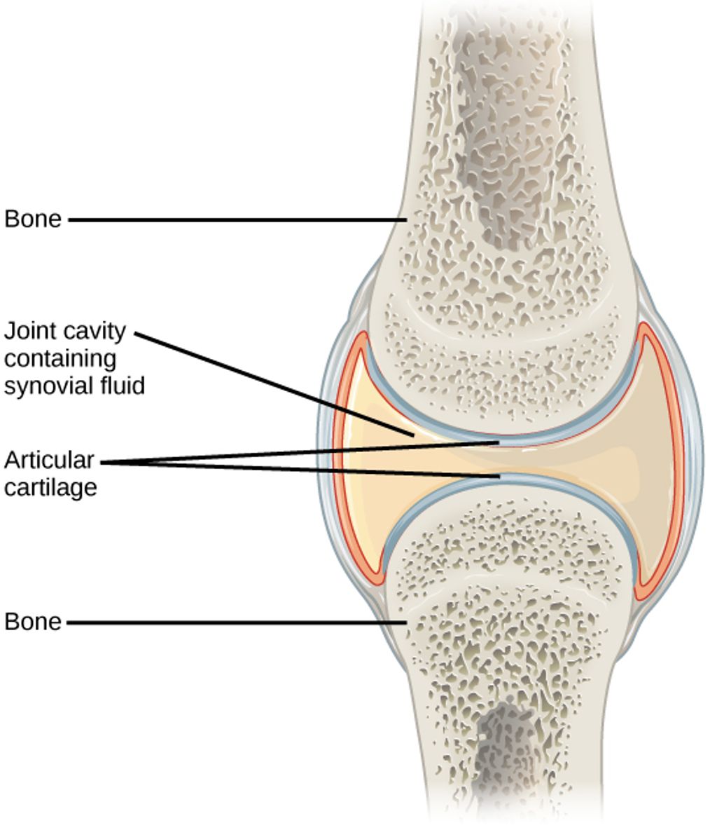

The knee joint is a complex structure that plays a crucial role in supporting the body and facilitating movement. Understanding the anatomy of the knee joint, including its bones and surrounding structures, is essential for both medical professionals and individuals seeking to maintain optimal joint health. In this article, we will explore the structure of the knee joint, focusing on the femur and tibia bones, the patella bone, the ligaments and tendons, and the articular cartilage.
The femur and tibia bones are the primary bones that form the knee joint. These bones play a crucial role in supporting the body’s weight and facilitating movement. The femur is the longest and strongest bone in the body, while the tibia is the second longest and provides stability to the knee joint. The interaction between these two bones is essential for the proper functioning of the knee joint.
Moving beyond the femur and tibia, we encounter the patella, or kneecap, which plays a crucial role in the knee joint’s function. The patella is a small, triangular bone that protects the knee joint and improves the leverage of the thigh muscles, which are essential for walking, running, and jumping.
The patella’s posterior surface is lined with articular cartilage, which aids in smooth movement against the femur. This cartilage is vital for absorbing stress and reducing friction during knee motion.
Remember, the health of the patella’s articular cartilage is key to maintaining knee mobility and reducing the risk of injury.
After discussing the ligaments and tendons, we must emphasize the importance of proper treatment for common knee injuries. Common knee injuries requiring surgery include ACL tears, fractures in the kneecap, torn meniscus, and patellar tendonitis. Proper treatment is crucial to avoid chronic pain and complications. It is essential to consult a healthcare professional for accurate diagnosis and personalized treatment plans. Additionally, rehabilitation and physical therapy play a vital role in the recovery process. We cannot stress enough the significance of early intervention and adherence to the prescribed treatment regimen. We must prioritize the long-term health and functionality of the knee joint.
As we delve deeper into the knee joint’s anatomy, we encounter the articular cartilage, a pivotal element in facilitating smooth joint movement. This specialized structure coats the surfaces of the bones within the joint, notably the femur, tibia, and patella, providing a slick, frictionless interface that aids in the effortless bending and extending of the knee.
The health of the articular cartilage is crucial for maintaining knee function. Over time, it can wear down or become damaged, leading to conditions such as osteoarthritis. To preserve the integrity of this cartilage, it is essential to understand its composition and the factors that affect its well-being.
Tip: Regular exercise and maintaining a healthy weight can help reduce the stress on knee cartilage, potentially slowing the progression of degenerative conditions.
In our comprehensive guide to knee anatomy, we also explore the morphology and function of the knee joint, as well as common issues that can arise. Effective management of knee health includes understanding over-the-counter solutions for knee pain and adopting strategies for managing discomfort during activities such as running and walking.
In conclusion, the anatomy of the knee joint bones is a complex and intricate system that plays a crucial role in human mobility and stability. Understanding the structure and function of these bones is essential for medical professionals, researchers, and individuals seeking to maintain optimal joint health. Further exploration of the interplay between the various components of the knee joint bones holds promise for advancements in orthopedic medicine and the treatment of musculoskeletal conditions.
The femur and tibia bones form the major weight-bearing structure of the knee joint and are responsible for stability and movement.
The patella bone acts as a protective covering for the knee joint and provides leverage for the quadriceps muscles.
Ligaments are tough bands of tissue that connect bones to each other, providing stability to the knee joint. Tendons are fibrous cords that attach muscles to bones, allowing movement of the joint.
Articular cartilage covers the ends of the bones in the knee joint, providing a smooth and low-friction surface for movement and absorbing shock during weight-bearing activities.
The knee joint supports the body by distributing the weight and forces from activities such as walking, running, jumping, and standing.
Common knee joint injuries and conditions include ligament tears (such as ACL or MCL tears), meniscus tears, arthritis, and patellar dislocation.
Discover how to alleviate knee pain when kneeling on carpet with our expert guide. Learn…
We understand knee pain after gardening; our step-by-step guide offers effective relief and prevention strategies…
Table of Contents Introduction Understanding Knee Pain Triggers How Smartwatch Technology Monitors Knee Pain Top…
Discover how to alleviate knee pain after sleeping on side with our expert guide. Learn…
Discover the causes and treatments for knee pain when bending but not swelling. We explore…
Knee pain relief for nurses is achievable. We share our expertise on managing knee pain,…