
You’ve probably heard someone say that children seem to grow bigger overnight evidence gathered from extensive research suggests that this is probably true. Many people have a busy schedule that prevents them from sleeping well. However, you should not forget that good sleep is good for your mental state And physical health. This is how sleep affects your bone health.
The Healing Power of Zzz’s: Sleep and Bone Remodeling
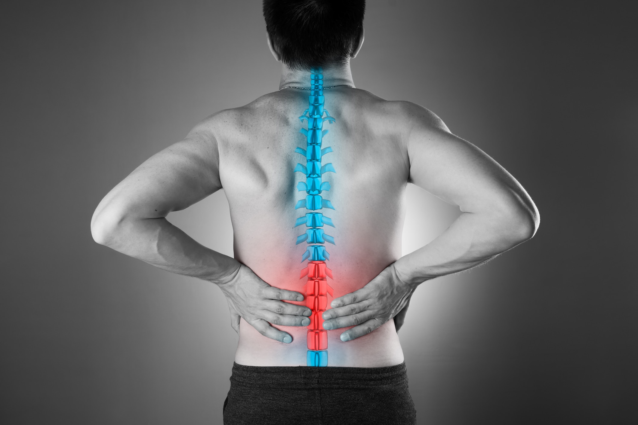
Your body constantly renews, repairs and grows its bones. There is a direct correlation between the amount of sleep you get and the health of your bones. Researchers have made the link individuals who receive shorter sleep duration have lower bone mineral density and a higher risk of osteoporosis. The study, conducted in postmenopausal women, found that women who get five or fewer hours of sleep per night have lower bone mineral density in the spine, neck, hip and even across the body.
Several healthy processes are affected by the amount of sleep you get, and one of these processes is bone remodeling. Your body’s special bone cells, osteocytes, manage bone remodeling. These cells cause various actions in the body, such as helping your bones maintain optimal mineral levels and healing damaged areas. For example, the cells will activate other cells known as osteoclasts, causing them to remove minerals from your bones if your calcium levels get too low. The cells also give rise to osteoblasts, which help them rebuild and repair your bones if you suffer several fractures. The bone remodeling processes are likely to be less effective for individuals who do not get enough rest.
Level up: Improve your sleep hygiene for better bone health
The general rule of thumb is that individuals who sleep and rest longer tend to have healthier bones than those who don’t. Bone growth and repair are facilitated by a good night’s sleep, as the rest gives your body enough time to repair and reshape itself.
Signs of poor sleep hygiene include having trouble sleeping, experiencing daytime sleepiness, and experiencing sleep disturbances. These are the most telling signals; However, another concern to consider is persistently poor sleep quality. Over time, poor sleep hygiene can cause these problems to persist and potentially worsen other health problems.
Creating one healthy sleep routine is important for both your physical and mental health. It improves your productivity and quality. Good sleep hygiene is vital for children and adults; however, it is even more important for individuals likely to be affected by bone-related conditions.
Good sleep habits are good for your health because they create consistency and positive reinforcement for all aspects of life. Good sleep hygiene can be the result of adapting your environment to your needs and establishing the right routines.
Pillow Talk: tips for bone-strengthening sleep
There are many of them steps you can take to improve your sleep experience. You need to optimize your sleep schedule, daily routines, and bedtime routines to help you get better sleep quality. Eat rightget enough fysical activityand create a pleasant environment in which you can relax and fall asleep easily. Here are a few more tips to help you improve your sleep routine:
-
Prioritize your sleep: You need to prioritize sleep if you want healthy bones, body and mind. Calculate your ideal sleep duration by taking into account the time you wake up and make this a regular part of your daily routine.
-
Set a fixed alarm time: Keeping a consistent wake-up time, regardless of the day of the week, can help you regulate your sleep patterns and improve sleep quality.
-
Maintain a regular sleep schedule: Consistency helps synchronize your body’s internal clock.
-
Create a sleep-conducive environment: Keep your bedroom dark, quiet and cool. The ideal temperature is about 65-68 degrees.
-
Limit screen time before bed: Exposure to blue light from screens can disrupt melatonin production.
-
Make gradual adjustments: Try to adjust your sleep pattern gradually. Make adjustments of half an hour or an hour each day until you adjust to your schedule.
-
Don’t take many naps: Avoid taking many naps during the day as this can affect your sleep patterns. Keep naps short and limit them to the early afternoon.
-
Prioritize nutrition: Avoid heavy meals and caffeine right before bed, opting for sleep-supporting snacks instead.
-
Add physical activity: Regular exercise can improve sleep quality, but avoid vigorous exercise before bed.
-
Deal with stress: Techniques such as meditation and deep breathing can alleviate sleep-disrupting stressors.
Incorporating these practical tips into your daily life can make a significant difference in both the quality of your sleep and the health of your bones. By taking proactive steps to improve your sleep habits, you’re investing in a healthier, more resilient future for your bones. So go ahead, prioritize sleep and let your body do its nightly magic for stronger, healthier bones. Good night!