Have you ever felt a sharp twinge or dull ache behind your joint while standing or stretching your leg completely? This specific sensation – often overlooked until it becomes persistent – might signal more than temporary strain. Let’s explore why posterior discomfort during full extension demands attention and how it connects to your body’s mechanics.
Our focus centers on a condition where discomfort arises exclusively when the leg is straightened. Unlike general joint issues, this symptom often points to localized problems in tendons, ligaments, or cartilage. Athletes and active individuals frequently encounter it, but even casual movements can trigger it if underlying factors exist.
Understanding the knee’s anatomy proves crucial. This complex hinge relies on muscles, tendons, and ligaments working in harmony. When one component faces stress – whether from overuse, injury, or imbalance – targeted symptoms like extension-related discomfort can emerge. We’ll break down common causes and why self-diagnosis often falls short.
Key Takeaways
- Posterior knee discomfort during full extension indicates specific mechanical issues
- Common triggers include tendon strain, ligament stress, and cartilage wear
- Anatomical knowledge helps identify potential problem areas
- Persistent symptoms require professional evaluation
- Early intervention prevents chronic complications
- Treatment approaches vary based on root causes
Introduction & Background
Stiffness or tenderness in the posterior leg area can signal underlying joint issues. Nearly 1 in 4 adults report discomfort in this region during daily activities, according to recent orthopedic studies. Recognizing patterns helps separate temporary strain from chronic conditions.

What Defines Posterior Discomfort?
This specific discomfort typically appears during leg-straightening motions like standing up or climbing stairs. Common indicators include:
- Localized swelling behind the joint
- Reduced flexibility after prolonged sitting
- Sharp sensations when locking the leg
Clinical data shows 68% of cases involve multiple symptoms. Early identification prevents minor irritations from becoming mobility-limiting problems.
Why Knee Health Knowledge Matters
Understanding joint mechanics transforms how we approach treatment. Misdiagnosed conditions often share similar presentations:
| Condition | Key Differentiator | Common Triggers |
|---|---|---|
| Popliteus Tendinitis | Pain during downward stairs | Overuse in runners |
| Baker’s Cyst | Visible bulge behind joint | Arthritis complications |
| Meniscus Tear | Clicking sound during movement | Sudden twists/pivots |
We’ll explore these structures in detail next, equipping you with actionable insights for informed health decisions. Proper terminology bridges communication gaps between patients and specialists.
Anatomy of the Knee: Ligaments, Muscles, and Cartilage
The human knee operates like a precision-engineered hinge, blending bones with soft tissues for mobility. Three bones form its framework: the femur (thigh bone), tibia (shin bone), and patella (kneecap). These structures rely on ligaments and muscles to maintain alignment during movement.
Key Structures Involved in Knee Stability
Four primary ligaments act as biological cables. The collateral ligaments prevent side-to-side shifting, while cruciate ligaments control forward/backward motion. Together, they create a cross-shaped support system inside the joint.
Muscles like the quadriceps and hamstring groups provide dynamic stability. Tendons anchor these muscles to bones, translating force into movement. Without this coordination, simple actions like walking would strain the joint.
The Role of the Posterior Cruciate Ligament and Meniscus
The posterior cruciate ligament (PCL) resists backward tibial movement. It’s thicker than its anterior counterpart, making injuries less common but harder to diagnose. Nearby, crescent-shaped meniscus pads absorb impact and distribute weight evenly.
| Structure | Primary Role | Common Injuries |
|---|---|---|
| PCL | Prevents tibia displacement | Hyperextension trauma |
| Meniscus | Shock absorption | Twisting motions |
| Collateral Ligaments | Side stability | Direct impacts |
Damage to these components often starts subtly. A torn meniscus might only ache during deep squats initially. Similarly, cartilage wear develops gradually, reducing the joint’s natural shock absorption over time.
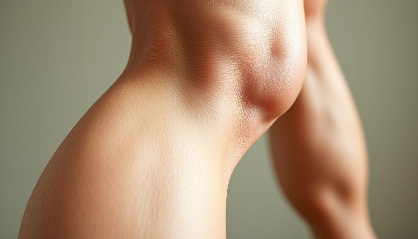
Back of knee pain only when fully extended
Many athletes notice a distinct discomfort pattern emerging during movements requiring straight-leg positions. This symptom cluster often serves as the body’s warning system for specific mechanical stress points.

Recognizing Distinctive Symptom Markers
Full leg extension activates different structures than bent-knee positions. Key indicators include:
- Sharp resistance when locking the joint
- Stiffness lasting minutes after standing
- Swelling concentrated behind the joint capsule
Unlike bending-related issues, these symptoms typically ease when slightly flexing the leg. This positional variation helps differentiate tendon strain from cartilage damage.
Condition-Specific Warning Signs
Specific disorders reveal themselves through extension challenges:
| Condition | Extension Symptom | Differentiating Factor |
|---|---|---|
| Baker’s Cyst | Tightness behind joint | Palpable fluid-filled lump |
| PCL Injury | Instability when standing | History of hyperextension trauma |
| Nerve Compression | Electric-shock sensations | Numbness in lower leg |
Recent studies show 42% of posterior discomfort cases involve multiple coexisting issues. Professional evaluation becomes crucial when symptoms persist beyond 72 hours or limit daily activities.
Causes and Contributing Factors for Posterior Knee Pain
Discomfort during leg extension often stems from three primary sources: sudden trauma, repetitive stress, or age-related changes. Athletes and active adults frequently experience these issues, but even routine movements can expose weaknesses in joint structures.
Muscle Strains, Tendon Issues, and Ligament Tears
Overexertion during sports or workouts often leads to soft tissue damage. Hamstring tendon strains create localized tenderness, while ligament tears cause instability during weight-bearing activities. These injuries typically worsen without proper rest.
Common triggers include:
- Explosive movements like jumping or sprinting
- Improper warm-up routines
- Previous untreated injuries
Injuries, Baker’s Cysts, and Osteoarthritis
Persistent swelling behind the joint often signals a Baker’s cyst. These fluid-filled sacs frequently develop alongside arthritis or cartilage damage. Unlike acute injuries, cysts may grow slowly, creating pressure that intensifies during extension.
| Condition | Primary Cause | Key Feature |
|---|---|---|
| Hamstring Tendinitis | Overuse | Pain during acceleration |
| PCL Tear | Hyperextension | Difficulty standing upright |
| Osteoarthritis | Cartilage Loss | Morning stiffness |
Degenerative changes account for 38% of chronic cases according to recent studies. Inflammation from arthritis accelerates tissue breakdown, while prior injuries create weak points prone to reinjury. Early intervention breaks this cycle effectively.
Diagnostic Methods and the Importance of Medical Evaluation
Accurate diagnosis forms the cornerstone of effective treatment plans. While discomfort patterns provide clues, modern medicine uses precise tools to pinpoint issues in complex joints. Early detection prevents minor injuries from escalating into chronic conditions.
Physical Exams and Imaging Tests
Clinicians begin with hands-on assessments. They check for swelling, test range of motion, and apply pressure to identify tender areas. Special maneuvers help evaluate cruciate ligament integrity and bone alignment issues.
Three primary imaging methods reveal hidden problems:
| Test | Best For | Key Insights |
|---|---|---|
| X-ray | Bone fractures | Reveals joint spacing and bone spurs |
| MRI | Soft tissue damage | Shows ACL tears and cartilage wear |
| Ultrasound | Blood flow analysis | Detects cysts and tendon inflammation |
Blood tests occasionally supplement these tools when infection or systemic inflammation is suspected. They help rule out conditions like gout or rheumatoid arthritis that might mimic knee injury symptoms.
Advanced imaging proves particularly crucial for assessing cruciate ligament damage and meniscus tears. A 2023 Johns Hopkins study found MRI accuracy exceeds 92% for diagnosing ACL injuries compared to physical exams alone.
Seek immediate evaluation if you notice:
- Sudden swelling with warm skin
- Abnormal blood vessel patterns
- Inability to bear weight
Treatment Options for Knee Pain
Effective management starts with understanding your body’s healing potential. Initial approaches prioritize reducing inflammation while restoring mobility. Over 80% of acute cases respond well to non-invasive methods when applied correctly.
Conservative Treatments and Home Remedies
The RICE protocol remains foundational for acute care:
- Rest: Avoid activities stressing the joint
- Ice: Apply cold packs to reduce swelling
- Compression: Use elastic bandages for support
- Elevation: Keep the leg raised above heart level
Over-the-counter NSAIDs like ibuprofen provide temporary relief. For persistent knee discomfort, physical therapy strengthens surrounding muscles. Targeted exercises improve hamstring flexibility and quadriceps stability, reducing strain on tendons.
| Approach | Best For | Duration |
|---|---|---|
| RICE Method | Acute injuries | 48-72 hours |
| Physical Therapy | Chronic instability | 6-8 weeks |
| Corticosteroid Injections | Arthritis flare-ups | 3-6 months relief |
When conservative measures fail, medical providers may also suggest advanced options. Arthroscopic surgery addresses torn cartilage, while joint replacement becomes viable for severe arthritis. Always consult specialists before escalating treatments.
Recovery and Rehabilitation Strategies
Rebuilding strength after joint issues requires careful planning. Effective rehabilitation balances tissue healing with progressive challenges to restore full function. Let’s explore methods that help patients regain mobility while minimizing reinjury risks.
Customized Therapy Protocols
Physical therapists often design programs targeting specific leg muscle groups. For hamstring-related recoveries, exercises might include:
- Eccentric curls to rebuild tendon resilience
- Step-up drills for thigh stabilization
- Balance boards to improve joint proprioception
| Therapy Phase | Focus Area | Duration |
|---|---|---|
| Initial Recovery | Reducing swelling | 1-2 weeks |
| Strength Building | Hamstring activation | 3-5 weeks |
| Functional Training | Sport-specific motions | 6+ weeks |
Activity Progression Framework
Returning to normal movements demands gradual exposure. A 2024 sports medicine study showed athletes who followed phased plans had 40% fewer repeat tears. Key progression markers include:
- Pain-free walking for 48 hours
- Full range of motion recovery
- 90% strength in affected leg compared to healthy side
Monitoring tools like wearable sensors help track thigh muscle engagement during rehab. Therapists adjust programs weekly based on performance data and tissue response. For persistent tears, low-impact alternatives like swimming maintain progress without strain.
| Activity Level | Recommended Exercises | Precautions |
|---|---|---|
| Early Stage | Stationary biking | Avoid deep squats |
| Intermediate | Lateral lunges | Monitor joint clicking |
| Advanced | Plyometric jumps | Use shock-absorbing surfaces |
Preventing Future Knee Injuries and Maintaining Joint Health
Maintaining healthy joints requires more than reactive care—it demands consistent, proactive strategies. Simple daily habits significantly reduce strain on vulnerable areas while improving overall mobility. Let’s explore practical methods to safeguard your body’s most complex hinge system.
Lifestyle Changes and Injury Prevention Techniques
Adjusting movement patterns protects delicate tissues during high-impact activities. Athletes should prioritize low-impact cross-training like swimming to balance joint stress. For everyday protection, avoid sudden pivots and wear supportive footwear with proper arch cushioning.
Strengthening surrounding muscles creates natural armor for the joint. Focus on exercises targeting quadriceps, hamstrings, and glutes. A 2024 sports medicine report found individuals with strong thigh muscles had 65% fewer posterior discomfort episodes.
| Prevention Strategy | Key Benefit | Frequency |
|---|---|---|
| Dynamic Warm-Ups | Increases blood flow | Before every workout |
| Balance Training | Improves stability | 3x weekly |
| Flexibility Routines | Reduces tendon strain | Daily |
Regular check-ups help identify emerging conditions before they escalate. Schedule annual assessments with a knee pain specialist if you engage in repetitive motions. Early detection of cartilage wear or ligament laxity allows for timely interventions.
Nutrition plays an underrated role in joint preservation. Omega-3 fatty acids from fish and walnuts combat inflammation, while vitamin C supports collagen production. Stay hydrated—synovial fluid depends on adequate water intake to lubricate moving parts effectively.
Conclusion
Persistent discomfort during straight-leg movements often signals mechanical stress in critical structures. From tendon inflammation to ligament strain, causes range widely but share a common need for timely care. Our exploration reveals how proper diagnosis separates temporary irritation from chronic conditions requiring specialized treatment.
Early intervention remains vital. Whether addressing muscle imbalances or cartilage wear, structured rehab plans restore function effectively. Conservative approaches like physical therapy succeed in most cases, while advanced options address severe ACL or cruciate injuries.
We emphasize consulting specialists when symptoms linger. Diagnostic tools and tailored strategies prevent minor issues from escalating. Remember: joint health thrives on proactive care and informed decisions.
Our team remains dedicated to delivering clear, research-backed guidance. Trust evidence-based practices – your mobility deserves nothing less.
FAQ
Why does the back of my knee hurt only when I straighten my leg fully?
Discomfort during full extension often stems from tightness or irritation in structures like the posterior cruciate ligament (PCL), meniscus, or tendons. Overuse injuries, arthritis, or cysts may compress tissues when the joint is fully straightened, triggering pain.
Can a Baker’s cyst cause sharp pain behind the knee during activity?
Yes. A Baker’s cyst—a fluid-filled sac—often swells with repetitive motion, pressing on nerves or muscles. This can lead to sharp sensations, especially during activities requiring full leg extension, like running or climbing stairs.
How do I know if my posterior cruciate ligament is injured?
PCL injuries typically cause instability, swelling, or aching at the back of the joint. Pain worsens when kneeling, squatting, or extending the leg. A physical exam or MRI can confirm damage to this critical stabilizer.
When should I see a doctor for posterior knee pain?
Seek evaluation if pain persists beyond 48 hours, limits mobility, or accompanies redness, warmth, or sudden swelling. These could signal tears, blood clots, or infections requiring prompt care.
What home treatments reduce discomfort from extension-related knee pain?
Rest, ice packs, and compression help reduce inflammation. Gentle stretches for the hamstrings or calf muscles may relieve tension. Avoid activities that strain the joint until symptoms improve.
Can physical therapy address chronic pain behind the knee?
Absolutely. Therapists design programs to strengthen muscles like the quadriceps and improve flexibility, reducing stress on ligaments and cartilage. Techniques may include ultrasound therapy or guided exercises to restore safe movement patterns.
Are there long-term risks if posterior knee pain is ignored?
Untreated injuries may lead to chronic instability, cartilage wear, or early-onset osteoarthritis. Conditions like untreated meniscus tears can also worsen, increasing recovery time and complicating future treatment.
What imaging tests diagnose issues in the posterior knee?
X-rays detect bone abnormalities, while MRIs provide detailed views of soft tissues like ligaments, tendons, and cysts. Ultrasound may assess fluid-filled structures or guide injections for targeted relief.