Chronic pain affects millions of people around the world. It is a pain that lasts for months or even years. There are many reasons why someone might have chronic pain. In this article, we will look at some of the main causes of chronic pain.
Key Takeaways
Chronic pain can be caused by problems with the nerves, such as nerve damage or central sensitization.
Inflammation from autoimmune diseases, infections, or chronic inflammatory conditions can lead to long-term pain.
Musculoskeletal issues like arthritis, fibromyalgia, and muscle strain are common causes of chronic pain.
Neurological Factors
Nerve Damage
Nerve damage can lead to chronic pain. When nerves are injured, they might send incorrect signals to the brain, causing pain even without a clear reason. This can happen due to accidents, surgeries, or diseases like diabetes.
Central Sensitization
Central sensitization is when the nervous system becomes overly sensitive. This heightened sensitivity makes even normal touch or movement feel painful. It can result from ongoing pain or injury, making the body react more strongly to pain signals.
Neuropathic Pain
Neuropathic pain is a type of pain caused by nerve problems. It often feels like burning, tingling, or shooting pain. People with conditions like shingles or multiple sclerosis might experience this. Treating neuropathic pain can be challenging, as it doesn’t always respond to regular painkillers.
Chronic pain from neurological factors can be hard to manage, but understanding the causes can help in finding the right treatment.
Inflammatory Causes
Autoimmune Diseases
Autoimmune diseases occur when the body’s immune system mistakenly attacks its own tissues. This can lead to chronic pain as the inflammation persists. Conditions like rheumatoid arthritis and lupus are common examples. Managing these diseases often requires long-term treatment.
Infections
Infections can also cause chronic pain. When the body fights off an infection, inflammation can linger, causing ongoing discomfort. For instance, some bacterial or viral infections can lead to conditions like post-infectious arthritis. It’s important to treat infections promptly to avoid long-term issues.
Chronic Inflammatory Conditions
Chronic inflammatory conditions, such as Crohn’s disease or ulcerative colitis, involve ongoing inflammation in the body. This persistent inflammation can cause significant pain and discomfort. Effective management of these conditions often includes medication, lifestyle changes, and sometimes surgery.
Chronic inflammation can severely impact quality of life, making daily activities challenging.
Musculoskeletal Issues
Arthritis
Arthritis is a common cause of chronic pain. It involves inflammation of the joints, which can lead to stiffness, swelling, and pain. Osteoarthritis and rheumatoid arthritis are the most common types. Managing arthritis often requires a combination of medication, physical therapy, and lifestyle changes.
Fibromyalgia
Fibromyalgia is a condition characterized by widespread musculoskeletal pain. People with fibromyalgia often experience fatigue, sleep disturbances, and memory issues. The exact cause is unknown, but it is believed to involve a combination of genetic and environmental factors. Treatment typically includes medication, exercise, and stress management techniques.
Muscle Strain
Muscle strain occurs when muscles are overstretched or torn. This can happen due to overuse, improper use, or injury. Symptoms include pain, swelling, and limited movement. Rest, ice, compression, and elevation (RICE) are common treatments for muscle strain. In severe cases, physical therapy may be needed to regain strength and flexibility.
Chronic musculoskeletal issues can significantly impact daily life, making it essential to seek appropriate treatment and management strategies.
Psychological Influences
Stress and Anxiety
Stress and anxiety can make chronic pain worse. When you’re stressed, your body releases chemicals that can increase pain. Managing stress is important for people with chronic pain.
Depression
Depression and chronic pain often go hand in hand. Feeling sad or hopeless can make pain feel worse. It’s important to treat both the pain and the depression to feel better.
Trauma and PTSD
People who have been through trauma or have PTSD might feel more pain. The brain and body remember the trauma, which can make pain last longer. Getting help for trauma can also help with pain.
It’s important to understand that the mind and body are connected. Taking care of your mental health can help reduce chronic pain.
Conclusion
Chronic pain can come from many different causes. It might be from an old injury, a disease, or even stress. Understanding what causes chronic pain is important. It helps doctors find the best ways to treat it. If you or someone you know has chronic pain, it’s important to talk to a doctor. They can help find the cause and suggest ways to feel better. Remember, you don’t have to live with pain. There are many treatments that can help.
Living with chronic pain can be tough. It’s a pain that lasts for a long time, and it can make everyday life difficult. But there are ways to manage it. This article will help you understand more about chronic pain and how to deal with it.
Key Takeaways
Chronic pain can come from many different causes and affects daily life in many ways.
There are treatments that don’t involve medicine, like physical therapy and talking with a therapist.
Changing your lifestyle, like eating better and exercising, can help manage pain.
Understanding Chronic Pain
Types of Chronic Pain
Chronic pain can be classified into several types, including nociceptive pain, neuropathic pain, and psychogenic pain. Nociceptive pain arises from tissue damage, while neuropathic pain is due to nerve damage. Psychogenic pain, on the other hand, is linked to psychological factors.
Causes and Risk Factors
There are many causes and risk factors for chronic pain. Injuries, surgeries, and diseases like arthritis can lead to chronic pain. Risk factors include age, genetics, and lifestyle choices such as smoking or lack of exercise.
Impact on Daily Life
Chronic pain can greatly affect daily life. It can make simple tasks difficult and reduce the quality of life. People with chronic pain often experience fatigue, sleep problems, and mood changes.
Living with chronic pain can be challenging, but understanding its types, causes, and impacts can help in managing it better.
Non-Pharmacological Treatments
Physical Therapy
Physical therapy is a common approach to managing chronic pain. It involves exercises and stretches that help improve mobility and reduce pain. Regular sessions can lead to significant improvements in strength and flexibility. Therapists may also use techniques like massage and heat therapy.
Cognitive Behavioral Therapy
Cognitive Behavioral Therapy (CBT) helps people change the way they think about pain. This therapy teaches coping strategies and problem-solving skills. CBT can be very effective in reducing the emotional distress that often accompanies chronic pain.
Alternative Therapies
Alternative therapies include methods like acupuncture, chiropractic care, and yoga. These treatments can offer relief for some people. They are often used alongside other treatments to provide a more comprehensive approach to pain management.
Combining different non-pharmacological treatments can often lead to better outcomes for those suffering from chronic pain.
Pharmacological Treatments
Over-the-Counter Medications
For many people, over-the-counter (OTC) medications are the first line of defense against chronic pain. These include common drugs like acetaminophen and nonsteroidal anti-inflammatory drugs (NSAIDs) such as ibuprofen. OTC medications can be effective for mild to moderate pain and are easily accessible.
Prescription Medications
When OTC medications are not enough, doctors may prescribe stronger drugs. These can include opioids, antidepressants, and anticonvulsants. Prescription medications are often tailored to the specific type of pain and the patient’s overall health. It’s important to follow the doctor’s instructions carefully to avoid complications.
Potential Side Effects
All medications come with potential side effects. Common side effects of pain medications can include drowsiness, nausea, and constipation. More serious risks, like addiction and liver damage, can occur with long-term use. Always discuss the risks and benefits with your healthcare provider.
Managing chronic pain often requires a combination of treatments. It’s essential to work closely with your healthcare team to find the best approach for you.
Lifestyle Modifications for Pain Management
Diet and Nutrition
Eating a balanced diet can help manage chronic pain. Foods rich in omega-3 fatty acids, like fish, can reduce inflammation. Staying hydrated is also important. Avoiding processed foods and sugars can make a big difference.
Exercise and Physical Activity
Regular exercise can improve your overall health and reduce pain. Activities like walking, swimming, or yoga can be very helpful. Start slow and gradually increase your activity level. Consistency is key to seeing benefits.
Stress Management Techniques
Managing stress is crucial for pain management. Techniques like deep breathing, meditation, and mindfulness can help. Joining a support group or talking to a therapist can also be beneficial.
Making small changes in your daily routine can have a big impact on managing chronic pain. It’s important to find what works best for you and stick with it.
Conclusion
In the end, managing chronic pain is about finding what works best for each person. There are many ways to help, like medicine, physical therapy, and talking to a counselor. It’s important to try different things and see what helps the most. Remember, everyone is different, and what works for one person might not work for another. Keep talking to your doctor and stay hopeful. With the right plan, you can live a better life even with chronic pain.
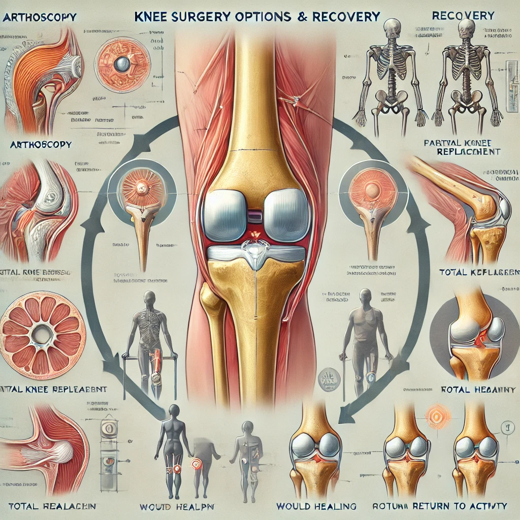
During arthroscopic debridement, small incisions are made around the knee to insert a tiny camera and instruments to remove loose fragments of cartilage or bone and smooth roughened articular surfaces. It provides short-term pain relief but doesn’t address underlying arthritis progression. This outpatient procedure has a relatively quick recovery.
Osteotomy
A knee osteotomy realigns the leg and redistributes knee loads to unload damaged areas. The surgeon cuts and reshapes the tibia or femur, then fixes it in place with plates and screws. This delays the need for knee replacement in younger arthritis patients. Recovery takes several months with restricted weightbearing.
Joint Replacement Recovery
Hospital Stay
After total knee replacement surgery, patients typically stay in the hospital for 1-3 days for pain control and initial recovery. Antibiotics prevent infection and blood thinner injections reduce clotting risk. Occupational therapy starts knee range of motion exercises. Post-op knee swelling is common.
Rehabilitation
Formal physical therapy generally begins 2-3 weeks after knee replacement once surgical wounds have healed adequately. The therapist helps improve flexibility, gait, and strength through graduated exercises. Progress is gradual over 3-6 months as swelling resolves and mobility improves. Home exercise continues after formal therapy ends. Full recovery takes up to a year.
Knee joint pain is a pervasive issue that affects individuals across all age groups and lifestyles. From athletes pushing their physical limits to older adults navigating the challenges of aging joints, knee pain can significantly impact daily activities and overall quality of life. Understanding the underlying causes of knee pain is crucial for accurate diagnosis and effective treatment. This comprehensive guide explores various mechanical problems, types of arthritis, and other potential causes, along with risk factors and diagnostic procedures.
[Image: An anatomical illustration of the knee joint, highlighting its complex structure including bones, cartilage, ligaments, and tendons, with labels pointing to common areas of pain.]
The knee, being one of the largest and most complex joints in the human body, is susceptible to a wide range of issues. Its intricate structure, comprising bones, cartilage, ligaments, and tendons, works in harmony to support our body weight and facilitate movement. However, this complexity also makes it vulnerable to various forms of injury and degeneration.
Mechanical Problems
Mechanical problems in the knee often result from injury or wear and tear on the joint’s components. These issues can cause pain, instability, and reduced range of motion.
Ligament Injuries
Ligaments are tough, elastic bands of tissue that connect bones to each other and provide stability to joints. The knee has four main ligaments, each susceptible to injury:
Anterior Cruciate Ligament (ACL)
Posterior Cruciate Ligament (PCL)
Medial Collateral Ligament (MCL)
Lateral Collateral Ligament (LCL)
[Image: A diagram showing the four main ligaments of the knee, with a side-by-side comparison of a healthy knee and one with a torn ACL.]
ACL Injuries
The ACL is one of the most commonly injured knee ligaments, especially among athletes. ACL tears often occur during activities that involve:
Sudden stops or changes in direction
Pivoting with the foot planted
Landing incorrectly from a jump
ACL injuries can range from mild sprains to complete tears. A characteristic “popping” sound often accompanies the injury, followed by rapid swelling and instability in the knee.
MCL Injuries
The MCL is frequently injured in contact sports or activities that involve quick changes in direction. MCL tears typically result from:
A direct blow to the outer part of the knee
Twisting or rotating the knee while the foot is planted
MCL injuries often cause pain and swelling on the inner side of the knee and may lead to instability when the knee is bent.
Meniscus Tears
The meniscus is a C-shaped piece of cartilage that acts as a cushion between the thighbone (femur) and shinbone (tibia). Each knee has two menisci:
Medial meniscus (inner side of the knee)
Lateral meniscus (outer side of the knee)
[Image: A cross-section view of the knee showing the location and shape of the menisci, with an example of a torn meniscus.]
Meniscus tears can occur due to:
Twisting or rotating the knee, especially when putting full weight on it
Aging and degenerative changes in older adults
Sports injuries, particularly in contact sports
Symptoms of a meniscus tear include:
Pain, especially when twisting or rotating the knee
Swelling and stiffness
Catching or locking of the knee
Difficulty fully straightening the knee
The severity of meniscus tears can vary, from minor tears that heal on their own to more severe tears that may require surgical intervention.
Patellar Tendinitis
Patellar tendinitis, also known as “jumper’s knee,” is an overuse injury affecting the tendon that connects the kneecap (patella) to the shinbone. This condition is common among athletes, especially those involved in sports that require frequent jumping.
[Image: An illustration showing patellar tendinitis, highlighting the inflamed patellar tendon and its connection to the kneecap and shinbone.]
Causes of patellar tendinitis include:
Repetitive stress on the patellar tendon
Sudden increases in training intensity or frequency
Inadequate rest between intense physical activities
Misalignment of the kneecap
Symptoms typically include:
Pain below the kneecap, especially during activities like jumping or climbing stairs
Tenderness when pressing on the affected area
Stiffness, particularly after periods of inactivity
If left untreated, patellar tendinitis can progress from an acute condition to a chronic problem, potentially leading to tendon degeneration and increased risk of rupture.
Arthritis
Arthritis is a common cause of knee pain, especially in older adults. There are several types of arthritis that can affect the knee joint, each with its unique characteristics and treatment approaches.
Osteoarthritis (OA)
Osteoarthritis is the most common form of arthritis affecting the knee. It’s a degenerative condition characterized by the breakdown of cartilage in the joint, leading to pain, stiffness, and reduced mobility.
[Image: A comparison of a healthy knee joint versus one affected by osteoarthritis, showing the cartilage breakdown, bone spurs, and narrowing of the joint space.]
Key features of osteoarthritis include:
Gradual onset of symptoms, typically developing over years
Pain that worsens with activity and improves with rest
Morning stiffness that typically lasts less than 30 minutes
Creaking or grinding sensation in the knee (crepitus)
Development of bone spurs (osteophytes)
Risk factors for developing knee osteoarthritis include:
Advanced age
Obesity
Previous joint injuries
Repetitive stress on the joint
Genetic predisposition
As osteoarthritis progresses, it can lead to significant pain and disability, potentially necessitating joint replacement surgery in severe cases.
Rheumatoid Arthritis (RA)
Rheumatoid arthritis is an autoimmune condition where the body’s immune system mistakenly attacks the synovial membrane, causing inflammation and joint damage. Unlike osteoarthritis, RA often affects both knees simultaneously.
[Image: An illustration comparing a normal knee joint to one affected by rheumatoid arthritis, highlighting synovial inflammation and joint erosion.]
Characteristics of rheumatoid arthritis in the knee include:
Symmetrical joint involvement (both knees often affected)
Pain, swelling, and warmth in the affected joints
Morning stiffness lasting more than an hour
Fatigue and general feeling of illness
Potential for joint deformity in advanced stages
RA is a systemic disease, meaning it can affect other parts of the body beyond the joints, including the skin, eyes, lungs, and blood vessels.
Gout and Pseudogout
Gout and pseudogout are types of arthritis caused by the deposition of crystals within the joint space.
Gout
Gout results from the accumulation of uric acid crystals in the joint. While it most commonly affects the big toe, knee involvement is not uncommon.
[Image: A microscopic view of uric acid crystals associated with gout, alongside an illustration of a gouty knee joint.]
Gout attacks are characterized by:
Sudden onset of severe pain, often occurring at night
Redness, warmth, and swelling in the affected joint
Extreme tenderness, even to light touch
Limited range of motion
Risk factors for gout include:
High levels of uric acid in the blood
Obesity
Excessive alcohol consumption
Diet high in purines (e.g., red meat, organ meats, some seafoods)
Certain medications (e.g., diuretics)
Pseudogout
Pseudogout, also known as calcium pyrophosphate deposition disease (CPPD), is caused by calcium pyrophosphate crystals forming in the joint.
Characteristics of pseudogout include:
Sudden attacks of pain and swelling, similar to gout
More common in older adults
Often affects larger joints like the knee
May be associated with other medical conditions or joint trauma
Both gout and pseudogout can lead to long-term joint damage if left untreated, emphasizing the importance of proper diagnosis and management.
Other Causes
While mechanical problems and arthritis are common culprits, several other conditions can cause knee joint pain.
Infections
Joint infections, also known as septic arthritis, can cause significant knee pain and require immediate medical attention.
[Image: An illustration showing a knee joint affected by septic arthritis, highlighting increased joint fluid and inflammatory changes.]
Causes of knee joint infections include:
Bacterial infections entering the joint through the bloodstream
Direct inoculation through injury or surgery
Spread from nearby infected tissues
Symptoms of a knee joint infection include:
Sudden onset of severe pain
Marked swelling and redness
Warmth around the joint
Fever and chills
Inability to bear weight on the affected leg
Prompt diagnosis and treatment with antibiotics and sometimes surgical drainage are crucial to prevent permanent joint damage.
Bone Tumors
While relatively rare, bone tumors can cause knee pain and swelling. These tumors can be benign (non-cancerous) or malignant (cancerous).
Types of bone tumors that can affect the knee include:
Osteochondromas: Benign bone tumors that typically develop in adolescents and young adults
Giant cell tumors: Usually benign but locally aggressive tumors
Osteosarcoma: A malignant bone cancer that can occur around the knee, especially in children and young adults
[Image: A series of X-ray or MRI images showing different types of bone tumors that can occur around the knee joint.]
Symptoms of bone tumors may include:
Persistent pain, often worse at night
Swelling or visible lump
Fractures due to weakened bone
Limited range of motion
Early detection and proper diagnosis are crucial for effective treatment of bone tumors.
Referred Pain
Sometimes, knee pain may not originate in the knee itself but can be referred from problems in other parts of the body, particularly the hip or lower back.
[Image: A diagram showing how pain from the hip or lower back can be referred to the knee, with nerve pathways highlighted.]
Characteristics of referred knee pain:
Pain patterns that don’t match typical knee injury or arthritis symptoms
Accompanying symptoms in the hip, lower back, or along the leg
Pain that doesn’t respond to typical knee treatments
Proper diagnosis of referred pain is essential to address the underlying cause and provide effective treatment.
Risk Factors
Several factors can increase an individual’s risk of developing knee joint pain. Understanding these risk factors can help in prevention and early intervention.
Age
As we age, the risk of developing knee pain increases due to:
Natural wear and tear on joint cartilage
Decreased muscle strength and flexibility
Higher likelihood of developing osteoarthritis
Accumulated effects of previous injuries
Gender
Gender can play a role in the development of knee pain:
Women are more prone to certain knee problems, such as patellofemoral pain syndrome
Hormonal changes, particularly during menopause, can affect joint health
Anatomical differences, such as wider hips in women, can affect knee alignment and stress
Obesity
Excess weight places additional stress on knee joints, significantly increasing the risk of knee pain and osteoarthritis.
Each pound of body weight exerts about 4 pounds of pressure on the knees when walking
Weight loss can dramatically reduce knee pain and slow the progression of osteoarthritis
[Image: An illustration showing how excess weight increases stress on the knee joint, with comparative figures for normal weight vs. obese individuals.]
High-Risk Activities
Certain activities and occupations can increase the risk of knee problems:
Jobs requiring repetitive knee stress (e.g., construction, carpet laying)
Activities involving frequent kneeling or squatting
While these activities don’t necessarily need to be avoided, proper training, technique, and protective equipment can help reduce the risk of knee injuries.
Diagnostic Procedures
Accurate diagnosis is crucial for effective treatment of knee joint pain. Healthcare providers use a combination of physical examination, imaging tests, and sometimes laboratory analysis to determine the underlying cause of knee pain.
Physical Examination
A thorough physical examination is the first step in diagnosing knee pain. The healthcare provider will:
Observe gait and standing posture
Palpate the knee to check for areas of tenderness, swelling, or warmth
Assess range of motion and stability
Perform specific tests to evaluate ligaments and menisci (e.g., McMurray test, Lachman test)
[Image: A series of photos demonstrating various physical examination techniques for knee assessment.]
Imaging Tests
Various imaging modalities can provide detailed information about the structures within and around the knee joint.
X-rays
X-rays are often the first imaging test performed. They can show:
Bone alignment
Joint space narrowing (indicative of cartilage loss)
Bone spurs (osteophytes)
Fractures
Magnetic Resonance Imaging (MRI)
MRI provides detailed images of soft tissues, including:
Ligaments and tendons
Cartilage and menisci
Bone marrow changes
[Image: Side-by-side comparison of a knee X-ray and MRI, highlighting the different structures visible in each.]
Computed Tomography (CT)
CT scans can be useful for:
Detailed bone imaging
Evaluating complex fractures
Guiding interventional procedures
Ultrasound
Ultrasound can be helpful for:
Evaluating soft tissue structures in real-time
Guiding injections or aspirations
Assessing inflammation in tendons and bursae
Lab Tests
In some cases, laboratory tests may be necessary to diagnose or rule out certain conditions:
Blood Tests
Erythrocyte sedimentation rate (ESR) and C-reactive protein (CRP) to assess inflammation
Rheumatoid factor and anti-CCP antibodies for rheumatoid arthritis
Uric acid levels for gout
Joint Fluid Analysis
Aspiration of joint fluid (arthrocentesis) can help diagnose:
Infections (by culturing the fluid)
Crystal-induced arthritis (by identifying uric acid or calcium pyrophosphate crystals)
Inflammatory conditions (by analyzing cell counts and other markers)
[Image: A microscopic view of joint fluid analysis, showing different types of crystals associated with gout and pseudogout.]
Conclusion
Understanding the various causes of knee joint pain is crucial for both patients and healthcare providers. The knee’s complex structure makes it susceptible to a wide range of issues, from acute injuries to chronic degenerative conditions. By recognizing the signs and symptoms associated with different causes of knee pain, individuals can seek appropriate care more promptly.
It’s important to remember that knee pain can often result from a combination of factors. For instance, a minor injury in a person with underlying osteoarthritis can lead to a significant exacerbation of symptoms. Similarly, lifestyle factors like obesity can compound the effects of age-related joint changes.
Proper diagnosis is key to effective treatment. While some causes of knee pain, such as minor strains or overuse injuries, may resolve with rest and home care, others require professional medical intervention. Persistent or severe knee pain should always be evaluated by a healthcare provider to ensure appropriate management and prevent long-term complications.
By staying informed about the potential causes of knee pain and being proactive about joint health, individuals can take steps to maintain healthy, pain-free knees throughout their lives. Regular exercise, maintaining a healthy weight, using proper techniques during physical activities, and seeking timely medical attention when problems arise are all crucial components of long-term knee health.
[Image: A motivational image showing people of various ages engaged in knee-friendly activities like swimming, cycling, and low-impact exercises, emphasizing the importance of staying active for knee health.]
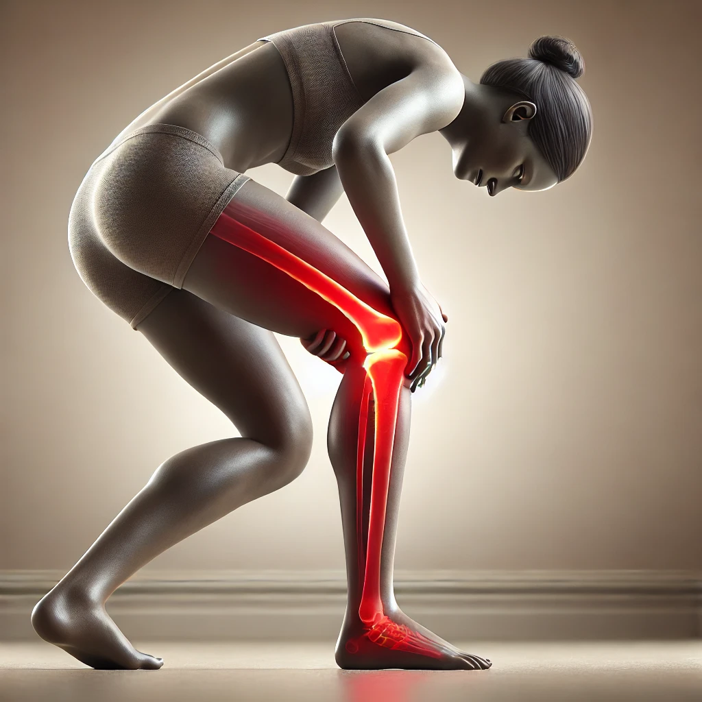
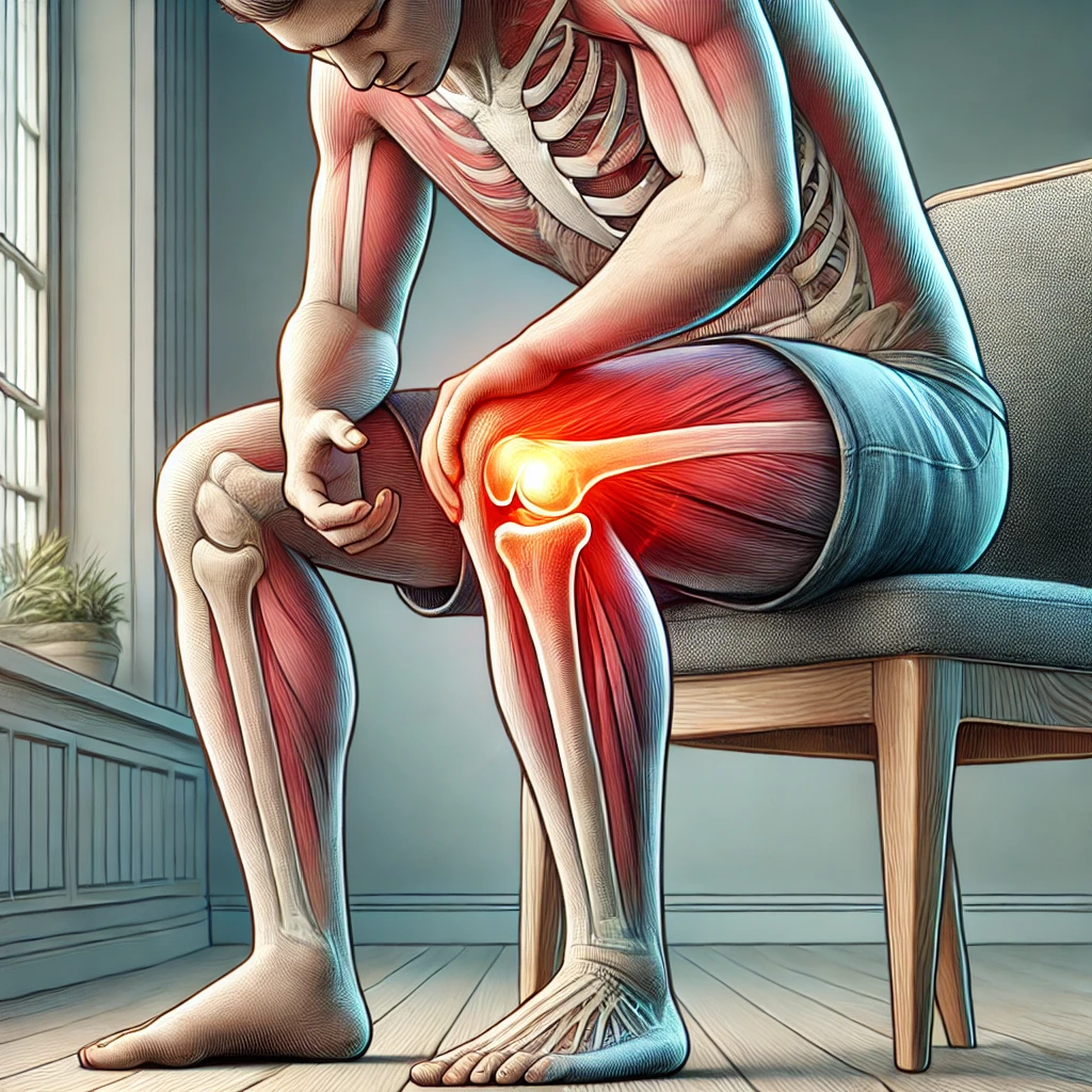
Knee pain when bending is a common issue that many people face. This discomfort can be mild or severe and can affect daily activities. Understanding the causes and treatments for this type of pain is important to find relief and improve quality of life. In this article, we’ll explore the various reasons behind knee pain when bending and discuss ways to manage and prevent it.
Common Causes of Right Side Knee Pain When Bending
Knee pain when bending is a common problem for many people and can have numerous underlying causes. Osteoarthritis, meniscus tears, bursitis, ligament strain, and tendonitis are some of the most common causes. Bone fractures or infections in the joint may also cause knee pain. Often, knee pain can occur due to overuse or an unexpected increase in physical activity. Additionally, obesity or misalignment of the hips (such as hip aberration) can lead to increased pressure and tension on the knees that can cause pain.
Sharp Pain in the Knee When Bending
Torn Ligament or Meniscus
A torn ligament or meniscus can cause sharp pain when bending the knee. This injury often happens during sports or physical activities. You might also notice swelling and difficulty moving your knee.
Knee or Patellar Fracture
A fracture in the knee or patella can lead to severe pain when bending. This type of injury usually results from a direct blow or fall. Symptoms include intense pain, swelling, and an inability to straighten the leg.
Osteoarthritis
Osteoarthritis is a common cause of knee pain, especially in older adults. This condition occurs when the cartilage in the knee joint wears down over time. Symptoms include pain, stiffness, and swelling, which can worsen when bending the knee.
If you experience sharp pain in your knee when bending, it’s important to consult a healthcare professional for a proper diagnosis and treatment plan.
Pain at the Top of the Kneecap When Bending
Knee Bursitis
Knee bursitis happens when the small fluid-filled sacs, called bursae, become inflamed. These sacs help reduce friction and cushion pressure points between your bones and the tendons, muscles, and skin near your joints. When they get irritated, you might feel pain at the top of your kneecap, especially when bending your knee.
Quadriceps Tendonitis
Quadriceps tendonitis is an inflammation of the tendon that connects your quadriceps muscles to your kneecap. This condition often results from overuse, especially in activities that involve a lot of jumping or running. Pain at the top of the kneecap is a common symptom, and it can worsen when you bend your knee.
Osteoarthritis
Osteoarthritis is a type of arthritis that occurs when the cartilage that cushions the ends of your bones wears down over time. This can lead to pain and stiffness in the knee, particularly at the top of the kneecap. Bending the knee can become especially painful as the condition progresses.
If you experience persistent pain at the top of your kneecap when bending, it’s important to consult a healthcare professional for a proper diagnosis and treatment plan.
Diagnosing Knee Pain When Bending
Medical History and Symptoms
When diagnosing knee pain, doctors first look at your medical history and symptoms. They ask about when the pain started, what makes it worse, and if you have any other health issues. This helps them understand the possible causes of your pain.
Physical Examination
Next, the doctor will do a physical exam. They will check your knee for swelling, tenderness, and range of motion. They might also move your knee in different ways to see what hurts. This can help them find out if you have a specific injury or condition.
Imaging Tests
Sometimes, doctors need more information to diagnose your knee pain. They might order imaging tests like X-rays, MRIs, or CT scans. These tests can show details inside your knee, like bones, cartilage, and ligaments. This helps doctors see if there is any damage or other issues causing your pain.
If you are experiencing knee pain, especially when you bend it to walk, kneel, sit, squat, and more, it is likely that you have a condition or injury that needs treatment. It is incredibly difficult to self-diagnose correctly, so seeing an orthopedic specialist is critical.
Treatment Options for Knee Pain When Bending
Medications
For knee pain, doctors often suggest medications to help manage discomfort and reduce inflammation. Over-the-counter pain relievers like ibuprofen or acetaminophen can be effective. In some cases, a doctor might prescribe stronger medications or injections to provide relief.
Physical Therapy
Physical therapy is a common treatment for knee pain. A physical therapist can guide you through exercises that improve strength, flexibility, and mobility in your knee. These exercises are tailored to your specific condition and can help you recover faster.
Surgical Interventions
When other treatments don’t work, surgery might be necessary. Procedures can range from minimally invasive arthroscopy to more complex operations like knee replacement. The type of surgery depends on the cause and severity of your knee pain.
It’s important to consult with a healthcare provider to determine the best treatment plan for your knee pain. They can help you weigh the benefits and risks of each option.
Preventing Knee Pain When Bending
Proper Warm-Up and Stretching
Warming up and stretching before any physical activity is crucial. Always stretch your legs before and after exercising. This helps to prepare your muscles and joints for the activity, reducing the risk of injury. Avoid sudden increases in the intensity of your exercise routine; instead, gradually work your way up.
Strengthening Exercises
Practicing regular strengthening exercises that target the muscles supporting your knees can significantly reduce the risk of injury. Focus on exercises that strengthen your quadriceps, hamstrings, and calves. These muscles help stabilize your knee joint.
Maintaining a Healthy Weight
Maintaining a healthy weight is essential for reducing the strain on your knees. If you are overweight, losing excess weight can make a big difference. This reduces the pressure on your knee joints, making injuries less likely.
Consistent exercise and a balanced diet are key to keeping your knees healthy and pain-free.
When to See a Doctor for Knee Pain
Persistent Pain
If your knee pain doesn’t go away after a few days, it’s time to see a doctor. Chronic pain that lasts for weeks or even months needs medical attention to find out what’s wrong and how to fix it.
Swelling and Instability
If your knee is swollen or feels like it might give out, you should get it checked. Swelling can mean there’s something serious going on inside your knee, and instability can make it hard to walk or do everyday things.
Limited Range of Motion
When you can’t bend or straighten your knee like you used to, it’s a sign you need to see a doctor. Limited movement can be a sign of a bigger problem that needs treatment.
Don’t ignore knee pain. Seeing a doctor early can help you get the right treatment and avoid more problems later.
Conclusion
Knee pain when bending can be a real hassle, affecting your daily activities and overall quality of life. It’s important to remember that there are many possible causes, from minor injuries to more serious conditions like osteoarthritis or torn ligaments. If you’re experiencing this kind of pain, it’s best to consult a doctor for a proper diagnosis and treatment plan. In the meantime, simple measures like rest, ice, and over-the-counter pain relievers can help manage the discomfort. Stay proactive about your knee health, and don’t ignore persistent pain. Taking early action can make a big difference in your recovery and long-term well-being.
Key Takeaways
Knee pain when bending can be caused by different factors like injuries, arthritis, and tendonitis.
Sharp knee pain might indicate a serious issue like a torn ligament or fracture.
Pain at the top of the kneecap could be due to bursitis or tendonitis.
Proper diagnosis often requires a combination of medical history, physical exams, and imaging tests.
Treatment options range from medications and physical therapy to surgical interventions.
Frequently Asked Questions
Why does my knee hurt when I bend it?
Knee pain when bending can be caused by various conditions such as Baker’s cyst, hamstring tendonitis, or a knee injury. Sharp pain might indicate a torn ligament or meniscus, a knee fracture, or osteoarthritis.
What should I do if I have sharp knee pain when bending?
If you experience sharp knee pain when bending, it’s important to see a doctor. They can diagnose the issue, which could be a torn ligament, meniscus, or a fracture. Avoid putting weight on the knee until you get a diagnosis.
Can I treat knee pain at home?
Some mild knee pain can be managed at home with rest, ice, compression, and elevation (RICE). Over-the-counter pain relievers may also help. However, if the pain is severe or persists, you should see a doctor.
How can I prevent knee pain when bending?
To prevent knee pain, make sure to warm up and stretch before activities, strengthen your leg muscles, and maintain a healthy weight. Wearing proper footwear and avoiding sudden increases in physical activity can also help.
When should I see a doctor for knee pain?
You should see a doctor if your knee pain is persistent, causes swelling or instability, or limits your range of motion. These could be signs of a more serious condition that needs medical attention.
What are the treatment options for knee pain?
Treatment options vary depending on the cause and severity of the pain. They can include medications, physical therapy, and in some cases, surgical interventions. Your doctor will recommend the best course of action for your specific situation.
Have you ever felt knee pain while sitting down? You’re not alone. Many people experience this discomfort, and it can range from a minor annoyance to a sign of a bigger issue. Understanding why your knee hurts when sitting is the first step toward finding relief. This article will explore common causes, symptoms, and preventive measures for knee pain related to sitting, as well as treatment options and when to seek professional help.
Common Causes of Knee Pain When Sitting
Knee pain from sitting is a common issue with several potential causes. Understanding these causes can help in managing and preventing the discomfort.
Poor Posture and Positioning
Sitting in an awkward position or with poor posture can strain your knees. When your chair or surface doesn’t provide proper support, it can lead to pain. Improper support is a key factor here.
Underlying Health Conditions
Sometimes, knee pain while sitting can be due to underlying health conditions. These conditions might include arthritis or other joint issues. It’s important to consider these possibilities if the pain persists.
Prolonged Sitting
Sitting for long periods can also cause knee pain. When you stay in one position for too long, it can put stress on your knees. Taking regular breaks to move around can help alleviate this issue.
Symptoms Associated with Knee Pain from Sitting
Identifying Discomfort
When you sit for long periods, you might notice a dull ache or sharp pain in your knees. This discomfort can be a sign that your sitting posture or chair isn’t providing the right support. Pay attention to when the pain starts and how long it lasts. This can help you figure out if sitting is the main cause.
Signs of Poor Support
If your chair doesn’t support your knees well, you might feel pain. Look for signs like your knees being higher or lower than your hips, or your feet not touching the ground. These can all lead to knee pain. Adjusting your chair or using a footrest can help.
When to Seek Medical Advice
Sometimes, knee pain can be a sign of a bigger problem. If the pain doesn’t go away or gets worse, it’s time to see a doctor. Other signs to watch for include swelling, redness, or a feeling of warmth in the knee. These could mean there’s something more serious going on.
If knee pain from sitting is affecting your daily life, don’t ignore it. Simple changes can make a big difference, but sometimes you need a professional to help you find the right solution.
Impact of Sitting Positions on Knee Health
Sitting Cross-Legged
Sitting with your legs crossed might feel comfortable, but it can put a lot of strain on your knees. This position can overstretch the muscles and ligaments around the knee, leading to pain and stiffness. Changing your sitting habits to keep your feet flat on the floor can help reduce this strain.
Sitting on Heels
When you sit on your heels, you place a lot of pressure on your knee joints. This can limit the range of motion and cause discomfort over time. It’s better to sit with your knees at a right angle and your feet flat on the ground to avoid this pressure.
Improper Chair Height
Using a chair that is too high or too low can also affect your knee health. If your chair is too high, your feet may dangle, putting extra pressure on your knees. If it’s too low, your knees may be bent too much, causing strain. Adjusting your chair so that your feet rest comfortably on the floor can make a big difference.
Remember, small changes in how you sit can have a big impact on your knee health. Always aim for a position that supports your body well and reduces unnecessary strain.
Preventive Measures to Avoid Knee Pain
Ergonomic Sitting Solutions
One of the best ways to prevent knee pain is by using ergonomic sitting solutions. Adjust your chair height so that your feet rest flat on the floor and your knees are at a 90-degree angle. Consider using a footrest if needed. Ensure your chair provides good lumbar support to maintain proper posture.
Regular Movement and Breaks
Sitting for long periods can strain your knees. Make it a habit to stand up and move around every 30 minutes. Simple activities like stretching or a short walk can help. Regular movement keeps your joints flexible and reduces stiffness.
Strengthening Exercises
Incorporate exercises that strengthen the muscles around your knees. Strong muscles provide better support and reduce the risk of pain. Focus on exercises like leg lifts, hamstring curls, and calf raises. Always warm up before exercising to prevent injury.
Taking proactive steps to care for your knees can make a significant difference in your overall comfort and mobility. Prioritize these preventive measures to maintain knee health and avoid pain.
Treatment Options for Knee Pain When Sitting
Home Remedies
For mild knee pain caused by sitting, home remedies can be quite effective. Applying ice packs to the affected area can reduce swelling and numb the pain. Over-the-counter pain relievers like ibuprofen can also help. Additionally, gentle stretching exercises can improve flexibility and reduce discomfort.
Physical Therapy
If home remedies aren’t enough, physical therapy might be the next step. A physical therapist can design a personalized exercise plan to strengthen the muscles around your knee. This can help improve your posture and reduce pain. They may also use techniques like massage or ultrasound therapy to alleviate discomfort.
Medical Interventions
In more severe cases, medical interventions may be necessary. Your doctor might recommend corticosteroid injections to reduce inflammation. In rare cases, surgery could be an option to correct underlying issues. Always consult a healthcare professional to determine the best course of action for your specific condition.
It’s important to address knee pain early to prevent it from becoming a chronic issue. Regular check-ups and following your treatment plan can make a significant difference in your knee health.
When to Consult a Healthcare Professional
Persistent Pain
If your knee pain doesn’t go away after a few days, it’s time to see a healthcare professional. Persistent pain can be a sign of a more serious issue that needs medical attention. Don’t ignore it, especially if it keeps you from doing your daily activities.
Associated Symptoms
Look out for other symptoms that come with your knee pain. These can include swelling, redness, or a feeling of warmth around the knee. If you notice any of these, it’s important to get checked out. Sometimes, these symptoms can mean there’s an infection or another condition that needs treatment.
Diagnostic Procedures
When you visit a healthcare provider, they might suggest some tests to find out what’s causing your knee pain. These tests can include X-rays, MRIs, or blood tests. These diagnostic procedures help in understanding the root cause and planning the right treatment for you.
It’s always better to be safe and get a professional opinion if you’re unsure about your knee pain. Early diagnosis can make treatment easier and more effective.
Conclusion
Knee pain while sitting is a common issue that many people face. It can be caused by various factors such as poor posture, sitting for long periods, or underlying health conditions. The good news is that most of these causes can be addressed with simple changes in your sitting habits and environment. If the pain persists, it’s important to consult a healthcare professional to rule out any serious conditions. By paying attention to how you sit and making necessary adjustments, you can significantly reduce or even eliminate knee pain while sitting.
Key Takeaways
Knee pain when sitting can be caused by poor posture, underlying health conditions, or prolonged sitting.
Common symptoms include discomfort, lack of support, and pain when bending the knee.
Sitting positions like cross-legged or on heels can negatively impact knee health.
Preventive measures include ergonomic seating, regular breaks, and strengthening exercises.
Consult a healthcare professional if the pain is persistent or accompanied by other symptoms.
Frequently Asked Questions
Why does my knee hurt when I sit for a long time?
Knee pain when sitting for long periods can be due to poor posture, lack of support, or underlying health conditions like arthritis or tendonitis.
Is it normal to feel knee pain while sitting?
Yes, it’s quite common. Sitting in awkward positions or for too long can put stress on your knee joints, causing discomfort.
What can I do to prevent knee pain when sitting?
To prevent knee pain, try to maintain good posture, use ergonomic chairs, take regular breaks to move around, and do exercises to strengthen your knees.
When should I see a doctor about knee pain from sitting?
If your knee pain is persistent, severe, or accompanied by other symptoms like swelling or redness, it’s best to consult a healthcare professional.
Can sitting cross-legged cause knee pain?
Yes, sitting cross-legged can put extra pressure on your knee joints, leading to pain, especially if done for extended periods.
What are some home remedies for knee pain from sitting?
Home remedies include applying ice, taking over-the-counter pain relievers, doing gentle stretches, and using cushions or ergonomic chairs for better support.
Knee pain while sitting is a common issue that many people face. Whether it’s due to poor posture, lack of support, or sitting for too long, this discomfort can affect your daily life. Understanding the causes and finding ways to prevent and treat this pain can help you stay comfortable and healthy.
Common Causes of Knee Pain When Sitting
Knee pain from sitting is a common issue with several potential causes. Poor posture and positioning can put extra stress on your knees, leading to discomfort. When you sit in an awkward position, your knees may not be aligned properly, causing strain.
Another cause is the lack of proper support. If your chair or seating surface doesn’t support your legs and knees well, it can lead to pain. This is especially true if you sit for long periods without changing positions.
Lastly, underlying health conditions can also contribute to knee pain when sitting. Conditions like arthritis or previous injuries can make your knees more sensitive to the stresses of sitting.
It’s important to pay attention to your sitting habits and make adjustments as needed to avoid knee pain.
How Sitting Too Long Affects Your Knees
Impact on Joint Health
Sitting for extended periods can lead to joint stiffness and discomfort. When you sit for too long, your knees remain in a bent position, which can cause the muscles and tendons around the joint to tighten. This can result in reduced flexibility and increased pain.
Circulation Issues
Remaining seated for long durations can also affect blood flow. Poor circulation can lead to swelling and discomfort in the knee area. It’s important to move around periodically to keep the blood flowing properly.
Muscle Stiffness
Extended sitting can cause your muscles to become stiff and sore. This is especially true for the muscles around your knees. Regular movement and stretching can help alleviate this stiffness and prevent pain.
Recognizing Symptoms of Knee Pain from Sitting
Identifying Discomfort
Knee pain from sitting can be tricky to identify. Common signs include a dull ache or sharp pain in the knee area. You might also feel stiffness or a burning sensation. Pay attention to when the pain starts and if it worsens after sitting for long periods.
When to Seek Medical Advice
If your knee pain persists or gets worse, it’s important to see a doctor. Look out for swelling, redness, or if the pain is severe enough to limit your daily activities. These could be signs of a more serious issue.
Common Misconceptions
Many people think knee pain from sitting is always due to poor posture. While that’s a common cause, other factors like lack of proper support or underlying health conditions can also contribute. It’s not just about how you sit, but also about how long you stay in one position.
Recognizing the symptoms early can help you take steps to prevent further discomfort and potential damage.
Preventive Measures for Knee Pain When Sitting
Choosing the Right Furniture
Selecting the right furniture is crucial. Opt for chairs that provide good support to your lower back and knees. Adjustable chairs can help you find the perfect height and angle. Make sure your feet rest flat on the floor or on a footrest.
Practicing Good Posture
Maintaining good posture can prevent knee pain. Sit up straight with your back against the chair. Keep your knees at a right angle and avoid crossing your legs. Proper alignment helps distribute your weight evenly, reducing strain on your knees.
Taking Regular Breaks
Taking breaks is essential. Stand up and stretch every 30 minutes to an hour. Walk around for a few minutes to improve circulation and reduce stiffness. These small actions can make a big difference in preventing knee pain.
Regular movement and good posture are key to avoiding knee pain when sitting for long periods.
Treatment Options for Knee Pain
Home Remedies
For mild knee pain, home remedies can be quite effective. Resting the knee and applying ice can help reduce swelling. Over-the-counter pain relievers like ibuprofen or acetaminophen can also be useful. Gentle exercises and stretches can improve flexibility and strength.
Physical Therapy
A healthcare provider may suggest physical therapy to strengthen the muscles around the knee. This can include exercises, electronic muscle stimulation, and ultrasound treatments. Physical therapy is often combined with other treatments for better results.
Medical Interventions
For more severe pain, medical interventions might be necessary. Injections, such as corticosteroids, can help reduce inflammation and pain. In some cases, surgery might be required to address underlying issues. It’s important to consult a healthcare professional for a proper diagnosis and treatment plan.
If knee pain persists despite trying these treatments, it’s crucial to seek medical advice. Ignoring the pain can lead to more serious issues down the line.
When to Consult a Healthcare Professional
If your knee pain doesn’t go away after a few days of home care, it might be time to see a doctor. Chronic pain can be a sign of a more serious issue that needs medical attention.
Look out for other symptoms like swelling, redness, or a fever. These could mean there’s an infection or another problem that needs treatment.
A healthcare professional can run tests to find out what’s causing your knee pain. This might include X-rays, MRIs, or blood tests to get a clear picture of your knee’s health.
Don’t ignore persistent knee pain. Early diagnosis and treatment can help prevent more serious problems later on.
Conclusion
Knee pain while sitting is a common issue that many people face. It can be caused by various factors such as poor posture, lack of support, or underlying health conditions. The good news is that most of these problems can be fixed with simple changes like improving your sitting habits or seeking medical advice. Remember, paying attention to your body and making small adjustments can go a long way in keeping your knees healthy and pain-free. If the pain persists, don’t hesitate to consult a healthcare professional for proper diagnosis and treatment.
Key Takeaways
Poor posture and lack of proper support are common causes of knee pain when sitting.
Sitting for long periods can negatively impact joint health, circulation, and muscle flexibility.
Recognizing the symptoms of knee pain early can help in seeking timely medical advice.
Choosing the right furniture and practicing good posture can prevent knee pain.
If knee pain persists, it’s important to consult a healthcare professional for proper diagnosis and treatment.
Frequently Asked Questions
Why does my knee hurt when I’m sitting?
Knee pain when sitting can be caused by poor posture, lack of proper support, sitting for too long, or underlying health conditions.
Is sitting too long bad for my knees?
Yes, sitting for extended periods can negatively impact your knee joints, reduce circulation, and cause muscle stiffness.
What are some symptoms of knee pain from sitting?
Symptoms include discomfort, stiffness, and pain in the knee area, especially after sitting for long periods.
How can I prevent knee pain when sitting?
You can prevent knee pain by using supportive furniture, practicing good posture, and taking regular breaks to move around.
What are some home remedies for knee pain?
Home remedies include applying ice, taking over-the-counter pain relievers, and doing gentle stretches and exercises.
When should I see a doctor for knee pain?
You should consult a healthcare professional if you have persistent pain, swelling, or if the pain is associated with other symptoms.
Knee pain while sitting is a common issue that many people face. It can be caused by various factors, including poor posture, sitting for too long, or underlying health problems. Addressing this pain often involves making changes to how you sit and seeking advice from a healthcare provider.
Common Causes of Knee Pain When Sitting
Knee pain from sitting is a common issue with several potential causes. Poor posture and positioning can put extra stress on your knees. Sitting with your legs crossed or in an awkward position can lead to discomfort. Additionally, the chair or surface you sit on might not provide the proper support your legs and knees need.
Poor Posture and Positioning
Sitting in a way that strains your knees can cause pain. For example, crossing your legs or sitting with your knees bent for too long can lead to discomfort. It’s important to maintain a good posture to avoid putting extra stress on your knees.
Underlying Health Conditions
Certain health conditions, like arthritis or patellofemoral pain syndrome, can make your knees hurt when you sit. These conditions can cause inflammation and pain, especially if you sit for long periods.
Prolonged Sitting
Sitting for too long without moving can also cause knee pain. When you stay in one position for a long time, your muscles can get stiff and sore. It’s important to take breaks and move around to keep your knees healthy.
The johns hopkins medicine website offers medical research, patient care, and education. Highlighted by new dean appointment and pediatric care advancements.
Symptoms Associated with Knee Pain from Sitting
Knee pain from sitting can be quite uncomfortable and may interfere with daily activities. Recognizing the symptoms can help in addressing the issue effectively.
Treatment Options for Knee Pain When Sitting
Home Remedies and Lifestyle Changes
For many, knee pain from sitting can be managed with simple home remedies and lifestyle adjustments. Regular movement is key; try to stand up and stretch every 30 to 60 minutes. Applying ice packs can help reduce swelling and discomfort. Over-the-counter pain relievers like ibuprofen can also be effective.
Medical Treatments
If home remedies don’t provide relief, it might be time to consult a healthcare professional. They may recommend physical therapy to strengthen the muscles around the knee. In some cases, corticosteroid injections can help reduce inflammation. For more severe conditions, surgical options might be considered.
When to See a Doctor
It’s important to know when to seek professional help. If your knee pain is severe, persistent, or accompanied by other symptoms like swelling or redness, it’s time to see a doctor. They can perform diagnostic tests to determine the underlying cause and develop a tailored treatment plan.
Don’t ignore persistent knee pain. Early intervention can prevent more serious issues down the line.
Preventive Measures to Avoid Knee Pain While Sitting
To keep knee pain at bay while sitting, it’s important to take some preventive steps. Mindfulness of your sitting posture can make a big difference. Regularly check how you’re sitting and make adjustments to avoid discomfort.
Ergonomic Sitting Solutions
Using ergonomic furniture can help a lot. Chairs with proper support for your back and knees can reduce strain. Make sure your feet are flat on the ground and your knees are at a right angle.
Regular Movement and Stretching
Taking breaks to move around is crucial. Stand up and walk for a few minutes every hour. Stretching your legs can also help keep your knees flexible and pain-free.
Strengthening Exercises
Doing exercises to strengthen the muscles around your knees can provide better support. Simple exercises like leg lifts and squats can be very effective.
Remember, staying active and mindful of your posture can go a long way in preventing knee pain.
When to Seek Professional Help for Knee Pain
Signs of Serious Conditions
If your knee pain is stopping you from doing daily activities or exercise, it’s time to see a healthcare provider. Persistent knee problems should be evaluated, especially if you have new pain, worsening pain, or pain that has lasted for several days. If you have an injury or are unsure of the cause, it’s best to get it checked out.
Diagnostic Procedures
When you have sharp or recurring knee pain, it’s important to get a professional diagnosis. This usually involves looking at your medical history and doing a physical exam. Doctors might also do some physical tests to see how well you can move your knee and how much it hurts.
Treatment Plans
Depending on how bad your knee pain is, a doctor might suggest different treatments. They could recommend home remedies, lifestyle changes, or medical treatments. If your pain is severe or chronic, your primary care provider might send you to a specialist.
If your knee pain is severe, doesn’t get better in a few weeks, or comes with swelling, locking, or clicking, you should see a doctor right away.
Impact of Furniture on Knee Pain
Choosing the Right Chair
The chair you use can greatly affect your knee pain. A supportive chair can do wonders to reduce discomfort, especially if you sit for long periods. Make sure your chair is not too low, as this can cause your knees to bend too much, leading to pain. It’s also important to position your chair correctly at your desk to avoid straining your knees and back.
Importance of Proper Support
The surface you sit on plays a big role in whether you feel knee pain. If your seat is too low, your knees will be in a more strenuous bent position, which can cause pain. Look for seats with an ergonomic design that provides good support, especially if you will be sitting for four or more hours at a time.
Adjusting Your Workspace
Your workspace setup can also impact your knee health. If your desk and chair are not at the right height and distance, you might end up in an awkward position that can lead to knee pain over time. Make sure your workspace is ergonomically arranged to keep your knees and back comfortable.
If you have knee pain while sitting, consider using a standing desk or taking breaks to stretch and move every 30 to 60 minutes.
Conclusion
Knee pain when sitting is a common issue that many people face. It can be caused by poor posture, sitting for too long, or even underlying health conditions. To prevent and treat this pain, it’s important to practice good sitting habits and consult a healthcare professional if the pain persists. Simple changes like adjusting your sitting position, taking breaks to move around, and using supportive furniture can make a big difference. Remember, if your knee pain is frequent or severe, it’s best to seek medical advice to rule out any serious conditions. Taking these steps can help you stay comfortable and pain-free while sitting.
Key Takeaways
Knee pain from sitting can stem from poor posture, prolonged sitting, or health issues.
Common symptoms include pain when bending the knee, discomfort in certain positions, and pain during transitions.
Home remedies and lifestyle changes can help alleviate knee pain, but medical treatment may be necessary for severe cases.
Preventive measures like ergonomic seating, regular movement, and exercises can reduce the risk of knee pain.
It’s important to consult a doctor if knee pain persists or worsens, as it may indicate a serious condition.
Frequently Asked Questions
Why does my knee hurt when I sit for a long time?
Knee pain from sitting for long periods can be caused by poor posture, improper leg positioning, or even the type of chair you’re using. Sometimes, underlying health conditions like arthritis can also cause knee pain.
Is knee pain when sitting normal?
Yes, knee pain when sitting is fairly common. It can happen due to sitting in an uncomfortable position or for too long. However, if the pain is frequent and severe, you should consult a doctor.
What can I do to relieve knee pain when sitting?
To relieve knee pain when sitting, try adjusting your sitting position, using a chair with proper support, and taking frequent breaks to move around and stretch. If the pain persists, consider seeing a healthcare professional.
When should I see a doctor for knee pain when sitting?
You should see a doctor if your knee pain is severe, frequent, or accompanied by swelling, redness, or difficulty moving the knee. These could be signs of a more serious condition that needs medical attention.
Can the type of chair I use affect my knee pain?
Yes, the type of chair you use can significantly impact your knee pain. Chairs that lack proper support or are not ergonomically designed can contribute to knee discomfort. Choosing a chair with good support can help alleviate pain.
Are there exercises to prevent knee pain when sitting?
Yes, regular movement and stretching exercises can help prevent knee pain when sitting. Strengthening exercises for the muscles around the knee can also provide better support and reduce pain.
Kneecap injuries, also known as patellar injuries, are common, especially among athletes and active individuals. These injuries can happen due to various reasons such as falls, accidents, or sports activities. The kneecap, or patella, plays a crucial role in the knee joint, helping with leg movement and providing protection. Understanding the causes, types, symptoms, and treatment options for kneecap injuries can help in managing and preventing them effectively.
Common Causes of Kneecap Injuries
Kneecap injuries can happen for many reasons. Here are some of the most common causes:
Trauma and Accidents
A direct hit to the knee can cause serious damage. Falls on hard surfaces like concrete, car accidents where the knee hits the dashboard, and even gunshot wounds can lead to kneecap injuries. The patella, located at the front of the knee, is especially vulnerable during these events.
Sports-Related Injuries
Playing sports can also put your kneecap at risk. Activities where the knee might get hit by a ball, bat, or stick are common culprits. Sudden movements or sharp impacts can also cause injuries, especially if the quadriceps muscle pulls on the kneecap too hard.
Anatomical Issues
Sometimes, the way your body is built can make you more likely to hurt your kneecap. Problems within the knee joint itself can lead to injuries over time. This can include issues like misalignment or weak muscles around the knee.
Kneecap injuries are often the result of a combination of factors, making it important to understand the various causes to prevent them effectively.
Types of Kneecap Injuries
Patellar Fractures
Patellar fractures occur when the kneecap bone breaks. This can happen due to a direct blow or a fall onto the knee. These fractures can be very painful and may limit your ability to straighten your knee. Treatment often involves immobilization with a cast or brace, and in severe cases, surgery may be required.
Dislocations
A dislocated kneecap happens when the patella is forced out of its normal position in the groove at the end of the femur. This can damage nearby soft tissues and cause significant pain and swelling. Immediate medical attention is usually necessary to realign the kneecap, followed by physical therapy to strengthen the surrounding muscles.
Tendon Tears
Tendon tears involve the ripping of the tendons that connect the kneecap to the muscles of the thigh and shin. These injuries can result from sudden, forceful movements or overuse. Symptoms include pain, swelling, and difficulty moving the knee. Treatment options range from rest and physical therapy to surgical repair, depending on the severity of the tear.
Kneecap injuries can vary widely in severity, but early diagnosis and appropriate treatment are crucial for effective recovery.
Symptoms of Kneecap Injuries
Pain and Swelling
Pain is the most common symptom of a kneecap injury. It can be sharp or dull and often worsens with movement. Swelling usually follows, making the knee appear red and puffy.
Locking and Instability
The knee might lock up, making it hard to straighten. It can also feel unstable, as if it might give way, which is often due to patellofemoral instability.
Difficulty in Movement
Injuries can make bending or straightening the knee difficult. In severe cases, you might not be able to put any weight on the leg at all.
Diagnosis Methods for Kneecap Injuries
Physical Examination
A physical examination is often the first step in diagnosing a kneecap injury. During this exam, the doctor will check for signs of pain, swelling, and instability. They may also perform manual tests, such as trying to extend your knee against gravity, to assess the damage to the kneecap or tendons. The straight leg raise test can reveal issues with the extensor mechanism, which includes the quadriceps tendon, patella, and patellar tendon.
Imaging Tests
Imaging tests are crucial for a more detailed look at the injury. Common imaging tests include X-rays, MRI scans, and CT scans. These tests help to identify fractures, dislocations, and other structural problems. An X-ray is often the first imaging test done to check for broken bones. If more detail is needed, an MRI or CT scan may be used to get a clearer picture of the soft tissues and bones.
Medical History Review
Reviewing the patient’s medical history is another important step. The doctor will ask about any previous knee injuries, surgeries, or conditions that might affect the current injury. They will also inquire about the circumstances leading to the injury, such as a fall or accident, and any symptoms experienced at the time, like hearing a pop or feeling the knee give way.
Quick and accurate diagnosis is key to effective treatment and recovery. If you suspect a kneecap injury, seek medical attention promptly.
Treatment Options for Kneecap Injuries
Home Remedies and Rest
For minor kneecap injuries, the RICE method is often recommended. This includes rest, ice, compression, and elevation. Over-the-counter anti-inflammatory medications like ibuprofen can help reduce pain and swelling. It’s important to avoid putting weight on the injured knee to allow it to heal properly.
Medical Interventions
More serious injuries may require medical interventions. These can include wearing a cast or brace to keep the leg stable while the bone heals. Pain medications, including opioids for severe pain, may be prescribed initially, followed by non-opioid options. Physical therapy is often recommended to help regain strength and mobility.
Surgical Procedures
In cases where the injury is severe or doesn’t respond to other treatments, surgery may be necessary. Surgical options can include repairing torn tendons or ligaments, or fixing fractures with screws and plates. Recovery from surgery will typically involve a period of immobilization followed by physical therapy to restore function.
Preventing Kneecap Injuries
Protective Gear
Wearing the right protective gear can make a big difference. Knee pads and braces can help shield your kneecaps during activities that put them at risk. Always choose gear that fits well and is appropriate for your specific sport or activity.
Strengthening Exercises
Doing exercises to strengthen your thigh muscles, like quadriceps and hamstrings, can help keep your knees stable. Strong muscles support your kneecaps better, reducing the chance of injury. Include both strength training and flexibility exercises in your routine.
Safe Practices in Sports
Practicing safe techniques in sports is crucial. Warm up before you start, and gradually increase the intensity of your workouts. Avoid sudden, intense movements that can strain your knees. If something causes knee pain, it’s best to stop and rest.
Taking simple steps like wearing the right shoes and maintaining a healthy weight can also help protect your knees. These actions lower the stress on your kneecaps and keep them healthy.
Recovery and Rehabilitation
Physical Therapy
Physical therapy is essential for regaining movement and strength in the knee. Rehabilitation focuses on improving range of motion, building up muscle strength, and decreasing knee stiffness. Recovery can take several months, depending on the severity of the injury.
Pain Management
Managing pain is crucial during the recovery process. If your pain is severe, your doctor may suggest a prescription-strength medication for a few days. Over-the-counter pain relievers can also be effective.
Long-term Care
Long-term care involves regular check-ups and possibly ongoing physical therapy. This ensures that the knee continues to heal properly and helps prevent future injuries. Staying active and following your doctor’s advice are key components of long-term care.
Whether your treatment is surgical or nonsurgical, rehabilitation will play a vital role in getting you back to your daily activities.
Conclusion
Kneecap injuries are common, especially among active individuals and athletes. They can range from minor bruises to severe fractures and dislocations. It’s important to recognize the symptoms early and seek appropriate medical attention. While some injuries can be managed with rest and home care, others may require more intensive treatments like braces or even surgery. Understanding the causes and types of kneecap injuries can help in preventing them and ensuring a quicker recovery if they do occur. Always consult with a healthcare provider for a proper diagnosis and treatment plan.
Key Takeaways
Kneecap injuries often result from trauma, sports activities, or anatomical problems.
Common types of kneecap injuries include fractures, dislocations, and tendon tears.
Symptoms of kneecap injuries usually involve pain, swelling, and difficulty in movement.
Diagnosis typically involves a physical exam and imaging tests like x-rays.
Treatment can range from home remedies and rest to medical interventions and surgery.
Frequently Asked Questions
What are common causes of kneecap injuries?
Kneecap injuries can happen due to falls, sports activities, or car accidents. Anything that hits your knee hard can cause an injury.
What types of kneecap injuries are there?
There are several types of kneecap injuries, including fractures, dislocations, and tendon tears.
How do I know if I have a kneecap injury?
Common signs include pain, swelling, difficulty moving your knee, and feeling like your knee might give out.
How are kneecap injuries diagnosed?
Doctors usually do a physical exam and may use imaging tests like x-rays to see the injury clearly.
What are the treatment options for kneecap injuries?
Treatment can range from rest and home care for minor injuries to medical interventions like braces or surgery for more serious cases.
How can I prevent kneecap injuries?
Wearing protective gear, doing exercises to strengthen your knees, and following safe practices in sports can help prevent injuries.
Knee pain when sitting is a common issue that many people face. Whether it’s a dull ache or sharp discomfort, this type of pain can make it difficult to relax or focus on tasks. Understanding the causes, symptoms, and preventive measures can help you manage and alleviate this pain effectively.
Common Causes of Knee Pain When Sitting
Improper Support
One of the main reasons for knee pain when sitting is improper support. If the chair or surface you are sitting on does not provide adequate support to your legs and knees, it can lead to discomfort and pain. Ensuring that your seating arrangement offers proper support can help alleviate this issue.
Poor Sitting Posture
Poor sitting posture is another common cause of knee pain. When you sit in a position that puts strain on your knees, it can result in pain and discomfort. It’s important to maintain a good posture to avoid putting unnecessary stress on your knees.
Prolonged Sitting
Sitting for extended periods can also contribute to knee pain. When you remain in a sitting position for too long, it can cause stiffness and discomfort in your knees. Taking regular breaks to stand up and move around can help prevent this problem.
Remember, making small adjustments to your sitting habits can make a big difference in preventing knee pain.
Symptoms Associated with Knee Pain from Sitting
Identifying Discomfort
When you sit for long periods, you might start to feel a dull ache or sharp pain in your knees. This discomfort can be a sign that your sitting habits are affecting your knee health. Pay attention to when and where the pain occurs to help identify the cause.
Recognizing Stiffness
Stiffness in the knees after sitting is another common symptom. You might find it hard to straighten your legs or feel like your knees are locked in place. This stiffness can make it difficult to move around after getting up.
Noting Swelling
Swelling around the knee area can also occur from sitting too long. This can be due to fluid buildup or inflammation. If you notice your knees look puffy or feel warm to the touch, it could be a sign of swelling.
It’s important to monitor these symptoms and make changes to your sitting habits to prevent further knee issues.
How Sitting Position Affects Knee Health
Cross-Legged Sitting
Sitting with your legs crossed can put pressure on your kneecaps. This position keeps your knees bent and can lead to stiffness and pain. Changing how you sit can help reduce discomfort. Try to keep your feet on the floor and your back straight for better posture.
Sitting on Heels
When you sit on your heels, your knees are under a lot of pressure. This can limit your knee’s range of motion and cause pain. It’s important to avoid sitting like this for long periods.
Bent Knee Positions
Any position that keeps your knees bent for too long can cause problems. This includes sitting in awkward positions or kneeling. These positions can make your knees stiff and sore. To prevent this, try to change your position often and avoid staying in one place for too long.
Mindfulness of Positioning: If you like to sit with your legs crossed, be aware of how your knees feel. Change positions if you start to feel a dull ache or pain.
Preventive Measures for Knee Pain When Sitting
Choosing the Right Chair
Selecting a chair with proper support is crucial. Ensure your chair has good lumbar support and allows your feet to rest flat on the floor. This helps in maintaining a natural posture and reduces strain on your knees.
Maintaining Good Posture
Sitting with a straight back and shoulders relaxed can prevent knee pain. Avoid slouching or leaning forward for long periods. Mindfulness of your posture can make a significant difference in how your knees feel.
Taking Regular Breaks
Taking breaks to stand up and move around is essential. Aim to stand up and stretch every 30 minutes. This simple action can alleviate knee pain and improve overall circulation.
Regular movement and proper support are key to preventing knee pain when sitting. Make these small changes to your routine to keep your knees healthy and pain-free.
Treatment Options for Knee Pain from Sitting
Home Remedies
For mild knee pain caused by sitting, home remedies can be quite effective. Applying ice packs to the affected area can reduce swelling and numb the pain. Over-the-counter pain relievers like ibuprofen can also help. Gentle stretching exercises can improve flexibility and reduce stiffness.
Medical Treatments
If home remedies don’t work, it’s important to consult a healthcare professional. They may prescribe stronger medications or recommend injections to relieve pain and inflammation. In some cases, they might suggest wearing a knee brace for added support.
Physical Therapy
Physical therapy is another effective treatment option. A physical therapist can design a personalized exercise program to strengthen the muscles around the knee, improving stability and reducing pain. They may also use techniques like ultrasound or electrical stimulation to promote healing.
If knee pain persists despite these treatments, it’s crucial to seek further medical advice to rule out any serious underlying conditions.
When to Seek Medical Advice
Persistent Pain
If your knee pain doesn’t go away or keeps coming back, it’s time to see a doctor. Persistent knee problems can be a sign of something more serious. Don’t ignore it if the pain sticks around for several days or gets worse over time.
Difficulty in Movement
When moving your knee becomes hard or painful, you should talk to a healthcare provider. This includes trouble bending or straightening your knee, or if it feels weak or unstable. If you find it hard to put weight on your knee, it’s important to get it checked out.
Signs of Underlying Conditions
Sometimes, knee pain can be a sign of other health issues. Look out for swelling, redness, or warmth around the knee. If your knee locks, clicks, or buckles, these could be signs of underlying conditions that need medical attention.
If you keep getting stabs of pain or moving your legs becomes a chore, you might need a professional evaluation.
Conclusion
Knee pain while sitting is a common issue that many people face. It can be caused by poor posture, lack of proper support, or sitting for extended periods. While it may seem like a minor inconvenience, it can lead to more serious problems if not addressed. Simple changes like improving your sitting posture, using supportive furniture, and taking regular breaks to move around can make a big difference. If the pain persists, it’s important to consult a healthcare professional to rule out any underlying conditions. By paying attention to your body and making small adjustments, you can alleviate knee pain and improve your overall comfort.
Key Takeaways
Improper support and poor posture are common causes of knee pain when sitting.
Symptoms to watch for include discomfort, stiffness, and swelling.
Sitting positions like cross-legged or on your heels can affect knee health.
Choosing a good chair and maintaining proper posture can prevent knee pain.
Home remedies, medical treatments, and physical therapy are available for managing knee pain.
Frequently Asked Questions
Why does my knee hurt when I sit for a long time?
Knee pain from sitting too long can be caused by poor support, bad posture, or staying in one position for too long. These factors can strain your knee joints.
Can sitting in certain positions cause knee pain?
Yes, sitting cross-legged, on your heels, or with bent knees can put stress on your knee joints and cause pain.
How can I prevent knee pain when sitting?
To prevent knee pain, choose a chair with good support, maintain good posture, and take regular breaks to move around.
What are some home remedies for knee pain from sitting?
Home remedies include applying ice, using over-the-counter pain relievers, and doing gentle stretches to relieve knee pain.
When should I see a doctor for knee pain when sitting?
You should see a doctor if you have persistent pain, difficulty moving, or signs of a more serious condition like swelling or redness.
Can poor posture really cause knee pain?
Yes, poor posture can lead to knee pain because it puts extra pressure on your knee joints. Sitting with proper alignment helps reduce this pressure.