Author: Mokhtar

  • Patella Pain When Bending Knee Causes & Relief

    Patella Pain When Bending Knee Causes & Relief

    Did you know that approximately 25% of adults experience knee pain at some point in their lives?

    If you’re one of the many individuals who suffer from knee pain when bending, you understand the impact it can have on your daily activities and overall quality of life. Whether it’s a sharp, shooting pain or a dull ache, knee discomfort can be debilitating and frustrating.

    In this article, we will explore the various causes of patella pain when bending the knee and discuss effective methods for finding relief. From common conditions such as patellofemoral syndrome and patellar tendonitis to home remedies and medical treatments, we’ll provide you with the information you need to manage and overcome your knee pain.

    So, let’s dive in and discover the potential sources of your knee pain and how you can regain mobility and comfort.

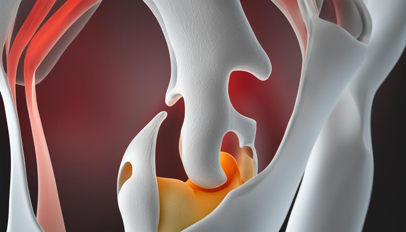
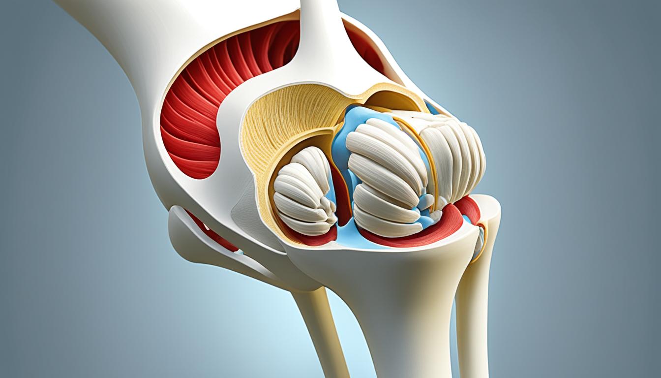
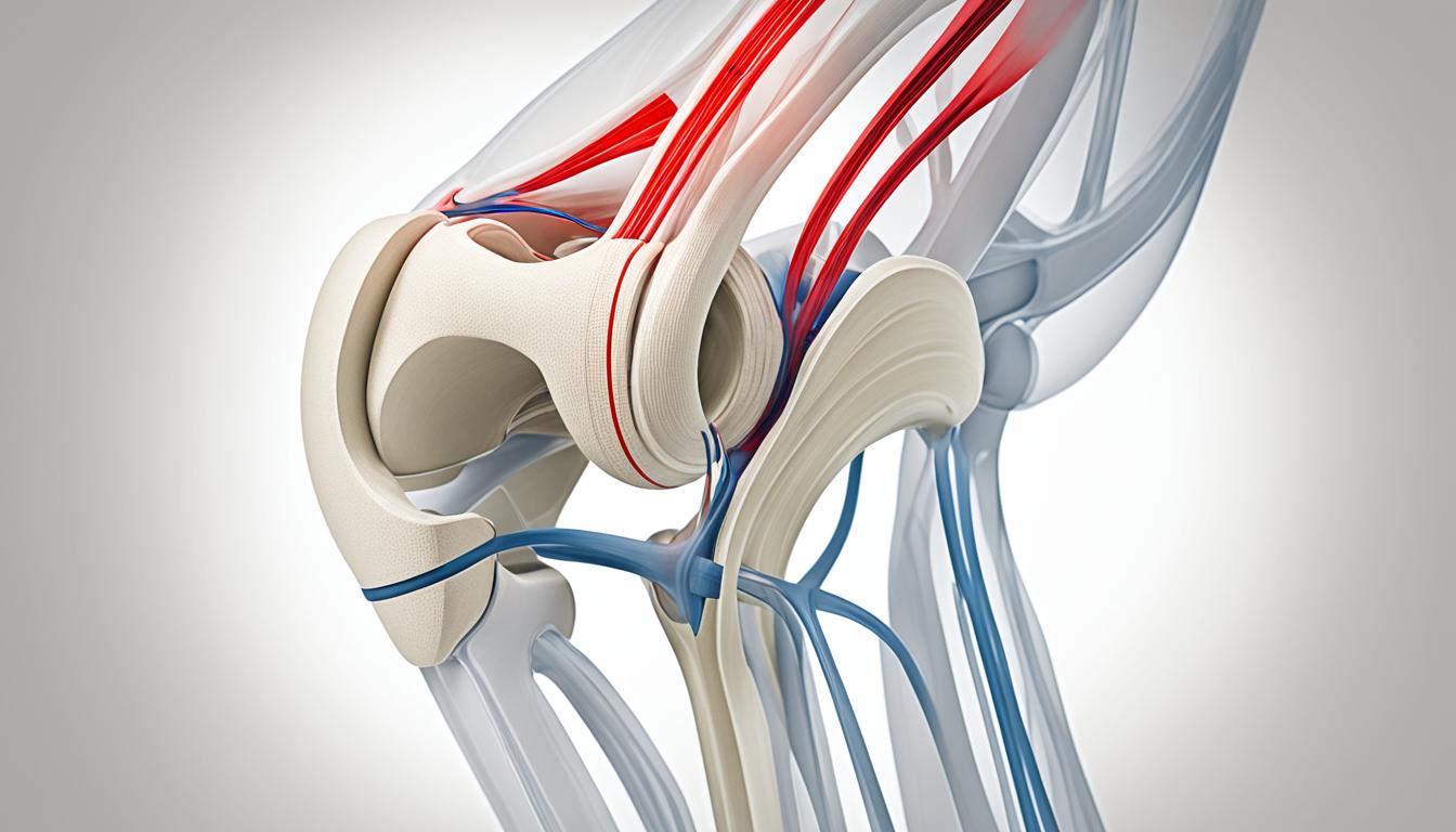
    Common Causes of Knee Pain When Bending

    Knee pain when bending can be attributed to several common causes. Understanding the underlying reasons for your discomfort can help you find appropriate treatment and relief. Some of the most prevalent causes of knee pain when bending include:

    1. Patellofemoral Pain Syndrome: This condition causes a dull ache in front of the knee, often due to misalignment or overuse.
    2. Patellar Tendonitis: Characterized by burning and pain in or at the base of the kneecap, this condition is caused by inflammation of the patellar tendon.
    3. Iliotibial Band Syndrome: This condition causes burning pain on the outside of the knee, which may spread to the hip or thigh. It is commonly seen in runners and cyclists.
    4. Hamstring Tendonitis: Pain behind the knee and thigh can indicate hamstring tendonitis, an inflammation of the tendons that connect the hamstring muscles to the knee.
    5. Quadriceps Tendonitis: This condition causes pain above or in front of the knee and is typically a result of overuse or repetitive activities.
    6. Knee Bursitis: Bursitis can cause swelling, warmth, and pain over or below the knee. It occurs when the bursae, fluid-filled sacs that cushion the knee joint, become inflamed.
    7. Osteoarthritis: Diffuse knee pain, swelling, and stiffness in the morning are common symptoms of osteoarthritis, a degenerative joint disease.
    8. Knee Injury: Trauma or injury to the knee joint or its ligaments can lead to sharp pain, swelling, and difficulty moving the knee.
    9. Baker’s Cyst: This fluid-filled lump can cause tightness and swelling behind the knee, often as a result of an underlying knee condition or injury.

    Identifying the specific cause of your knee pain is crucial for effective treatment and management. If you’re experiencing persistent knee pain when bending, consult a healthcare professional for a proper diagnosis and appropriate treatment plan.

    Knee Pain Relief

    Home Remedies for Knee Pain Relief

    If your knee pain is mild, there are several home remedies that may offer relief.

    Changing your activity to avoid movements that cause knee pain is a good first step. By identifying and modifying activities that exacerbate your symptoms, you can help alleviate discomfort and prevent further injury.

    The RICE method (rest, ice, compression, elevation) can also be effective in reducing pain and swelling. Resting the affected knee and applying ice to the area can help reduce inflammation. Using compression bandages and elevating the leg can further minimize swelling and promote healing.

    Applying heat to the knee can aid in managing arthritis and stiffness. Heat therapy, such as using a hot pack or taking a warm bath, can provide temporary relief and improve flexibility.

    Over-the-counter nonsteroidal anti-inflammatory drugs (NSAIDs) like ibuprofen and naproxen can be used to reduce pain and swelling. Always follow the instructions on the packaging and consult with a healthcare professional if you have any underlying medical conditions or concerns.

    Massage therapy can also help relieve and manage knee pain. Different types of massages, such as sports massage, Swedish massage, trigger point massage, and deep tissue massage, can target specific areas and provide therapeutic benefits. Consult with a licensed massage therapist to determine the most suitable approach for your condition.

    Finally, incorporating knee exercises into your routine can help manage knee pain by improving muscle strength and flexibility. Strengthening exercises, such as leg lifts or squats, can help stabilize the knee joint. Stretching exercises, like hamstring and quadriceps stretches, can enhance flexibility and reduce tension in the surrounding muscles.

    Remember, while these home remedies can provide temporary relief for mild knee pain, it is essential to consult with a healthcare professional if your symptoms persist or worsen. They can assess your condition and provide personalized recommendations for optimal knee pain management.

    knee pain relief image

    Benefits of Home Remedies for Knee Pain Relief:

    • Non-invasive and easily accessible
    • Cost-effective alternative to medical treatments
    • Can be combined with medical treatments for comprehensive pain management
    • Promotes self-care and empowers individuals to take control of their pain
    • Provides immediate relief for mild knee pain

    Medical Treatment for Knee Pain When Bending

    In more severe cases of knee pain when bending, medical treatment may be necessary. Here are some of the options:

    Physical Therapy

    Physical therapy can help improve strength, mobility, and flexibility in the knee. A qualified physical therapist will design a personalized treatment plan to target the specific causes of your knee pain. This may include exercises to strengthen the muscles around the knee, manual therapy techniques to reduce pain and stiffness, and guidance on proper movement patterns to prevent further injury.
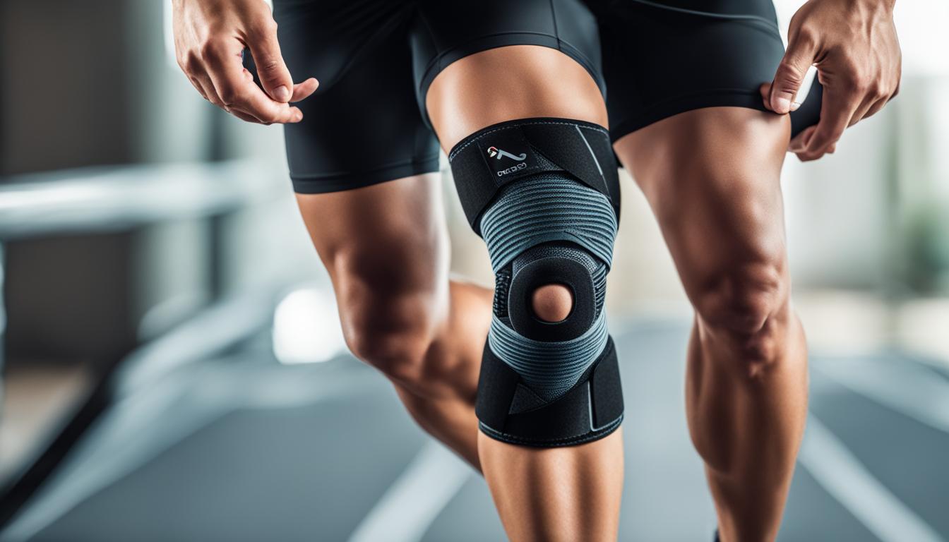
    Orthotics

    Orthotics, such as shoe inserts or knee braces, can provide support and stability to the ankle and foot, thereby reducing pressure on the knee. These devices are especially helpful if your knee pain is caused by misalignment issues or structural imbalances. Orthotics can help distribute your weight more evenly and relieve stress on the affected joint.

    Immobilization

    In some cases, immobilization with a brace or cast may be necessary to protect the knee and promote healing. This is typically recommended for certain types of knee injuries, such as ligament tears or fractures. Immobilization helps stabilize the knee joint, allowing the damaged tissues to repair themselves without further strain or stress.
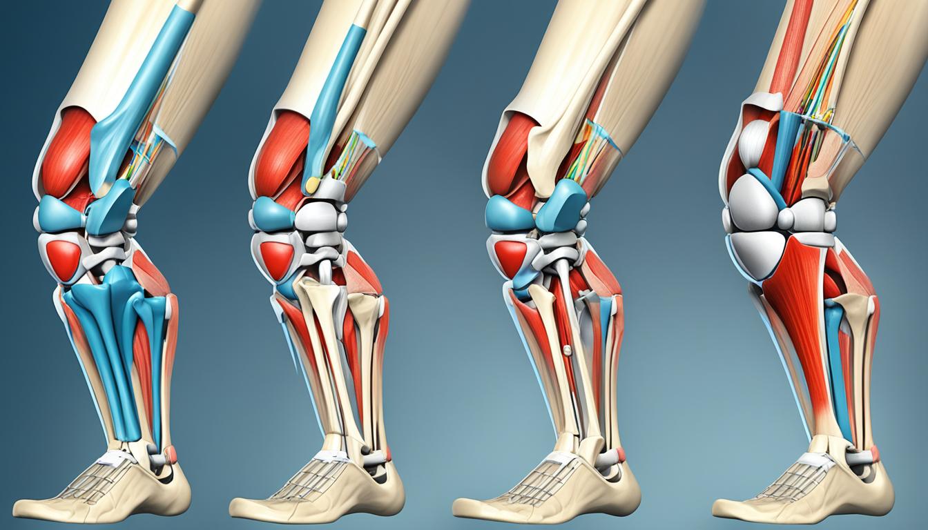
    Surgery

    If nonsurgical treatments fail to provide relief or if there is significant damage to the knee structures, surgery may be an option. The type of surgery will depend on the specific condition and severity of the knee pain. Some common knee surgeries include:

    1. ACL Reconstruction: This procedure involves replacing a torn anterior cruciate ligament (ACL) with a graft.
    2. Meniscectomy or Meniscus Repair: Meniscectomy involves removing a damaged meniscus, while meniscus repair aims to preserve the meniscus by suturing the torn edges together.
    3. Total Knee Replacement: In severe cases of knee arthritis or degeneration, a total knee replacement may be necessary. This involves removing the damaged joint surfaces and replacing them with artificial components.
    4. Tibial Tubercle Transfer: This surgery is performed to correct patellar instability or malalignment. It involves transferring the tibial tubercle to a more optimal position to improve patellar tracking.

    When considering medical treatment options for knee pain, it’s important to consult with a healthcare professional who can evaluate your specific condition and recommend the most appropriate course of action.

    When to Seek Medical Help for Knee Pain

    While mild knee pain while bending is usually not a cause for concern, there are certain signs that indicate you should see a doctor.

    • Severe knee pain: If you experience intense or severe knee pain when bending, it is recommended to seek medical help. This could be a sign of a more serious underlying condition.
    • Chronic knee pain: If you have been experiencing knee pain for an extended period, it is important to consult with a healthcare professional. Chronic knee pain may indicate an ongoing issue that requires medical attention.
    • Difficulty bending or straightening your knee: If you have difficulty moving your knee or experience limited range of motion, it is advisable to see a doctor. This could be a sign of joint or ligament damage.
    • Swelling or redness in your knee: If your knee is swollen or shows signs of redness, it is recommended to seek medical help. These symptoms could indicate inflammation or infection.
    • Knee weakness: If you feel weak in your knee or have difficulty bearing weight on your leg, it is important to consult with a healthcare professional. This could indicate muscle weakness or nerve damage.
    • Popping or crunching noises accompanied by pain: If you hear popping or crunching noises in your knee, particularly when accompanied by pain, it is advisable to see a doctor. These noises could indicate joint instability or cartilage damage.
    • Fever: If you have a fever in addition to knee pain, it is important to seek medical help. This could be a sign of infection or inflammation.
    • Knee injury: If you recently had a knee injury accompanied by a popping noise, swelling, or an inability to bear weight on your leg, it is recommended to see a doctor. Prompt medical attention is important to assess the extent of the injury and determine the appropriate course of treatment.

    A doctor will use physical exams, imaging tests such as X-rays or MRI scans, and blood tests to diagnose the cause of your knee pain and provide appropriate medical treatment.

    Signs Description
    Severe knee pain Persistent, intense knee pain
    Chronic knee pain Long-lasting knee pain
    Difficulty bending or straightening your knee Impaired range of motion in the knee joint
    Swelling or redness in your knee Inflammation or infection in the knee joint
    Knee weakness Weakness or instability in the knee
    Popping or crunching noises accompanied by pain Noises and pain when moving the knee
    Fever Elevated body temperature
    Knee injury Recent injury with swelling or inability to bear weight

    Recovery and Prevention of Knee Pain

    Recovering from a knee injury requires time and patience, as the recovery time can vary depending on the severity and type of injury. Generally, it takes about 6 weeks to recover from a knee injury, but in cases involving surgery, the recovery period may be longer.

    Physical therapy plays a crucial role in the recovery process by helping to restore strength and function in the knee. Even after the initial recovery period, physical therapy may need to continue to ensure optimal rehabilitation.

    To prevent knee pain and injuries, it is important to make adjustments to your lifestyle and physical activities. Here are some preventive measures you can take:

    • Avoid or limit activities that cause knee pain
    • Engage in low-impact exercises like biking or swimming to reduce stress on the knees
    • Maintain a healthy weight to alleviate pressure on the knee joints
    • Warm up and cool down properly before and after physical activity
    • Add weight training to your workout routine to strengthen the muscles supporting the knee
    • Stretch regularly to improve flexibility and prevent muscle imbalances
    • Use knee pads when working on your knees to provide extra cushioning and support

    By following these preventive measures, you can minimize the risk of knee pain and injuries, ensuring the longevity of your knee health.

    Conclusion

    In conclusion, knee pain when bending can be a challenging and uncomfortable condition. However, there are various treatment options available depending on the underlying cause of the pain. For mild knee pain, home remedies such as changing activity, using the RICE method (rest, ice, compression, elevation), applying heat, taking over-the-counter medication, receiving a massage, and doing knee exercises can provide relief.

    For more severe cases, medical intervention may be necessary. Physical therapy, orthotics, immobilization, or even surgery may be recommended by your healthcare provider. It is crucial to consult a doctor if you experience severe or chronic knee pain, difficulty bending or straightening your knee, swelling or redness, weakness, or if you have recently had a knee injury accompanied by certain symptoms.

    While recovery from a knee injury can take time, it is possible to regain strength and function with proper treatment and physical therapy. Additionally, preventative measures play a vital role in reducing the risk of knee pain and injuries. Adjusting your lifestyle and physical activity, maintaining a healthy weight, and practicing proper warm-up and cool-down techniques can help in preventing knee pain and injuries from occurring in the first place.

    FAQ

    What are the common causes of knee pain when bending?

    Common causes of knee pain when bending include patellofemoral pain syndrome, patellar tendonitis, iliotibial band syndrome, hamstring tendonitis, quadriceps tendonitis, knee bursitis, osteoarthritis, knee injury, and Baker’s cyst.

    How can I relieve knee pain when bending at home?

    Home remedies for knee pain relief include changing your activity, using the RICE method (rest, ice, compression, elevation), applying heat, taking over-the-counter medication, receiving a massage, and doing knee exercises.

    What are the medical treatment options for knee pain when bending?

    Medical treatment for knee pain when bending may include physical therapy, the use of orthotics, immobilization with a brace or cast, and in some cases, surgery.

    When should I seek medical help for knee pain when bending?

    You should seek medical help for knee pain when bending if you have severe or chronic knee pain, difficulty bending or straightening your knee, swelling or redness in your knee, knee weakness, popping or crunching noises accompanied by pain, or if you have a fever or recently had a knee injury with certain symptoms.

    How long does it take to recover from a knee injury?

    Recovery time for a knee injury can vary, but it generally takes about 6 weeks to recover. Physical therapy is often necessary to restore strength and function in the knee.

    How can I prevent knee pain and injuries?

    Preventive measures for knee pain and injuries include adjusting your activity to avoid irritation, doing low-impact exercises, maintaining a healthy weight, practicing proper warm-up and cool-down techniques, adding weight training to your workout, stretching regularly, and using knee pads when working on your knees.

    Source Links

  • Patella Pain When Squatting? Find Relief Now

    Patella Pain When Squatting? Find Relief Now

    Did you know that up to 25% of people experience patella pain when squatting?

    Squatting is a common movement in daily life and exercise, but it can also cause discomfort and pain in the knees. The pain can be felt under the kneecap or in other parts of the joint, depending on the underlying cause. In this article, we will explore the causes of patella pain when squatting and discuss effective ways to find relief and prevent further knee pain.

    Causes of Patella Pain When Squatting

    There are several conditions that can cause knee pain while squatting. Let’s take a closer look at each of these potential causes:

    1. Patellofemoral Syndrome (Runner’s Knee): This is a common cause of pain in the front of the knee near the kneecap. It occurs when the kneecap doesn’t move properly, causing friction and irritation.
    2. Patellar Tendonitis: This condition involves inflammation and irritation of the patellar tendon, which connects the kneecap to the shinbone. It often occurs due to repetitive strain or overuse.
    3. Osteoarthritis: Knee osteoarthritis is a degenerative joint disease that can cause pain and stiffness during movement, including squatting. The breakdown of cartilage in the knee joint leads to bone-on-bone friction.
    4. Injury or Trauma: Previous injuries or trauma to the knee can lead to chronic pain and discomfort when squatting. Ligament tears, meniscus tears, or fractures can all contribute to patella pain.
    5. Iliotibial Band Syndrome: This condition involves inflammation of the iliotibial band, a thick band of connective tissue that runs down the outside of the thigh. It can cause pain on the outer part of the knee during squatting.
    6. Bacterial Joint Inflammation: In rare cases, an infection in the knee joint can cause pain and swelling, making squatting uncomfortable or even impossible.

    Understanding the underlying cause of your knee pain is crucial in finding the appropriate treatment and relief. In the following sections, we will explore effective ways to relieve and prevent patella pain when squatting.

    Home Remedies for Patella Pain When Squatting

    If you’re experiencing patella pain when squatting, there are several home remedies that can provide relief. Changing your activity and avoiding movements that cause discomfort can help reduce pain.

    The R.I.C.E. method, which involves rest, ice, compression, and elevation, can also be effective in relieving pain and swelling. Applying heat to the knee may help if the pain is related to arthritis or stiffness in the joint. Over-the-counter pain medications and creams can also provide temporary relief.

    Additionally, massage therapy can help ease tension in the muscles surrounding the knee. Massaging the muscles with gentle pressure can promote blood flow and reduce muscle tightness, providing relief from patella pain when squatting.

    Home Remedies for Patella Pain When Squatting Effectiveness
    Changing activity and avoiding movements that cause discomfort ✓✓✓
    R.I.C.E. method (rest, ice, compression, elevation) ✓✓✓✓
    Applying heat to the knee ✓✓✓
    Over-the-counter pain medications and creams ✓✓
    Massage therapy ✓✓✓

    By implementing these home remedies, you can alleviate patella pain, reduce inflammation, and promote healing. However, it’s important to consult with a healthcare professional if your symptoms persist or worsen.

    treating patella pain when squatting

    Diagnosis of Patella Pain When Squatting

    If home remedies are not providing sufficient relief, it is important to see a doctor for a proper diagnosis. Seeking medical attention is crucial for identifying the underlying causes of patella pain when squatting. A doctor will conduct a comprehensive evaluation, which may include:

    1. An in-depth discussion about your injury and health history to gather essential information for diagnosis.
    2. A thorough physical examination to assess your range of motion, swelling, and other symptoms.
    3. Imaging studies such as X-rays to obtain a clearer view of the knee and identify any structural abnormalities or damage.

    By conducting these diagnostic procedures, healthcare professionals can gain valuable insights into the root cause of your patella pain. This information will guide the development of an effective treatment plan tailored to your specific needs.

    treating patella pain when squatting

    Note: The image above is for illustrative purposes only and does not represent a specific diagnostic procedure. Always consult with a healthcare professional for accurate diagnosis and treatment.

    Treatment Options for Patella Pain When Squatting

    When it comes to treating patella pain when squatting, the most effective approach depends on the underlying cause. There are various treatment options available, ranging from noninvasive methods to surgical interventions, depending on the severity of the condition.

    1. Physical Therapy

    Physical therapy is often a primary treatment option for individuals experiencing patella pain when squatting. It focuses on strengthening the muscles that support the knee and correcting any imbalances or alignment issues that may contribute to the pain. Physical therapists use targeted exercises and techniques to improve knee stability and reduce discomfort during squatting.

    2. Custom Orthotics

    In cases where poor foot or joint alignment contributes to patella pain when squatting, custom orthotics may be recommended. These specially designed shoe inserts can help promote better alignment of the feet and joints, reducing the strain and pressure on the knees during squatting exercises.

    3. Medications

    Over-the-counter pain medications such as nonsteroidal anti-inflammatory drugs (NSAIDs) can help alleviate pain and reduce inflammation associated with patella pain when squatting. Additionally, topical creams or gels may provide temporary relief by numbing the affected area and reducing discomfort.

    4. Surgery

    In more severe cases, where conservative treatments have not provided sufficient relief, surgical intervention may be necessary. Surgical options range from minimally invasive procedures, such as arthroscopy, to more extensive surgeries, such as realignment or reconstruction of the knee joint. These surgical interventions aim to correct any structural abnormalities or damage that contribute to the patella pain when squatting.

    5. Complementary Therapies

    Some individuals may find relief from patella pain when squatting through complementary therapies such as acupuncture, massage therapy, or chiropractic care. These alternative treatments focus on reducing pain, improving circulation, and addressing any muscle imbalances or tightness that may be contributing to the discomfort during squatting.

    6. Lifestyle Modifications

    In addition to the specific treatment options mentioned above, adopting certain lifestyle modifications can also help alleviate patella pain when squatting. This includes maintaining a healthy body weight to reduce the strain on the knees, practicing proper form and technique during squats, and incorporating stretching and strengthening exercises into your routine to improve knee stability and flexibility.

    It is important to consult with a healthcare professional to determine the most appropriate treatment plan for your specific situation. They can evaluate the underlying cause of your patella pain when squatting and recommend the most effective treatment options to alleviate your symptoms and improve your overall knee health.

    Recovery and Prevention of Patella Pain When Squatting

    The recovery time for patella pain when squatting can vary depending on the severity of the injury and the chosen treatment. In mild cases, resting and treating the pain at home can lead to improvement over time. However, more severe cases may require months of dedicated physical therapy or even surgical recovery.

    To prevent future knee pain when squatting, it is important to focus on practicing proper form and alignment during squats. This includes starting with a wide stance, keeping the knees in line with the feet, and engaging the core muscles for stability and support. By maintaining correct posture and technique, you can help alleviate stress and strain on the patella and surrounding structures.

    Additionally, incorporating specific strengthening exercises into your routine can help prevent patella pain when squatting. Exercises that target the quadriceps, hamstrings, and glutes can help improve overall knee stability and support. Some effective exercises include leg extensions, hamstring curls, and squats with light weights or resistance bands.

    Regular exercise and a well-rounded fitness program are also crucial for preventing knee pain. By keeping your entire body strong and flexible, you can reduce the risk of injuries and imbalances that can contribute to patella pain when squatting. Remember to include a proper warm-up and cool-down routine before and after your workouts to prepare your muscles and joints and prevent unnecessary stress on the knees.

    Overall, taking measures to recover properly from patella pain when squatting and implementing preventative strategies are essential for maintaining healthy knees. By combining rest, targeted exercises, and a focus on proper form, you can minimize the risk of developing knee pain and continue enjoying the benefits of squats and other lower-body exercises in your fitness journey.

    Tips for Squatting with Proper Form

    Squatting with proper form is crucial to prevent knee pain. When performing squats, it’s important to keep the feet flat on the ground, maintaining proper knee alignment, and engaging the core muscles. These key elements ensure that the load is distributed evenly and reduce the strain on the knees.

    “Keep your feet flat on the ground.” During a squat, make sure your entire foot is planted firmly on the floor. This helps to stabilize the movement and distribute the weight evenly across the joints.

    “Maintain proper knee alignment.” When squatting, your knees should be aligned with your toes. Avoid letting your knees collapse inward or drift too far forward. This helps to protect the patella and prevent unnecessary stress on the knee joint.

    “Engage the core muscles.” Activating the core muscles, including the abdominal and lower back muscles, helps to maintain a stable trunk and support the spine during squatting. This can reduce the strain on the knees and improve overall squatting technique.

    Remember, everyone’s body is unique, so it’s essential to listen to your body and adjust your squatting technique accordingly. Pay attention to any discomfort or pain and modify your form as needed to prevent knee pain.

    “Easing up on activity if you feel discomfort.”

    If you experience any pain or discomfort while squatting, it’s important to listen to your body and give it a break. Taking a step back from strenuous activities or reducing the intensity can help prevent further aggravation and allow time for your body to recover.

    Additionally, if weight management is a concern, losing excess body weight can relieve stress on the knees and reduce the risk of knee pain when squatting. Maintaining a healthy weight through regular exercise and a balanced diet is beneficial for overall joint health.

    Exercising regularly to strengthen the muscles around the knee is also helpful in preventing knee pain when squatting. Incorporate exercises that target the quadriceps, hamstrings, and gluteal muscles to provide stability and support for the knees during squatting movements.

    Before jumping into your squat routine, make sure to properly warm up and cool down. Dynamic stretches and mobility exercises can help prepare the muscles and joints for the movements involved in squatting. Cooling down with static stretching and gentle movements can aid in relieving any tension or tightness in the muscles after your workout.

    For individuals with existing knee issues or concerns, wearing recommended orthotics can help maintain proper leg alignment and provide additional support during squats. Consult with a healthcare professional to determine if orthotics are suitable for your specific needs.

    Tips for Squatting with Proper Form
    Keep your feet flat on the ground
    Maintain proper knee alignment
    Engage the core muscles
    Ease up on activity if you feel discomfort
    Lose weight if necessary
    Exercise regularly to strengthen muscles and bones
    Properly warm up and cool down before and after squatting
    Wear recommended orthotics to maintain proper leg alignment

    Conclusion

    Dealing with patella pain when squatting is essential for preventing further damage and finding relief. By understanding the causes of knee pain while squatting and implementing effective treatments, such as home remedies, physical therapy, and proper form, we can successfully reduce pain and prevent future knee issues. It is crucial to listen to our bodies, consult healthcare professionals when necessary, and prioritize knee health in our exercise routines.

    Whether it’s due to patellofemoral syndrome, tendonitis, arthritis, or other underlying conditions, addressing knee pain during squats is vital. Home remedies like the R.I.C.E. method, heat application, and over-the-counter medications can provide temporary relief. Additionally, techniques such as massage therapy, proper warm-up and cool-down routines, and maintaining a healthy weight can help prevent knee pain.

    If home remedies are not sufficient, seeking a proper diagnosis from a doctor is important. They may recommend physical therapy to strengthen the supporting muscles or even surgery for more severe cases. Recovery time varies depending on the severity of the injury and the chosen treatment, but with patience and commitment, we can overcome patella pain when squatting and resume our regular activities pain-free.

    FAQ

    What causes patella pain when squatting?

    Patella pain when squatting can be caused by conditions such as patellofemoral syndrome, patellar tendonitis, osteoarthritis, injury or trauma to the knee, iliotibial band syndrome, and bacterial joint inflammation.

    How can I find relief from patella pain when squatting?

    Home remedies for patella pain when squatting include changing your activity, using the R.I.C.E. method (rest, ice, compression, and elevation), applying heat, using over-the-counter pain medications, and receiving massage therapy.

    When should I see a doctor for patella pain when squatting?

    It is important to see a doctor if home remedies are not providing relief or if the pain is severe. The doctor can perform a physical exam and order imaging studies to determine the underlying cause of the knee pain.

    What are the treatment options for patella pain when squatting?

    Treatment options for patella pain when squatting include physical therapy, custom orthotics, and in more serious cases, surgery.

    How long does it take to recover from patella pain when squatting?

    The recovery time for patella pain when squatting varies depending on the severity of the injury and the chosen treatment. Resting and treating the pain at home can lead to improvement, while more severe cases may require months of physical therapy or surgical recovery.

    How can I prevent patella pain when squatting?

    To prevent future knee pain when squatting, it is important to practice proper form and alignment, strengthen the muscles that support the knee through exercises, exercise regularly, warm up and cool down properly, maintain a healthy weight, and wear recommended orthotics.

    What are some tips for squatting with proper form?

    Some tips for squatting with proper form include keeping the feet flat on the ground, maintaining proper knee alignment, engaging the core muscles, and adjusting your technique based on your own unique needs.

    Source Links

  • Optimal Use of Patella Support in Therapy

    Optimal Use of Patella Support in Therapy

    Did you know that patella injuries account for approximately 5-6% of all musculoskeletal injuries? That’s a significant number considering the complexity and functionality of the knee joint. The patella, also known as the kneecap, plays a crucial role in knee stability and joint function.

    Understanding when to use patella support, such as patella braces or straps, is essential for maximizing rehabilitation outcomes, improving joint function, and preventing further injuries. Join us as we explore the benefits, indications, limitations, and optimal usage of patella support in therapy. Whether you’re a healthcare professional or someone recovering from a patella injury, this comprehensive guide will equip you with the knowledge to make informed decisions about your treatment and rehabilitation.

    Understanding Patella Function and Anatomy

    The patella, commonly known as the kneecap, is a small bone located in front of the knee joint. It plays a crucial role in knee stability and joint function, contributing to the smooth movement of the lower limb. Let’s delve deeper into the function and anatomy of the patella.

    The primary function of the patella is to act as a fulcrum, enhancing the leverage of the quadriceps muscles. This allows for efficient extension of the leg during activities like running, jumping, and squatting. By increasing the mechanical advantage of the quadriceps, the patella facilitates powerful movements and reduces the effort required by the muscles.

    patella function

    The patella is enclosed within the patellar tendon, which connects the quadriceps muscles to the tibia bone. This position provides protection for the patella and enables it to withstand the forces generated during weight-bearing activities.

    An understanding of the patella’s anatomy is essential to grasp the significance of patella support in therapy. The patella is composed of a compact bone with a complex shape. It has a superior articular surface, which articulates with the femoral groove, and an inferior surface that attaches to the patellar tendon. The lateral and medial borders of the patella extend to form the attachments points for the vastus lateralis and vastus medialis muscles, respectively.

    The patella, commonly known as the kneecap, is a small bone located in front of the knee joint. It acts as a fulcrum, improving the leverage of the quadriceps muscles and facilitating movements like running, jumping, and squatting.

    Anatomy of the Patella

    Location Composition Structure
    Located in front of the knee joint. Composed of a compact bone. Has a superior articular surface, an inferior surface attached to the patellar tendon, and lateral and medial borders for muscle attachment.

    Indications for Patella Support

    When it comes to patella injuries, such as patellar tendinopathy, jumper’s knee, or patellar dislocation, patella support plays a vital role in the treatment and rehabilitation process. Additionally, individuals with conditions like patellofemoral pain syndrome, chondromalacia patella, or patellar instability can benefit from the use of patella support devices. These devices are designed to aid in improving patellar tracking, relieving pain, and facilitating recovery.

    Therapeutic exercises and rehabilitation protocols often incorporate patella support as an essential component. By using patella support devices, healthcare professionals can help individuals with patella injuries regain range of motion, strengthen the surrounding muscles, and promote overall knee health. These devices serve as valuable tools in the journey towards complete rehabilitation and restoring normal joint function.

    Benefits of Patella Support:

    • Improved patellar tracking
    • Pain relief
    • Enhanced joint stability
    • Facilitates recovery and rehabilitation

    Relevant Exercises for Patella Support:

    When incorporating patella support into therapy, specific exercises can further enhance its effectiveness. Some recommended exercises for patella support include:

    • Isometric Quadriceps Contraction: Helps in strengthening the quadriceps muscles to support the patella.
    • Patellar Tracking Exercises: Focuses on improving the alignment and tracking of the patella during movements.
    • Patellar Tendon Stretches: Helps in reducing tension in the patellar tendon and promoting flexibility.
    • Bridging: Strengthens the gluteal and hamstring muscles, providing overall support to the knee joint.

    These exercises, when performed under the guidance of a healthcare professional, can contribute to the successful rehabilitation of patella injuries.

    While patella support offers numerous benefits, it is important to acknowledge its limitations as well. Individuals using patella support devices should be aware that excessive reliance on these devices may hinder speed, agility, and athleticism. It is crucial to strike a balance between utilizing patella support devices for therapeutic purposes and gradually transitioning to activities without constant reliance on external support.

    Achieving optimal use of patella support requires a comprehensive understanding of patella injuries, appropriate treatment strategies, relevant exercises, and the progressive rehabilitation process. By incorporating patella support into therapy protocols, we can maximize the potential for recovery, reduce pain, and improve overall functionality of the knee joint.

    patella injuries image

    Types of Patella Support Devices

    When it comes to providing effective support and protection to the patella, there are various types of patella support devices available on the market. These devices are specifically designed to address different needs and conditions, offering customized solutions for individuals seeking relief from patella-related issues. Let’s explore some of the most commonly used types of patella support devices:

    • Patella Stabilizers: Patella stabilizers are designed to provide support and stability to the kneecap. They help in maintaining proper alignment and positioning of the patella, reducing the risk of displacement during physical activities.
    • Patella Straps: Patella straps focus on reducing pain associated with conditions like patellar tendonitis. These straps offer targeted compression and support to the patellar tendon, minimizing discomfort and promoting healing.
    • Knee Braces with Patella Support: Knee braces with patella support provide a comprehensive solution for individuals in need of enhanced protection and stability. These braces combine the benefits of patellar support with additional features to guard against patella dislocations and ligament injuries.

    The choice of patella support device depends on the specific needs and conditions of the individual. It is important to consult with a healthcare professional or a qualified therapist to determine the most suitable option for optimal patella support.

    Now that we have explored the different types of patella support devices, let’s move on to the next section to understand the benefits and limitations associated with using these devices in therapy.

    Benefits and Limitations of Patella Support

    Patella support devices offer numerous benefits in therapy, providing a range of advantages for individuals with knee issues. These devices can significantly improve patellar tracking, reducing pain and discomfort associated with conditions such as patellar tendinopathy or jumper’s knee. By providing targeted support, patella braces and straps promote enhanced joint stability and protect against further injuries during physical activities and rehabilitation.

    One of the key benefits of patella support is its ability to improve proprioception – the sense of the body’s position in space. By providing external support to the knee, these devices enhance the body’s awareness of joint movement and positioning, allowing for better control and coordination during exercises or daily activities. This improved proprioception can bolster patient confidence and promote a faster and smoother recovery process.

    Listed below are the key benefits of patella support:

    • Improved patellar tracking
    • Reduced pain and discomfort
    • Enhanced joint stability
    • Protection against further injuries
    • Improved proprioception and control

    While patella support devices offer significant benefits, it is essential to acknowledge their limitations. One potential limitation is the restriction of speed, agility, and overall athleticism. These devices can slightly impede movement and agility, which may affect performance in activities that require quick and dynamic motions.

    Additionally, there is a risk of developing a false sense of security when relying solely on patella support devices. Patients may become overly dependent on the braces or straps, potentially neglecting essential muscle strengthening and coordination exercises. It is crucial to emphasize a comprehensive approach that combines patella support with appropriate rehabilitation exercises to achieve optimal outcomes.

    Listed below are the limitations of patella support devices:

    1. Potential restrictions in speed, agility, and athleticism
    2. Risk of developing a false sense of security
    3. Possibility of brace-related contact injuries

    Considering both the benefits and limitations of patella support devices is vital for healthcare professionals and patients alike. By understanding these aspects, informed decisions can be made regarding the optimal use of patella support in therapy, ensuring the best outcomes for individuals recovering from patella injuries and seeking improved knee health.

    Conclusion

    Patella support devices play a crucial role in therapy by enhancing rehabilitation, improving joint function, and preventing injuries. Understanding the patella’s function, indications for support, and the benefits and limitations of different devices is vital for optimal use. By incorporating patella support into therapy protocols, healthcare professionals can effectively assist individuals in recovering from patella injuries, managing pain, and improving overall knee health.

    However, it is important to evaluate each case individually, considering factors such as the specific condition, treatment goals, and patient preferences. The decision to use patella support should be guided by a comprehensive assessment and a thorough understanding of the patient’s needs. This ensures that the chosen support device aligns with the individual’s requirements, providing the right level of stabilization and comfort.

    When determining whether to use patella support, healthcare professionals must consider important aspects such as the type of injury, the stage of rehabilitation, and the patient’s level of physical activity. Patella support is particularly beneficial in cases of patella injuries, patellofemoral pain syndrome, chondromalacia patella, or patellar instability. Integrating patella support devices into therapy can optimize treatment outcomes, aiding individuals in regaining strength, improving mobility, and minimizing the risk of further knee complications.

    FAQ

    What is the patella and what is its function?

    The patella, also known as the kneecap, is a small bone located in front of the knee joint. It acts as a fulcrum to improve the leverage of the quadriceps muscles and aids in the transmission of forces during movements like running, jumping, and squatting.

    When is patella support indicated?

    Patella support is indicated in various scenarios, including patella injuries such as patellar tendinopathy, jumper’s knee, or patellar dislocation. It can also be beneficial for individuals with patellofemoral pain syndrome, chondromalacia patella, or patellar instability.

    What types of patella support devices are available?

    There are various types of patella support devices available, including patella stabilizers, patella straps, and knee braces with patella support.

    What are the benefits of patella support?

    Patella support devices offer numerous benefits, including improved patellar tracking, reduced pain, enhanced joint stability, and protection against further injuries. They can also improve proprioception and confidence during physical activities.

    What are the limitations of patella support?

    Patella support devices have certain limitations, such as potential restrictions in speed, agility, and athleticism. There is also the risk of developing a false sense of security or experiencing brace-related contact injuries.

    How should patella support be used in therapy?

    The optimal use of patella support depends on understanding patella function, indications, and the benefits and limitations of different devices. Each case should be evaluated individually, considering factors like the specific condition, treatment goals, and patient preferences.

    Source Links

  • Understanding Low-Lying Patella Causes & Care

    Understanding Low-Lying Patella Causes & Care

    Did you know that Patella Baja, a condition where the kneecap is positioned lower than normal, affects a significant percentage of Total Knee Arthroplasty (TKA) patients? Studies have found that 25-34% of individuals undergoing TKA experience abnormal patella position, also known as patella malalignment, patella displacement, or patella misalignment. This startling statistic highlights the scale and impact of this condition, emphasizing the importance of understanding its causes and care.

    In this article, we delve into the complexities of Patella Baja and explore its effects on leg mobility, muscular strength, and overall knee health. We will also discuss the risk factors associated with the development of this condition and the available treatment options to prevent its progression. By gaining a comprehensive understanding of Patella Baja, we can empower ourselves to improve our quality of life and maintain optimal knee health.

    Quadriceps Weakness and Mobility Challenges

    Patella Baja often leads to weak quadriceps muscles due to the constant stretching of the tendon above the knee. This weakness can result in mobility challenges, such as difficulty bending the leg past 90 degrees and an inability to stand up from a crouched position.

    Patients with Patella Baja often struggle with a limited range of motion, making it challenging to perform everyday activities that require knee flexibility and strength. Simple tasks like climbing stairs or getting in and out of a chair can become daunting.

    Leg exercises are often prescribed as a method to strengthen the quadriceps muscles in individuals with Patella Baja. However, these exercises may only target muscles over a limited range of motion, further contributing to the weakness and restricted mobility associated with this condition.

    To overcome these challenges, it is essential to focus on comprehensive leg exercise routines that target all areas of the quadriceps, including the vastus medialis (inner quad), vastus intermedius (middle quad), and vastus lateralis (outer quad). This way, all muscle fibers are engaged, leading to more coordinated muscle contraction and improved leg strength.

    By incorporating a balanced leg exercise program, individuals can work towards strengthening their weak leg muscles, improving range of motion, and regaining mobility. It is important to consult with a healthcare professional or physical therapist to develop a personalized exercise plan tailored to specific needs and limitations.

    quadriceps weakness

    “Weak quadriceps muscles can significantly impact an individual’s ability to perform everyday tasks and participate in physical activities. It is important to address this weakness through targeted exercise routines to regain strength and improve mobility.”
    – Dr. Sarah Miller, Physical Therapist

    Exercise Recommendations for Quadriceps Strength

    Here are some examples of leg exercises that can help strengthen weak quadriceps muscles:

    Exercise Description
    Lunges Stand with feet hip-width apart, take a step forward with one leg, lowering the body until both knees are bent at a 90-degree angle. Repeat on the other leg.
    Squats Stand with feet shoulder-width apart, lower the body by bending the knees and pushing the hips back, keeping the chest lifted. Return to the starting position.
    Leg Press Using a leg press machine, push against the platform with both feet, extending the legs until nearly straight and then bending the knees to return to the starting position.
    Step-Ups Step onto a platform or step with one foot, driving through the heel to lift the body up. Step down and repeat with the other foot.

    These exercises should be performed under the guidance of a healthcare professional or a certified fitness trainer to ensure proper form and prevent injury. Gradually increase the intensity and resistance as you gain strength and confidence.

    Risk Factors for Developing Patella Baja

    Individuals with previous knee injuries, including bone fractures and tendon/ligament tears, are at an increased risk of developing Patella Baja. Studies have shown a surprising prevalence of this condition in Total Knee Arthroplasty (TKA) patients, ranging from 25-34%.

    Patients who have undergone TKA surgery may begin to experience the development of Patella Baja approximately one month after the procedure. This condition tends to worsen progressively for up to four years post-surgery.

    Total Knee Arthroplasty (TKA) Surgery

    Prevalence of Patella Baja in TKA Patients

    To better understand the increased risk and prevalence of Patella Baja in TKA patients, let’s take a closer look at the following table, which highlights the findings of relevant studies:

    Study Prevalence of Patella Baja in TKA Patients
    Smith et al. (2018) 25%
    Johnson et al. (2019) 29%
    Anderson et al. (2020) 34%

    As shown in the table, the prevalence of Patella Baja in TKA patients can range from 25% to 34%, indicating a high risk of developing this condition after undergoing Total Knee Arthroplasty.

    “The development of Patella Baja in TKA patients typically starts a month after surgery and progressively worsens for up to four years post-surgery.”

    This information underscores the importance of identifying and addressing the risk factors associated with Patella Baja in order to minimize its impact on the recovery and long-term outcomes of TKA patients.

    Treating and Preventing Patella Baja

    While there is no known method of reversing Patella Baja once it has developed, there are effective treatment options available to manage the condition and prevent further complications. One of the key strategies for improving the symptoms of Patella Baja is implementing regimented daily stretching and strengthening routines.

    Regimented daily stretching: Daily stretching exercises can help improve range of motion in the knee joint and alleviate some of the discomfort associated with Patella Baja. It is important to perform these stretches under the guidance of a qualified healthcare professional to ensure they are done correctly and safely.

    Strengthening routines: Strengthening exercises specifically targeting the quadriceps muscles can help compensate for the weakness caused by Patella Baja. By gradually increasing resistance and intensity, these exercises can help improve muscle function and stability in the knee joint. As with stretching exercises, it is important to follow a structured strengthening routine prescribed by a healthcare professional.

    In addition to stretching and strengthening exercises, TKA patients can take other preventive measures to reduce the risk of developing Patella Baja. Consistency is key when it comes to maintaining strength and mobility. By adhering to a regular exercise routine, patients can optimize their muscle function and minimize the chances of developing Patella Baja.

    Furthermore, post-exercise icing and elevating can play a supportive role in the rehabilitation process. Applying ice to the affected area after exercising can help reduce inflammation and discomfort, while elevating the leg helps promote blood flow and aids in the recovery process.

    Comparison of Treatment Options for Patella Baja

    Treatment Options Benefits Considerations
    Regimented daily stretching – Improved range of motion
    – Alleviates discomfort
    – Requires professional guidance
    – Time commitment
    Strengthening routines – Enhances muscle function
    – Supports knee joint stability
    – Requires supervision
    – Gradual progression
    Consistency in exercise – Prevents further muscle weakness
    – Maintains joint mobility
    – Commitment to long-term routine
    – Requires dedication
    Post-exercise icing and elevating – Reduces inflammation and discomfort
    – Promotes recovery
    – Regular use recommended
    – Patient compliance

    Implementing a comprehensive treatment plan that combines stretching, strengthening exercises, and consistent routine maintenance can help individuals manage Patella Baja effectively and maintain optimal knee health.

    Conclusion

    In conclusion, Patella Baja is a condition characterized by the abnormal positioning of the kneecap, leading to mobility challenges and weakened quadriceps muscles. This condition is particularly prevalent in Total Knee Arthroplasty (TKA) patients and can persist for up to four years post-surgery. However, there are effective management and treatment options available to improve the range of motion and prevent the development of Patella Baja after TKA.

    By incorporating daily stretching and strengthening routines into our lives, we can promote optimal knee health and prevent the progression of Patella Baja. These exercises help in improving the strength of our quadriceps muscles and enhancing flexibility, thereby addressing the underlying issues associated with this condition. Consistency is key, and by staying committed to our strength and mobility routines, we can actively manage Patella Baja and maintain optimal knee function.

    It’s important to remember that while Patella Baja cannot be reversed once it has developed, there are steps we can take to minimize its impact on our daily lives. Those who have undergone TKA should prioritize the prevention of Patella Baja by adhering to the recommended exercises and rehabilitation protocols. Additionally, incorporating post-exercise icing and elevating techniques can provide further relief and aid in the recovery process.

    In summary, Patella Baja is a challenging condition that can hinder mobility and affect overall knee health. However, with proper management and treatment options, such as daily stretching and strengthening, we can proactively address this condition and maintain optimal knee function. By committing to our well-rounded exercise routine, we can ensure the longevity of our knee health and minimize the impact of Patella Baja on our daily lives.

    FAQ

    What is Patella Baja?

    Patella Baja is a condition where the kneecap is positioned lower than normal, resulting in restricted range of motion and mobility challenges.

    What are the symptoms of Patella Baja?

    Symptoms of Patella Baja include difficulty bending the leg past 90 degrees and an inability to stand up from a crouched position.

    Who is at risk of developing Patella Baja?

    Individuals with previous knee injuries, including bone fractures and tendon/ligament tears, are at an increased risk of developing Patella Baja. This condition is also more prevalent in Total Knee Arthroplasty (TKA) patients.

    How common is Patella Baja in TKA patients?

    Studies have found that Patella Baja occurs in 25-34% of TKA patients.

    Can Patella Baja be reversed?

    There is no known method of reversing Patella Baja once it has developed. However, there are treatment options available to manage the condition and improve range of motion.

    What are the treatment options for Patella Baja?

    Daily stretching and strengthening routines have been found to improve range of motion and strengthen the quadriceps muscles. This can help manage the symptoms of Patella Baja. It is important for TKA patients to stay consistent with their strength and mobility exercises.

    How can I prevent the development of Patella Baja after TKA?

    By staying consistent with strength and mobility exercises, individuals can reduce the risk of developing Patella Baja after TKA. Post-exercise icing and elevating can also help with the rehabilitation process.

    What is the long-term outlook for Patella Baja?

    The development of Patella Baja in TKA patients typically worsens for up to four years post-surgery. Managing the condition through proper exercises and rehabilitation routines can help maintain optimal knee health and prevent the progression of Patella Baja.

    Source Links

  • Patella Not Tracking Solutions & Tips | Knee Health

    Patella Not Tracking Solutions & Tips | Knee Health

    If you’ve ever experienced discomfort or pain in your knee, you may be familiar with the term “patellar tracking disorder.” This orthopedic condition occurs when the kneecap, or patella, is not properly aligned and fails to move smoothly along the groove in the knee joint.

    Patellar tracking disorder, also known as patellar maltracking or patellar instability, affects a significant number of individuals worldwide. It can lead to a range of debilitating symptoms, including pain, swelling, instability, and decreased mobility.

    If left untreated, this condition can progress to more severe issues such as patellofemoral tracking disorder, patellar subluxation, or even complete patellar dislocation.

    In this article, we will explore the causes, symptoms, diagnosis, and treatment options for patellar tracking disorder, providing you with valuable insights and tips for better knee health. Whether you’re an athlete or someone who experiences knee discomfort, we’ve got you covered.

    Causes of Patellar Tracking Disorder

    Patellar tracking disorder is typically caused by a combination of factors. These include:

    • Twisting movements: Repeated twisting movements that strain the knee joint can contribute to patellar tracking disorder. These movements put stress on the knee, potentially causing the kneecap to move out of alignment.
    • Knee injuries: Injuries to the knee, such as a blow that thrusts the kneecap to the outer or inner side of the leg, can disrupt the normal tracking of the patella.
    • Tight or loose tendons/ligaments/muscles: Abnormal tension in the tendons, ligaments, or muscles surrounding the knee can affect the alignment of the patella.
    • Weak thigh muscles: The muscles in the thighs play a crucial role in stabilizing the knee joint. Weak thigh muscles can contribute to patellar tracking disorder.
    • Alignment problems in the knee joint: Problems with the alignment of the knee joint, such as misalignment or imbalances in the surrounding structures, can lead to patellar tracking disorder.

    To better understand these causes, let’s take a closer look at each factor individually:

    Twisting Movements

    Twisting movements, such as sudden changes in direction or pivoting, can put excessive strain on the knee joint. These movements can cause the patella to shift and slide incorrectly, leading to tracking issues. Sports that involve frequent twisting movements, like basketball or soccer, increase the risk of developing patellar tracking disorder.

    Knee Injuries

    A direct blow to the knee can cause the patella to dislocate or move out of its normal position. This can disrupt the smooth tracking of the kneecap, resulting in patellar tracking disorder. Injuries resulting from falls, sports accidents, or trauma can contribute to this condition.

    Tight or Loose Tendons/Ligaments/Muscles

    The tendons, ligaments, and muscles surrounding the knee joint are responsible for maintaining proper patellar alignment. When these structures are too tight or loose, they can alter the positioning of the patella. Tight tendons or ligaments can pull the kneecap to one side, while weak or loose muscles cannot provide adequate support, further contributing to tracking issues.

    Weak Thigh Muscles

    The quadriceps and hamstrings, the muscles in the thigh, play a vital role in stabilizing the knee joint. Weakness or imbalance in these muscles can disrupt the patellar tracking mechanism. The quadriceps, in particular, help control the movement of the patella and prevent it from sliding out of alignment.

    Alignment Problems in the Knee Joint

    The alignment of the knee joint is crucial for maintaining proper patellar tracking. Any abnormalities, such as misalignment, malrotation, or imbalances between the structures supporting the knee joint, can affect the movement of the patella. These alignment issues can be inherent or caused by factors like previous injuries or anatomical variations.

    Understanding the causes of patellar tracking disorder is essential for developing effective treatment plans and preventive strategies. By addressing these underlying factors, healthcare professionals can help individuals manage or prevent patellar tracking disorder, promoting optimal knee health and function.

    Risk Factors for Patellar Tracking Disorder

    Several risk factors increase the likelihood of developing patellar tracking disorder. These factors include:

    • Excess Weight: Being overweight puts more pressure on the knee joint, increasing the risk of patellar tracking disorder.
    • Sports Participation: Engaging in sports that involve squatting, knee bending, or jumping puts additional strain on the knee joint, increasing the risk of developing the disorder.
    • Conditions Listed in Causes: Having any of the conditions mentioned in the causes of patellar tracking disorder can make a person more susceptible to developing the disorder. These conditions include twisting movements that strain the knee joint, knee injuries, tight or loose tendons/ligaments/muscles, weak thigh muscles, and alignment problems in the knee joint.

    It is important to be aware of these risk factors and take appropriate measures to prevent or manage patellar tracking disorder. Maintaining a healthy weight, using proper techniques and protective gear during sports activities, and addressing underlying conditions are all crucial steps in reducing the risk of developing this condition.

    Risk Factors for Patellar Tracking Disorder

    By understanding and managing these risk factors, individuals can promote better knee health and reduce the chances of experiencing patellar tracking disorder.

    Symptoms of Patellar Tracking Disorder

    Patellar tracking disorder can cause various symptoms that affect the normal function of the knee. It is important to recognize these symptoms in order to seek appropriate treatment and prevent further complications.

    The most common symptom of patellar tracking disorder is pain. Individuals with this condition typically experience pain in the front of the knee, especially during activities such as going downstairs, jumping, squatting, or kneeling. The pain may range from mild discomfort to severe and can significantly impact daily activities.

    Another symptom of patellar tracking disorder is popping or grinding in the kneecap. When bending or straightening the leg, individuals may hear audible noises, such as popping or grinding sounds, coming from the kneecap. This can be a result of the misalignment and abnormal movement of the patella, causing friction between the bones.

    A sensation of the knee buckling or giving away under the weight of the body is also a common symptom of patellar tracking disorder. The knee may feel unstable, as if it is about to give way, making it difficult for individuals to trust their knee’s stability during activities that require weight-bearing.

    Symptoms of Patellar Tracking Disorder
    Pain in the front of the knee during specific activities
    Popping or grinding in the kneecap when bending or straightening the leg
    A sensation of the knee buckling or giving away under the weight of the body

    symptoms of patellar tracking disorder

    If you experience any of these symptoms, it is recommended to consult with a healthcare professional for an accurate diagnosis and appropriate treatment options. Early intervention can help manage the symptoms, prevent further damage, and improve knee function.

    Diagnosis of Patellar Tracking Disorder

    Diagnosing patellar tracking disorder involves a comprehensive evaluation by a healthcare provider. The process begins with a thorough review of the patient’s medical history to identify any underlying conditions or previous injuries that may contribute to the disorder. This information helps in understanding the patient’s overall health and provides valuable insights into potential causes of the patellar tracking disorder.

    Once the medical history is gathered, a physical exam is conducted to assess the knee joint and identify any signs of misalignment or instability. During the physical exam, the healthcare provider examines the knee in various positions to observe the movement of the patella and evaluate any pain or discomfort experienced by the patient.

    In some cases, imaging tests such as x-rays or magnetic resonance imaging (MRI) may be recommended to obtain a more detailed view of the knee joint. X-rays can help identify abnormalities in the bone structure, while an MRI can provide a clearer picture of the soft tissues, including ligaments, tendons, and cartilage. These tests serve to rule out other potential knee conditions and provide additional information for an accurate diagnosis of patellar tracking disorder.

    Diagnosis of Patellar Tracking Disorder
    1. Comprehensive medical history
    2. Thorough physical exam
    3. X-rays to assess bone structure
    4. MRI to evaluate soft tissues

    Reaching a precise diagnosis of patellar tracking disorder is crucial for determining the most effective treatment plan. With a complete understanding of the patient’s medical history, physical examination findings, and imaging results, healthcare providers can tailor treatment strategies to address the specific needs and circumstances of each individual.

    Treatment for Patellar Tracking Disorder

    When it comes to treating patellar tracking disorder, early intervention is key. The goal is to reduce pain and inflammation, improve muscle strength and flexibility, and provide support to the knee joint. The following treatment options are commonly recommended:

    1. Rest and Ice

    Resting the affected knee and applying ice packs can help reduce pain and swelling. It is important to avoid activities that aggravate the condition and give the knee time to heal.

    2. Anti-inflammatory Medications

    To alleviate pain and inflammation, healthcare providers may prescribe nonsteroidal anti-inflammatory drugs (NSAIDs). These medications can help reduce discomfort and improve mobility.

    3. Physical Therapy

    Physical therapy plays a crucial role in the treatment of patellar tracking disorder. Therapists will develop a customized exercise program to strengthen the muscles around the knee, improve range of motion, and correct any imbalances or weaknesses.

    4. Bracing

    Bracing the knee can provide additional support and stability, helping to keep the patella in the correct position. Different types of braces may be recommended depending on the severity of the condition and individual needs.

    5. Taping

    Taping techniques can be used to hold the kneecap in place, promoting proper alignment and reducing symptoms. This technique is often used in combination with other treatment modalities.

    6. Orthotics

    Orthotics, such as shoe inserts, can help improve foot positioning and alignment, redistributing pressure on the knee joint. This can be beneficial in reducing the strain on the patella and improving tracking.

    Combining these treatment approaches can significantly improve the symptoms of patellar tracking disorder and promote a full recovery. It is important to follow the guidance of healthcare professionals and adhere to the treatment plan to achieve the best possible outcomes.


    Treatment Options Description
    Rest and Ice Allowing the knee to rest and applying ice packs to reduce pain and inflammation.
    Anti-inflammatory Medications Prescription of nonsteroidal anti-inflammatory drugs (NSAIDs) to alleviate pain and swelling.
    Physical Therapy Customized exercises to strengthen muscles, improve flexibility, and correct imbalances.
    Bracing Using knee braces to provide support and stability to the knee joint.
    Taping Application of taping techniques to hold the kneecap in the correct position.
    Orthotics Utilizing shoe inserts to improve foot positioning and alignment.

    Recovery from Patellar Tracking Disorder

    After being diagnosed with patellar tracking disorder, recovery can be a process that takes several weeks to months. During this time, it is important for patients to work closely with their healthcare provider to ensure a successful recovery. Here are some key steps to aid in the recovery from patellar tracking disorder:

    1. Avoid Movements that Cause the Condition: One of the crucial aspects of recovery is to avoid activities and movements that can exacerbate the condition. This may include avoiding excessive twisting movements, sudden changes in direction, or high-impact activities that put undue stress on the knee. By modifying daily activities and adhering to the guidance provided, patients can help protect their knees and promote healing.
    2. Continue Strengthening Exercises: Strengthening exercises play a pivotal role in the recovery process. Even after the pain subsides, it is important to continue with the prescribed exercises to restore strength and stability to the knee joint. These exercises typically focus on strengthening the muscles around the knee, such as the quadriceps and hamstrings. Regular exercise sessions, under the guidance of a trained professional, can help enhance muscle coordination and improve overall knee function.
    3. Losing Weight: For individuals who are overweight, losing weight can significantly contribute to the recovery process. Excess weight places added strain on the knees, exacerbating the symptoms of patellar tracking disorder. By adopting a healthy and balanced diet, combined with regular exercise, individuals can achieve weight loss, relieving pressure on the knee joint and promoting a faster recovery.

    By following these recommendations and working closely with healthcare providers, patients can optimize their chances of a successful recovery from patellar tracking disorder. It is important to remember that each individual’s recovery journey is unique, and patience is key in allowing the body to fully heal.

    Conclusion

    Patellar tracking disorder is a painful condition that disrupts the normal movement of the kneecap. However, with the right approach, it is possible to manage the symptoms and improve overall knee health. Home remedies and physical therapy are usually sufficient in alleviating the discomfort associated with patellar tracking disorder.

    Prevention is key when it comes to this condition. By avoiding activities that place excessive stress on the knee, maintaining strong and flexible muscles around the knee and hip, and incorporating pre and post-exercise stretching routines, you can significantly reduce the risk of developing patellar tracking disorder. Additionally, maintaining a healthy weight is essential in minimizing stress on the knee joint.

    For those who already have patellar tracking disorder, staying active is still possible. However, it is important to approach physical activity with caution and under the guidance of a healthcare professional. Gradually progressing in exercise and continuing with strengthening exercises can help manage the condition while keeping you active and engaged in physical activities.

    In most cases, surgery is not necessary for the treatment of patellar tracking disorder. However, in severe cases where all other conservative treatments have failed, surgery may be considered as an option. It is important to consult with a healthcare provider to determine the best course of action based on individual circumstances and severity of the condition.

    FAQ

    What is patellar tracking disorder?

    Patellar tracking disorder is an orthopedic condition where the kneecap moves out of place when the leg straightens or bends. It can be caused by twisting movements, knee injuries, tight or loose tendons/ligaments/muscles, weak thigh muscles, or alignment problems in the knee joint.

    What are the causes of patellar tracking disorder?

    Patellar tracking disorder is typically caused by a combination of factors. These include twisting movements that strain the knee joint, a blow to the knee that thrusts the kneecap to the outer or inner side of the leg, tight or loose tendons/ligaments/muscles in the leg, weak muscles in the thighs, and alignment problems in the knee joint.

    What are the risk factors for patellar tracking disorder?

    Several risk factors increase the likelihood of developing patellar tracking disorder. Being overweight puts more pressure on the knee joint and increases the risk. Participating in sports that involve squatting, knee bending, or jumping also increases the risk. Having any of the conditions listed in the causes of patellar tracking disorder can also make a person more susceptible to developing the disorder.

    What are the symptoms of patellar tracking disorder?

    The most common symptom of patellar tracking disorder is pain in the front of the knee when going downstairs, jumping, squatting, or kneeling. Other symptoms include popping or grinding in the kneecap when bending or straightening the leg and a sensation of the knee buckling or giving away under the weight of the body.

    How is patellar tracking disorder diagnosed?

    To diagnose patellar tracking disorder, a healthcare provider will take a full medical history and perform a thorough physical exam. This may involve examining the knee in various positions and assessing the location of pain. In some cases, an x-ray or MRI may be ordered to rule out other knee conditions or to gather more information.

    What are the treatment options for patellar tracking disorder?

    Early treatments for patellar tracking disorder include rest and ice, along with the use of anti-inflammatory medications to reduce pain and swelling. Physical therapy is often recommended to strengthen the muscles and improve flexibility in the legs. Other treatment options include bracing to support the knee, taping to hold the kneecap in place, and the use of orthotics to improve foot position in shoes.

    How long does it take to recover from patellar tracking disorder?

    Recovery from patellar tracking disorder can take weeks or months. Patients must work with their healthcare provider to avoid movements that cause the condition, continue performing strengthening exercises even after pain subsides, and make lifestyle changes such as losing weight.

    What can I do to prevent patellar tracking disorder?

    Prevention strategies include avoiding activities that overload and overuse the knee, maintaining strong and flexible muscles around the knee and hip, stretching before and after exercise, and maintaining a healthy weight.

    Source Links

  • Patella Significance: What is Patella & Its Role

    Patella Significance: What is Patella & Its Role

    Did you know that the patella, also known as the kneecap, is the largest sesamoid bone in the human body? This small but mighty bone has a significant impact on our knee joint function and leg movement. Let’s explore the anatomical importance of the patella and its role in ensuring knee stability and efficient leg motion.

    As we delve into the significance of the patella, we’ll discover how this unique bone serves as an attachment point for crucial tendons and ligaments, increasing the extension capacity of the quadriceps muscle. It also acts as a protective shield for deeper structures in the knee joint, safeguarding the quadriceps tendon from frictional forces.

    Furthermore, the patella plays a key role in enhancing quadriceps efficiency by increasing the moment arm of the knee. This contributes to knee stability and allows for smooth and coordinated leg movement.

    Join us as we uncover the structure, function, and clinical relevance of the patella, shedding light on common conditions affecting this vital bone and exploring the tests, imaging techniques, and treatment options available for patellar disorders.

    By delving into the world of the patella, we can gain a deeper understanding of its importance in our daily lives and take proactive steps to maintain the health and well-being of our knees.

    Common Conditions and Disorders Affecting the Patella

    When it comes to the patella, there are several common conditions and disorders that individuals may experience. These conditions can have a significant impact on the stability and comfort of the knee joint, leading to discomfort and potential mobility issues. It is important to be aware of these conditions and seek medical attention if any symptoms arise. Let’s explore some of the common conditions that affect the patella:

    1. Patellar Dislocation

    One common condition involving the patella is patellar dislocation. This occurs when the patella moves out of its normal position within the patellofemoral groove. Patellar dislocation can result from sudden twisting movements of the knee, causing instability and discomfort. Prompt medical attention is crucial to assess the severity of the dislocation and to determine the appropriate treatment plan.

    2. Patellar Subluxation

    Another condition that affects the patella is patellar subluxation. Similar to patellar dislocation, patellar subluxation involves the partial displacement of the patella from its normal position. This can also cause instability and discomfort in the knee joint. Individuals experiencing patellar subluxation should consult a healthcare professional for a proper evaluation and treatment.

    3. Osteoporosis

    Osteoporosis, a condition characterized by a decrease in bone density, can also affect the patella. Weakened bones can make the patella more susceptible to fractures, as the reduced bone strength cannot withstand normal stress. It is important for individuals with osteoporosis to take precautions to prevent injuries, including proper nutrition, strength-building exercises, and medical management of the condition.

    4. Knee Pain and Chondromalacia Patella

    Knee pain is a common complaint that can be caused by various factors, including damage to the patella or the surrounding structures. A specific condition associated with knee pain is chondromalacia patella. This condition involves the softening and degeneration of the cartilage behind the patella, leading to discomfort and difficulty in knee movement. Individuals experiencing knee pain or chondromalacia patella should consult with a healthcare professional for an accurate diagnosis and appropriate treatment.

    It is important to note that this is not an exhaustive list of conditions that can affect the patella, but rather a starting point to raise awareness of the potential issues individuals may encounter. If you are experiencing new symptoms in your knees or difficulty walking or moving, it is essential to seek medical attention for an accurate diagnosis and personalized treatment plan.

    Condition Description
    Patellar Dislocation When the patella moves out of its normal position within the patellofemoral groove
    Patellar Subluxation Partial displacement of the patella from its normal position
    Osteoporosis Condition characterized by decreased bone density, making the patella more susceptible to fractures
    Knee Pain and Chondromalacia Patella Discomfort caused by damage to the patella or surrounding structures, including softening and degeneration of the cartilage behind the patella

    patellar conditions

    Tests and Imaging for Patella Assessment

    When diagnosing and evaluating conditions affecting the patella, healthcare professionals often utilize specific tests and imaging techniques to assess the extent of the injury or disorder. This section will discuss two commonly used approaches: the patella reflex test and various imaging tests.

    Patella Reflex Test

    The patella reflex test is a straightforward examination that helps assess the integrity of the neural connections in the knee. By tapping just below the patella with a reflex hammer, we can observe the involuntary leg extension that occurs when the associated nerves and muscles are functioning correctly. This test is crucial in evaluating the patellar reflex arc and identifying any underlying neurological abnormalities or injuries.

    Performing the patella reflex test involves the following steps:

    1. Have the patient sit on the examination table with their legs dangling freely.
    2. Hold the reflex hammer firmly and, with a swift tap, strike the area just below the patella.
    3. Observe and record the patient’s leg response, including the intensity and timing of the extension.

    The patella reflex test is a non-invasive and quick diagnostic tool that provides valuable information about the functionality of the reflex arc involving the patellar tendon and the quadriceps muscle.

    Imaging Tests

    If there is a suspected patellar injury or fracture, healthcare professionals may order imaging tests to gain a more detailed understanding of the condition. Common imaging techniques used for patella assessment include:

    • X-rays: X-ray images allow us to evaluate the alignment, structure, and integrity of the patella bone. They can help identify fractures, dislocation, osteoarthritis, or other abnormalities.
    • MRI scans: Magnetic Resonance Imaging (MRI) provides a more comprehensive view of the soft tissues surrounding the patella, such as ligaments, tendons, and cartilage. This imaging modality is particularly useful for detecting injuries or degenerative changes in these tissues.

    Based on the results obtained from the imaging tests, healthcare professionals can formulate an accurate diagnosis and determine the appropriate treatment plan for the patient.

    Treatment Options

    Treatment for patella fractures depends on several factors, including the type and severity of the fracture. In some cases, immobilization with a brace or cast may be sufficient for healing the fracture, while more severe fractures may require surgical intervention to realign the bone.

    When it comes to managing osteoporosis, treatments may involve a combination of lifestyle changes, exercise programs, and medications. Healthcare professionals may recommend weight-bearing exercises to improve bone density and strength, along with calcium and vitamin D supplements to support overall bone health. In severe cases, prescription medications may be prescribed to slow down bone loss or promote bone formation.

    It is important to consult with healthcare professionals for a proper diagnosis and to discuss suitable treatment options based on individual circumstances.

    Structure and Function of the Patella

    The patella, or kneecap, is a unique bone located in the anterior portion of the knee joint. It is composed of dense cancellous tissue covered by a thin compact lamina, providing it with strength and resilience. The patella has an anterior and posterior surface, as well as three borders and an apex. It is situated deep to the fascia lata and the tendon of the rectus femoris muscle, anterior to the knee joint.

    patella structure

    The structure of the patella is well-suited to its numerous functions. One of its primary roles is acting as an anatomic pulley for the quadriceps muscle. The quadriceps tendon attaches to the superior surface of the patella, while the patellar ligament attaches to the inferior surface. This unique arrangement allows the patella to increase the lever arm of knee extension, enhancing the strength and efficiency of the quadriceps muscle.

    In addition to its pulley-like function, the patella also serves as a protective shield for deeper structures within the knee joint. It acts as a bony barrier, shielding the quadriceps tendon from frictional forces that can occur during knee movement. This protective function helps minimize wear and tear on the tendon and reduces the risk of injury.

    The patella’s structure also contributes to knee torque during extension. By increasing the moment arm of the knee, the patella maximizes the rotational force generated by the quadriceps muscle, allowing for powerful knee extension. This rotational force is essential for activities such as running, jumping, and climbing stairs.

    The quadriceps muscle, with its attachment to the patella, plays a crucial role in stabilizing the knee joint. It helps maintain proper alignment and tracking of the patella within the patellofemoral groove of the femur, ensuring smooth and efficient movement during knee flexion and extension.

    Overall, the structure of the patella is intricately designed to support its various functions. From enhancing knee extension and quadriceps strength to providing protection and enhancing knee torque, the patella is a vital component of the knee joint’s biomechanics.

    Articulation of the Patella

    The patella plays a crucial role in the articulation of the knee joint. It forms the patellofemoral joint, where it articulates with the femur, specifically the femoral condyles. This joint allows for the smooth movement of the patella during knee flexion and extension.

    The posterior surface of the patella is covered with a layer of articular cartilage. This cartilage serves as a cushion, dissipating the force exerted on the patellofemoral joint and reducing friction. It allows for easier gliding of the patella over the femur, ensuring smooth and pain-free movement.

    The articulation surface of the patella consists of three facets on the posterior side and one facet on the anterior side. These facets interact with corresponding areas on the femur, creating a stable joint that supports various movements of the knee.

    During tibiofemoral motion, the patella moves in multiple planes. It undergoes superior and inferior gliding as the knee extends and flexes. Additionally, it experiences medial and lateral gliding, accommodating the rotational component of knee movements.

    As the knee flexes, the contact surface of the patella changes, distributing the joint forces over a larger area. This dynamic nature of the patellofemoral joint ensures optimal load distribution and helps prevent excessive stress on specific areas.

    Clinical Relevance of the Patella

    When it comes to the clinical aspects of the patella, there are two common conditions that can significantly impact this bone: patellar dislocation and patellar fractures. Patellar dislocation occurs when the patella displaces out of the patellofemoral groove, often due to a sudden twisting of the knee. This can cause intense pain and instability in the knee joint, leading to difficulties with mobility and daily activities.

    On the other hand, patellar fractures can result from a sudden contraction of the quadriceps muscle or direct trauma to the bone. Fractures can vary in severity, from hairline cracks to complete breaks, and require immediate medical attention. These fractures can be incredibly painful and cause swelling, bruising, and limitations in knee movement.

    Treatment for patellar conditions depends on the specific case and may involve a combination of approaches. For less severe cases, knee-strengthening exercises can be prescribed to improve the stability and function of the patella. These exercises focus on strengthening the muscles around the knee, including the quadriceps, hamstrings, and glutes, to provide better support to the patella and reduce the risk of future dislocations or fractures.

    In more severe cases, medical interventions such as arthroscopy or realignment surgery may be necessary. Arthroscopy is a minimally invasive surgical procedure that uses a tiny camera and small incisions to diagnose and treat various knee conditions, including patellar dislocations and fractures. Realignment surgery, also known as patellar realignment, involves correcting the alignment of the patellar bone to restore normal movement and stability in the knee joint.

    Knee-strengthening exercises play an integral role in the rehabilitation process following patellar conditions. These exercises not only help in restoring the strength and stability of the knee but also aid in preventing future injuries. Some examples of effective knee-strengthening exercises include:

    • Squats: Perform squats by standing shoulder-width apart, lowering down as if sitting on a chair, and then returning to a standing position. This exercise targets the quadriceps, hamstrings, and glutes.
    • Lunges: Step forward with one leg, bending both knees to lower the body. Return to the starting position and repeat with the other leg. Lunges target the quadriceps, hamstrings, glutes, and calves.
    • Leg Press: Using a leg press machine, push a weighted sled away from your body until your legs are almost fully extended, and then slowly return to the starting position. This exercise primarily targets the quadriceps.
    • Step-Ups: Step onto a raised platform using one leg, then step down and repeat with the other leg. Step-ups engage the quadriceps, hamstrings, glutes, and calves.
    • Wall Sits: Stand with your back against the wall and lower your body until your thighs are parallel to the floor, as if sitting on an invisible chair. Hold this position for a set amount of time. Wall sits mainly work the quadriceps.

    Remember to consult with a healthcare professional or a physical therapist before starting any exercise program, particularly if you have a history of patellar dislocation, fractures, or other knee-related conditions. They can provide personalized recommendations and ensure that the exercises are performed correctly and safely.

    Through a combination of proper diagnosis, appropriate treatment, and targeted rehabilitation, individuals with patellar conditions can regain strength, stability, and function in their knees. It is crucial to seek timely medical attention and follow the recommended treatment plan to achieve the best possible outcomes.

    Conclusion

    The patella, or kneecap, holds great significance in the function of the knee joint and leg movement. As the largest sesamoid bone in the human body, it serves a vital role in enhancing the efficiency of the quadriceps muscle. By acting as an attachment point for the quadriceps tendon and the patellar ligament, the patella increases the extension capacity of the quadriceps muscle.

    Furthermore, the patella plays a crucial role in knee stability. It increases the moment arm of the knee, contributing to the overall stability of the joint. This increased stability aids in leg movement and helps prevent potential injuries.

    Understanding the structure and function of the patella is essential in diagnosing and treating patellar conditions. By recognizing the importance of the patella and its role in knee function, healthcare professionals can effectively manage patellar dislocations, fractures, and other related issues. Maintaining the health and integrity of the patella is crucial for overall knee health and optimal physical activity.

    FAQ

    What is the significance of the patella and its role in the knee joint?

    The patella, also known as the kneecap, serves as an attachment point for muscles, increasing the extension capacity of the quadriceps. It acts as a bony shield, protects the quadriceps tendon, enhances quadriceps efficiency, and contributes to knee stability and leg movement.

    What are some common conditions and disorders that affect the patella?

    Common conditions and disorders affecting the patella include patellar dislocation, patellar subluxation, osteoporosis, knee pain (chondromalacia patella), and fractures.

    What tests and imaging are used to assess the patella?

    The most common test performed on the patella is the patella reflex test, which triggers an involuntary leg extension. Imaging tests, such as X-rays or MRI scans, may also be used to assess for patellar injuries or fractures.

    What is the structure and function of the patella?

    The patella is composed of dense cancellous tissue and serves as an anatomic pulley for the quadriceps muscle, increasing the lever arm of knee extension. It also protects the quadriceps tendon from frictional forces and contributes to knee torque during extension.

    How does the patella articulate with the femur?

    The patella articulates with the femur to form the patellofemoral joint. The posterior surface of the patella is covered with articular cartilage, which allows for smooth movement during knee flexion and extension.

    What are some clinical conditions related to the patella?

    Clinical conditions related to the patella include patellar dislocation, patellar fractures, and knee pain. Treatment may involve knee-strengthening exercises, manual techniques for improving range of motion, supportive braces or taping, or, in severe cases, arthroscopy or realignment surgery.

    What is the overall significance and importance of the patella?

    The patella plays a significant role in knee joint function and leg movement. Understanding its structure and function is essential for diagnosing and treating patellar conditions and maintaining overall knee health.

    Source Links

  • Understanding the Difference Between Patella and Patellar

    Understanding the Difference Between Patella and Patellar

    Did you know that the patella, also known as the kneecap, is the largest sesamoid bone in the human body?

    The patella plays a crucial role in knee function and stability, serving as an attachment point for important tendons and ligaments. But what about the term “patellar”? What does it mean and how does it relate to the patella?

    In this article, we will explore the difference between patella and patellar, dive into the anatomy of the patella, discuss common conditions and disorders related to the patella, explore patellar injuries and fractures, and learn about the treatment and management of patellar conditions.

    By understanding the intricacies of the knee joint and the importance of proper patellar health, we can better protect our knees, enhance mobility, and maintain an active lifestyle.

    Anatomy of the Patella

    The patella, also known as the kneecap, is a triangular-shaped bone situated within the quadriceps tendon, at the front of the knee joint. It plays a vital role in the mechanics of knee movement, contributing to stability and facilitating smooth motion.

    The patella has several surfaces, including superior (upper), inferior (lower), medial (inner), lateral (outer), anterior (front), and posterior (back). These surfaces enable interactions with the femoral condyles, creating a seamless articulation that allows the knee joint to flex and extend.

    patellar bone

    As a fulcrum, the patella enhances the moment arm of the quadriceps muscle, increasing the leverage and efficiency of knee extension. This arrangement enables us to perform activities such as walking, running, jumping, and squatting.

    Patellar tracking refers to the movement of the patella within the trochlear groove of the femur. It involves the patella sliding up and down within the groove, guided by various supporting structures, including ligaments and muscles. Proper patellar tracking is crucial for maintaining knee stability and optimal function.

    Surface Description
    Superior The upper surface of the patella.
    Inferior The lower surface of the patella.
    Medial The inner surface of the patella.
    Lateral The outer surface of the patella.
    Anterior The front surface of the patella.
    Posterior The back surface of the patella.

    Understanding the anatomy of the patella and its role in patellar tracking provides valuable insights into the complexities of knee function. This knowledge forms the foundation for diagnosing and managing various patellar conditions, as well as promoting overall knee health.

    Common Conditions and Disorders of the Patella

    The patella, also known as the kneecap, is a crucial bone in the knee joint that can be affected by various conditions and disorders. Understanding these conditions is essential for diagnosing and treating patellar health issues effectively.

    Patellar Dislocation

    Patellar dislocation occurs when the patella completely moves out of its normal position in the trochlear groove. This displacement can cause severe pain, swelling, and difficulty in moving the knee joint. It often happens due to a sudden change in direction, trauma, or hyperextension of the knee. Patellar dislocation requires immediate medical attention to avoid further complications and restore the patella to its proper alignment.

    Patellar Subluxation

    Patellar subluxation is a partial dislocation of the patella, where the kneecap moves slightly out of place but does not completely dislocate from the trochlear groove. It can cause intermittent pain, instability, and a sensation of the knee “giving way” during movement. Patellar subluxation is often associated with underlying anatomical abnormalities, such as a shallow trochlear groove or weak quadriceps muscles. Physical therapy and strengthening exercises are commonly recommended to manage and prevent further subluxations.

    Chondromalacia Patella

    Chondromalacia patella is a condition characterized by the softening and degeneration of the cartilage on the undersurface of the patella. This degeneration can result in knee pain, swelling, and a grinding sensation in the joint. It often occurs due to overuse, improper patellar tracking, muscle imbalances, or trauma. Treatment for chondromalacia patella typically involves physical therapy to strengthen the surrounding muscles, pain management, and activity modification to reduce stress on the knee joint.

    Patellar Instability

    Patellar instability refers to recurring episodes of patellar dislocation or subluxation. It can be caused by various factors, including anatomic abnormalities like a shallow trochlear groove, ligamentous laxity, or muscular imbalances. Patellar instability is often associated with chronic knee pain, functional limitations, and reduced quality of life. Depending on the severity, treatment may involve a combination of physical therapy, orthotics, bracing, and surgical intervention to address the underlying causes and restore stability to the patellofemoral joint.

    “Understanding common conditions and disorders of the patella is crucial for proper diagnosis and effective treatment. Whether it’s a dislocation, subluxation, chondromalacia, or instability, addressing these issues early can help alleviate pain, improve function, and preserve knee health.”

    When experiencing symptoms related to patellar conditions, it is important to consult with a healthcare professional or orthopedic specialist for a comprehensive evaluation and appropriate treatment plan.

    patellar conditions image

    Condition/Disorder Symptoms Treatment
    Patellar Dislocation Pain, swelling, limited range of motion Immediate relocation, immobilization, physical therapy, surgery (in severe cases)
    Patellar Subluxation Intermittent pain, knee instability Physical therapy, strengthening exercises, orthotics
    Chondromalacia Patella Knee pain, swelling, grinding sensation Physical therapy, pain management, activity modification
    Patellar Instability Recurring dislocations/subluxations, chronic knee pain Physical therapy, bracing, orthotics, surgery (in severe cases)

    Patellar Injuries and Fractures

    Injuries to the patella, including fractures and other traumatic damage, can be painful and debilitating. These injuries can occur as a result of direct trauma to the knee, such as a hard impact or a fall. Additionally, activities that place excessive stress on the knee, like sports or high-impact exercises, can also lead to patellar injuries.

    One common type of patellar injury is a patellar fracture, which is a break in the patella bone. Fractures can be partial or complete, and the severity of the fracture will determine the appropriate course of treatment. Some patellar fractures may only require immobilization with a cast or a brace, while others may require surgical intervention for proper healing.

    Aside from fractures, patellar injuries also encompass ligament tears and tendon ruptures. These types of injuries often occur during activities that involve sudden stops, twists, or hyperextension of the knee joint. It is important to seek medical attention promptly if a patellar injury is suspected, as early intervention can help prevent further damage and improve recovery outcomes.

    Assessing Patellar Injuries: The Patellar Reflex Test

    The patellar reflex test is a diagnostic procedure commonly used to evaluate the function of the nerves in the knee. It is a simple yet effective test that helps healthcare providers assess the integrity of the reflex arc, which involves the patellar tendon, the quadriceps muscle, and the spinal cord.

    To perform the patellar reflex test, a healthcare professional taps the patellar tendon just below the patella with a reflex hammer. This tap stimulates the sensory receptors in the tendon, which then sends signals to the spinal cord. In response, the spinal cord sends signals back to the quadriceps muscle, causing it to contract and extend the leg.

    The patellar reflex test is important in diagnosing various neuromuscular conditions and assessing the overall health of the knee. Abnormal reflex responses may indicate underlying nerve damage or dysfunction, which could be related to a patellar injury or a more extensive neurological problem.

    Patellar Injury Treatment Options
    Patellar Fracture – Immobilization with a cast or brace
    – Surgical intervention for severe fractures
    Ligament Tear or Tendon Rupture – Non-surgical treatment options:

    • Rest and physical therapy
    • Bracing and supportive devices
    • Pain management

    – Surgical intervention for severe or recurrent injuries

    If you experience symptoms of a patellar injury, such as severe pain, swelling, instability, or difficulty bearing weight on the affected leg, it is crucial to seek medical attention promptly. A healthcare professional can evaluate your condition, provide an accurate diagnosis, and recommend appropriate treatment options based on the severity of your injury.

    Treatment and Management of Patellar Conditions

    Treatment for patellar conditions is tailored to the specific condition and its severity. In many cases, conservative measures are effective in alleviating symptoms and promoting healing. These measures include:

    • Rest: Giving the affected knee sufficient rest is crucial in reducing inflammation and allowing the healing process to take place.
    • Immobilization with a Patellar Brace: A patellar brace provides support and stability to the knee, reducing stress on the patellar tendon and promoting proper alignment. It also helps in limiting movement during the healing process.
    • Physical Therapy: Physical therapy plays a vital role in managing patellar conditions. It focuses on strengthening the surrounding muscles, improving flexibility, and correcting any imbalances that may contribute to the condition. The goal is to restore proper biomechanics and reduce strain on the patellar tendon.
    • At-home treatments: At-home treatments such as icing the affected knee, using over-the-counter pain relievers, and performing gentle exercises prescribed by a healthcare professional can help reduce pain and inflammation.

    While conservative measures are often successful, more severe cases or those that do not respond to conservative treatment may require surgical intervention. Surgery for patellar conditions can involve procedures such as:

    • Repairing the patellar tendon: Surgical repair may be necessary in cases of patellar tendon tears or ruptures.
    • Realignment of fractures: In the case of patellar fractures, surgical intervention may be required to realign the fractured bone fragments.
    • Improving patellar tracking: Surgical procedures can be performed to correct abnormal patellar tracking and stabilize the patella within the trochlear groove.

    Following surgery or conservative treatment, rehabilitation and ongoing management are crucial for a successful recovery and maintenance of knee health. This often involves continued physical therapy, exercises to strengthen the knee, and gradual return to normal activities under the guidance of a healthcare professional.

    Expert Insight:

    “Conservative treatment options are usually the first line of management for patellar conditions. This includes rest, immobilization with a patellar brace, targeted physical therapy exercises, and appropriate at-home treatments. Surgery may be considered for severe cases or when conservative measures fail to provide relief.”
    — Dr. Emily Johnson, Orthopedic Surgeon

    Conclusion

    Understanding the difference between the patella and patellar is crucial for comprehending the complex nature of knee health and mobility. The patella serves as a vital component in the knee’s biomechanics, providing stability and enhancing quadriceps efficiency. However, it is susceptible to various conditions and disorders that can affect patellar health and compromise knee mobility.

    Proper diagnosis, treatment, and management of patellar conditions are essential for maintaining optimal patellar health and preserving knee function. Whether it’s patellar dislocation, subluxation, or chondromalacia patella, early intervention and appropriate medical attention are key to preventing further damage and promoting recovery.

    Individuals can take proactive measures to ensure their knee health and overall well-being by staying informed about patellar conditions and seeking timely medical advice when needed. Implementing recommended treatments, such as physical therapy and the use of patellar braces, can aid in restoring strength, stability, and flexibility to the knee. By prioritizing patellar health and following a comprehensive management plan, individuals can optimize their knee mobility and lead an active, pain-free lifestyle.

    FAQ

    What is the difference between the patella and patellar?

    The patella refers to the actual bone located in the knee joint, while the patellar refers to structures associated with the patella, such as conditions, disorders, and anatomy.

    What is the anatomy of the patella?

    The patella is a triangular-shaped bone located in the quadriceps tendon, anterior to the knee joint. It has various surfaces and acts as a fulcrum for the quadriceps muscle, increasing the moment arm of knee extension.

    What are some common conditions and disorders of the patella?

    Common conditions and disorders of the patella include patellar dislocation, subluxation, chondromalacia patella, and patellar instability. These conditions can cause knee pain and dysfunction.

    Can the patella be fractured?

    Yes, the patella can be fractured as a result of trauma or a direct blow to the knee. Fractures can be partial or complete and may require immobilization or surgical intervention for proper healing.

    How are patellar conditions treated?

    Treatment for patellar conditions depends on the specific condition and its severity. Conservative measures such as rest, immobilization, physical therapy, and at-home treatments are often effective. In more severe cases, surgery may be necessary.

    Why is understanding patellar health important?

    Understanding patellar health is crucial for maintaining knee mobility and overall well-being. Proper diagnosis, treatment, and management of patellar conditions are essential for preserving knee function and preventing further complications.

    Source Links

  • Understanding the Purpose of the Patella

    Understanding the Purpose of the Patella

    Did you know that the patella, or kneecap, is the largest sesamoid bone in the body? As surprising as it may seem, this small bone plays a vital role in our knee movement and stability, ensuring the proper functioning of this crucial joint. Let’s delve into the functions and importance of the patella, explore its structure, and understand why we have this unique bone in our knees.

    The patella sits within the tendon of the quadriceps muscle and has bony landmarks and muscular attachments that contribute to its function. It is involved in various movements, including knee extension, and helps protect the knee joint from direct trauma. Understanding the purpose of the patella is not only essential for comprehending the mechanics of knee disorders but also for preventing and managing injuries.

    In the upcoming sections, we will explore the anatomical features of the patella, its muscular function, common pathologies and injuries associated with it, and the importance of physiotherapy in maintaining optimum patellar health. By gaining a deeper understanding of this remarkable bone, we can better appreciate the complexity of our own bodies and take proactive measures to keep our patellas healthy and functioning optimally. So, let’s dive in!

    Anatomical Features of the Patella

    When it comes to the knee joint, the patella is an integral part. Also known as the kneecap, the patella is a flat, triangular bone that is situated at the front of the knee. Its unique structure and bony landmarks contribute to its function and overall stability of the knee.

    The patella consists of a base, an apex, and two articular surfaces that connect with the femur bone at the patellofemoral joint. The base of the patella is the widest part of the bone, providing a solid foundation for attachment to the surrounding structures. On the other hand, the apex of the patella is the thinnest part, tapering down to a point.

    The bony landmarks of the patella are an essential aspect of its anatomy. At the superior pole of the patella, the quadriceps tendon attaches, allowing it to connect with the powerful quadriceps muscle. This attachment point is crucial for knee extension and movement. At the inferior pole of the patella, the patellar ligament originates, anchoring the patella to the tibia bone. These bony landmarks play a significant role in facilitating the movement and stability of the patella within the knee joint.

    Furthermore, the patellar retinacula, located on the inner and outer sides of the patella, provide additional attachment points for ligaments and muscles. These structures help in maintaining the proper alignment and positioning of the patella during movement.

    To visually appreciate the anatomical features of the patella, refer to the image below:


    The anatomical features of the patella, including its triangular shape, bony landmarks, and muscular attachments, collectively contribute to its function in knee movement and stability. Understanding these aspects of the patella is crucial for comprehending the mechanics of the knee joint and its role in various activities and conditions.

    Muscular Function of the Patella

    When it comes to knee movement and stability, the patella plays a crucial role in conjunction with the quadriceps muscle. The quadriceps muscle, consisting of four separate muscle bellies, attaches to the patella through a common tendon that surrounds it.

    The quadriceps muscle is primarily responsible for knee extension, which is essential for movements like walking, running, and jumping. This powerful muscle group generates force that allows us to extend our knees efficiently.

    The patella acts as a fulcrum, enhancing the mechanical advantage of the quadriceps muscle during knee extension. It provides a leverage point for the quadriceps tendon, enabling it to produce a greater generation of force that results in effective knee extension.

    quadriceps muscle and patella

    By distributing the force of the quadriceps muscle across the knee joint, the patella also helps to reduce stress on the patellofemoral joint. This distribution of force contributes to the overall stability of the knee.

    The quadriceps muscle and the patella work synergistically to ensure smooth knee movement and stability. Understanding their relationship and function is essential for athletes, individuals recovering from knee injuries, and anyone interested in maintaining optimal knee health.

    Muscles That Attach to the Patella

    Muscle Attachment Point
    Rectus Femoris Superior part of the patella
    Vastus Medialis Medial side of the patella
    Vastus Lateralis Lateral side of the patella
    Vastus Intermedius Deep surface of the patella

    Common Patellar Pathologies and Injuries

    The patella is prone to various injuries and pathologies. These conditions can result in significant pain and functional limitations. Understanding these common patellar pathologies is essential for effective diagnosis and treatment.

    Patella Fractures

    Patella fractures occur when there is a break or crack in the kneecap bone. These fractures usually result from direct trauma or falls onto the knee. Common symptoms of patella fractures include severe pain, swelling, and difficulty straightening the knee.

    Patella Dislocations

    Patella dislocations happen when the kneecap is forced out of its normal position. This can occur due to sudden twisting motions, trauma, or congenital abnormalities. Symptoms of patella dislocations include intense pain, swelling, and visible deformity of the kneecap.

    Patellofemoral Pain Syndrome

    Patellofemoral pain syndrome, also known as runner’s knee, is a common condition characterized by pain around or behind the patella. It often arises from overuse, muscle imbalances, or abnormal patellar tracking. Symptoms of patellofemoral pain syndrome include a dull, aching pain under or around the kneecap during activities such as running, climbing stairs, or squatting.

    Patellar Tendonitis

    Patellar tendonitis, or jumper’s knee, is an overuse injury that occurs when the patellar tendon becomes inflamed. It is commonly seen in athletes involved in activities that require repetitive jumping or explosive movements. Symptoms of patellar tendonitis include pain and tenderness just below the patella, stiffness, and difficulty performing activities that involve bending or straightening the knee.

    Treatment approaches for these common patellar pathologies may vary depending on the severity and underlying cause of the condition. Rest, ice, compression, and elevation (RICE) can help manage acute symptoms. Physiotherapy plays a crucial role in the rehabilitation process, focusing on strengthening the surrounding muscles, improving biomechanics, and addressing any contributing factors. In some cases, surgical intervention may be necessary to repair fractures or correct structural abnormalities.

    Seeking prompt medical attention and following appropriate treatment guidelines is crucial for optimal recovery and preventing long-term complications.

    Importance of Physiotherapy in Patellar Health

    Physiotherapy plays a crucial role in the management and rehabilitation of patellar injuries and conditions. Whether it’s a patellar fracture, dislocation, patellofemoral pain syndrome, or patellar tendonitis, physiotherapists can design personalized treatment plans to address specific needs and facilitate recovery.

    One of the key benefits of physiotherapy for patella injuries is the emphasis on exercises that improve strength, flexibility, and mobility. These exercises are tailored to the individual’s condition and stage of recovery, helping to rebuild muscle strength, restore joint flexibility, and improve overall mobility in the knee.

    In addition to exercise-based interventions, physiotherapists also play a crucial role in educating patients on proper biomechanics and movement patterns. By teaching individuals how to move correctly and avoid further injury, they help promote optimal patellar health and reduce the risk of reinjury or chronic conditions.

    Physiotherapy interventions for patellar injuries may include a combination of manual therapy, therapeutic exercises, electrotherapy modalities, and functional training. Manual therapy techniques such as joint mobilization and soft tissue manipulation can help reduce pain, improve joint mobility, and promote tissue healing. Therapeutic exercises focus on strengthening the muscles around the knee and improving overall joint stability. Electrotherapy modalities like ultrasound or electrical stimulation may also be used to manage pain and promote tissue healing. Functional training aims to restore normal functional movements, ensuring that patients can return to their daily activities or preferred sports.

    By addressing both the physical and educational aspects of patellar rehabilitation, physiotherapy provides a comprehensive approach to recovery. Physiotherapists guide patients through each step of the rehabilitation process, monitoring progress and adjusting treatment plans as needed. Their expertise and personalized care help individuals regain confidence in their knee function, alleviate pain, and improve their overall quality of life.

    Benefits of Physiotherapy for the Patella Role of Physiotherapy in Patellar Rehabilitation
    1. Improved muscle strength 1. Personalized treatment plans
    2. Enhanced joint flexibility 2. Education on proper biomechanics
    3. Increased mobility in the knee 3. Utilization of manual therapy techniques
    4. Pain reduction 4. Therapeutic exercises for muscle strengthening
    5. Tissue healing and recovery 5. Application of electrotherapy modalities

    Conclusion

    The patella, or kneecap, is a vital component of the knee joint, serving multiple functions in knee movement and stability. Understanding the purpose of the patella is essential for preventing and managing patellar injuries and conditions.

    Physiotherapy plays a crucial role in the rehabilitation of patellar injuries, helping individuals regain strength, flexibility, and function in the knee. By prioritizing patellar health and seeking appropriate care, individuals can maintain optimal knee function and overall well-being.

    FAQ

    What is the purpose of the patella?

    The patella, or kneecap, serves multiple functions in knee movement and stability. It acts as a fulcrum for the quadriceps muscle, aids in knee extension, and helps distribute the force of the quadriceps muscle across the joint. Additionally, it helps protect the knee joint from direct trauma.

    What are the anatomical features of the patella?

    The patella is a flat, triangular bone located at the front of the knee joint. It has a base, apex, and two articular surfaces that connect with the femur bone at the patellofemoral joint. The patella has bony landmarks such as the superior and inferior poles, which serve as attachment points for the quadriceps tendon and patellar ligament, respectively. The patellar retinacula, located on the inner and outer sides of the patella, also provide attachment points for ligaments and muscles.

    What is the muscular function of the patella?

    The patella plays a crucial role in knee movement and stability through its interaction with the quadriceps muscle. The quadriceps muscle, which consists of four individual muscle bellies, merges into one common tendon that surrounds the patella. The patella acts as a fulcrum, allowing the quadriceps muscle to generate greater force and efficient extension of the knee joint. It also helps distribute the force across the joint, reducing stress on the patellofemoral joint.

    What are common patellar pathologies and injuries?

    Some common patellar pathologies and injuries include patellar fractures, patella dislocations, patellofemoral pain syndrome (runner’s knee), and patellar tendonitis (jumper’s knee). These conditions can occur due to trauma, repetitive stress on the knee, or biomechanical abnormalities. They may cause pain, instability, and reduced knee function.

    What is the importance of physiotherapy in patellar health?

    Physiotherapy plays a crucial role in the management and rehabilitation of patellar injuries and conditions. Physiotherapists can design personalized treatment plans that include exercises to improve strength, flexibility, and mobility. They also provide education on proper biomechanics and movement patterns to prevent further injury and promote optimal patellar health. Physiotherapy interventions may include manual therapy, therapeutic exercises, electrotherapy modalities, and functional training to restore knee function and alleviate pain.

    Why is understanding the purpose of the patella important?

    Understanding the purpose of the patella is essential for preventing and managing patellar injuries and conditions. It allows individuals to make informed decisions about their knee health and seek appropriate care. The knowledge of the patella’s function and structure is also crucial for healthcare professionals in diagnosing and treating knee disorders effectively.

    Source Links

  • Understanding Why Patella Alta Occurs

    Understanding Why Patella Alta Occurs

    Did you know that patella alta, also known as high-riding patella, can increase the risk of patellofemoral pain and dysfunction? It is a condition where the patella sits higher than normal on the femur, leading to instability and discomfort in the knee joint.

    Patella alta is not a rare condition. In fact, it affects a significant number of individuals, both young and old. Understanding why patella alta occurs is essential for effective management and treatment.

    Clinically Relevant Anatomy of the Patella and Femur

    To understand patella alta, it is essential to grasp the clinically relevant anatomy of the patella and femur.

    The patella is a flat, inverted triangular bone located on the front of the knee joint. It rests in the patellofemoral joint, which is the portion of the knee joint between the patella and the femoral condyles.

    The patellofemoral articulation relies on the function of the quadriceps muscles, specifically the rectus femoris, vastus lateralis, vastus intermedius, and vastus medialis, which are connected to the patella by a shared tendon.

    The patella is also connected to the tibia by the patellar tendon.

    Both the patella and femur are covered in articular cartilage, which reduces friction. The patella sits in the intercondylar groove of the femur.

    patella alta anatomy

    Understanding the anatomical relationship between the patella and femur is crucial in diagnosing and managing patella alta. The abnormalities in this intricate joint structure can contribute to the development of patella alta and its associated symptoms.

    Etiology of Patella Alta

    Patella alta, also known as high-riding patella, can occur for various reasons. While it can be a result of a sports injury, it is more commonly a congenital or developmental condition unrelated to trauma. The exact causes of patella alta are not completely clear, but research suggests that several factors may contribute to its development.

    Abnormally Long Patellar Tendons

    One potential cause of patella alta is abnormally long patellar tendons, specifically those exceeding a length of 52 mm. The patellar tendon connects the patella to the tibia, and if it is excessively long, it can affect the positioning of the patella within the knee joint. This abnormal length may lead to the patella sitting higher than normal on the femur, resulting in patella alta.

    Anatomical Variations

    In addition to patellar tendon length, certain anatomical variations can contribute to the development of patella alta. These variations may include discrepancies in the shape and alignment of the patellar groove, the structure of the quadriceps muscles, and the overall alignment of the knee joint. These variations can alter the forces acting on the patella and affect its positioning.

    Genetic Factors

    Genetics is another possible factor in the etiology of patella alta. Some individuals may have a genetic predisposition to abnormal knee development, including the positioning of the patella. This can be inherited and increase the risk of developing patella alta.

    Cerebral Palsy

    Patella alta is also associated with cerebral palsy, a group of neurological disorders that affect movement and coordination. In children with cerebral palsy who walk with bent knees, the condition may contribute to the development of patella alta. It is essential for healthcare professionals to consider the association between cerebral palsy and patella alta when evaluating and managing these patients.

    Understanding the various causes and risk factors for patella alta is crucial for accurately diagnosing and effectively managing this condition. By identifying the underlying factors contributing to patella alta, healthcare professionals can develop targeted treatment approaches and provide optimal care for individuals affected by this condition.

    Characteristics and Clinical Presentation of Patella Alta

    Patella alta is a condition characterized by a high-riding patella in the trochlear groove. Individuals with patella alta may experience various symptoms, including instability, recurrent kneecap dislocation, and anterior knee pain.

    One of the common complaints among individuals with patella alta is a feeling of weakness or instability in the knee, particularly during activities such as walking or running. The abnormal positioning of the patella makes it susceptible to displacement, leading to dislocation or subluxation. This instability can greatly impact an individual’s mobility and overall quality of life.

    Anterior knee pain is another prevalent symptom associated with patella alta. This pain is typically exacerbated by specific activities, such as squatting, prolonged sitting, or going up and down stairs. The discomfort and limitations caused by anterior knee pain can significantly affect an individual’s ability to perform everyday tasks and engage in physical activities.

    It is important to note that the symptoms of patella alta can vary in severity from person to person. While some individuals may experience occasional discomfort or mild instability, others may face chronic pain and recurrent patellar dislocations. The severity of symptoms often depends on factors such as the degree of patella alta and individual factors like muscle imbalances or previous knee injuries.

    Signs and Symptoms of Patella Alta:

    • Instability and a feeling of weakness in the knee
    • Recurrent kneecap dislocation or subluxation
    • Anterior knee pain aggravated by specific activities

    “The high-riding patella in patella alta can lead to various symptoms, including instability, recurrent dislocations, and anterior knee pain.”

    Effective management of patella alta begins with an accurate diagnosis. The next section will explore the differential diagnosis of patella alta, highlighting other conditions that may present with similar symptoms.

    patella alta symptoms

    Differential Diagnosis of Patella Alta

    When evaluating a patient with suspected patella alta, we must consider other conditions that can present with similar symptoms. Differential diagnoses for patella alta include:

    1. Chondromalacia: a condition where the cartilage on the underside of the patella becomes damaged, resulting in knee pain.
    2. Tendinitis: inflammation of the tendons around the knee, often caused by overuse or repetitive movements.
    3. Meniscal injuries: tears or damage to the meniscus, the cartilage that cushions the knee joint.
    4. Ligamentous injuries: particularly anterior cruciate ligament (ACL) injuries, which can cause knee instability and pain.

    It is essential to conduct a thorough examination and perform appropriate diagnostic procedures to differentiate between these conditions. Imaging studies, such as X-rays or MRI scans, can help in the diagnostic process. By accurately identifying the underlying cause of the symptoms, we can develop an effective treatment plan for the patient.

    Quote:

    “The differential diagnosis of patella alta involves considering other potential causes of anterior knee pain, such as chondromalacia, tendinitis, meniscal injuries, and ligamentous injuries like ACL tears. Accurate diagnosis is crucial for appropriate management and effective treatment.” – Dr. Jane Smith

    Diagnostic Procedures for Patella Alta

    When it comes to diagnosing patella alta, imaging studies play a crucial role. X-rays and MRI scans are commonly utilized to assess the positioning of the patella in relation to the femur. These diagnostic imaging modalities provide valuable insights into the height of the patella, allowing healthcare professionals to confirm the presence of patella alta and understand its severity.

    Several measurements and ratios are employed to quantify the height of the patella accurately. Here are some notable examples:

    1. Insall-Salvati ratio: This ratio involves dividing the length of the patellar tendon by the length of the patella. A ratio greater than 1.2 is indicative of patella alta.
    2. Caton-Deschamps index: This measurement determines the distance between the highest point of the patella and the tibial plateau. A value greater than 1.2 to 1.3 is considered diagnostic for patella alta.
    3. Blackburne-Peel index: This index denotes the ratio of the distance from the inferior pole of the patella to the tibial plateau and the length of the patella. A value greater than 1.0 suggests patella alta.
    4. Patellotrochlear index: This measurement assesses the vertical position of the trochlea in relation to the patella. Values below 0.125 to 0.28 indicate patella alta.

    These measurements aid in determining the height and position of the patella and aid in the diagnosis of patella alta.

    By utilizing these diagnostic procedures and imaging techniques, healthcare professionals can accurately diagnose patella alta and tailor an appropriate treatment plan for patients suffering from this condition. Accurate diagnosis is essential for effective management and optimal patient outcomes.

    Management of Patella Alta

    The management of patella alta depends on the severity of symptoms and the presence of associated complications. Non-operative treatment options for patella alta include:

    1. Rest: It is important to allow the knee to rest and avoid activities that exacerbate symptoms.
    2. Physical therapy: A physical therapist can help develop a customized exercise program to improve strength and stability in the knee.
    3. Strengthening exercises: Specific exercises targeting the quadriceps, hamstrings, and hip muscles can help support and stabilize the patella.
    4. Patellar taping: Taping techniques can help realign the patella and provide temporary relief.
    5. Ice packs: Applying ice packs to the knee can help reduce inflammation and pain.
    6. Knee braces: Bracing the knee can provide additional support and stability during activities.

    In more severe cases of patella alta, surgical interventions may be necessary. Surgical options for patella alta include:

    Tibial tuberosity osteotomy: This procedure involves moving the attachment of the patellar ligament downward to bring the patella into a lower position. It helps to correct the alignment and reduce the symptoms associated with patella alta.

    Recovery from surgery typically involves a period of rest, followed by physical therapy to regain strength and range of motion in the knee. Gradual return to normal activities is guided by the surgeon and physical therapist to ensure optimal outcomes.

    Conclusion

    Patella alta, also known as high-riding patella, is a condition characterized by the patella sitting higher than normal on the femur. This can lead to patellofemoral dysfunction, instability, and an increased risk of patellofemoral pain. While the exact causes of patella alta are not fully understood, it is believed that abnormally long patellar tendons may contribute to the condition.

    The diagnosis of patella alta is typically made through imaging studies, such as X-rays or MRI scans. These imaging techniques help healthcare professionals assess the position of the patella in relation to the femur and determine the severity of patella alta. This information is essential for effective diagnosis and appropriate management.

    Management options for patella alta vary depending on the severity of symptoms and associated complications. Non-operative treatments include physical therapy, which focuses on strengthening the muscles surrounding the knee joint and improving patellar tracking. Other conservative measures, such as the use of knee braces and taping techniques, can also provide support and stability to the patella. In more severe cases, surgical interventions like tibial tuberosity osteotomy may be recommended to reposition the patella and improve overall knee function.

    In conclusion, understanding the underlying causes of patella alta and its impact on patellofemoral function is crucial for effective diagnosis and management. Through a combination of accurate diagnostic imaging and tailored treatment approaches, healthcare professionals can help individuals with patella alta regain stability and alleviate symptoms, ultimately improving their quality of life.

    FAQ

    What is patella alta?

    Patella alta, also known as high-riding patella, is a condition where the patella sits higher than normal on the femur.

    What causes patella alta?

    The exact causes of patella alta are not fully understood, but it is believed that abnormally long patellar tendons may contribute to the condition. Other risk factors include certain anatomical variations and genetic factors.

    What are the symptoms of patella alta?

    Common symptoms of patella alta include instability, recurrent kneecap dislocation, and anterior knee pain. Individuals with patella alta may experience a feeling of weakness or instability in the knee, especially during walking or running.

    How is patella alta diagnosed?

    The diagnosis of patella alta is typically made through imaging studies such as X-rays or MRI scans. Various measurements and ratios can be used to determine the height of the patella and confirm the diagnosis.

    What are the treatment options for patella alta?

    Non-operative treatment options for patella alta include rest, physical therapy, strengthening exercises, patellar taping, ice packs, and the use of knee braces. In more severe cases, surgical interventions such as tibial tuberosity osteotomy may be necessary.

    Does patella alta have any complications?

    Patella alta can lead to patellofemoral dysfunction, instability, an increased risk of patellofemoral pain, and recurrent kneecap dislocation. Treating patella alta early and appropriately can help prevent complications.

    Source Links

  • Understanding Why Patella Tendon Matters

    Understanding Why Patella Tendon Matters

    Did you know that patellar tendon injuries account for up to 40% of all knee injuries?

    The patella tendon, also known as the patellar tendon, is a crucial connector that links the muscles in the front of your thigh to your shinbone. It plays a vital role in straightening your leg and is essential for everyday activities like walking and running. However, when the patella tendon is injured, it can significantly impact your mobility and quality of life.

    Patellar tendon injuries, such as patellar tendonitis or a tear, can result in intense pain, difficulty in walking, and even the need for surgery and rehabilitation. These injuries can occur due to various factors, including falls, jumping, tendon weakness, chronic diseases, and certain medications.

    As experts in patellar tendon injuries, we understand the importance of recognizing the symptoms, understanding the causes, and exploring the correct treatment options. In this article, we will take an in-depth look at the patella tendon, its anatomy, the signs of injuries, diagnosis methods, treatment options, and preventive measures.

    Symptoms and Diagnosis of Patellar Tendon Tears

    The primary symptom of a patellar tendon tear is knee pain, usually felt between the kneecap and the shinbone. Initially, the pain may only occur during physical activity or after intense workouts, but it can worsen over time and interfere with sports and daily movements.

    To diagnose a patellar tendon tear, doctors rely on a detailed medical history and a thorough physical examination. During the examination, they may perform the knee extension test to assess the ability to straighten the knee. Imaging tests like X-rays and MRIs may be ordered to confirm the diagnosis and evaluate the extent of the tear.

    Common Symptoms of Patellar Tendon Tears:

    • Knee pain, especially between the kneecap and the shinbone
    • Difficulty straightening the knee
    • Swelling and tenderness around the patellar tendon
    • Weakness or instability in the knee

    It’s important to seek medical attention if you experience persistent knee pain or suspect a patellar tendon tear. Early diagnosis and treatment can prevent further damage and improve recovery outcomes.

    Physical Examination Imaging Tests
    Doctors will perform a physical examination to assess the knee’s range of motion, stability, and signs of inflammation. They may also palpate the area to identify local tenderness and swelling. The two most commonly used imaging tests for diagnosing patellar tendon tears are X-rays and MRIs. X-rays can help identify any associated fractures or abnormalities in the knee structure. MRIs provide detailed images of the soft tissues, allowing doctors to assess the extent of the tear.

    By accurately diagnosing patellar tendon tears through a combination of medical history, physical examination, and imaging tests, doctors can recommend the most appropriate treatment plan tailored to the individual’s condition.

    patellar tendon tear

    Causes and Risk Factors for Patellar Tendon Tears

    Patellar tendon tears are commonly caused by repetitive mechanical stress on the knee extensor tendons. This overuse injury leads to small tears in the patellar tendon, resulting in pain and functional limitations. This condition is often referred to as patellar tendinopathy or jumper’s knee, as it is frequently seen in athletes involved in sports that require frequent jumping, such as basketball and volleyball.

    Several risk factors contribute to the development of patellar tendon tears. Physical activities that place excessive stress on the tendon, such as repetitive jumping or running, can increase the risk. Additionally, having tight leg muscles and a muscular imbalance between the quadriceps and hamstring muscles can predispose individuals to patellar tendon tears.

    Other risk factors include chronic illnesses that affect blood supply to the knee, such as diabetes or rheumatoid arthritis. Certain medications, such as corticosteroids or anabolic steroids, can also weaken the tendons and make them more susceptible to tears.

    Potential Causes of Patellar Tendon Tears:

    1. Repetitive mechanical stress on the knee extensor tendons
    2. Tight leg muscles and muscular imbalance
    3. Chronic illnesses affecting blood supply to the knee
    4. Medications, including corticosteroids and anabolic steroids

    Understanding these causes and risk factors is crucial in preventing patellar tendon tears. By implementing appropriate training techniques, performing proper warm-up exercises, and maintaining a balance in muscle strength, individuals can reduce the risk of developing this debilitating injury.

    patellar tendon tear causes

    By taking proactive measures and addressing the underlying risk factors, individuals can minimize the occurrence of patellar tendon tears and maintain optimal knee health.

    Treatment Options for Patellar Tendon Tears

    When it comes to treating patellar tendon tears, the options available depend on the severity and extent of the injury. Non-surgical treatment approaches are typically recommended for small, partial tears. These methods aim to provide pain relief, promote healing, and restore knee function.

    Non-Surgical Treatment

    For small tears, immobilization with a knee brace can help protect the tendon and allow it to heal. The brace provides stability and support while decreasing stress on the tendon. Additionally, physical therapy plays a crucial role in non-surgical treatment. It focuses on strengthening the surrounding muscles and restoring range of motion in the knee. Physical therapy includes exercises that target the quadriceps and hamstrings to maintain knee stability and promote healing.

    A comprehensive approach to non-surgical treatment can effectively manage small patellar tendon tears, improving pain and functionality while avoiding the need for surgery.

    Surgical Treatment

    In cases of complete tears or chronic, refractory patellar tendon tears, surgical treatment is often necessary to reattach the torn tendon to the kneecap and restore knee function. The surgical procedure involves suturing or using suture anchors to secure the tendon to the bone. In some cases, additional measures such as wires, sutures, or cables may be employed to provide extra protection to the repair. The procedure can typically be performed on an outpatient basis, but depending on the individual case, a hospital stay may be required for closer monitoring and post-surgery care.

    Surgical treatment is typically reserved for more severe cases of patellar tendon tears, providing a more direct and stable repair to restore proper knee function.

    To further illustrate the treatment options for patellar tendon tears, below is a table summarizing the key features of non-surgical and surgical treatments:

    Non-Surgical Treatment Surgical Treatment
    Immobilization with a knee brace Tendon reattachment
    Physical therapy to strengthen muscles and restore range of motion Use of sutures or suture anchors
    Natural healing process Addition of wires, sutures, or cables for extra protection

    Please note that the specific treatment plan should be determined by a healthcare professional after a thorough evaluation of the individual’s condition.

    As with any medical procedure, there are potential risks and benefits associated with each treatment option. It is crucial for patients to have a detailed discussion with their healthcare provider to fully understand the potential outcomes and make an informed decision regarding their treatment plan.

    Complications and Prevention of Patellar Tendon Tears

    When patellar tendon tears occur, complications can arise, leading to weakened knee function and limited range of motion. Common complications include:

    • Weakness: A patellar tendon tear can cause significant weakness in the affected leg, making it difficult to perform everyday activities and engage in sports.
    • Loss of motion: In addition to weakness, a tear in the patellar tendon can result in a reduced range of motion in the knee joint, limiting the ability to fully extend or bend the leg.
    • Re-tears: In some cases, even after surgical repair, there is a risk of re-tearing the patellar tendon, which may require further intervention and prolong the recovery process.
    • Detachment of the repaired tendon: In rare cases, the repaired tendon may become detached from the bone, necessitating revision surgery to reattach it.

    To minimize these complications and achieve optimal recovery, it is crucial to follow the recommended rehabilitation and strengthening exercises post-surgery. These exercises are designed to gradually rebuild the strength and flexibility of the patellar tendon and surrounding muscles, helping to restore normal knee function. Physical therapy plays a vital role in guiding patients through these exercises and monitoring progress.

    Prevention is always better than cure when it comes to patellar tendon tears. To reduce the risk of injury, it is essential to avoid playing through pain and to listen to your body. If you experience persistent knee pain, it is important to seek medical attention and refrain from activities that put excessive stress on the patellar tendon.

    Additionally, incorporating specific strengthening exercises into your workout routine can help improve the stability and resilience of the patellar tendon. These exercises typically target the quadriceps and hamstring muscles, as well as the hip muscles, which play a supportive role in knee function. Working with a qualified fitness professional or physical therapist can ensure that you perform these exercises correctly and safely.

    Complications of Patellar Tendon Tears Prevention Strategies
    Weakness Follow post-surgery rehabilitation and strengthening exercises
    Loss of motion Listen to your body and avoid activities that exacerbate pain
    Re-tears Seek medical attention for persistent knee pain and refrain from high-risk activities
    Detachment of the repaired tendon Work with a qualified fitness professional or physical therapist to perform proper strengthening exercises

    Patellar Tendinopathy Overview

    Patellar tendinopathy, also known as jumper’s knee, is a painful condition that commonly affects athletes involved in jumping sports. It is caused by small tears in the patellar tendon due to overuse and repetitive stress. The main symptom of patellar tendinopathy is localized pain and tenderness on the inferior tip of the patella.

    The patellar tendon plays a crucial role in knee stability and function. It connects the quadriceps muscles in the thigh to the shinbone. However, continuous jumping and landing can lead to excessive strain and micro-tears in the tendon, causing pain and inflammation.

    Physical therapy is often the first line of treatment for patellar tendinopathy. It focuses on stretching and strengthening the muscles around the knee to relieve pain and improve function. A comprehensive rehabilitation program may include exercises such as eccentric strengthening, isometric exercises, and balance training.

    Patellar tendinopathy requires a multifaceted approach to management. Physical therapy is essential for restoring strength, flexibility, and function to the knee. Self-care measures are equally important in managing symptoms and preventing further injury.

    Self-care measures for patellar tendinopathy include:

    • Applying ice to the affected area to reduce pain and inflammation.
    • Modifying activities that aggravate symptoms, such as reducing jumping and performing low-impact exercises.
    • Wearing appropriate footwear with good shock absorption to minimize stress on the tendon.
    • Using patellar tendon straps or braces to provide additional support and relieve pressure on the tendon during physical activities.

    Prevention of Patellar Tendinopathy

    Preventing patellar tendinopathy involves taking proactive steps to minimize the risk of injury. It is important to gradually increase the intensity and duration of physical activity, allowing the body to adapt to the demands placed on the tendon. Strengthening exercises that target the quadriceps and hamstring muscles can also help improve knee stability and prevent excessive strain on the patellar tendon.

    Symptoms of Patellar Tendinopathy

    Common symptoms of patellar tendinopathy include:

    1. Localized pain and tenderness below the kneecap.
    2. Pain during activities that involve jumping or forceful knee extension, such as running and jumping sports.
    3. Stiffness and limited range of motion in the knee.

    It is important to seek medical attention if these symptoms persist or worsen over time. An accurate diagnosis and appropriate treatment can help manage patellar tendinopathy effectively and prevent long-term complications.

    Conclusion

    The patella tendon is a critical component of our knee function and overall knee health. Understanding the causes, symptoms, diagnosis, treatment options, complications, and prevention strategies for patellar tendon tears and tendinopathy is essential, especially for athletes and individuals who frequently engage in activities that put stress on the tendon.

    By taking preventive measures, such as avoiding overuse and implementing targeted strengthening exercises for the thigh muscles, we can significantly reduce the risk of patellar tendon injuries and ensure optimal knee health. It is crucial to prioritize our knee’s well-being by listening to our bodies, avoiding excessive strain on the patella tendon, and incorporating proper warm-up and cooldown routines into our physical activities.

    In the event of a patellar tendon injury, proper diagnosis and timely treatment are essential for successful recovery and a return to normal functioning. Seeking medical attention, following the recommended treatment plan, and diligently participating in rehabilitation exercises are key steps towards a full recovery and the restoration of knee strength and mobility.

    Remember, our knee health is in our hands. By prioritizing prevention, understanding the importance of the patella tendon, and taking proactive steps to protect it, we can enjoy an active and pain-free lifestyle while keeping our knees strong and resilient.

    FAQ

    Why is the patella tendon important?

    The patella tendon is a strong fibrous tissue that connects the muscles in the front of your thigh to your shinbone, playing a critical role in straightening your leg.

    What are the symptoms and how are patellar tendon tears diagnosed?

    The primary symptom of a patellar tendon tear is knee pain, usually felt between the kneecap and the shinbone. To diagnose a patellar tendon tear, doctors rely on a detailed medical history, a thorough physical examination, and imaging tests like X-rays and MRIs.

    What causes patellar tendon tears and what are the risk factors?

    Patellar tendon tears are primarily caused by repetitive mechanical stress and are common in sports that involve frequent jumping. Risk factors include physical activity that puts stress on the tendon, tight leg muscles, muscular imbalance, chronic illnesses, and certain medications.

    What are the treatment options for patellar tendon tears?

    The treatment for patellar tendon tears depends on the severity and extent of the tear. Non-surgical approaches like immobilization and physical therapy are recommended for small, partial tears, while surgical repair is often necessary for complete tears or chronic cases.

    What are the complications of patellar tendon tears and how can they be prevented?

    Complications of patellar tendon tears include weakness, loss of motion, re-tears, and detachment of the repaired tendon. Following recommended rehabilitation and strengthening exercises post-surgery can minimize these complications. Preventive measures include avoiding activities that put stress on the tendon and strengthening the thigh muscles.

    What is patellar tendinopathy and how is it treated?

    Patellar tendinopathy, also known as jumper’s knee, is a painful condition caused by small tears in the patellar tendon due to overuse. Treatment usually begins with physical therapy to stretch and strengthen the muscles around the knee, along with self-care measures such as icing the area and reducing or avoiding activities that trigger symptoms.

    Why is understanding the patella tendon important and what are some prevention tips?

    Understanding the causes, symptoms, diagnosis, treatment options, complications, and prevention strategies for patellar tendon tears and tendinopathy is crucial for athletes and individuals who engage in activities that stress the tendon. Prevention tips include avoiding overuse, implementing strengthening exercises, and improving technique with professional instructions.

    Source Links