Author: Mokhtar

  • Importance of the Patella in Knee Function

    Importance of the Patella in Knee Function

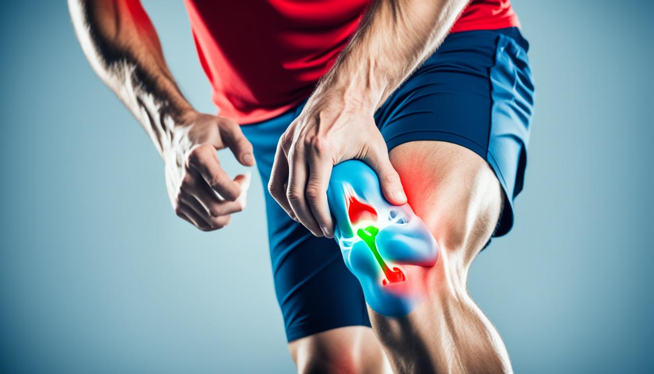
    Did you know that the patella, also known as the kneecap, is the largest sesamoid bone in the body? Its size alone speaks to the crucial role it plays in knee function. Located within the quadriceps tendon, anterior to the knee joint, the patella serves multiple functions that are vital for maintaining knee joint stability and efficient movement.

    The patella acts as an attachment point for the quadriceps tendon and the patellar ligament, increasing the effective extension capacity of the quadriceps muscle. It also protects the quadriceps tendon from frictional forces and acts as a bony shield for deeper structures in the knee joint. Without a properly functioning patella, knee stability is compromised, and movement becomes inefficient.

    Understanding the importance of the patella and its role in knee function is essential for maintaining knee joint stability and preventing knee pain. In this article, we will explore the biomechanics of the patella, common conditions that affect it, evaluation methods for patellar health, treatment options, and the overall significance of the patella in knee joint stability. Let’s dive deeper into the fascinating world of the patella and its impact on our knee health.

    Importance of Patellar Biomechanics in Knee Function

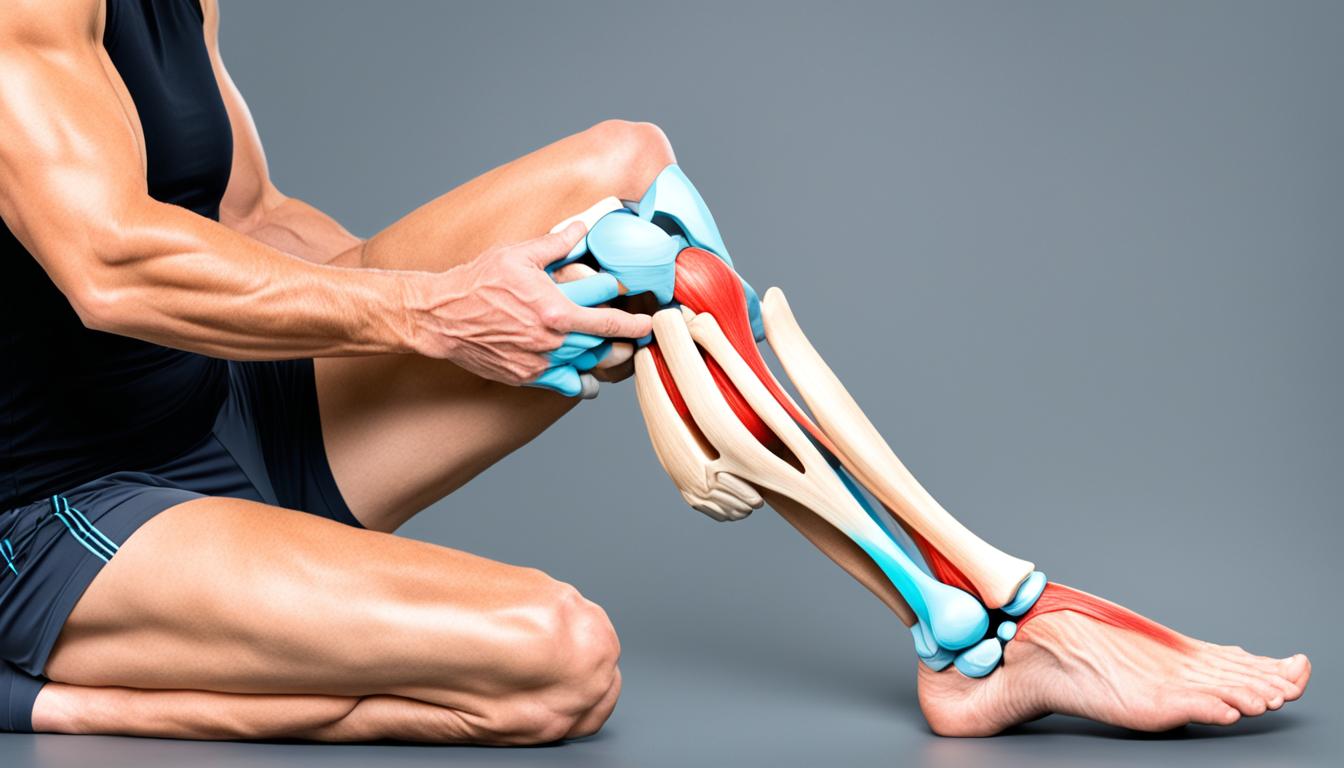
    Patellar biomechanics play a significant role in maintaining proper knee function and promoting overall knee health. The function of the patella goes beyond just being a bony structure in the knee joint. It acts as a crucial fulcrum, enhancing the efficiency of knee extension and reducing the force required for full extension.

    patellar biomechanics

    When the knee is extended, the patella increases the moment arm, resulting in a greater lever arm for the quadriceps muscle to generate torque on the tibia. This mechanism allows for smoother and more effortless movement during knee extension, preventing excessive strain on the joint.

    In addition to its role in knee extension, the patella also plays a crucial part in patellar tracking within the patellofemoral joint. Proper patellar tracking ensures smooth movement of the knee during flexion and extension, reducing the risk of patellofemoral joint dysfunction and associated knee pain.

    Correct patellar biomechanics are essential for preventing knee pain and maintaining optimal patellofemoral joint health. When the patella is functioning optimally, the joint experiences minimal wear and tear, reducing the risk of degenerative conditions and enhancing long-term joint health.

    Benefits of Proper Patellar Biomechanics:

    • Efficient knee extension with reduced force requirements
    • Improved patellar tracking for smooth knee movement
    • Prevention of patellofemoral joint dysfunction
    • Reduced risk of knee pain and discomfort
    • Enhanced patellofemoral joint health and longevity

    Understanding the importance of patellar biomechanics highlights its integral role in knee function and the prevention of knee pain. By prioritizing proper patellar tracking and biomechanics, we can take proactive steps to maintain patellofemoral joint health and prevent potential knee problems in the future.

    Common Conditions and Disorders that Affect the Patella

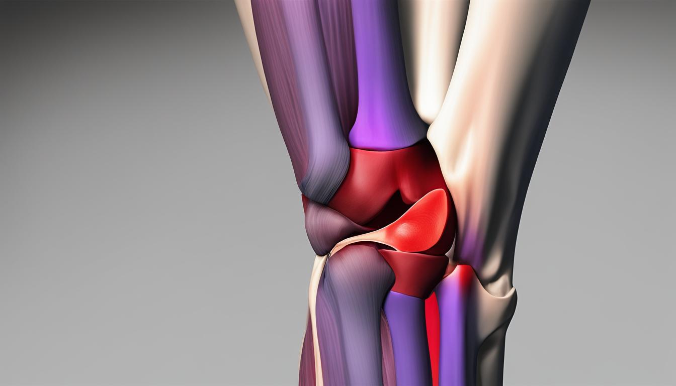
    The patella, also known as the kneecap, is susceptible to various conditions and disorders that can cause knee pain and affect its function. Understanding these common issues can help individuals recognize the symptoms and seek appropriate medical attention for early intervention and treatment.

    1. Patellar Dislocation

    One common condition that affects the patella is patellar dislocation. This occurs when the patella completely moves out of its normal position within the patellofemoral groove. Patellar dislocation can result from traumatic injuries, such as falls or direct blows to the knee, or from underlying joint instability.

    When the patella dislocates, individuals may experience severe pain, swelling, and an inability to straighten the knee. Prompt medical attention is necessary to relocate the patella and prevent further damage. In some cases, surgical intervention may be required to stabilize the patella and prevent future dislocations.

    2. Patellar Subluxation

    Another condition that affects the patella is patellar subluxation. This occurs when the patella partially moves out of its normal position but returns to its original place on its own. Patellar subluxation can also be caused by traumatic injuries or joint instability.

    Individuals experiencing patellar subluxation may feel a sensation of the patella “slipping” or “giving way” during physical activity. While the patella returns to its normal position, it can lead to pain, instability, and difficulty with knee movements. Treatment options for patellar subluxation may include physical therapy exercises to strengthen the muscles around the knee joint and reduce the risk of further subluxations.

    3. Osteoporosis and Patellar Fractures

    Osteoporosis is a condition characterized by weakened bones, making them more prone to fractures. While osteoporosis commonly affects the spine and hips, it can also impact the patella and increase the risk of fractures in this area.

    Fractures of the patella can cause severe pain, swelling, and limited mobility. Treatment options for patellar fractures depend on the severity of the fracture but may include immobilization with a brace, physical therapy, or surgical repair.

    It is important to seek medical attention if experiencing knee pain or symptoms of patellar instability. Early diagnosis and proper management can help prevent complications and promote optimal knee health.

    Patellar Conditions

    Condition Symptoms Treatment
    Patellar Dislocation Pain, swelling, inability to straighten the knee Relocation, immobilization, physical therapy, possible surgery
    Patellar Subluxation Sensation of patella “slipping,” pain, instability Physical therapy, exercises to strengthen muscles around the knee joint
    Osteoporosis and Patellar Fractures Pain, swelling, limited mobility Brace immobilization, physical therapy, surgical repair if necessary

    Tests and Imaging for Evaluating Patellar Health

    To evaluate the health of the patella, various tests and imaging techniques are used. These tests help determine the appropriate treatment plan for patellar conditions and disorders.

    One of the most common tests used to assess patellar health is the patella reflex test. In this test, the knee is tapped below the patella to elicit an involuntary reflex. The presence and strength of the reflex can provide insight into the functioning of the patella and its associated nerves.

    In addition to the patella reflex test, imaging tests are frequently employed to further evaluate patellar health. X-rays are a commonly used imaging technique to assess for patellar injuries, fractures, or the presence of osteoporosis. X-rays can provide detailed images of the patella and surrounding structures, helping healthcare professionals make accurate diagnoses and develop appropriate treatment plans. Other imaging tests, such as MRIs or CT scans, may be utilized in more complex cases or when further evaluation is needed.

    These tests and imaging techniques play a crucial role in understanding the extent of patellar conditions and disorders. By providing detailed insights into the health and structural integrity of the patella, healthcare professionals can make informed decisions regarding treatment options, including conservative approaches or surgical interventions.

    Treatments for Patellar Conditions and Disorders

    Treatment options for patellar conditions and disorders vary depending on the specific condition and its severity. In many cases, conservative treatments may be sufficient, such as wearing a brace or immobilizing device, resting, and engaging in physical therapy exercises to strengthen the surrounding muscles.

    **At-home treatments** like **icing** and **over-the-counter pain relievers** can also provide relief for patellar conditions. Icing the affected area helps reduce inflammation and relieve pain. Over-the-counter pain relievers, such as nonsteroidal anti-inflammatory drugs (NSAIDs), can help manage pain and inflammation as well.

    Proper rest and avoiding activities that worsen the condition are crucial for the recovery process. It is important to listen to your body and give it the time it needs to heal.

    In cases of **patellar fractures** or **osteoporosis**, more intensive treatment approaches may be necessary. **Fracture treatment** typically involves immobilization with a cast or a brace to allow the bone to heal properly. In severe cases, surgery may be required to realign the fragments and stabilize the patella.

    **Osteoporosis treatment** focuses on strengthening the bones and preventing further bone loss. This may involve a combination of medications, lifestyle changes, and nutritional adjustments to promote bone density and overall bone health.

    Each patellar condition is unique, and the treatment plan should be tailored to individual needs. It is important to consult with a healthcare professional to determine the most appropriate treatment options for your specific situation.

    Treatment Options for Patellar Conditions and Disorders

    Treatment Description
    Braces or immobilizing devices Wearing a brace or immobilizing device to provide support and stability to the patella and surrounding structures.
    Rest Taking a break from activities that aggravate the patellar condition to allow for healing and recovery.
    Physical therapy exercises Engaging in prescribed exercises to strengthen the surrounding muscles and improve joint stability.
    At-home treatments Utilizing techniques such as icing and over-the-counter pain relievers to reduce inflammation and manage pain.
    Fracture treatment Immobilizing the patella with a cast or a brace to promote proper healing. Surgery may be required for severe fractures.
    Osteoporosis treatment Focusing on strengthening the bones and preventing further bone loss through medication, lifestyle changes, and nutritional adjustments.

    The Role of Patella in Knee Joint Stability and Prevention of Knee Pain

    The patella, also known as the kneecap, plays a crucial role in maintaining knee joint stability and preventing knee pain. It serves as a bony shield, protecting the knee joint from external forces and distributing them evenly across the joint. This reduces the risk of injury and instability, keeping the knee joint strong and secure.

    Moreover, the patella improves the efficiency of knee extension by increasing the moment arm of the knee. This allows the quadriceps muscles to generate torque on the tibia, facilitating smooth and powerful movements. By optimizing knee extension, the patella helps prevent excessive strain on the knee, minimizing the risk of pain and discomfort.

    In addition to its protective and biomechanical functions, the patella also plays a key role in proper patellar tracking. It ensures that the patella glides smoothly along the trochlear groove of the femur during knee flexion and extension. This reduces friction and wear on the joint surfaces, promoting joint health and longevity.

    Understanding the importance of the patella allows individuals to take proactive steps in maintaining knee health and preventing pain. By adopting habits that support knee joint stability, such as engaging in regular exercise to strengthen the surrounding muscles and avoiding activities that put excessive stress on the knee, one can significantly reduce the risk of knee pain and injury.

    Proper care and attention to the patella, along with overall knee joint health, can lead to a pain-free and active lifestyle. So, let’s prioritize the well-being of our patella and ensure the stability and longevity of our knee joints.

    Conclusion

    In conclusion, the patella plays a vital role in knee function and joint stability. Its proper biomechanics and tracking are essential for efficient movement and the prevention of knee pain. By understanding the importance of the patella, we can better care for our knee health and seek appropriate treatment when needed. By prioritizing the health of our patella, we can maintain a healthy and functional knee joint, leading to an improved quality of life.

    Whether it’s engaging in regular exercise, wearing proper protective gear during physical activities, or seeking medical attention for any symptoms or conditions related to the patella, taking care of our knee health should always be a priority. The patella’s role in knee joint stability cannot be overstated, and by maintaining its proper function, we can minimize the risk of injury and maximize our mobility.

    Remember, a healthy patella means a healthier knee joint overall. By incorporating simple preventive measures into our daily lives and staying proactive about our knee health, we can ensure that the patella continues to fulfill its important role in supporting our knee function and maintaining joint stability. Let’s prioritize our patella and enjoy a life of pain-free movement.

    FAQ

    Why is the patella important in knee function?

    The patella plays a crucial role in knee function by acting as an attachment point for the quadriceps tendon and the patellar ligament. It also increases the effective extension capacity of the quadriceps muscle, protects the quadriceps tendon from frictional forces, and acts as a bony shield for deeper structures in the knee joint.

    What is the function of the patella?

    The patella increases the moment arm of the knee during extension, allowing the quadriceps to generate torque on the tibia. This improves the efficiency of knee extension and reduces the amount of force required for full extension. The patella also contributes to the proper tracking of the patellofemoral joint, ensuring smooth movement of the knee during flexion and extension.

    How does the patella contribute to knee joint stability?

    The patella acts as a bony shield and helps distribute forces evenly across the knee joint. It improves the stability of the knee by reducing the risk of injury and instability. Its proper tracking and biomechanics also prevent excessive friction and wear on the joint surfaces.

    What are some common conditions and disorders that affect the patella?

    Common conditions that affect the patella include patellar dislocation, patellar subluxation, and osteoporosis. Patellar dislocation and subluxation can occur due to injuries or joint instability, while osteoporosis weakens the bones, including the patella, and increases the risk of fractures.

    How is the health of the patella evaluated?

    The health of the patella can be evaluated through various tests and imaging techniques. The most common test is the patella reflex test, where the knee is tapped below the patella to elicit an involuntary reflex. Imaging tests, such as X-rays, may be performed to assess patellar injuries, fractures, or the presence of osteoporosis.

    What are the treatment options for patellar conditions and disorders?

    The treatment options for patellar conditions and disorders depend on the specific condition and its severity. In many cases, conservative treatments such as wearing a brace or immobilizing device, resting, and engaging in physical therapy exercises to strengthen the surrounding muscles may be sufficient. At-home treatments like icing and over-the-counter pain relievers can also provide relief. However, more intensive treatment approaches, including surgery, may be necessary for patellar fractures or osteoporosis.

    How does the patella contribute to the prevention of knee pain?

    The patella helps distribute forces evenly across the knee joint, reducing the risk of excessive stress and strain on the surrounding structures. Its proper tracking and biomechanics ensure smooth movement of the knee, preventing excessive friction and wear on the joint surfaces. This contributes to the prevention of knee pain and the maintenance of patellofemoral joint health.

    Source Links

  • Understanding Why Patella Pain Occurs

    Understanding Why Patella Pain Occurs

    Did you know that patellofemoral pain syndrome, commonly known as runner’s knee, affects up to 25% of active individuals? That’s a staggering number of people experiencing pain and discomfort in their knees while engaging in various physical activities.

    Patellofemoral pain syndrome is characterized by pain at the front of the knee, specifically around the kneecap. It is more prevalent in individuals who participate in running and sports involving running and jumping. The pain can be triggered by activities such as running, walking up or down stairs, sitting for long periods, or squatting.

    In this article, we will delve into the causes of patella pain, the role of overuse and muscle imbalances, the impact of injury, the risk factors involved, preventive measures, and the diagnosis and treatment options available. Our goal is to provide you with a comprehensive understanding of patellofemoral pain and empower you to prevent and manage this condition effectively.

    So, let’s dive in and uncover the reasons behind patella pain and how you can find relief from this debilitating condition.

    The Role of Overuse in Patella Pain

    In the realm of patellofemoral pain syndrome, overuse plays a significant role in the development of this condition. Activities that involve running and jumping, such as sports, can subject the knee joint to repeated stress, leading to irritation under the kneecap. This repetitive stress on the knee joint can cause inflammation and pain in the patella region, contributing to patellofemoral pain syndrome.

    To prevent overuse injuries and minimize patella pain, it is essential to adopt proper training techniques and gradually increase activity levels. Listening to your body’s signals and not pushing yourself too hard too soon is crucial in maintaining the health of your patella.

    overuse and patella pain

    Repetitive Stress and Its Impact on Patella Health

    Repetitive stress is a key factor in the development of patella pain. When the knee joint is subjected to repeated stress from activities like running and jumping, it can lead to inflammation and irritation under the kneecap, resulting in patellofemoral pain syndrome. The constant pounding and strain on the knee joint can cause wear and tear, leading to overuse injuries and patella pain.

    Repetitive stress on the knee joint from activities such as running can cause inflammation and irritation under the kneecap.

    Incorporating rest days into your training routine and engaging in low-impact activities can help alleviate stress on the knee joint and reduce the risk of developing patellofemoral pain. It is crucial to strike a balance between challenging your body and allowing it to rest and recover.

    Preventing Overuse Injuries and Patella Pain

    To prevent overuse injuries and minimize the risk of patella pain, it is important to follow these preventive measures:

    • Gradually increase the intensity and duration of your workouts to allow your body to adapt to the demands placed on it.
    • Include proper warm-up and cool-down routines in your exercise regimen to prepare your muscles and joints for activity and promote efficient recovery.
    • Listen to your body’s signals and take rest days when needed to prevent overloading the knee joint.
    • Incorporate cross-training activities that are less stressful on the knees, such as swimming or cycling, to reduce the repetitive impact on your joints.
    • Wear appropriate footwear that provides adequate support and cushioning for your feet and knees.

    By adopting these preventive measures, you can reduce the risk of overuse injuries and patella pain, allowing you to remain active and pain-free.

    Muscle Imbalances and Weaknesses

    Muscle imbalances and weaknesses are important factors to consider when addressing patellofemoral pain. In particular, the quadriceps and the muscles around the hip and knee play a key role in knee stability and patella alignment.

    When these muscles are imbalanced or weak, they fail to adequately support the knee joint, leading to patella misalignment and subsequent pain. For example, if the quadriceps are stronger on one side of the knee than the other, it can cause the kneecap to track incorrectly.

    Addressing these muscle imbalances and weaknesses is essential for preventing and managing patellofemoral pain. Strengthening the key muscle groups, such as the quadriceps, hamstrings, and glutes, can help improve knee stability and patella alignment.

    The Role of Quadriceps Strength

    Strong quadriceps muscles are crucial for maintaining proper knee alignment and preventing patella pain. These muscles, located at the front of the thigh, provide stability and support to the knee joint. When the quadriceps are weak, the kneecap is more likely to move out of alignment, causing pain and discomfort.

    A study conducted by Wang et al. found that individuals with weaker quadriceps muscles were at a higher risk of developing patellofemoral pain syndrome. Furthermore, a review by Witvrouw et al. highlighted the importance of quadriceps strengthening exercises in both preventing and treating patellofemoral pain.

    Quadriceps Strengthening Exercises Description
    Lunges Stand with one leg forward and one leg back, then lower your body by bending your knees. Return to the starting position and repeat on the other side.
    Squats Stand with your feet shoulder-width apart, then lower your body down as if sitting back into a chair. Return to the starting position and repeat.
    Step-ups Step up onto a raised platform with one leg, then step back down. Repeat with the other leg.

    The Importance of Hip and Knee Alignment

    In addition to quadriceps strength, proper alignment of the hip and knee joints is crucial for preventing patellofemoral pain. When the knee collapses inward during activities like squatting or running, it can put excessive stress on the patella and lead to pain and dysfunction.

    “Maintaining proper hip and knee alignment is essential for optimal knee function and injury prevention.” – Dr. Sarah Johnson

    Strengthening the hip abductor and external rotator muscles can help maintain better alignment and reduce the risk of patellofemoral pain. Exercises such as lateral leg raises, clamshells, and glute bridges target these muscles and improve hip stability.

    Hip Strengthening Exercises Description
    Lateral Leg Raises Lie on your side with your legs stacked. Lift the top leg upward, keeping it straight. Lower back down and repeat on the other side.
    Clamshells Lie on your side with your hips and knees bent. Keeping your feet together, lift the top knee as far as you can without rotating your hips. Lower back down and repeat on the other side.
    Glute Bridges Lie on your back with your knees bent and feet flat on the ground. Lift your hips upward, squeezing your glutes, and then lower back down.

    By focusing on strengthening the quadriceps and improving hip and knee alignment, individuals can address muscle imbalances and weaknesses that contribute to patellofemoral pain. These exercises, performed regularly and with proper form, can help maintain knee stability, patella alignment, and prevent the onset or recurrence of patella pain.

    muscle imbalances

    The Impact of Injury on Patella Health

    Trauma to the kneecap can have a significant impact on its health, leading to conditions such as patellofemoral pain syndrome. Injuries like patella dislocation or fractures can cause the kneecap to get out of place or break, resulting in patella pain and discomfort.

    Patella dislocation occurs when the kneecap moves or slides out of its normal position, often caused by a forceful impact or sudden change in direction. This displacement can cause severe pain, swelling, and instability in the knee joint. Similarly, patella fractures involve a break in the kneecap bone, resulting in intense pain, bruising, and difficulty moving the knee.

    Knee surgery, especially procedures that utilize the patient’s own patellar tendon as a graft in anterior cruciate ligament repair, can also contribute to patellofemoral pain. These surgical interventions, while necessary for treating certain knee conditions, can disrupt the natural stability and alignment of the patella, leading to persistent pain and discomfort.

    To minimize the likelihood of developing patella pain, it is essential to take appropriate preventive measures and engage in timely treatment for patella injuries. Utilizing proper protective gear and equipment during physical activities can help reduce the risk of knee trauma. Additionally, seeking professional medical attention and rehabilitation after an injury can facilitate proper healing and recovery.

    It is important to note that the severity and long-term impact of patella injuries can vary. Some individuals may experience mild discomfort and recover fully with conservative treatment, while others may require more extensive interventions, such as surgery or physical therapy, to manage their symptoms and regain optimal knee function.

    Key Points:

    • Trauma to the kneecap, such as patella dislocation or fractures, can cause patella pain and discomfort.
    • Knee surgery, especially procedures involving the use of the patellar tendon, increases the risk of developing patellofemoral pain.
    • Preventing and treating patella injuries appropriately is crucial to minimizing the likelihood of developing patella pain.

    Understanding the Risk Factors for Patella Pain

    Several risk factors can increase an individual’s likelihood of developing patellofemoral pain. Age and sex play significant roles in the occurrence of patella pain. Teenagers and young adults are more prone to experiencing patellofemoral pain, while women have a higher prevalence compared to men.

    Women’s wider pelvises contribute to the increased angle at which the bones in the knee joint meet, leading to a higher risk of patella pain. Engaging in certain sports that involve running and jumping can further strain the knees, increasing the susceptibility to developing patellofemoral pain.

    Risk Factors Description
    Age Teenagers and young adults are commonly affected by patella pain.
    Sex Women have a higher prevalence of patellofemoral pain compared to men.
    Engaging in certain sports Participating in sports involving running and jumping increases the risk of patella pain.

    Preventive Measures for Patella Pain

    While knee pain can sometimes be unavoidable, there are steps that can be taken to prevent or lessen the likelihood of developing patellofemoral pain. Building strength in the leg and hip muscles helps maintain knee balance during activity. Avoiding deep squatting during weight training is advised. Moving safely with proper techniques, especially focusing on strengthening outer hip muscles, can help prevent the knee from caving inward during activities like squatting, jumping, or stepping down. Losing excess weight, warming up before exercise, practicing stretching exercises for flexibility, and gradually increasing workout intensity are also preventive measures for patella pain.

    By following these preventive measures, individuals can significantly reduce their risk of developing patella pain. Let’s dive into each measure in more detail:

    1. Building Strength in the Leg and Hip Muscles

    Strong leg and hip muscles play a crucial role in maintaining knee stability and reducing the strain on the patellofemoral joint. It is essential to incorporate exercises that target these muscle groups into your regular fitness routine. Examples of effective exercises include squats, lunges, leg presses, and hip abduction exercises.

    2. Avoiding Deep Squatting during Weight Training

    Deep squatting puts excessive stress on the patellofemoral joint and can exacerbate existing patella pain or lead to its development. Avoid deep squatting during weight training and focus instead on performing partial squats within a pain-free range of motion.

    3. Moving Safely and Using Proper Techniques

    Proper movement techniques during physical activities are crucial for preventing patella pain. For instance, when performing squats or lunges, it’s important to engage the outer hip muscles and avoid letting the knees cave inward. Consulting with a fitness professional or physical therapist can be helpful in learning the correct form and technique.

    4. Losing Excess Weight

    Excess weight can place additional stress on the patellofemoral joint and increase the risk of developing patella pain. Maintaining a healthy weight through a balanced diet and regular exercise can help alleviate pressure on the knee joint and reduce the likelihood of experiencing patellofemoral pain.

    5. Warming Up and Stretching

    Prior to engaging in any physical activity, it’s crucial to warm up properly to increase blood flow, elevate core body temperature, and loosen up the muscles. Warming up prepares the body for exercise and helps prevent injuries. Additionally, incorporating stretching exercises into your routine can improve flexibility and reduce the risk of muscle imbalances that contribute to patellofemoral pain.

    6. Gradually Increasing Workout Intensity

    When increasing the intensity or duration of your workouts, it’s important to do so gradually. Rapidly intensifying your exercise routine can overload the patellofemoral joint, leading to pain and potential injury. Gradually increasing intensity allows your body to adapt and build strength without placing excessive stress on the knee joint.

    By adopting these preventive measures for patella pain, individuals can reduce the risk and severity of patellofemoral pain syndrome. Taking care of your knees and following these guidelines will contribute to maintaining a healthy and pain-free lifestyle.

    Diagnosing and Treating Patella Pain

    Proper diagnosis and treatment are essential for managing patellofemoral pain. At our healthcare facility, we employ comprehensive methods to accurately diagnose the underlying causes of patella pain. Through a thorough physical examination, we assess the functionality and alignment of the knee joint. We also evaluate symptoms reported by patients, such as pain intensity, location, and aggravating factors.

    In addition to the physical examination, we may recommend imaging tests such as x-rays or MRI scans to further assess the condition. These tests provide detailed information about the structures of the knee, helping us identify any abnormalities or damage that could be causing the patella pain.

    Once the diagnosis is confirmed, we develop personalized treatment plans to address the specific needs of each individual. Treatment options for patella pain vary depending on the severity of the condition, but most cases can be effectively managed without the need for surgery.

    For nonsurgical management, we typically advise activity changes to reduce stress on the knee joint. This may involve modifications to exercise routines, avoiding high-impact activities, or implementing periods of rest to allow for recovery and pain relief.

    Ice packs and compression knee wraps can help reduce inflammation and provide temporary relief from pain. We may also recommend over-the-counter or prescription anti-inflammatory medications to alleviate discomfort.

    Stretching exercises for the muscles around the knee, as well as strengthening exercises for the quadriceps and hips, can improve knee stability and support overall patella alignment. Physical therapy sessions with our experienced therapists may be recommended to optimize range of motion, strength, and endurance.

    “Our goal is to provide comprehensive and effective treatment options for patella pain, helping our patients regain function and improve their quality of life.”

    In some cases, orthotics or shoe inserts may be prescribed to provide additional support to the foot and ankle, reducing stress on the knees.

    When necessary, we collaborate with orthopedic specialists to determine if surgical intervention is the best course of action. However, our primary focus is always on conservative, nonsurgical approaches to manage patella pain, ensuring that patients can recover and resume their normal activities as soon as possible.

    Diagnosing and Treating Patella Pain: Summary

    Properly diagnosing patellofemoral pain syndrome is a crucial step in effectively treating and managing the condition. Our healthcare professionals conduct physical examinations and utilize imaging tests to accurately identify the causes of patella pain. Through personalized treatment plans, we employ nonsurgical approaches such as activity changes, rest, ice packs, compression knee wraps, anti-inflammatory medication, stretching exercises, and strengthening exercises. Physical therapy and the use of orthotics or shoe inserts may also enhance recovery and provide long-term relief from patella pain.

    Conclusion

    In conclusion, patellofemoral pain is a common condition that often affects individuals who engage in activities such as running and jumping. It is essential to understand the causes and risk factors associated with patella pain to effectively prevent and manage this condition.

    By implementing preventive measures such as building strength, moving safely, losing weight, warming up, stretching, and gradually increasing workouts, individuals can significantly reduce their likelihood of developing patellofemoral pain. These proactive steps can help maintain knee balance, prevent muscle imbalances, and avoid overuse injuries.

    For individuals already experiencing patella pain, proper diagnosis and treatment are key. Activity changes, rest, ice, medication, and physical therapy can help alleviate pain, improve function, and facilitate recovery. It is also essential to pay attention to any structural problems or contributing factors that may require additional medical interventions.

    By staying proactive in preventing patella pain and seeking appropriate treatment when needed, individuals can minimize the impact of patellofemoral pain on their daily lives and maintain optimal knee health.

    FAQ

    Why does patella pain occur?

    Patella pain can occur due to various reasons, such as overuse, muscle imbalances, injury, and surgery. Repetitive stress on the knee joint, muscle weaknesses, trauma to the kneecap, and certain risk factors can contribute to the development of patellofemoral pain syndrome.

    What are the causes of patella pain?

    Patella pain can be caused by overuse injuries, muscle imbalances or weaknesses, trauma to the kneecap, and certain risk factors like age, sex, and participating in specific sports. Understanding the underlying causes is crucial for prevention and management of patella pain.

    What triggers patella pain?

    Patella pain can be triggered by activities such as running, walking up or down stairs, sitting for long periods, and squatting. These activities can aggravate the pain and discomfort experienced in the front of the knee, around the kneecap.

    How do muscle imbalances or weaknesses contribute to patella pain?

    When the muscles around the hip and knee, particularly the quadriceps, are imbalanced or weak, they fail to keep the kneecap properly aligned. This misalignment can result in patellofemoral pain syndrome and contribute to patella pain. Strengthening these muscles and maintaining proper knee alignment can help prevent and manage patella pain.

    What are the causes of patella injuries?

    Patella injuries can be caused by trauma to the kneecap, such as dislocation or fractures. Additionally, knee surgery, particularly the use of one’s own patellar tendon as a graft in anterior cruciate ligament repair, increases the risk of patellofemoral pain. Appropriate prevention and treatment methods are essential to minimize the likelihood of developing patella pain.

    What are the risk factors for developing patella pain?

    Age, sex, and participating in certain sports that involve running and jumping are risk factors for developing patellofemoral pain. Teenagers and young adults are more likely to experience patella pain, and women are twice as likely as men to develop the condition. Engaging in activities that add extra stress on the knees can also increase the risk.

    How can patella pain be prevented?

    Preventive measures for patella pain include building strength in the leg and hip muscles, moving safely with proper techniques, losing excess weight, warming up before exercise, practicing stretching exercises for flexibility, and gradually increasing workout intensity. These measures help maintain knee balance, prevent muscle imbalances, and minimize the risk of developing patellofemoral pain.

    How is patella pain diagnosed and treated?

    Healthcare providers can diagnose patellofemoral pain through physical examination, evaluation of symptoms, and imaging tests like x-rays or MRI scans. Treatment options vary but often involve nonsurgical approaches such as activity changes, rest, ice packs, compression knee wraps, anti-inflammatory medication, stretching exercises, and strengthening exercises. In some cases, physical therapy or orthotics may be recommended.

    Source Links

  • Why the Patella is a Sesamoid Bone Explained

    Why the Patella is a Sesamoid Bone Explained

    Did you know that the human body has over 200 bones? Yet, there is one bone that stands out for its unique function and structure. It’s the patella, also known as the kneecap, and it serves a remarkable role as a sesamoid bone.

    But what exactly makes the patella so special? Why is it classified as a sesamoid bone? And what function does it serve in our bodies?

    In this article, we will explore the intriguing world of the patella and uncover the reasons behind its significance. From its development during gestation to its role in the biomechanics of the lower extremities, we will dive into the fascinating details that make the patella a truly remarkable anatomical feature.

    Structure and Function of Sesamoid Bones

    Sesamoid bones play a crucial role in our musculoskeletal system, providing added strength to muscles and supporting tendon stability. These small bones can be found in various joints throughout the body. Let’s explore their structure, function, and the different types of sesamoid bones.

    Structure:

    Sesamoid bones are unique due to their location within tendons or muscles. They are typically small and oval-shaped, with a smooth surface to reduce friction. These bones can vary in size, with some being as small as a grain of rice and others as large as a golf ball.

    Function:

    The primary function of sesamoid bones, including the patella, is to enhance joint function and provide support to muscles and tendons. They act as pulleys, changing the direction of forces and improving the mechanical efficiency of the muscles they are associated with. The sesamoid bones’ position within the tendon also helps protect the tendon from excessive wear and tear, reducing the risk of injury.

    Types of Sesamoid Bones:

    Sesamoid bones are broadly classified into two types: Type A and Type B.

    Type A sesamoid bones: These bones are adjacent to a joint and become part of the joint capsule. They are firmly embedded within the tendon or muscle and move together with the joint. Examples of type A sesamoid bones include the patella and the pisiform bone found in the wrist.

    Type B sesamoid bones: These bones overlie a bony prominence and are separated from it by an underlying bursa. Unlike type A sesamoid bones, type B bones are not part of the joint capsule. The two most common examples of type B sesamoid bones are the fabella behind the knee joint and the sesamoid bones under the first metatarsophalangeal joint of the foot.

    Type Examples
    Type A Patella, pisiform bone
    Type B Fabella, sesamoid bones in the foot

    Note: The patella, as the largest sesamoid bone, deserves special attention due to its significant role in knee joint function. Its structure and function will be further explored in the following sections.

    sesamoid bone structure

    Understanding the structure and function of sesamoid bones helps us appreciate their importance in our body’s biomechanics. The next section will delve into the embryology and development of the patella, shedding light on its fascinating journey from a band of fibrous tissue to a fully-formed bone.

    Embryology and Development of the Patella

    The development of the patella begins during the first trimester of gestation. It starts as a band of fibrous tissue and gradually chondrifies, forming the early patella.

    By the 9th week of gestation, the embryological patella inserts into the quadriceps tendon. During the 14th week of gestation, the patella is entirely cartilaginous and resembles the adult patella structure by the 23rd week.

    Ossification of the patella occurs during childhood and continues into early adolescence, stabilizing into the adult patella during the pubertal years.

    ossification of the patella

    “The development of the patella is a fascinating process that starts early on in gestation. The initial fibrous tissue gradually transforms into a fully-formed bone through the stages of chondrification and ossification. This intricate process highlights the importance of the patella in the support and function of the knee joint.”

    Blood Supply and Nerves of the Patella

    The patella, being a crucial component of the lower extremity, boasts a robust blood supply that is vital for its function. The blood supply of the patella is facilitated by a comprehensive network of extraosseous and intraosseous blood vessels.

    The extraosseous blood vessels, including genicular arteries and anterior tibial recurrent arteries, contribute significantly to the blood supply of the patella.

    Meanwhile, the intraosseous blood supply comprises mid-patellar vessels and polar vessels, ensuring adequate vascularization within the patella.

    In terms of innervation, the patella is extensively supplied by different nerves, which are responsible for sensory and motor functions of the bone.

    The anterior aspect of the patella receives innervation from the infrapatellar branch of the saphenous nerve, while the medial aspect is innervated by the infrapatellar branch of the obturator nerve.

    For the lateral aspect, the nerve supply comes from the lateral femoral cutaneous nerve and the peroneal nerve.

    “The innervation of the patella ensures adequate nerve supply for various functions and contributes to the overall sensation and movement of the bone.” – Dr. Emily Johnson

    It is worth noting that other sesamoid bones, such as the hallux sesamoid bone, receive innervation from the plantar digital nerves.

    Blood Supply and Nerves of the Patella

    Aspect Blood Supply Innervation
    Anterior Infrapatellar branch of the saphenous nerve Infrapatellar branch of the saphenous nerve
    Medial Infrapatellar branch of the obturator nerve Infrapatellar branch of the obturator nerve
    Lateral Lateral femoral cutaneous nerve
    Peroneal nerve
    Lateral femoral cutaneous nerve
    Peroneal nerve

    Conclusion

    The patella, as a sesamoid bone in humans, serves a unique and crucial purpose in our musculoskeletal system. As the largest sesamoid bone in the body, the patella plays a significant role in the biomechanics and functionality of the knee joint.

    One of the main purposes of the patella is to increase joint leverage. By acting as a fulcrum, the patella enhances the mechanical advantage of the knee, making movements more efficient and coordinated. This allows for a wide range of motion, enabling activities such as walking, running, and jumping.

    In addition to its lever function, the patella provides protection to the muscles and tendons surrounding the knee joint. By distributing forces and alleviating stress, it helps prevent strain and injury, especially during weight-bearing activities.

    Throughout gestation, the patella undergoes a remarkable development process, starting as a band of fibrous tissue and gradually ossifying into a fully-formed bone. This intricate growth reflects the vital role of the patella in the form and function of the lower extremities.

    Understanding the significance of the patella as a sesamoid bone provides valuable insights into the complexities of our musculoskeletal system. By recognizing the interconnectedness of different anatomical structures, we can further appreciate the remarkable design and functionality of our bodies.

    FAQ

    Why is the patella considered a sesamoid bone?

    The patella is considered a sesamoid bone because it is a small bone embedded within the tendon of the quadriceps muscle. It functions as a pulley, increasing joint leverage and distributing forces within the muscle and tendon. This helps to alleviate stress and protect the knee joint.

    What is the function of the patella as a sesamoid bone?

    The patella plays a critical role in the functionality of the knee joint. As the largest sesamoid bone, it increases joint leverage, allowing for a wider range of motion and better control of knee movements. It also contributes to the knee’s extensor properties, enabling activities such as walking, running, and jumping.

    Where is the patella located in the body?

    The patella, also known as the kneecap, is located in the front of the knee joint. It sits within the quadriceps tendon, which connects the thigh muscles to the tibia bone. The patella moves along the femoral groove during knee movements.

    How does the development of the patella occur?

    The patella begins its development as a band of fibrous tissue during the first trimester of gestation. It gradually chondrifies and forms the early patella. By the 9th week of gestation, it inserts into the quadriceps tendon. Ossification of the patella occurs during childhood and continues into adolescence, resulting in the fully-formed adult patella.

    What is the blood supply and innervation of the patella?

    The patella has a rich blood supply, which comes from an extensive network of extraosseous and intraosseous blood vessels. The extraosseous network includes genicular arteries and anterior tibial recurrent arteries. The intraosseous blood supply consists of mid-patellar vessels and polar vessels. The patella is also extensively innervated, with different nerves supplying its anterior, medial, and lateral aspects.

    Source Links

  • Relief for Patella Pain: Causes & Solutions

    Relief for Patella Pain: Causes & Solutions

    Did you know that patella pain, also known as patellofemoral pain, affects approximately 25% of active individuals?

    When it comes to knee problems, patella pain is a common issue that can significantly impact daily activities and hinder one’s ability to engage in physical exercise and sports. This type of pain is typically felt around the front of the knee, where the knee connects with the lower end of the thigh bone (femur). If you are currently experiencing patella pain or want to learn more about how to prevent it, you’ve come to the right place.

    In this article, we will explore the causes of patella pain, effective treatment options, and preventive strategies to help you find relief and manage this condition. Whether you’re an athlete, a fitness enthusiast, or simply someone looking to take care of their knee health, understanding the causes and solutions for patella pain is crucial.

    By the end of this article, you will have a comprehensive understanding of patella pain and the tools necessary to alleviate discomfort, promote healing, and prevent future occurrences. So let’s dive in!

    Common Causes of Patella Pain

    Understanding the causes of patella pain is essential in developing effective treatment and prevention strategies. Patella pain, also known as patellofemoral pain, can be attributed to various factors that contribute to strain on the knee joint. By identifying these causes, we can address the root of the problem and find relief from the discomfort.

    Structural Defects

    Structural abnormalities, such as misalignment of the patella or femur, can lead to patella pain. These defects can put extra pressure on the knee joint, causing pain and discomfort. Diagnosing and addressing these issues is crucial for effective management.

    Improper Walking or Running Techniques

    The way we walk or run can have a significant impact on our knee health. Incorrect techniques, such as overpronation or supination, can place excessive stress on the patella, leading to pain. It is important to learn proper gait mechanics to prevent unnecessary strain on the knee joint.

    High Kneecap Position

    A higher than normal position of the kneecap can cause patella pain. This misalignment can disrupt the natural movement of the patella, leading to inflammation and discomfort. Addressing the position of the kneecap through appropriate exercises and techniques can help alleviate the pain.

    Weak Thigh Muscles

    The muscles surrounding the knee joint play a crucial role in maintaining its stability and supporting its movements. Weak quadriceps or hamstrings can lead to imbalances and strain on the patella, resulting in pain. Strengthening these muscles through targeted exercises can help alleviate patella pain.

    Tight Hamstrings

    Tight hamstrings can contribute to patella pain by altering the biomechanics of the knee joint. When the hamstrings are tight, they can pull on the patella, causing it to track incorrectly and leading to discomfort. Regular stretching and flexibility exercises can help relax the hamstrings and reduce patella pain.

    Poor Foot Support

    Inadequate foot support, such as wearing shoes with insufficient arch support, can affect the alignment of the entire lower extremity. This misalignment can put unnecessary stress on the patella, resulting in pain. Opting for footwear that provides proper cushioning and support can help alleviate patella pain.

    Excessive Training or Overuse

    Engaging in intense physical activity without proper rest and recovery can lead to overuse injuries, including patella pain. Constant repetitive movements and high impact activities can strain the knee joint and cause discomfort. Balancing training intensity with adequate rest is essential for preventing patella pain.

    Causes of Patella Pain

    Identifying the underlying causes of patella pain allows us to develop effective treatment and prevention strategies. By addressing structural defects, improving walking or running techniques, optimizing kneecap position, strengthening thigh muscles, stretching tight hamstrings, ensuring proper foot support, and avoiding excessive training or overuse, we can alleviate patella pain and promote knee health.

    Effective Treatment for Patella Pain

    The treatment of patella pain often begins with simple measures. Resting the knee and avoiding activities that exacerbate the pain, such as climbing stairs or squatting, is recommended. Over-the-counter pain relievers, such as acetaminophen or ibuprofen, can be taken for temporary relief. Physical therapy exercises, including rehabilitation exercises and strengthening exercises for the muscles that support the knee, can be beneficial in relieving patella pain. Other treatment options include the use of supportive braces, taping techniques, icing after exercise, and the use of orthotics or shoe inserts. In more severe cases, surgical procedures such as arthroscopy or realignment may be recommended.

    When it comes to treating patella pain, a holistic approach that combines various methods often yields the best results. Let’s take a closer look at some effective treatment options:

    1. Rest and Avoidance of Aggravating Activities

    One of the first steps in managing patella pain is to give the knee adequate rest. This means avoiding activities that put strain on the knee joint and worsen the pain. Climbing stairs, squatting, and participating in high-impact sports should be avoided until the pain subsides.

    2. Over-the-Counter Pain Relievers

    Over-the-counter pain relievers can provide temporary relief from patella pain. Medications such as acetaminophen or ibuprofen can help alleviate pain and reduce inflammation in the knee joint. However, it’s important to follow the recommended dosage and consult a healthcare professional if the pain persists or worsens.

    3. Physical Therapy Exercises

    Physical therapy exercises play a crucial role in the treatment of patella pain. These exercises focus on rehabilitating and strengthening the muscles that support the knee joint, such as the quadriceps and hamstrings. A physical therapist can guide individuals through a personalized exercise program that targets the specific areas of weakness and imbalance.

    4. Supportive Braces and Taping Techniques

    Supportive braces and taping techniques can provide additional stability to the knee joint and alleviate patella pain. These devices help distribute the forces exerted on the knee more evenly, reducing strain on the patella. A healthcare professional can recommend the appropriate type of brace or taping technique based on the individual’s condition and needs.

    5. Icing and Orthotics

    After engaging in physical activity, icing the knee can help reduce inflammation and relieve patella pain. Applying an ice pack for 15-20 minutes at a time, several times a day, can be effective. Additionally, the use of orthotics or shoe inserts can provide support and alignment to the feet, reducing stress on the knee joint.

    6. Surgical Interventions

    In severe cases where conservative measures do not provide sufficient relief, surgical interventions may be considered. Arthroscopic procedures can be used to diagnose and treat underlying issues within the knee joint, while realignment surgeries aim to correct any structural abnormalities that contribute to patella pain. These procedures should be discussed with a healthcare professional to determine the most appropriate course of action.

    By combining these treatment options, individuals can effectively manage and find relief from patella pain. It’s important to consult with healthcare professionals for an accurate diagnosis and personalized treatment plan that addresses the underlying causes of patella pain.

    patella pain treatment

    Preventive Strategies for Patella Pain

    Preventing patella pain involves taking certain preventive measures. By implementing these strategies, we can reduce the risk of developing patella pain and maintain optimal knee health.

    1. Maintain a healthy weight: Excess weight puts additional stress on the knee joints, increasing the likelihood of patella pain. By maintaining a healthy weight through proper nutrition and regular exercise, we can alleviate strain on the knees.
    2. Stretch before physical activities: Prior to engaging in physical activities, it is important to properly warm up the muscles and joints. Incorporate stretching exercises that target the quadriceps, hamstrings, and calf muscles to prepare the knees for movement.
    3. Gradually increase activity levels: When starting a new exercise program or intensifying physical activity, it is essential to gradually increase the duration and intensity. Sudden, intense workouts can lead to overuse and strain on the patella, causing pain. Take it slow and listen to your body’s signals.
    4. Wear proper footwear with good support: Investing in well-fitting shoes with adequate cushioning and arch support is crucial for maintaining proper alignment and reducing stress on the knees. Choose athletic shoes that are specifically designed for the type of activity you engage in.
    5. Practice correct techniques while running or exercising: Applying proper form and technique during physical activities is essential in preventing knee injuries and patella pain. Consult with a fitness professional or coach to ensure you are using the correct techniques and postures.

    In addition to these preventive measures, paying attention to leg alignment during activities that put strain on the knees, such as going up and down stairs or squatting, can significantly reduce the risk of developing patella pain.

    To further illustrate the importance of preventive strategies, let’s take a look at the following table:

    Activity Risk of Patella Pain
    Running with proper form and footwear Low
    Running with improper form and unsupportive footwear High
    Increased activity level gradually Low
    Sudden intense workouts High
    Proper leg alignment during activities Low

    “Prevention is the key to maintaining healthy knees and avoiding patella pain. By implementing these strategies, we can protect our knees and enjoy an active, pain-free lifestyle.”

    Conclusion

    Patella pain is a challenging condition that can greatly impact individuals who are physically active. To find relief and effectively manage patella pain, it is crucial to understand its causes and implement appropriate treatment and prevention strategies. By following a comprehensive approach that includes rest, medication, physical therapy exercises, supportive braces or taping, and preventive measures, individuals can experience relief from patella pain and reduce the risk of future occurrences.

    Consulting with healthcare professionals is paramount for an accurate diagnosis and personalized treatment plan. They can provide guidance on the most suitable pain relief options, such as over-the-counter pain relievers, and recommend specific exercises to strengthen the muscles supporting the knee joint. Additionally, healthcare professionals can offer valuable insight into preventive strategies, which may include maintaining a healthy weight, practicing proper running or exercise techniques, and wearing appropriate footwear with good support.

    Remember, managing patella pain is a multifaceted process that requires commitment and consistency. By diligently following the recommended treatment plan and incorporating preventive measures into your daily routine, you can effectively alleviate patella pain, improve your knee health, and maintain an active lifestyle. Don’t let patella pain hold you back—take control of your knee health today!

    FAQ

    What causes patella pain?

    Patella pain can be caused by various factors, including structural defects, improper walking or running techniques, high kneecap position, weak thigh muscles, tight hamstrings, poor foot support, and excessive training or overuse.

    How can I find relief from patella pain?

    To find relief from patella pain, it is important to rest the knee and avoid activities that exacerbate the pain. Over-the-counter pain relievers, physical therapy exercises, supportive braces or taping techniques, icing after exercise, and the use of orthotics or shoe inserts can also provide relief.

    What are some treatment options for patella pain?

    Treatment options for patella pain include rest, over-the-counter pain relievers, physical therapy exercises, supportive braces or taping techniques, icing after exercise, and in severe cases, surgical procedures such as arthroscopy or realignment may be recommended.

    How can I prevent patella pain?

    Preventive strategies for patella pain include maintaining a healthy weight, stretching before physical activities, gradually increasing activity levels, wearing proper footwear with good support, and practicing correct techniques while running or exercising. Paying attention to leg alignment during activities that put strain on the knees can also help prevent the development of patella pain.

    Can patella pain be managed effectively?

    Yes, patella pain can be managed effectively by following a comprehensive approach that includes rest, medication, physical therapy exercises, supportive braces or taping techniques, and preventive measures. By consulting with healthcare professionals for an accurate diagnosis and personalized treatment plan, individuals can experience relief from patella pain and reduce the risk of future occurrences.

    Source Links

  • Understanding Lateral Patella Dislocations

    Understanding Lateral Patella Dislocations

    Did you know that patellar dislocation accounts for approximately 2 to 3% of knee injuries? That’s right, this common condition affects a significant number of individuals, particularly young and active ones, including adolescent females and athletes.

    But what exactly is a lateral patella dislocation, and what are the causes and risk factors associated with it? In this article, we will delve into the world of patellar instability and explore the various factors that contribute to the occurrence of lateral patella dislocations.

    Whether you’re a healthcare professional, a patient, or simply curious about knee injuries, understanding the causes and risk factors for lateral patellar dislocation is crucial for prevention, diagnosis, and effective management. Let’s dive in and discover the ins and outs of this condition and how it impacts individuals.

    Risk Factors for Lateral Patellar Dislocation

    Understanding the risk factors associated with lateral patellar dislocation is crucial for effective prevention and management. Several factors can increase an individual’s susceptibility to this condition.

    Anatomical Risk Factors

    Anatomical variations in the knee joint can predispose individuals to lateral patellar dislocation. These include:

    • Patella Alta: An unusually high position of the patella, resulting in an increased risk of dislocation.
    • Trochlear Dysplasia: Shallow or malformed groove in the femur, leading to poor patellar stability.
    • Lateral Patella Tilt: Excessive lateral tilt of the patella, making it more prone to dislocation.

    Ligamentous Laxity

    Ligamentous laxity refers to increased looseness or flexibility of the ligaments in the knee joint. It is commonly seen in females and individuals with connective tissue disorders, such as Ehlers-Danlos syndrome. Ligamentous laxity can contribute to patellar instability and raise the risk of dislocation.

    Muscle Imbalances

    Muscle imbalances, particularly weakness of the vastus medialis oblique (VMO), can also predispose individuals to lateral patellar dislocation. The VMO plays a crucial role in stabilizing the patella, and its weakness can result in patellar instability.

    Activity-Related Factors

    Certain activities increase the risk of lateral patellar dislocation. These include:

    • High-Impact Sports: Activities involving jumping, pivoting, or sudden changes in direction, such as basketball, soccer, and skiing.
    • Dancing: Recurrent and forceful movements in dance styles that place stress on the knee joint.

    Demographic Factors

    Demographic factors can also influence the risk of lateral patellar dislocation:

    • Teenagers: Adolescents, especially those experiencing rapid growth, may have looser joints and ligaments, making them more susceptible to patellar dislocation.
    • Women: Women, with their wider hips and looser ligaments, may experience increased lateral stress on the knee, increasing the risk of dislocation.
    • Big and Tall Men: Individuals with a larger body size may experience increased pressure on the knee joints, putting them at higher risk of patellar dislocation.

    By understanding these risk factors, healthcare professionals can implement targeted prevention strategies and develop personalized treatment plans for individuals with lateral patellar dislocation.

    risk factors for patellar dislocation

    Anatomical Risk Factors Ligamentous Laxity Muscle Imbalances Activity-Related Factors Demographic Factors
    Patella Alta Ligamentous laxity in females and individuals with connective tissue disorders Weakness of the vastus medialis oblique (VMO) muscle High-impact sports Teenagers
    Trochlear Dysplasia Dancing Women
    Excessive lateral patella tilt Big and tall men

    Signs and Symptoms of Lateral Patellar Dislocation

    Common signs and symptoms of lateral patellar dislocation include:

    • An audible pop or snapping sound at the time of dislocation.
    • Buckling or instability of the knee.
    • Intense pain in the knee.
    • Sudden swelling and bruising at the knee.
    • Locking of the knee joint.
    • Inability to walk or bear weight on the affected leg.
    • Visual dislocation of the kneecap, which may be visibly out of place.

    When a lateral patellar dislocation occurs, it is often accompanied by an audible pop or snapping sound. This distinct sound is indicative of the displacement of the kneecap from its normal position. In addition to the audible pop, individuals may experience a sensation of buckling or instability in the knee joint. This instability can make it difficult to maintain balance and perform regular activities.

    The most prominent symptom of a lateral patellar dislocation is intense pain in the knee. The pain is often immediate and can be severe, making it challenging to bear weight on the affected leg. Swelling and bruising are also common after a patellar dislocation, as the soft tissues surrounding the knee joint may become inflamed due to the trauma.

    Another sign of a lateral patellar dislocation is the locking of the knee joint. This occurs when the kneecap becomes trapped outside of its normal position and cannot be easily moved back into place. The locked knee joint can cause a significant limitation in mobility and may require medical intervention to restore proper alignment.

    Visually, a lateral patellar dislocation can be identified by the visible dislocation of the kneecap. The kneecap may appear shifted or out of place, providing a clear indication of the dislocation. This visual dislocation is often accompanied by swelling and bruising, further underscoring the severity of the injury.

    Recognizing these signs and symptoms is essential for prompt diagnosis and appropriate management of a lateral patellar dislocation. If any of these symptoms are experienced after a knee injury, seeking medical attention is crucial to prevent further damage and facilitate effective treatment.

    lateral patellar dislocation symptoms

    Diagnosis and Evaluation of Lateral Patellar Dislocation

    Diagnosing lateral patellar dislocation involves a comprehensive approach, combining the patient’s medical history, physical examination, and imaging studies. This allows healthcare professionals to accurately assess the extent of the injury and determine the most appropriate treatment plan.

    History: Patients with lateral patellar dislocation often recall a specific traumatic event that triggered the injury, such as a non-contact twisting injury or a direct blow to the knee. They typically experience sudden pain, swelling, and joint instability.

    Physical Examination: During a physical examination, physicians evaluate several key factors to confirm the diagnosis. This includes assessing the knee for signs of joint effusion or hemarthrosis (blood in the joint), alignment abnormalities, tenderness, and irregularities along the poles of the patella. Range of motion restrictions are also assessed, and specific tests like the patellar apprehension test and patellar tracking assessment may be performed to evaluate stability and tracking of the patella.

    Imaging Studies: X-rays are commonly used to identify any associated fractures, malalignment, or anatomical risk factors that may contribute to patellar instability. Additional imaging, such as CT scans or MRI, may be necessary to evaluate the full extent of the injury, measure the tuberosity tibia-trochlear groove distance, and assess for potential osteochondral lesions.

    The combination of history, physical examination, and imaging studies allows healthcare professionals to make an accurate diagnosis of lateral patellar dislocation. This comprehensive evaluation is crucial in developing an effective treatment plan that addresses the specific needs and circumstances of the individual patient.

    Treatment and Management of Lateral Patellar Dislocation

    When it comes to treating lateral patellar dislocation, there are several approaches that can be taken based on the severity of the condition and the individual’s specific needs. In many cases, conservative management techniques can effectively treat primary acute patellar dislocations without the need for surgery. This typically involves immobilization of the knee, physiotherapy sessions to strengthen the surrounding muscles and improve stability, and the use of bracing to prevent re-dislocation. Additionally, medications may be prescribed to manage pain and inflammation during the recovery process.

    However, if the dislocations become recurrent or chronic, or if there is significant ligamentous or bony damage, surgical intervention may be necessary. There are various surgical options available, including lateral release, medial patellofemoral ligament reconstruction, and distal realignment or anteromedialisation. These procedures aim to restore proper alignment and stability to the patella, reducing the risk of future dislocations and improving overall knee function.

    Physiotherapy plays a critical role in the management of lateral patellar dislocation, regardless of whether conservative or surgical treatment is pursued. An individualized physiotherapy program may include exercises to improve range of motion, strengthen the quadriceps and other surrounding muscles, enhance proprioception, and promote proper patellar tracking. These exercises and techniques are designed to support the recovery process and help patients regain full function and stability in their knee joint.

    Preventing lateral patellar dislocation is also a key aspect of treatment and management. Addressing risk factors is essential, such as providing appropriate training and conditioning for athletes, focusing on muscle balance and stability, and utilizing protective bracing when necessary. By taking proactive measures to reduce the likelihood of dislocation, individuals can safeguard their knee health and minimize the need for additional treatment or surgery.

    Recovering from a lateral patellar dislocation requires a comprehensive rehabilitation program that includes ongoing monitoring and follow-up care. Range of motion exercises, strength-building exercises, and stability training are essential components of this program, along with gradually reintroducing functional activities. The goal is to restore full range of motion, build strength, and ensure a stable knee joint, allowing individuals to return to their normal activities with confidence.

    FAQ

    Why does the patella dislocate laterally?

    Acute lateral patella dislocations are typically caused by trauma, such as a non-contact twisting injury or a direct blow to the knee. Recurrent subluxation and chronic laxity of the patella can also lead to lateral patellar dislocations. Anatomic variations, ligamentous laxity, muscle imbalances, and certain activities can also contribute to patellar instability.

    What are the risk factors for lateral patellar dislocation?

    Risk factors for lateral patellar dislocation include anatomical variations such as patella alta and trochlear dysplasia, ligamentous laxity (more common in females and individuals with connective tissue disorders), and muscle imbalances. Certain activities, rapid growth in teenagers, wider hips and looser ligaments in women, and increased pressure on the joints in big and tall men can also increase the risk of dislocation.

    What are the common signs and symptoms of lateral patellar dislocation?

    Common signs and symptoms of lateral patellar dislocation include an audible pop or snapping sound at the time of dislocation, buckling or instability of the knee, intense pain, sudden swelling and bruising, locking of the knee joint, inability to walk or bear weight on the affected leg, and visual dislocation of the kneecap.

    How is lateral patellar dislocation diagnosed and evaluated?

    Diagnosis of lateral patellar dislocation involves a combination of history, physical examination, and imaging studies. History includes a traumatic event followed by pain, swelling, and joint instability. Physical examination assesses the knee for abnormalities, tenderness, and range of motion restrictions. Imaging studies, such as X-rays, CT scans, or MRI, are used to identify associated fractures, malalignment, anatomical risk factors, and osteochondral lesions.

    What are the treatment and management options for lateral patellar dislocation?

    Primary acute patellar dislocations can often be managed conservatively with immobilization, physiotherapy, bracing, and medication for pain and inflammation. Surgical intervention may be necessary for recurrent or chronic dislocations, significant ligamentous or bony damage. Surgical options include lateral release, medial patellofemoral ligament reconstruction, and distal realignment or anteromedialisation. Physiotherapy is crucial for rehabilitation and includes range of motion exercises, strengthening exercises, proprioception training, and patellar tracking exercises. Prevention involves addressing risk factors, proper training, and using protective bracing. Recovery requires a comprehensive rehabilitation program and close monitoring.

    Source Links

  • Understanding Why the Patella is a Sesamoid Bone

    Understanding Why the Patella is a Sesamoid Bone

    Did you know that the patella, commonly known as the kneecap, is the largest sesamoid bone in the human body? Sesamoid bones are small bones embedded within muscles or tendons near joint surfaces, and the patella is a prime example of this unique bone type.

    Sesamoid bones are found throughout the body, but the patella holds special significance due to its crucial role in the biomechanics of the knee joint. Connected to the muscles via tendons, sesamoid bones act as pulleys, alleviating stress on the muscles and tendons and enhancing joint leverage.

    In this article, we will explore the anatomy and functions of the patella, its role in the knee joint, and the rehabilitation strategies for sesamoid bone injuries, shedding light on the importance of this small but mighty bone. So, let’s dive in and unravel the mysteries of the remarkable patella!

    Structure and Function of the Patella

    The patella, also known as the kneecap, is an essential bone located within the patellar tendon, connecting the quadriceps muscles to the tibia. It plays a crucial role in the biomechanics of the knee joint and the extensor mechanism, enabling the extension of the knee.

    Functionally, the patella acts as an attachment point for the quadriceps tendon and the patellar ligament, enhancing the extension capacity of the quadriceps and providing mechanical advantage to the thigh muscles. During activities such as walking and running, the patella guides the knee into a flexed position, ensuring smooth and efficient movement.

    Proper patellar alignment is critical for optimal knee function. Disruption in the patellar tracking can lead to conditions like patellofemoral pain syndrome, characterized by anterior knee pain. To address these issues, closed-chain eccentric exercises are often recommended. These exercises involve eccentric muscle contractions during knee extension, helping to strengthen the quadriceps muscles and improve muscle-tendon coordination without placing excessive force on the patella and the knee joint.

    Understanding the structure and function of the patella is vital in diagnosing and treating various knee conditions. By optimizing patellar alignment and implementing appropriate rehabilitation strategies, we can enhance knee joint biomechanics and minimize the risk of patellofemoral pain and other related problems.

    Knee Extension: The Role of the Patella

    Knee extension is a fundamental movement facilitated by the patella. During knee extension, the quadriceps muscles contract, pulling on the patellar tendon and extending the knee. The patella acts as a fulcrum, increasing the mechanical advantage of the quadriceps and allowing for more efficient force transmission.

    patellar tracking

    To ensure proper knee extension, the patella must track smoothly within the patellar groove of the femur. Patellar tracking refers to the alignment and movement of the patella during knee motion. Deviations in patellar tracking can cause patellar malalignment and lead to knee issues, such as patellofemoral pain syndrome.

    By strengthening the quadriceps and improving patellar tracking through targeted exercises, such as closed-chain eccentric exercises, we can promote optimal knee extension and reduce the risk of patellofemoral pain. Implementing a comprehensive rehabilitation program that addresses muscle imbalances, improves patellar alignment, and enhances biomechanics is crucial for successful treatment.

    Benefits of Closed-Chain Eccentric Exercise for Patellofemoral Pain: Guidelines for Performing Closed-Chain Eccentric Exercise:
    1. Strengthening quadriceps muscles
    2. Improving muscle-tendon coordination
    3. Alleviating knee pain and discomfort
    4. Enhancing knee joint stability
    1. Start with low resistance
    2. Gradually increase resistance as tolerated
    3. Perform exercises under the guidance of a healthcare professional
    4. Maintain proper form and technique

    The Role of Sesamoid Bones in the Hand and Foot

    Sesamoid bones are not only found in the knee but also in the hand and foot. In the hand, most people have five sesamoid bones, with two located at the base of the thumb. These hand sesamoid bones play a crucial role in providing stability and enabling fine motor movements of the thumb. However, injuries to these hand sesamoids can be challenging to diagnose due to their small size and complex anatomy.

    When hand sesamoid injuries occur, they should not be overlooked as they can cause significant pain and lead to decreased hand function. Treatment for hand sesamoid injuries will vary depending on the severity of the injury, but it may involve surgical intervention, immobilization with braces or splints, and rehabilitation exercises to restore normal movement and strength.

    In the foot, the tibial and fibular sesamoids are located within the big toe. These foot sesamoids play a critical role in weight-bearing and force transfer during activities such as walking and running. However, injuries to these sesamoids, commonly known as turf toe, can cause short- and long-term discomfort and affect the overall functionality of the foot.

    Rehabilitation for foot sesamoid injuries typically involves a comprehensive approach. Pain management techniques, such as icing and anti-inflammatory medication, may be used initially to alleviate discomfort. Additionally, taping and bracing can help provide support and stabilize the sesamoids while they heal. Calf stretching exercises are often recommended to maintain flexibility and prevent muscle imbalances.

    foot sesamoid bones

    Furthermore, recommending orthotic shoes with firm soles can help redistribute weight throughout the foot, reducing unnecessary pressure on the sesamoids. Strengthening the structures surrounding the foot sesamoids, such as the calf muscles and intrinsic foot muscles, through specific exercises is vital for successful rehabilitation. Isolated movements that target these muscles, combined with functional exercises that simulate walking or running, can help restore balance and overall foot function.

    Overall, the role of sesamoid bones in the hand and foot cannot be understated. Injuries to these sesamoids can significantly impact daily activities and require specialized rehabilitation to restore normal function. By understanding the unique anatomy and function of hand and foot sesamoid bones, healthcare professionals can develop effective treatment plans that address the specific needs of each patient.

    Sesamoid Bones in the Hand and Foot

    Location Number
    Hand 5 (2 at the base of the thumb)
    Foot Tibial and fibular sesamoids in the big toe

    Treatment and Rehabilitation Strategies for Sesamoid Injuries

    Treating sesamoid injuries requires a tailored approach based on the specific location and severity of the injury. Whether it’s patellar tendonitis, hand sesamoid injuries, or foot sesamoid injuries, a combination of treatment modalities and rehabilitation strategies can help alleviate pain and restore function.

    Treating Patellar Tendonitis

    Patellar tendonitis, a common sesamoid-related condition, can be effectively managed by addressing biomechanical factors that contribute to inflammation and pain. One widely recommended approach is closed-chain eccentric exercise, focusing on the quadriceps muscles and patellar ligament. This type of exercise strengthens the muscles and tendons without placing excessive force on the knee joint, promoting healing and reducing symptoms.

    Hand Sesamoid Rehab Techniques

    Rehabilitation strategies for hand sesamoid injuries typically involve a combination of isolated movements and functional exercises. Isolated movements target the hand and wrist, such as grip and pinch exercises, to improve strength and mobility. These exercises are followed by functional exercises that simulate real-life movements, helping restore normal use and function of the hand.

    Foot Sesamoid Rehab Techniques

    Rehabilitation for foot sesamoid injuries often involves a comprehensive approach to pain management and functional recovery. Techniques such as taping, bracing, and calf stretching can help alleviate pain and promote healing. Additionally, recommending orthotic shoes with firm soles provides support and reduces stress on the foot sesamoids. Strengthening the structures surrounding the injury and addressing any deficits within the kinetic chain are crucial for full rehabilitation.

    Prehab Sessions and Pain Management

    Prehabilitation sessions, which involve strengthening and training the muscles before surgery, can significantly improve post-surgery recovery outcomes. These sessions help optimize muscle function and enhance the body’s ability to recover from surgery or injury. Pain management strategies, tailored to the specific injury and individual, play a vital role in the rehabilitation process. Effective pain management techniques can include physical therapy, medications, and alternative therapies.

    Overall, a comprehensive treatment and rehabilitation plan for sesamoid injuries may include a combination of isolated movements, functional exercises, pain management strategies, bracing, and the use of orthotic shoes. Working with a healthcare professional who specializes in musculoskeletal injuries can help ensure the most effective and personalized approach to recovery.

    Treatment and Rehabilitation Strategies for Sesamoid Injuries Benefits
    Closed-chain eccentric exercise – Strengthen muscles and tendons without excessive force on the knee joint
    – Promote healing of patellar tendonitis
    Hand sesamoid rehab techniques – Improve hand and wrist strength and mobility
    – Restore normal use and function
    Foot sesamoid rehab techniques – Alleviate foot sesamoid pain
    – Promote healing and functional recovery
    Prehab sessions and pain management – Optimize post-surgery recovery outcomes
    – Minimize pain during the rehabilitation process

    Conclusion

    Sesamoid bones, such as the patella, play essential roles in the biomechanics and range of motion of the human body. They function as leverage points, reducing friction, and dispersing forces within muscles and tendons, safeguarding them from strain and injury. Understanding the structure and function of sesamoid bones, including their significance in specific joints like the knee, hand, and foot, is crucial for effective treatment and rehabilitation of sesamoid bone injuries.

    Rehabilitation strategies for sesamoid injuries typically involve a comprehensive approach. This includes managing pain, implementing targeted strengthening exercises, performing functional movements, and addressing biomechanical factors that contribute to the injury. By recognizing the importance of sesamoid bones in everyday movement and prioritizing proper care and rehabilitation, individuals can maintain mobility and overall musculoskeletal health.

    From the patella’s role in the knee joint to the sesamoid bones found in the hand and foot, these small yet significant skeletal structures deserve attention and care. Whether it be through pain management, exercise, or preventive measures, promoting the health and functionality of sesamoid bones is vital. By understanding their functions and implementing appropriate rehabilitation strategies, individuals can improve recovery outcomes and continue to lead active and pain-free lives.

    FAQ

    Why is the patella called a sesamoid bone?

    The patella is called a sesamoid bone because it is a small bone embedded within the tendon of a muscle near a joint surface, resembling a sesame seed. It is the largest sesamoid bone in the human body.

    What is the function of the patella?

    The patella plays a critical role in the biomechanics of the knee joint. It enhances joint leverage, contributes to the knee’s extensor properties, and guides the knee into a flexed position, ensuring smooth movement during activities like walking and running.

    How is the patella structured?

    The patella is formed through endochondral ossification during fetal development. It continues to grow and ossify throughout childhood and adolescence. It has a rich blood supply and extensive innervation, ensuring its nourishment and sensory function.

    What are sesamoid bones in the body?

    Sesamoid bones are small bones commonly found embedded within muscles or tendons near joint surfaces. Besides the patella, sesamoid bones are also found in the hand and foot. They play a crucial role in weight-bearing, force transfer, and joint stability.

    How do sesamoid bone injuries in the hand and foot occur?

    Injuries to sesamoid bones in the hand and foot can occur due to trauma, overuse, or repetitive stress. They can cause pain, decreased function, and instability in these areas.

    What are some rehabilitation strategies for sesamoid injuries?

    Rehabilitation for sesamoid injuries includes pain management, exercise, taping, bracing, and orthotic shoes. Techniques such as closed-chain eccentric exercise, isolated movements, and functional exercises are often recommended for effective rehabilitation.

    Why is the proper alignment of the patella important?

    Proper patellar alignment is essential for the normal functioning of the knee joint. It ensures optimal movement, stability, and weight-bearing capacity during activities. Disruption of patellar alignment can lead to conditions like patellofemoral pain syndrome.

    How can closed-chain eccentric exercise help with patellofemoral pain?

    Closed-chain eccentric exercise is often recommended for the treatment of patellofemoral pain. It helps strengthen the quadriceps muscles and improve muscle-tendon coordination without placing excessive force on the patella and the knee joint.

    What is the role of the patella as a sesamoid bone?

    The patella, as a sesamoid bone, plays a crucial role in the knee joint’s biomechanics and range of motion. It provides leverage, decreases friction, and distributes forces throughout the muscles and tendons, protecting them from strain and injury.

    Why should the care and rehabilitation of sesamoid injuries be prioritized?

    Proper care and rehabilitation of sesamoid injuries are crucial for maintaining mobility and overall musculoskeletal health. Sesamoid bones, such as the patella, are important for everyday movement, and addressing these injuries can prevent long-term discomfort and functional limitations.

    Source Links

  • Understanding Who Patella Dislocation Affects

    Understanding Who Patella Dislocation Affects

    Did you know that patellar dislocation accounts for approximately 2 to 3% of knee injuries? This common condition can cause significant pain and functional limitations, particularly among young and active individuals.

    Patellar instability is a spectrum of conditions, ranging from intermittent subluxations to dislocation. Acute patellar dislocations often occur due to trauma, while recurrent subluxation and chronic laxity can also contribute to this condition. Patellar dislocation affects a wide range of individuals, but it is particularly prevalent in adolescent females and athletes.

    Let’s delve into the causes, symptoms, treatment options, and prevention strategies for patellar dislocation, so we can better manage and prevent this debilitating knee injury.

    Patellar Dislocation Causes

    Acute patellar dislocations can be caused by various factors, ranging from traumatic injuries to anatomical predispositions. Understanding the causes of patellar dislocation is essential for effective diagnosis and treatment.

    Traumatic Injuries

    Non-contact twisting injuries and direct blows to the medial aspect of the knee are common causes of acute patellar dislocations. One common mechanism involves external tibial rotation with the foot fixed on the ground. This forceful movement can lead to the displacement of the patella from its normal position.

    Anatomical Predispositions

    Anatomical variations can increase the risk of patellar dislocation. Patella alta, a condition characterized by a high-riding patella, and trochlear dysplasia, an abnormality in the groove of the thighbone where the patella slides, are examples of such predispositions. These anatomical variations can impair the normal tracking and stability of the patella, increasing the likelihood of dislocation.

    Ligamentous Laxity

    Ligamentous laxity, also known as joint hypermobility, can contribute to patellar dislocation. Individuals with generalized ligamentous laxity are more prone to joint instability and dislocation. This laxity is often seen in females or those with connective tissue disorders.

    Muscular Imbalance

    Muscular imbalance, specifically weakness of the vastus medialis oblique (VMO), is another factor that can contribute to patellar instability. The VMO is a muscle responsible for stabilizing the patella during knee movement. When the VMO is weakened or imbalanced with other muscles, it can lead to patellar malalignment and dislocation.

    Understanding the causes of patellar dislocation can help healthcare professionals develop targeted treatment plans and preventive measures. By addressing the underlying factors contributing to patellar instability, patients can regain knee stability and reduce the risk of future dislocations.

    patellar dislocation causes

    Cause Description
    Traumatic Injuries Non-contact twisting injuries or direct blows to the medial aspect of the knee, often involving external tibial rotation with the foot fixed on the ground.
    Anatomical Predispositions Anatomical variations such as patella alta and trochlear dysplasia, which can lead to abnormal patellar tracking and stability.
    Ligamentous Laxity Generalized ligamentous laxity, commonly observed in females or individuals with connective tissue disorders, can contribute to joint instability and dislocation.
    Muscular Imbalance Weakness or imbalance in the vastus medialis oblique (VMO) muscle, responsible for stabilizing the patella, can result in patellar malalignment and dislocation.

    Patellar Dislocation Symptoms

    When it comes to patellar dislocation, understanding the symptoms is crucial for early detection and appropriate treatment. Here are the key symptoms to watch out for:

    • Audible pop: Many individuals report hearing a distinct popping sound at the time of the dislocation.
    • Buckling of the knee: The knee may give way or buckle, causing instability and difficulty in maintaining balance.
    • Intense pain: Patellar dislocation typically results in severe pain at the site of the injury.
    • Sudden swelling and bruising: The knee may immediately swell and show signs of bruising due to the trauma.
    • Locking of the knee: In some cases, the dislocated patella can get stuck in an abnormal position, causing temporary immobility or locking of the knee joint.
    • Inability to walk: Individuals may have difficulty walking or putting weight on the affected leg.
    • Visual misplacement of the kneecap: The patella may appear visibly dislocated or out of its normal position.

    Additional symptoms may include pain, clicking, and catching sensations during movement, with symptoms typically worsening during flexion and kneeling. It is important to note that these symptoms can manifest in both acute and chronic cases of patellar dislocation.

    Proper recognition of these symptoms can help individuals seek timely medical attention and receive appropriate treatment to address the dislocation and prevent further damage.

    patellar-dislocation-symptoms

    Placing the image above centers it within the content, making it visually appealing and relevant to the topic of patellar dislocation symptoms.

    Patellar Dislocation Treatment

    Treatment for patellar dislocation depends on the severity and recurrence of the condition. The goal of initial treatment is to reduce the dislocation and address the underlying causes of instability. This may involve a combination of non-surgical and surgical interventions.

    Non-Surgical Treatment

    Non-surgical treatment options for patellar dislocation include:

    • Physical therapy: Strengthening the quadriceps muscles can help improve knee stability and reduce the risk of future dislocations.
    • Bracing or taping: These techniques can be used to control the movement of the kneecap and provide additional support.

    Non-surgical treatment is usually recommended for patients with mild or first-time dislocations, as well as those who do not have significant pain or functional limitations. It is important to note that non-surgical treatment may not be sufficient for individuals with severe symptoms or repeated dislocations.

    Surgical Treatment

    Surgery may be necessary for patients with significant pain, recurrent dislocations, or structural abnormalities that contribute to patellar dislocation. Surgical interventions aim to correct the displacement of the patella and restore normal alignment and function.

    Common surgical procedures for patellar dislocation include:

    • Lateral release: This procedure involves cutting the tight lateral ligaments to allow the patella to return to its normal position.
    • Realignment of muscles and kneecap: In some cases, the muscles and kneecap may need to be repositioned to prevent future dislocations.

    Surgical treatment is often recommended for patients who have not responded to non-surgical options or who have structural abnormalities that require correction. It is important to consult with a healthcare professional to determine the most appropriate treatment plan based on individual needs and circumstances.

    Rehabilitation

    Rehabilitation plays a crucial role in the treatment of patellar dislocation. After surgery or as part of non-surgical treatment, a structured rehabilitation program is necessary to restore knee stability, strength, and functionality.

    A healthcare professional, such as a physical therapist, will guide patients through a progressive rehabilitation program that may include:

    • Range-of-motion exercises to improve knee flexibility.
    • Strength-training exercises to target the quadriceps and other muscles that support the knee.
    • Proprioception and balance exercises to enhance joint awareness and stability.
    • Functional exercises to improve agility, coordination, and sport-specific movements.

    Rehabilitation should be tailored to the individual’s specific needs and may vary in duration depending on the severity of the condition and the surgical intervention performed. Compliance with the rehabilitation program is crucial for achieving optimal outcomes.

    By combining appropriate treatment options, including both non-surgical and surgical interventions, along with a comprehensive rehabilitation program, individuals with patellar dislocation can regain knee stability, reduce the risk of future dislocations, and restore overall knee functionality.

    Patellar Dislocation Surgery

    For patients with severe symptoms or recurrent dislocations, patellar dislocation surgery may be considered. This surgical intervention aims to address the underlying causes of patellar instability and restore the kneecap to its normal position.

    A common surgical technique used in patellar dislocation surgery is lateral release. This involves cutting the tight lateral ligaments to allow the patella to resume its proper alignment. By releasing these tight ligaments, the kneecap can regain its stability and reduce the risk of future dislocations.

    In some cases, arthroscopy may be performed during the surgery. This minimally invasive procedure allows the surgeon to visualize the joint and assess its alignment. Through arthroscopy, the surgeon can accurately diagnose any additional issues contributing to patellar instability and address them accordingly.

    Individuals with repeated dislocations may require surgical realignment of the muscles and the kneecap. This procedure helps to provide long-term stability and prevent future instances of patellar dislocation.

    Risks and Recovery

    As with any surgery, there are risks involved in patellar dislocation surgery, including infection, blood clots, and nerve damage. Recovery from the surgery may vary depending on the individual and the extent of the procedure. Generally, rehabilitation will be necessary to restore strength and flexibility in the knee joint.

    In some cases, a period of immobilization and the use of crutches may be required following patellar dislocation surgery. Physical therapy will gradually be introduced to improve range of motion, strengthen the surrounding muscles, and enhance overall knee function.

    It is important for patients to follow the post-surgical guidelines provided by their healthcare team to ensure a successful recovery and minimize the risk of complications.

    Patellar Dislocation Rehabilitation

    Rehabilitation plays a crucial role in the effective treatment of patellar dislocation, aiming to restore knee stability and function. Physical therapy is a key component of the rehabilitation process, focused on strengthening the quadriceps and improving overall knee strength and flexibility. By targeting these specific areas, we can enhance patellar tracking and reduce the risk of future dislocations.

    During the rehabilitation period, healthcare professionals may utilize bracing and taping techniques to provide additional support to the kneecap. These assistive measures can help protect the joint and enhance the healing process. Additionally, bracing and taping can aid in the prevention of further dislocations, offering stability and promoting proper alignment.

    The rehabilitation process follows a gradual progression, guided by the healthcare professional, to ensure a safe and effective recovery. This approach involves a well-structured series of exercises and activities tailored to the individual’s needs and abilities. The exercises will focus on improving strength, flexibility, and proprioception, helping patients regain optimal knee function.

    Throughout the rehabilitation journey, patients will learn proper body mechanics and movement patterns to prevent future injuries. Education on proper landing techniques, jumping mechanics, and joint protection strategies will be provided to individuals engaged in physical activities or sports.

    Collaboration between the patient and healthcare professionals is essential for successful patellar dislocation rehabilitation. Open communication and adherence to the prescribed exercises and activities are critical for achieving desired outcomes. With dedication and commitment to the rehabilitation process, individuals can regain knee stability, reduce the risk of future dislocations, and resume their normal daily activities.

    Rehabilitation Goals:

    • Strengthen the quadriceps and surrounding muscles to support knee stability
    • Improve overall knee strength and flexibility
    • Enhance patellar tracking and reduce the risk of future dislocations
    • Ensure safe and effective recovery through a gradual progression of exercises
    • Teach proper body mechanics and movement patterns to prevent future injuries
    Rehabilitation Techniques Benefits
    Physical therapy – Strengthens quadriceps and improves knee function
    – Enhances patellar tracking
    – Provides guidance and support during the recovery process
    Bracing and taping – Offers additional stability and support to the kneecap
    – Protects the joint during rehabilitation
    – Reduces the risk of further dislocations
    Gradual progression of exercises and activities – Ensures a safe and effective recovery
    – Builds strength, flexibility, and proprioception
    – Promotes optimal knee function
    Education on proper body mechanics and movement patterns – Reduces the risk of future injuries
    – Enhances performance during physical activities and sports

    Rehabilitation is a comprehensive process that not only aids in the recovery from patellar dislocation but also offers valuable guidance for injury prevention. By emphasizing proper technique, strengthening key muscle groups, and promoting joint stability, we can help individuals regain their mobility and confidence.

    Patellar Dislocation Prevention

    While some risk factors for patellar dislocation, such as anatomic variations and ligamentous laxity, cannot be controlled, there are steps individuals can take to reduce the risk of patellar dislocation.

    1. Maintain proper muscle balance and strength: Strong and balanced quadriceps muscles help stabilize the patella and reduce the risk of dislocation. Incorporate exercises that target the quadriceps, such as squats and lunges, into your fitness routine. Consult with a physical therapist or fitness professional for a personalized exercise plan.

    2. Practice appropriate landing and jumping techniques: Learn and use proper landing and jumping techniques during physical activities, especially high-impact sports like basketball and volleyball. Bend your knees and land softly to distribute the force evenly and prevent excessive stress on the patella.

    3. Use protective equipment: When participating in activities that may put stress on the knees, such as contact sports or activities with sudden changes in direction, consider using knee braces or patellar stabilizers. These support devices help provide additional stability and reduce the risk of patellar dislocation.

    4. Avoid excessive stress on the knees: Be mindful of activities that place excessive stress on the knees, such as repetitive kneeling or squatting. Take breaks or modify movements as needed to reduce the strain on the patellar joint.

    5. Engage in regular physical exercise: Regular exercise helps strengthen the muscles surrounding the knee joint and maintain overall joint health. Low-impact activities like swimming or cycling can be particularly beneficial for individuals at risk of patellar dislocation.

    Remember to consult with a healthcare professional, such as an orthopedic specialist or physical therapist, for personalized advice and guidance on preventing patellar dislocation.

    Testimonial:

    “Following a structured exercise program and using knee braces during sports activities have significantly reduced my risk of patellar dislocation. I’ve regained confidence in my knee stability and can now enjoy my favorite activities without fear of injury.” – Sarah, avid hiker and sports enthusiast

    By incorporating these preventive measures and maintaining a proactive approach to knee health, individuals can minimize the risk of patellar dislocation and continue to lead active, fulfilling lives.

    Conclusion

    Patellar dislocation is a common knee injury that can cause significant pain and functional limitations. It affects a wide range of individuals, including athletes, dancers, teenagers, women, and individuals with preexisting patellar instability. Understanding the causes, symptoms, and treatment options for patellar dislocation is crucial for effective management and prevention of recurrent episodes.

    Rehabilitation plays a key role in restoring knee stability and functionality for individuals with patellar dislocation. Through targeted exercises and physical therapy, patients can strengthen their quadriceps and improve overall knee strength and flexibility. The gradual progression of rehabilitation exercises, guided by healthcare professionals, helps reduce the risk of future dislocations and provides a safe and effective recovery.

    Prevention is another important aspect of managing patellar dislocation. By maintaining proper muscle balance and strength, practicing appropriate landing and jumping techniques, and using protective equipment when necessary, individuals can minimize the risk of experiencing anterior knee dislocation. It is also important to avoid excessive stress on the knees through regular physical exercise and to prioritize overall joint health.

    FAQ

    Who does patellar dislocation affect?

    Patellar dislocation can affect a wide range of individuals, including athletes, dancers, teenagers, women, and individuals with preexisting patellar instability.

    What are the causes of patellar dislocation?

    Patellar dislocation can be caused by non-contact twisting injuries, direct blows to the medial aspect of the knee, ligamentous laxity, anatomic variations, and muscular imbalance.

    What are the symptoms of patellar dislocation?

    Symptoms of patellar dislocation include an audible pop, buckling of the knee, intense pain, sudden swelling, bruising at the knee, locking of the knee, inability to walk, and visual misplacement of the kneecap. Patients may also experience pain, clicking, and catching during movement, with symptoms worsening during flexion and kneeling.

    How is patellar dislocation treated?

    Treatment for patellar dislocation depends on the severity and recurrence of the condition. Initial treatment may involve physical therapy to strengthen the quadriceps and bracing or taping to control the movement of the kneecap. Surgery may be necessary for patients with significant pain or repeated dislocations.

    What is involved in patellar dislocation surgery?

    Patellar dislocation surgery aims to address the underlying causes of patellar instability and restore the normal position of the kneecap. Surgical techniques may include lateral release to correct patellar displacement, realignment of muscles and the kneecap, and arthroscopy to visualize the joint.

    How does rehabilitation help in the treatment of patellar dislocation?

    Rehabilitation plays a crucial role in the treatment of patellar dislocation to restore knee stability and functionality. Physical therapy focuses on strengthening the quadriceps and improving overall knee strength and flexibility. Rehabilitation exercises aim to improve patellar tracking and reduce the risk of future dislocations.

    Can patellar dislocation be prevented?

    While some risk factors for patellar dislocation cannot be controlled, there are steps individuals can take to reduce the risk. This includes maintaining proper muscle balance and strength, practicing appropriate landing and jumping techniques during physical activities, using protective equipment when necessary, and avoiding excessive stress on the knees.

    What is patellar instability and anterior knee dislocation?

    Patellar instability is a spectrum of conditions ranging from intermittent subluxations to dislocation. Anterior knee dislocation refers to the displacement of the patella to the front of the knee joint. These conditions can cause significant pain and functional limitations.

    Source Links

  • Understanding Patellar Ligament Attachment Points

    Understanding Patellar Ligament Attachment Points

    Did you know that the patellar ligament, also known as the attachment site of the patellar tendon, plays a crucial role in the stability and function of the knee joint? This small but mighty ligament connects the inferior third of the patella (kneecap) to the tibial tuberosity, anchoring the patella in place and allowing for efficient extension of the knee.

    Located anterior to the knee joint, the patellar ligament acts as a bridge between the quadriceps muscle and the tibia, providing stability and support during movement. Without the proper attachment of the patellar ligament, the knee joint would be prone to instability and decreased functionality.

    In this article, we will explore the anatomy and function of the patellar ligament, as well as its role in patellofemoral pain and pathology. We will also discuss the examination techniques used to assess the integrity of the patellar ligament and guide appropriate treatment strategies for conditions related to this important structure.

    Join us as we delve into the fascinating world of the patellar ligament and discover how this seemingly small attachment point has a significant impact on our knee function and overall mobility.

    Anatomy of the Patella and Knee Joint

    The patella, also known as the kneecap, is a vital anatomical structure of the patella and plays a crucial role in knee joint anatomy. It is positioned deep to the fascia lata and rectus femoris tendon, anterior to the knee joint. With a triangular transverse cross-section, the patella articulates with the trochlear groove of the distal femur.

    The patella possesses multiple articulating surfaces, including the medial and lateral facets, which facilitate smooth movement within the knee joint. Notably, the attachment points of the patellar ligament are of significant importance. The patellar ligament attaches to the tibial tuberosity, while the quadriceps tendon connects to the superior aspect of the patella.

    The patellar ligament envelops the inferior third of the patella, offering stability to the knee joint during movement. This anatomy of the patella and the knee joint elucidates the interdependence of various structures in maintaining the functionality and integrity of the knee.

    Function of the Patellar Ligament

    The patellar ligament plays a crucial role in the function of the patellofemoral joint. It enhances the efficiency of the quadriceps muscle by increasing the moment arm of the extended knee.

    The patella acts as a fulcrum for the quadriceps tendon, allowing for more effective torque generation on the tibia during knee extension. This increased torque is especially critical during the final 15° of knee extension, where twice as much torque is required compared to the initial phase of extension.

    The patellar ligament helps to increase the moment arm during this phase, resulting in an additional 60% torque generation.

    quadriceps efficiency

    Furthermore, the patellar ligament contributes to the protection of the quadriceps tendon from frictional forces and acts as a bony shield for deeper structures in the knee joint.

    Overall, the patellar ligament plays a significant role in optimizing quadriceps efficiency and maintaining stability within the patellofemoral joint.

    Static and Dynamic Patellar Alignment

    The alignment of the patella can vary between individuals and can significantly impact the function of the patellofemoral joint. Two important aspects to consider when assessing patellar alignment are static alignment and dynamic alignment.

    Static Patellar Alignment

    Static patellar alignment refers to the position of the patella in relation to the femur in a resting position. Ideally, the patella should be equidistant from the medial and lateral borders of the femur, ensuring optimal load distribution and minimizing excessive stress on any particular area. However, deviations from this ideal alignment can occur, leading to various issues and potential complications.

    An anterior or posterior tilt of the patella refers to the forward or backward inclination of the patella, respectively. This tilt can result from muscular imbalances or connective tissue abnormalities. In contrast, an inferior or superior tilt refers to the downward or upward tilting of the patella, respectively. Lateral or medial tilting indicates deviations to the right or left, respectively.

    These deviations from the optimal alignment can disrupt the smooth interaction between the patella and the femur, leading to patellofemoral pain, instability, and dysfunction. The precise assessment of static patellar alignment is crucial in diagnosing and managing patellofemoral issues.

    Dynamic Patellar Alignment

    Dynamic patellar alignment pertains to the movement of the patella during knee flexion and extension. As we move our knee joint, the patella can tilt and rotate. These dynamic adjustments help maintain optimal patellofemoral tracking and stability.

    During knee flexion and extension, the patella can tilt forward or backward, as well as rotate medially or laterally. These movements allow the patella to maintain proper alignment and engage in smooth articulation with the trochlear groove of the femur.

    Patella Alta and Patella Baja

    Patella alta and patella baja are two specific alignment variations that can impact the function and stability of the patellofemoral joint.

    Patella alta refers to a higher-riding patella, where the patella is positioned relatively higher in the knee joint. This anatomical discrepancy can result from factors such as a shallow trochlear groove or a patellar tendon that inserts higher on the patella. Patella alta can lead to patellar instability and an increased risk of patellar subluxation or dislocation.

    On the other hand, patella baja refers to a lower-riding patella, where the patella is positioned relatively lower in the knee joint. This alignment variation can occur due to factors such as a deep trochlear groove or a patellar tendon that inserts lower on the patella. Patella baja can cause restrictions in knee flexion and increased patellofemoral joint stress.

    Assessing static and dynamic patellar alignment is crucial in understanding and managing patellofemoral issues effectively. Proper alignment ensures optimal joint function, stability, and reduced risk of complications.

    Alignment Description Potential Complications
    Anterior tilt Forward inclination of the patella Patellofemoral pain, instability
    Posterior tilt Backward inclination of the patella Patellofemoral pain, instability
    Inferior tilt Downward tilting of the patella Patellofemoral pain, restricted knee flexion
    Superior tilt Upward tilting of the patella Patellofemoral pain, increased joint stress
    Lateral tilt Deviation to the right side Patellofemoral pain, altered tracking
    Medial tilt Deviation to the left side Patellofemoral pain, altered tracking
    Patella alta Higher-riding patella Patellar instability, subluxation, dislocation
    Patella baja Lower-riding patella Restricted knee flexion, increased joint stress

    static and dynamic alignment image

    Patellofemoral Pain and Pathology

    Patellofemoral pain is a common condition characterized by discomfort and pain in and around the patellofemoral joint, particularly during activities that involve lower-limb loading. This condition can be attributed to various factors, including patellar malalignment, overuse, muscle imbalances, and trauma. Individuals with patellofemoral pain may experience symptoms such as anterior knee pain, stiffness, and a sensation of grinding or popping in the knee.

    Patellofemoral osteoarthritis is a degenerative condition that affects the patellofemoral joint, leading to pain, stiffness, and reduced mobility. It is characterized by the breakdown of cartilage in the patellofemoral joint, resulting in inflammation and joint degeneration. This condition can cause significant discomfort and impact the quality of life, particularly in individuals who engage in activities that place stress on the knee joint.

    Patellar instability refers to the tendency of the patella to dislocate or subluxate from its normal position. This can occur as a result of patellar malalignment, ligament laxity, or trauma. Individuals with patellar instability may experience recurrent episodes of knee pain, swelling, and a feeling of instability in the knee joint. This condition can significantly impact daily activities and sports participation, requiring appropriate management and treatment.

    Patellar tendon rupture is a rare but severe injury that can occur when the patellar tendon is completely severed from the patella or the tibial tuberosity. This injury often requires surgical repair to restore the function and stability of the patellofemoral joint. Individuals with a patellar tendon rupture may experience sudden, severe pain, an inability to straighten the knee, and swelling around the kneecap.

    The patellar reflex is a neurological test used to assess the function of the patellar tendon and the reflex arc. It involves tapping the patellar tendon, which elicits a reflexive contraction of the quadriceps muscle and extension of the knee. This reflex provides information about the integrity and responsiveness of the neuromuscular pathways involved in knee extension.

    Condition Symptoms Treatment
    Patellofemoral Pain Anterior knee pain, stiffness, grinding or popping sensation in the knee Physical therapy, bracing, pain management, muscle strengthening exercises
    Patellofemoral Osteoarthritis Pain, stiffness, reduced mobility Non-surgical: medication, physical therapy, weight management; Surgical: arthroscopy, partial or total joint replacement
    Patellar Instability Recurrent dislocation, knee pain, instability Physical therapy, bracing, surgical stabilization procedures
    Patellar Tendon Rupture Sudden, severe pain, inability to straighten the knee Surgical repair, physical therapy, rehabilitation

    We encourage individuals experiencing any patellofemoral pain or pathology to seek proper medical evaluation and treatment. Early diagnosis and appropriate management can significantly improve outcomes and help individuals regain function and quality of life.

    Examination of the Patellar Ligament

    The examination of the patellar ligament is an essential component of assessing the alignment, function, and stability of the patellofemoral joint. By conducting a comprehensive physical examination, we can gather valuable information that guides proper treatment and rehabilitation strategies.

    Different examination techniques can be employed to evaluate the patellar ligament in various positions, including standing, sitting, and supine. These examinations allow us to assess different aspects of the joint, providing a comprehensive understanding of its condition.

    Standing Examination

    The standing examination is performed to evaluate static alignment, patellar height, and leg-length discrepancies. By observing the patient in an upright position, we can assess the positioning of the patella in relation to the femur and tibia. Any deviations from the normal alignment can be indicative of underlying issues that may require further investigation.

    Sitting Examination

    The sitting examination provides valuable information about patellar tracking and potential patellofemoral pain. By observing the patient’s knee movement while seated, we can assess the smoothness and stability of the patella as it glides in the trochlear groove of the femur. This examination helps identify any abnormalities or imbalances in the patellar movement.

    Supine Examination

    The supine examination allows for the assessment of patellar mobility, stability, and the presence of any palpable abnormalities. By manipulating the patella while the patient lies flat on their back, we can evaluate the range of motion, joint stability, and detect any signs of tenderness or palpable abnormalities. This examination is crucial for identifying potential underlying issues and determining the overall health of the patellar ligament.

    Special Tests for Patellar Ligament Assessment

    In addition to the standard physical examination, special tests can be performed to assess patellar instability or patellofemoral pain. These tests involve specific maneuvers designed to reproduce symptoms and assess joint stability. Two common special tests include:

    1. The apprehension test: This test is used to assess for patellar instability. By applying lateral force to the patella, the examiner can evaluate the patient’s response and detect any signs of apprehension or discomfort.
    2. The Clarke’s test: This test helps assess for patellofemoral pain. By applying pressure to the patella while the patient performs a quadriceps contraction, the examiner can determine the presence of patellar maltracking or pain.

    These special tests, along with the standard physical examination techniques, provide valuable insights into the condition of the patellar ligament and the overall health of the patellofemoral joint.

    Examination Technique Purpose
    Standing Examination Assess static alignment, patellar height, and leg-length discrepancies
    Sitting Examination Evaluate patellar tracking and detect patellofemoral pain
    Supine Examination Assess patellar mobility, stability, and palpable abnormalities

    Conclusion

    The patellar ligament plays a crucial role in the function and stability of the patellofemoral joint. It serves as an attachment point for the patella, allowing for efficient quadriceps muscle function and knee extension. Understanding the anatomy and function of the patellar ligament is essential for the diagnosis and management of patellofemoral pain and pathology.

    A comprehensive examination of the patellar ligament provides valuable information about alignment, stability, and function, which guides appropriate treatment and rehabilitation strategies. By properly assessing the patellar ligament, we can contribute to enhanced mobility and function of the knee joint, leading to improved quality of life for individuals experiencing patellofemoral issues.

    By focusing on the alignment, stability, and overall function of the patellar ligament, we can better address patellofemoral pain and pathology. This thorough understanding allows us to develop individualized treatment plans to optimize outcomes and restore optimal knee joint function for our patients. Together with other healthcare professionals, we can ensure that individuals experiencing patellofemoral pain receive the care they need.

    FAQ

    What is the patellar ligament attachment site?

    The patellar ligament attaches the inferior third of the patella to the tibial tuberosity.

    What is the function of the patellar ligament?

    The patellar ligament plays a crucial role in the function of the patellofemoral joint, enhancing the efficiency of the quadriceps muscle and providing stability to the knee joint during movement.

    How does patellar alignment affect the function of the patellofemoral joint?

    Patellar alignment variations can impact patellofemoral tracking and stability, potentially leading to patellofemoral pain or dysfunction.

    What are common patellofemoral pathologies?

    Common patellofemoral pathologies include patellofemoral pain, patellofemoral osteoarthritis, patellar instability, and patellar tendon rupture.

    How can the patellar ligament be examined?

    The patellar ligament can be examined through a comprehensive physical examination, including standing, sitting, and supine assessments, as well as special tests to assess stability and function.

    What is the significance of understanding the patellar ligament?

    Understanding the anatomy and function of the patellar ligament is essential for diagnosing and managing patellofemoral pain and pathology, guiding appropriate treatment and rehabilitation strategies.

    Source Links

  • Patella Location in the Human Body Explained

    Patella Location in the Human Body Explained

    Did you know that the patella, commonly known as the kneecap, is the largest sesamoid bone in the human body? This seemingly small but mighty bone holds immense significance for our mobility and protection, serving as the anchor point for crucial tendons and ligaments.

    Join us as we explore the intricate position and function of the patella in the human body. From its unique anatomy to the development variations and common conditions affecting it, we’ll uncover the secrets of this important bone. Discover where the patella is located within our bodies, how it contributes to knee extension, and how it protects the knee joint from friction and deeper structural damage. This is essential knowledge for understanding and addressing any potential issues related to the patella.

    Anatomy and Function of the Patella

    When it comes to the anatomy of the patella, this small yet significant bone is located deep to the fascia lata and the rectus femoris tendon, in the anterior region of the knee joint.

    The patella, also known as the kneecap, has a triangular shape with a pointed apex that is attached to the patellar ligament. The upper third of the patella serves as the attachment point for the quadriceps tendon, while the vastus medialis and lateralis muscles insert on the medial and lateral borders, respectively.

    The patella plays a crucial role in the function of the knee. One of its primary functions is to enhance the efficiency of the quadriceps muscle. The patella increases the moment arm, which allows for greater torque production during knee extension. This increased torque production leads to more efficient movement and improved force transmission.

    In addition to enhancing muscle efficiency, the patella also helps in stabilizing the knee joint during physical activities such as walking, running, and jumping. It acts as a protective shield for the deeper structures in the knee joint, providing support and stability during various movements.

    To help visualize the anatomy and function of the patella, refer to the table below:

    Anatomy of the Patella Function of the Patella
    – Located deep to the fascia lata and rectus femoris tendon
    – Triangular shape with a pointed apex
    – Attached to the patellar ligament
    – Quadriceps tendon attaches to the upper third
    – Enhances efficiency of the quadriceps muscle
    – Increases the moment arm for greater torque production during knee extension
    – Stabilizes the knee joint during physical activities
    – Acts as a bony shield for deeper structures in the knee joint

    Development and Variations of the Patella

    The development of the patella begins during childhood and continues into adulthood. It originates from a continuous band of fibrous connective tissue at the distal margin of the femur. Ossification, the process of bone formation, starts between the ages of 3 and 6 years and typically completes by adulthood.

    Variations in the patella are not uncommon and can have an impact on its alignment and function. One such variation is a bipartite patella, where the patella is split into two parts due to the ossification of a second cartilaginous layer. Although bipartite patella is usually asymptomatic, it can sometimes cause pain or discomfort.

    Another variation is patella alta, which refers to a high-riding patella. In this condition, the patella is positioned higher than normal within the quadriceps tendon. Patella baja, on the other hand, is characterized by a low-riding patella, where it sits lower than the usual position.

    These variations in the patella’s structure can affect the alignment and function of the knee joint. It’s important to consider these variations when diagnosing and treating conditions related to the patella.

    variations of the patella

    Did you know?
    The term “patella” comes from the Latin word for “small pan” or “dish.” It was named after its round and flat shape, resembling a dish.

    Common Conditions and Disorders of the Patella

    Several conditions and disorders can affect the patella, leading to knee pain and dysfunction. Patellar dislocation and patellar subluxation are common causes of knee pain, where the patella either fully dislocates or partially dislocates from its groove in the femur. When the patella dislocates, it moves out of its normal position, usually towards the outside of the knee. This can cause severe pain, swelling, and instability in the knee joint. Patellar subluxation, on the other hand, is a partial dislocation where the patella momentarily moves out of place but returns to its original position.

    Osteoporosis, a condition characterized by decreased bone density and increased fracture risk, can also affect the patella. As the patella is a bone, it can be weakened by osteoporosis, making it more susceptible to fractures. Osteoporosis-related fractures in the patella can cause significant pain, swelling, and hinder normal knee function. It is important to note that osteoporosis is a systemic condition that affects multiple bones throughout the body, including the patella.

    If you experience new symptoms in your knees, such as pain, swelling, grinding, or having trouble walking or moving, it is important to seek medical attention. A healthcare professional can properly diagnose and treat conditions affecting the patella, providing the necessary guidance and interventions for recovery and pain management.


    Common Conditions and Disorders of the Patella

    Condition Symptoms Treatment
    Patellar Dislocation Pain, swelling, instability in the knee Immobilization, physical therapy, surgery in severe cases
    Patellar Subluxation Intermittent pain, feeling of patella shifting or slipping Physical therapy, strengthening exercises, bracing
    Osteoporosis-related Patella Fracture Pain, swelling, limited mobility in the knee Rest, immobilization, pain management, lifestyle modifications

    Please note that the above table provides a general overview and may not capture all possible conditions and treatments. A healthcare professional should be consulted for an accurate diagnosis and personalized treatment plan.

    osteoporosis and the patella

    Tests and Treatments for the Patella

    When it comes to assessing the function of the patella and diagnosing patellar conditions, healthcare providers rely on various tests and imaging techniques. These tests help in identifying the underlying cause of pain or dysfunction and guide appropriate treatment strategies.

    Patella Reflex Test

    The patella reflex test is a simple yet effective diagnostic tool used to evaluate the patellar reflex and the integrity of the nervous system. During the test, the healthcare provider taps the patellar tendon just below the patella, causing a reflexive contraction of the quadriceps muscle. Abnormal reflex responses can indicate nerve damage or other neurological issues.

    Imaging Tests for the Patella

    In cases where a more detailed evaluation is required, imaging tests such as X-rays and magnetic resonance imaging (MRI) can provide valuable insights into the structure and condition of the patella. X-rays can help identify fractures, dislocations, or degenerative changes, while MRIs offer a more comprehensive view of soft tissue injuries, such as ligament tears or cartilage damage.

    Treatments for Patellar Conditions

    Treatment strategies for patellar conditions depend on the underlying cause and severity of the condition. Common treatment approaches include:

    • Wearing braces or using immobilizing devices to provide stability and support to the patella.
    • Resting the affected knee to reduce inflammation and allow for healing.
    • Engaging in physical therapy exercises to strengthen the surrounding muscles and improve joint stability.
    • Using at-home treatments like icing the knee and taking pain relievers to manage pain and discomfort.

    In more severe cases, such as patellar fractures, immobilization with a cast or brace may be necessary to facilitate proper healing. In some instances, surgical intervention might be required to realign the fractured bone segments or address other structural abnormalities.

    Osteoporosis Treatment and Patellar Care

    Osteoporosis, a condition characterized by weakened bones, can increase the risk of fractures in the patella. Treatment for osteoporosis focuses on strengthening bones and reducing the risk of fractures. This may involve lifestyle modifications, such as regular weight-bearing exercises, adequate calcium and vitamin D intake, and lifestyle changes to reduce fall risks. In more severe cases, medications may be prescribed to optimize bone health and reduce the risk of fractures.

    By implementing comprehensive treatment plans, healthcare providers aim to alleviate pain, improve function, and promote healing in individuals with patellar conditions. Regular monitoring, rehabilitation exercises, and patient education are essential components of successful treatment regimens.

    Conclusion

    The patella, located anterior to the knee joint in the human body, is a vital component for knee extension and stability. Its unique anatomical structure and function contribute to efficient movement and protection of the knee joint. Understanding the precise location and function of the patella is crucial for accurate diagnosis and effective treatment of conditions that affect it. Through proper medical evaluation and treatment, individuals experiencing patellar-related issues can find relief from pain and regain their mobility.

    Whether it’s providing attachment points for tendons and ligaments, increasing the extension capacity of the quadriceps muscle, or acting as a bony shield for deeper knee joint structures, the patella plays a significant role. Its position in the body, anterior to the knee joint, facilitates optimal function and biomechanics during various activities.

    By having a comprehensive understanding of the patella’s location and its contribution to knee function, healthcare providers can accurately diagnose and prescribe appropriate treatments for conditions such as patellar dislocation, subluxation, fractures, and complications related to osteoporosis. With timely and proper medical interventions, patients can experience significant improvements in their quality of life and regain their ability to engage in physical activities comfortably.

    FAQ

    Where is the patella located in the body?

    The patella, also known as the kneecap, is located in the anterior part of the knee joint, between the femur (thigh bone) and the tibia (shin bone).

    What is the function of the patella?

    The patella serves several important functions, including providing attachment points for the quadriceps tendon and the patellar ligament, increasing the extension capacity of the quadriceps muscle, protecting the quadriceps tendon from friction, and acting as a bony shield for deeper structures in the knee joint.

    How does the patella develop?

    The patella develops from a continuous band of fibrous connective tissue at the distal margin of the femur. Ossification of the patella begins around the age of 3–6 years and typically completes by adulthood.

    What are the common conditions that affect the patella?

    Common conditions that can affect the patella include patellar dislocation, patellar subluxation, and osteoporosis, which weakens the bones and increases the risk of fractures.

    How are patellar conditions diagnosed and treated?

    Patellar conditions can be diagnosed using imaging tests such as X-rays and MRIs. Treatments may include wearing braces or immobilizing devices, resting, physical therapy, and at-home treatments like icing and pain relievers. In cases of patellar fracture, immobilization and surgery may be required.

    Source Links

  • Torn Patella: What Is It & Healing Tips

    Torn Patella: What Is It & Healing Tips

    Did you know that a torn patella, also known as a patellar tear or knee cap tear, is a common knee injury that affects thousands of individuals each year?

    Whether it’s from participating in explosive sports, experiencing accidents or falls, or engaging in activities involving sudden acceleration, a torn patella can cause significant pain and limitations in knee function.

    In this article, we will explore the causes, symptoms, treatment options, rehabilitation process, and tips for returning to normal activities after a torn patella. By understanding the nature of this injury and implementing the right strategies, you can facilitate healing, regain strength, and get back to an active lifestyle.

    Causes and Symptoms of a Torn Patella

    A torn patella, also known as a patellar tear or knee cap tear, can be caused by various factors. Most commonly, it occurs due to a direct impact to the front of the knee, such as a fall or a blow. Additionally, activities that involve jumping, where the knee is bent and the foot is planted, can also lead to a torn patella. Individuals with weakened patellar tendons, such as those with patellar tendinitis or certain chronic diseases, may be more prone to experiencing this injury.

    Recognizing the symptoms of a torn patella is crucial for prompt diagnosis and appropriate treatment. Some common symptoms include:

    • A tearing or popping sensation at the time of injury
    • Swelling around the knee
    • Bruising
    • Tenderness
    • Difficulty walking or straightening the knee

    If you experience any of these symptoms, it is recommended to seek medical attention. A physical examination, along with imaging tests such as X-rays or MRI scans, is usually required to obtain an accurate diagnosis.

    Treatment Options for a Torn Patella

    When it comes to treating a torn patella, the approach depends on the severity of the tear. For small partial tears, nonsurgical options are often considered. These may include wearing a knee immobilizer or brace to provide stability and support to the injured knee. Physical therapy is also an essential component of the treatment plan for these types of tears. Through targeted exercises and rehabilitation, the goal is to strengthen the surrounding muscles and promote healing.

    In cases of more severe tears or complete ruptures, surgery is typically recommended. The surgical procedure involves repairing the torn tendon and reattaching it to the kneecap. This is done using sutures or suture anchors to ensure stability and proper alignment. Following surgery, the knee will need to be immobilized to allow for healing. Physical therapy and rehabilitation will be an integral part of the recovery process, helping to restore strength, range of motion, and stability in the knee.

    Recovery from a torn patella can be a lengthy process that requires patience and perseverance. It may take up to a year for complete healing, depending on the individual and the extent of the injury. During this time, regular follow-up appointments with the healthcare professional are crucial to monitor progress and ensure proper healing.

    Benefits of Surgery for Torn Patella

    While surgery may seem like an intimidating option, it can offer several benefits for individuals with a torn patella. By undergoing surgery, individuals have the opportunity to restore the function and stability of their knee. This can significantly improve their quality of life, allowing them to return to the activities they love without pain or limitation.

    “Surgery for a torn patella offers the chance to reattach the tendon, providing long-term stability and promoting proper healing.” – Dr. Emma Rodriguez, Orthopedic Surgeon

    In addition to repairing the torn tendon, surgery can also address any associated damage, such as cartilage injuries or loose fragments inside the joint. By addressing these issues, the chances of future complications or reinjury are greatly reduced.

    torn patella

    Rehabilitation and Recovery from a Torn Patella

    Rehabilitation plays a vital role in the recovery process following a torn patella. The journey to regain mobility, strength, and functionality in the knee begins immediately after surgery or the initial injury. It involves a comprehensive approach that includes various interventions and exercises tailored to the individual’s specific needs.

    Upon diagnosis, the healthcare professional may recommend wearing a knee brace or immobilizer to provide support and stability to the injured knee. Crutches may also be necessary to assist with mobility and prevent unnecessary strain on the knee during the early stages of recovery.

    Specific exercises aimed at strengthening the quadriceps muscles and improving range of motion are essential components of the rehabilitation process. These exercises are typically progressive in nature, gradually increasing in intensity and complexity as healing progresses.

    Physical therapy sessions are scheduled regularly to monitor progress and modify the rehabilitation program accordingly. The physiotherapist guides the individual through exercises, manual therapy techniques, and movement patterns to retrain the muscles and promote optimal healing.

    Rehabilitation for a torn patella can span several months, with the length of recovery time varying depending on the individual and the extent of the tear. It is crucial to strictly follow the prescribed guidelines provided by the healthcare professional to ensure proper healing and a successful recovery.

    Benefits of Rehabilitation for a Torn Patella Components of Rehabilitation
    • Restores strength and stability to the knee
    • Improves range of motion
    • Enhances balance and coordination
    • Prevents muscle imbalances and compensations
    • Facilitates a safe return to normal activities and sports
    1. Wearing a knee brace or immobilizer
    2. Using crutches for mobility
    3. Quadriceps-strengthening exercises
    4. Range-of-motion exercises
    5. Physical therapy sessions

    knee rehabilitation

    Returning to Normal Activities after a Torn Patella

    After recovering from a torn patella, it’s natural to want to return to your normal activities and sports. However, it’s important to approach this phase of your recovery with caution and care to prevent reinjury. The knee may never fully regain its pre-injury strength, and there may be a slight difference between the injured knee and the uninjured knee.

    Listen to your body and pay attention to any pain or discomfort during activities. It’s crucial to start slowly and gradually increase the intensity and duration of your physical activities. Building up strength, flexibility, and conditioning can help minimize the risk of future knee injuries and improve your overall performance.

    Here are some tips to navigate your return to activities after a torn patella:

    1. Consult with a healthcare professional or physical therapist: Before resuming your activities, it’s recommended to consult with a healthcare professional or physical therapist. They can assess your progress, provide personalized advice, and guide you on when and how to safely return to your desired activities.
    2. Follow a structured plan: Work with your healthcare professional or physical therapist to develop a structured plan for gradually increasing your activity level. This plan should consider your specific needs, goals, and the type of activities you wish to engage in.
    3. Warm up and cool down: Prior to engaging in any physical activity, make sure to warm up your muscles and joints to increase flexibility and blood flow. Afterward, cool down with stretches to help promote muscle recovery and prevent muscle tightness.
    4. Use appropriate protective gear: If your activity requires protective gear, such as knee braces or pads, make sure to use them. These can provide additional support and help prevent further injuries.
    5. Modify activities if needed: Depending on the nature of your activity, you may need to modify certain movements or techniques to reduce stress on your knee. Work with your healthcare professional or physical therapist to identify any necessary modifications for your specific activities.
    6. Listen to your knee: Pay attention to any warning signs or discomfort in your knee during and after activities. If you experience pain, swelling, or instability, it’s important to stop and seek medical advice.

    Remember, returning to normal activities after a torn patella is a gradual process. Give yourself time to build up your strength and confidence. By following proper precautions and seeking guidance from healthcare professionals, you can safely enjoy your favorite activities and maintain an active lifestyle.

    Benefits of a Structured Rehabilitation Program

    A structured rehabilitation program plays a crucial role in ensuring a successful return to normal activities after a torn patella. Here are some of the benefits:

    1. Targeted exercises: A structured program focuses on specific exercises that help strengthen the muscles around the knee, improve range of motion, and enhance stability. These exercises are tailored to your individual needs and recovery goals.
    2. Supervised guidance: Working with a physical therapist or healthcare professional provides you with expert guidance and supervision throughout your rehabilitation journey. They can monitor your progress, make necessary adjustments, and ensure that you’re performing the exercises correctly and safely.
    3. Gradual progression: A structured program ensures a gradual progression of exercises, gradually increasing in intensity and complexity as your knee continues to heal. This approach minimizes the risk of reinjury and allows your knee to adapt and become stronger over time.
    4. Monitoring and reassessment: Regular appointments with your physical therapist or healthcare professional allow for ongoing monitoring and reassessment of your knee’s progress. They can identify any potential issues or limitations and make appropriate modifications to your rehabilitation plan.
    5. Education and guidance: Alongside targeted exercises, a structured program provides education and guidance on proper body mechanics, injury prevention techniques, and strategies for managing pain and inflammation. This knowledge equips you with the tools to maintain your knee health long-term.

    A structured rehabilitation program enhances your recovery journey, ensuring that you regain confidence in your knee and safely return to the activities you love.

    Activity Level Recommendations
    Low impact activities Walking, swimming, stationary cycling, yoga, Pilates
    Moderate impact activities Elliptical machine, hiking, doubles tennis, dancing
    High impact activities Running, jumping sports (basketball, volleyball), soccer, skiing, jumping rope

    Conclusion

    A torn patella can be a challenging and disabling injury, requiring prompt treatment and a well-structured rehabilitation program. It is essential to seek proper medical care and follow the prescribed treatment plan to ensure optimal healing and recovery.

    Physical therapy plays a crucial role in the rehabilitation process, helping to restore mobility, strength, and function in the knee. By attending regular therapy sessions and committing to the recovery process, individuals can improve their chances of regaining their pre-injury level of activity.

    While the recovery time for a torn patella may vary depending on the individual and the extent of the injury, it is important to remember that patience and dedication are key. With time, effort, and the guidance of healthcare professionals, it is possible to return to an active and fulfilling lifestyle after a torn patella.

    FAQ

    What is a torn patella?

    A torn patella occurs when the patellar tendon, which connects the kneecap to the shinbone, tears.

    What causes a torn patella?

    A torn patella can be caused by a direct impact to the front of the knee, such as a fall or a blow. It can also occur during activities that involve jumping, where the knee is bent and the foot is planted.

    What are the symptoms of a torn patella?

    Symptoms of a torn patella include a tearing or popping sensation at the time of injury, swelling, bruising, tenderness, and difficulty walking or straightening the knee.

    What are the treatment options for a torn patella?

    The treatment for a torn patella depends on the severity of the tear. Small partial tears may be treated with nonsurgical options, such as wearing a knee immobilizer or brace, and undergoing physical therapy. More severe tears or complete ruptures usually require surgery to repair the torn tendon.

    How long does it take to recover from a torn patella?

    Recovery from a torn patella can take anywhere from 6 months to a year, depending on the individual and the extent of the injury.

    What is involved in the rehabilitation process for a torn patella?

    The rehabilitation process for a torn patella usually involves wearing a knee brace or immobilizer, using crutches for mobility, and performing specific exercises to strengthen the quadriceps muscles and improve range of motion.

    Can I return to normal activities after a torn patella?

    After a torn patella has healed and rehabilitation is completed, individuals can gradually return to normal activities. However, it is important to ease back into physical activity and sports to prevent reinjury.

    Can a torn patella be fully healed?

    The knee may never fully regain its pre-injury strength, and there may be a slight difference between the injured knee and the uninjured knee. However, with proper medical care, physical therapy, and commitment to the recovery process, individuals can regain mobility, strength, and function in the knee.

    Source Links