What if that occasional stiffness or dull ache in your joint isn’t normal wear and tear? Over 32 million Americans live with degenerative joint conditions, yet many dismiss early discomfort as “part of getting older.” But ignoring subtle changes could cost you mobility later.
Cartilage breakdown in joints rarely happens overnight. Research from the Cleveland Clinic shows gradual damage often starts with minor symptoms like morning stiffness or soreness after activity. These initial warnings matter—they’re your body’s way of signaling trouble.
We often see patients who waited until pain became constant or movement felt restricted. By then, treatment options shrink. Catching cartilage wear early helps slow progression and preserves flexibility. Simple lifestyle adjustments, like low-impact exercise, can make a significant difference.
Key Takeaways
Subtle joint stiffness or discomfort may indicate cartilage breakdown, not just aging.
Over 32 million U.S. adults manage osteoarthritis-related joint issues.
Morning soreness or post-activity pain often precedes severe mobility loss.
Early intervention reduces future dependence on aggressive treatments.
Lifestyle changes can protect joint function when started promptly.
Introduction to Knee Osteoarthritis and Early Detection
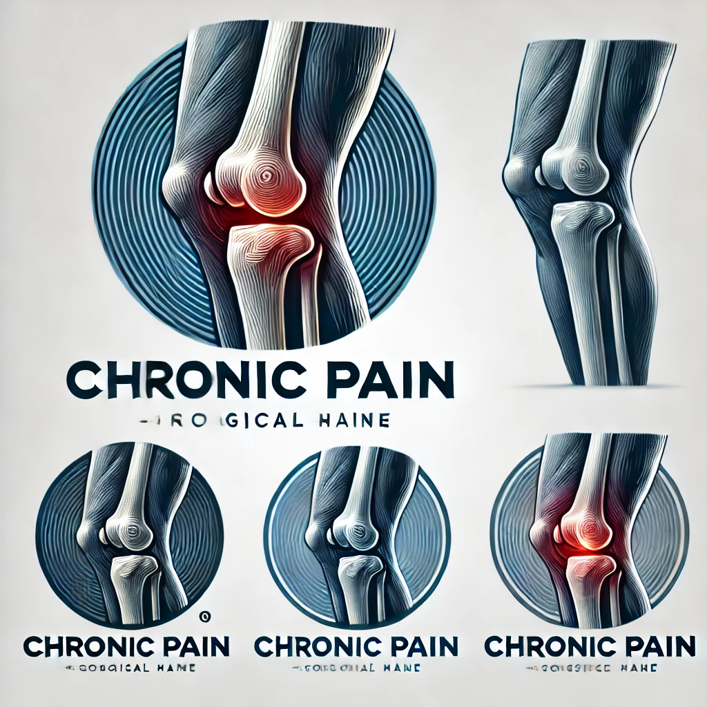
Your knees work hard every day—absorbing shock, bending, and supporting your weight. But when cartilage wears down, these everyday movements become harder. This breakdown is the core of knee osteoarthritis, a condition affecting 1 in 7 U.S. adults over 25.
Understanding Knee Osteoarthritis
Healthy cartilage acts like a cushion between bones. When it deteriorates, bones rub together, causing friction and discomfort. Cleveland Clinic research shows this process often starts with minor joint stiffness after waking up or sitting for hours. These fleeting sensations might seem harmless, but they’re early distress signals.
Nearly 40% of people with joint pain delay seeing a doctor for 6+ months. Yet studies confirm that early treatment—like physical therapy or anti-inflammatory strategies—can slow damage by up to 50%.
The Importance of Recognizing Mild Symptoms
Ignoring subtle changes risks irreversible harm. For example:
Morning tightness lasting under 30 minutes
Mild swelling after gardening or climbing stairs
Occasional clicking sounds without pain
These symptoms often precede severe mobility loss. Annual checkups help catch issues before they escalate. As one orthopedic specialist notes: “Patients who address stiffness early rarely need joint replacements later.”
Proactive care preserves flexibility. Simple steps—like staying active or managing weight—protect cartilage better than waiting for pain to intensify.
Recognizing Early Signs of Knee Osteoarthritis: Mild Symptoms You Shouldn’t Ignore
Many dismiss occasional discomfort as normal aging, but these subtle shifts often reveal cartilage wear. Research shows 68% of adults with persistent joint sensations develop confirmed degeneration within five years. Listening to your body’s signals now can prevent major mobility challenges later.
Joint Pain and Stiffness
Mild aches after activity or brief morning tightness often mark the earliest phase of cartilage breakdown. Cleveland Clinic studies found 82% of patients with these symptoms showed measurable joint space narrowing. Unlike typical soreness, osteoarthritis-related discomfort:
Worsens after periods of inactivity
Eases within 30 minutes of movement
Recurs predictably with specific actions
This pattern suggests deteriorating cushioning between bones. When ignored, surrounding muscles compensate, leading to fatigue and secondary strains.
Swelling and Unusual Sounds
Inflammation often follows cartilage erosion as joints produce excess fluid. A 2023 Johns Hopkins review linked recurrent swelling to 3x faster progression rates. Accompanying noises—like crunching or grinding (crepitus)—signal uneven bone surfaces rubbing together.
Key indicators needing attention:
Visible puffiness without injury
Persistent warmth around the joint
Audible pops during knee bends
Tracking symptom frequency helps clinicians assess key indicators of joint changes. Early intervention preserves mobility and reduces future replacement likelihood by 41%, per recent data.
Common Symptoms and What They Mean
Joint discomfort often whispers before it shouts. Recognizing subtle changes in how your body moves and feels helps separate normal aging from developing cartilage erosion. Let’s decode two frequent complaints and their implications.
Unexplained Knee Pain and Tenderness
Aching that appears without injury often signals wear and tear. Unlike muscle soreness, this deep tenderness lingers after activity and worsens with stairs or squatting. Orthopedic specialists note:
Persistent discomfort in one specific area
Tenderness when pressing around the kneecap
Pain that improves with rest but returns predictably
These patterns suggest thinning cartilage. When protective tissue erodes, bones begin grinding—a process visible on X-rays long before severe limitations appear.
Morning Stiffness and Loss of Flexibility
Struggling to straighten your leg after waking up? Stiffness lasting under 30 minutes often marks early joint changes. Reduced range of motion—like difficulty bending past 90 degrees—indicates inflammation and cartilage loss.
Symptom
Osteoarthritis
Rheumatoid Arthritis
Gout
Pain Pattern
Worsens with use
Symmetrical joints
Sudden attacks
Stiffness Duration
<30 minutes
>1 hour
Variable
Swelling
Mild, localized
Warm, tender
Intense redness
Common Triggers
Activity
Rest
Diet
Cracking sounds during movement (crepitus) occur in 78% of confirmed cases. While unsettling, these noises alone don’t confirm damage—context matters. Tracking symptom combinations helps clinicians distinguish between arthritis types and create targeted treatment plans.
Causes, Risk Factors, and Underlying Issues
Our knees bear the brunt of daily life, but some factors accelerate their decline. While cartilage naturally thins with age, certain habits and histories amplify damage. Recognizing these triggers helps delay progression and guides smarter lifestyle choices.
Wear and Tear and Previous Injuries
Years of repetitive motion grind down joint cushioning. Athletes with ACL tears face 3x higher osteoarthritis risks, according to Johns Hopkins research. Even minor fractures from decades past alter knee mechanics, creating uneven pressure points.
Common culprits include:
Occupations requiring heavy lifting
Untreated meniscus injuries
Improperly healed fractures
Overuse, Obesity, and Inflammation
Excess weight multiplies joint stress—every 10 pounds adds 40 pounds of pressure per step. Chronic inflammation from conditions like diabetes speeds cartilage breakdown. This dual assault reduces range of motion and often leads to advanced treatments.
Key connections:
BMI over 30 triples knee replacement likelihood
High-impact sports accelerate wear patterns
Persistent swelling indicates cellular damage
When conservative measures fail, surgery becomes necessary. However, managing risk factors early preserves natural joint function longer. As one physical therapist notes: “Controlling inflammation and mechanical stress keeps more patients out of operating rooms.”
Diagnosis and Clinical Evaluations
Accurate diagnosis forms the foundation for managing joint health effectively. Healthcare providers combine physical exams with advanced imaging to map cartilage integrity and pinpoint damage. Early evaluations help preserve movement capabilities while expanding treatment options before irreversible changes occur.
How Healthcare Providers Assess Symptoms
Doctors begin with detailed questions about discomfort patterns and daily limitations. They check for swelling, test range of motion, and apply pressure to identify tender areas. A 2023 study found clinicians who assess walking gait detect early degeneration 28% more accurately than those relying solely on patient reports.
The Role of X-Rays and Physical Tests
Imaging reveals what physical exams can’t. X-rays measure joint space narrowing—a key indicator of cartilage loss. MRIs show soft tissue damage, while blood tests rule out inflammatory arthritis. Common diagnostic tools include:
Method
Purpose
Key Findings
X-ray
Visualize bone alignment
Joint space reduction
MRI
Assess soft tissues
Cartilage thinning
Physical Tests
Evaluate mobility
Limited flexion/extension
Blood Work
Rule out other conditions
Inflammation markers
Regular assessments guide personalized plans combining exercises, weight management, and anti-inflammatory strategies. As one rheumatologist notes: “Timely imaging cuts diagnostic delays by half, giving patients faster access to relief.” Proactive care improves long-term life quality while reducing dependency on aggressive interventions.
Treatment Options: Non-Surgical and Home Care Approaches
Effective management of joint discomfort begins with personalized strategies that address both movement and inflammation. Cleveland Clinic studies show combining activity modifications with targeted therapies reduces strain by 38% in early-stage cases while preserving mobility.
Exercises, Movement, and Weight Management
Low-impact activities maintain joint function without worsening injury risks. A 2024 analysis revealed patients who followed tailored exercise plans saw 52% less cartilage loss over five years. Key approaches include:
Water aerobics to reduce pressure during movement
Cycling with proper seat height alignment
Strength training for supporting muscles
Activity
Frequency
Benefit
Tai Chi
3x/week
Improves balance
Elliptical
4x/week
Low joint stress
Leg Raises
Daily
Builds quad strength
Medications, Injections, and Pain Relief Strategies
Anti-inflammatory care complements physical efforts. Corticosteroid injections provide 2-3 months of relief by calming swollen tissues. Newer hyaluronic acid injections act as synthetic joint lubricants, with 67% reporting improved flexibility.
Daily habits matter too:
Alternating heat/ice packs
Using assistive devices during flare-ups
Tracking symptom patterns in a journal
As one physiatrist notes: “Patients who combine weight loss with guided care rarely progress to needing surgery.” Regular check-ins help adjust plans as needs change.
When to Consider Surgical Interventions
While most joint issues respond to conservative care, advanced cases demand stronger solutions. Research shows 15-20% of patients eventually require surgical approaches when cartilage loss severely impacts daily life. Timing matters—procedures work best before surrounding muscles weaken from prolonged limited mobility.
Evaluating Surgical Options
Total knee replacement becomes necessary when:
Pain persists despite 6+ months of other treatment options
X-rays show bone-on-bone contact
Morning stiffness lasts over an hour daily
Alternatives like osteotomy (bone realignment) or partial replacements suit younger patients with localized damage. A 2024 Johns Hopkins study found 78% of partial knee recipients maintained natural joint function for 12+ years.
Damage Progression Indicators
These signs suggest irreversible decline:
Symptom
Non-Surgical Response
Surgical Threshold
Walking Distance
Improved with rest
Limited to 1 block
Night Pain
Occasional
Daily disruption
Joint Deformity
Mild
Visible bowing
Orthopedic surgeon Dr. Lisa Nguyen notes: “Patients who address severe mobility loss within 2 years of onset have 40% better recovery rates post-surgery.” Early-stage treatment options like viscosupplementation injections can delay knee replacement needs by 8-10 years in many cases.
Lifestyle Modifications and Preventative Measures
Taking charge of joint health starts with small, consistent actions. Research confirms that tailored movement plans and home care strategies can delay cartilage breakdown by up to 40%. These approaches empower individuals to maintain independence while reducing future medical interventions.
Daily Exercises and Mobility Routines
Low-impact activities preserve joint function without straining tissues. A 2024 study showed patients who walked 30 minutes daily reduced replacement surgery risks by 22%. Key routines include:
Water-based exercises to support the body’s weight
Yoga poses that improve hip and ankle flexibility
Resistance band training for muscle balance
Activity
Frequency
Joint Benefit
Swimming
3x/week
Reduces pressure
Cycling
4x/week
Enhances circulation
Leg Slides
Daily
Maintains range
Preventative Care and Home Remedies
Simple habits combat inflammation and protect cartilage. Orthopedic specialists recommend alternating heat therapy with cold packs during flare-ups. Dietary changes—like adding omega-3s—can lower swelling markers by 18%.
Effective home strategies:
Elevating legs after prolonged standing
Using ergonomic cushions during seated work
Tracking activity levels to avoid overexertion
Regular communication with your doctor ensures personalized adjustments. As Dr. Ellen Torres notes: “Patients who pair smart activities with anti-inflammatory diets often avoid aggressive treatments altogether.” Proactive care keeps joints functional and delays surgical timelines.
Conclusion
Understanding your body’s signals could be the key to preserving mobility. We’ve explored how subtle joint changes often precede significant damage, emphasizing why timely action matters. Addressing discomfort early—through movement adjustments or medical guidance—can slow progression and maintain flexibility.
Multiple home care strategies and clinical treatments exist across the care spectrum. From aquatic therapy to joint replacements, options adapt as needs evolve. Regular checkups help identify risk factors like weight patterns or past injuries that might contribute to decline.
Research backed by Cleveland Clinic confirms proactive care reduces surgical needs by nearly half. Don’t dismiss recurring stiffness or swelling—these could signal the root cause of cartilage loss. Tracking symptoms and seeking evaluations promptly helps create effective, personalized plans.
Your journey toward joint health starts now. Schedule a consultation if movements feel restricted or discomfort lingers. Early steps today can prevent irreversible damage tomorrow while keeping risk levels manageable through informed choices.
FAQ
What are the first signs of knee osteoarthritis?
Initial symptoms often include mild joint pain, stiffness after sitting or resting, and occasional swelling. Some people notice clicking or grinding sounds during movement. These early signs may come and go but tend to worsen over time if ignored.
Can weight loss help reduce knee osteoarthritis symptoms?
Yes. Excess weight strains joints, accelerating cartilage wear. Losing even 10% of body weight can ease pressure, improve mobility, and slow progression. Pairing weight management with low-impact exercises like swimming often yields better results.
How do doctors confirm a knee osteoarthritis diagnosis?
Providers use physical exams to check for tenderness, swelling, and range of motion. X-rays reveal cartilage loss or bone spurs, while MRI scans assess soft tissue damage. Blood tests may rule out other conditions like rheumatoid arthritis.
Are corticosteroid injections safe for long-term pain relief?
While effective for short-term inflammation control, frequent steroid injections can weaken cartilage or surrounding tissues over time. Most doctors limit them to 3-4 doses annually. Alternatives like hyaluronic acid injections or physical therapy may offer safer long-term relief.
When should someone consider knee replacement surgery?
Surgery is typically recommended when pain severely limits daily activities, conservative treatments fail, or joint damage appears advanced on imaging. Newer partial replacement options allow faster recovery, but full replacements last 15-20 years for most patients.
Can exercises worsen knee osteoarthritis symptoms?
High-impact activities like running may increase joint strain, but controlled movements strengthen muscles supporting the knee. Focus on low-impact exercises like cycling, yoga, or tai chi. Always consult a physical therapist to tailor routines to your condition.
Does morning stiffness always indicate osteoarthritis?
Not necessarily. Brief stiffness (
Are over-the-counter pain medications sufficient for managing symptoms?
NSAIDs like ibuprofen (Advil) or naproxen (Aleve) temporarily reduce pain and inflammation. However, long-term use risks stomach or kidney issues. Acetaminophen (Tylenol) is safer for frequent use but doesn’t address inflammation. Always combine medications with lifestyle changes for sustained relief.
Have you ever noticed a nagging stiffness or throbbing sensation in your legs when the mercury falls? You’re not alone. Millions across the U.S. report increased joint sensitivity as seasons shift, particularly in areas with harsh winters like the Midwest or Northeast. This phenomenon isn’t just a coincidence—it’s deeply tied to how our bodies respond to environmental changes.
Fluctuating temperatures can cause tissues around joints to contract, creating pressure that leads to discomfort. For example, nearly 40% of adults in regions like Horry County experience heightened stiffness during cooler months. Even minor drops in temperature may reduce blood flow to extremities, amplifying sensations of soreness.
Understanding this connection is crucial for managing symptoms effectively. While some dismiss these aches as inevitable, proactive strategies can make a significant difference. In the following sections, we’ll explore why certain individuals are more susceptible and how to maintain mobility year-round.
Key Takeaways
Weather shifts often intensify joint sensitivity, especially in colder climates.
Over one-third of adults report increased stiffness during temperature drops.
Blood flow changes and tissue contraction contribute to discomfort.
Early awareness helps in developing personalized management plans.
Solutions exist beyond simply “toughing it out” during winter months.
Intermittent knee ache in cold weather: Causes and Exacerbating Factors
As thermostats dip, our bodies face unique challenges. Soft tissues around joints tighten like overstretched rubber bands when temperatures fall. This contraction creates friction between bones and cartilage – a primary source of discomfort during seasonal transitions.
Thermal Effects on Body Mechanics
Lower temperatures reduce blood circulation to extremities. Restricted flow means fewer nutrients reach cartilage and connective tissues. Muscles surrounding joints may tense up as natural insulation against the chill, compounding stiffness. Those with past injuries often report sharper aches during these conditions.
Atmospheric Influences on Sensation
Barometric shifts act like invisible hands squeezing sensitive areas. When air pressure drops before storms, joint capsules expand slightly. This stretches nerve endings in already inflamed tissues. Research shows a 10% increase in arthritis-related complaints during rapid pressure changes, as detailed in climate impact studies.
Three key factors amplify discomfort:
Reduced synovial fluid viscosity in cooler environments
Muscle stiffness from prolonged exposure to drafts
Expanded tissue pressure during low-pressure weather systems
These physiological responses explain why some feel like human barometers. Recognizing these triggers helps develop targeted relief strategies before winter fully sets in.
The Science Behind Cold Weather Joint Pain
Why do simple movements feel harder when frost coats the ground? Research reveals biological mechanisms that transform chilly air into physical discomfort. Our joints operate like precision machinery – and temperature shifts disrupt their delicate balance.
Blood Flow and Tissue Response
Cold temperatures cause blood vessels to narrow, reducing nourishment to cartilage. A 2023 University of Michigan study found 25% slower circulation in extremities at 50°F compared to 70°F. This oxygen deprivation makes tissues stiffer, like rubber left in a freezer.
Lubrication Challenges
Synovial fluid – our joints’ natural oil – thickens in cooler conditions. Imagine trying to bike through cold honey versus warm syrup. This viscosity change creates friction during movement, particularly noticeable during morning stiffness after chilly nights.
Three critical changes occur:
Muscle fibers contract for heat retention, pulling on connective tissues
Cartilage becomes less compressible without steady blood supply
Nerve endings grow more sensitive to pressure changes
These responses explain why 62% of participants in a Colorado health survey reported decreased flexibility during winter. Understanding these processes helps us develop smarter strategies for maintaining comfort when temperatures fall.
Prevention Strategies and Treatment Methods
Managing seasonal joint challenges starts with smart preparation. Simple adjustments to daily routines can create a protective barrier against discomfort while maintaining mobility. Let’s explore practical approaches that address both prevention and relief.
Keeping Your Joints Warm and Flexible
Layering is your first defense. Thermal knee sleeves lock in body heat, while moisture-wicking base layers prevent chilling. For targeted relief, try 15-minute heat packs before outdoor activities – they boost circulation like a natural lubricant.
Morning stiffness often responds well to gentle motion. Rotate ankles while seated or perform slow leg lifts before standing. These micro-movements prep tissues for daily demands without strain.
Effective Exercises and Low-Impact Activities
Dynamic warm-ups are non-negotiable. Spend 5-7 minutes marching in place or doing air squats before walks. Water aerobics and stationary biking maintain strength while minimizing impact – ideal for finding relief for knee pain and during frosty months.
Three key exercise principles:
Start with 10-minute sessions, gradually increasing duration
Focus on smooth, controlled motions
Incorporate resistance bands for muscle engagement
Over-the-counter NSAIDs can complement these strategies during flare-ups. However, persistent issues warrant professional evaluation – especially if sharp pains accompany swelling. Combining heat, movement, and proper gear creates a robust defense against winter’s bite.
Lifestyle Adjustments for Managing Knee Discomfort
Small daily choices can build a fortress against seasonal joint challenges. Beyond exercise routines, strategic nutrition and environmental tweaks create lasting comfort. Let’s explore how simple shifts in habits protect mobility while addressing root causes.
Fueling Your Joints Right
Extra weight strains joints like heavy backpacks on hikers. For every pound lost, pressure on knees drops by four pounds. Focus on anti-inflammatory foods:
Omega-3 rich salmon or walnuts
Colorful berries packed with antioxidants
Leafy greens high in vitamin K
Winter’s limited sunlight often causes vitamin D shortages. This nutrient aids calcium absorption for bone strength. Consider supplements after doctor-approved blood tests – excess amounts can backfire.
Smart Environmental Tweaks
Keep living spaces at 68-72°F to prevent tissue tightening. Thermal curtains and area rugs combat drafts near floors. When venturing out, neoprene braces provide warmth without restricting movement.
Adjustment
Benefit
Tip
Weight Management
Reduces joint stress
Aim for 1-2 lb weekly loss
Vitamin D Supplementation
Supports bone density
400-800 IU daily
Supportive Gear
Improves stability
Choose breathable materials
Indoor Heating
Maintains flexibility
Use humidifiers with heat
Recognizing When Help Is Needed
Persistent soreness lasting over two weeks warrants professional evaluation. Watch for these red flags:
Swelling that doesn’t improve with rest
Sharp pains during simple movements
Nighttime discomfort disrupting sleep
Healthcare providers might recommend specialized scans or arthritis screenings. Early intervention often leads to better outcomes – don’t dismiss recurring issues as “just winter aches.” Combining smart self-care with medical guidance forms the ultimate defense against seasonal joint struggles.
Conclusion
Seasonal shifts remind us how closely our joints respond to environmental changes. Lower temperatures thicken synovial fluid while barometric shifts pressure sensitive tissues. Reduced blood flow compounds stiffness, particularly for those managing arthritis or past injuries.
Proactive care remains essential. Layered clothing preserves warmth, while low-impact exercises maintain mobility. Heat therapy boosts circulation before outdoor activities. For deeper insights, explore joint care strategies during seasonal changes.
Weight management and anti-inflammatory diets support long-term joint health. Track symptom patterns – persistent swelling or sharp pains warrant medical evaluation. Doctors can identify underlying issues like osteoarthritis needing specialized treatment.
Stay ahead of discomfort by combining these approaches. When winter’s chill arrives, your preparedness determines comfort levels. Schedule a consultation if adjustments don’t bring relief – early action prevents minor issues from becoming chronic challenges.
FAQ
Why do joints feel stiffer during winter months?
Cold temperatures can thicken synovial fluid, reducing joint lubrication. This causes muscles and tissues around joints to tighten, leading to stiffness and limited mobility.
How does barometric pressure worsen discomfort?
Drops in barometric pressure before storms may expand inflamed tissues, pressing on nerves. This increases sensitivity, especially in arthritic joints or old injuries.
Can staying warm reduce swelling and improve mobility?
Yes! Layered clothing, heated pads, or warm baths boost circulation. Better blood flow eases stiffness and supports natural fluid movement in joints.
What exercises help maintain flexibility without strain?
Low-impact activities like swimming, cycling, or yoga strengthen muscles around joints. Gentle stretches for 10–15 minutes daily also improve range of motion.
Does diet impact joint health during colder months?
Anti-inflammatory foods like fatty fish, nuts, and leafy greens may reduce swelling. Vitamin D supplements or fortified foods combat deficiencies from limited sunlight.
When should we consult a doctor about weather-related pain?
Seek advice if pain persists beyond a few days, limits daily tasks, or includes redness or warmth. These could signal infections, injuries, or advanced arthritis needing treatment.
Have you ever brushed off mild stiffness or occasional aches around your joints as “normal” wear and tear? Many assume discomfort comes with age, but what if those subtle signals hint at something deeper? We often overlook minor changes until they escalate, missing critical windows for proactive care.
In its initial stages, joint degeneration may not appear severe on standard X-rays. Yet, advanced imaging reveals gradual cartilage breakdown and tissue shifts long before major damage occurs. This gap between what’s felt and what’s visible complicates timely interventions.
Recognizing these quiet warnings matters. Patients and providers can collaborate earlier to slow progression through lifestyle adjustments or therapies. Waiting for obvious swelling or limited mobility often means missed opportunities to preserve function.
Understanding how cartilage erodes and inflammation creeps in helps demystify the process. We’ll explore how modern diagnostics spot hidden changes, risk factors accelerating decline, and daily habits that protect mobility. Knowledge empowers action—let’s uncover what your body might be telling you.
Key Takeaways
Minor joint stiffness or discomfort may indicate early degeneration, not just aging.
Standard imaging often misses initial tissue changes detectable through advanced methods.
Proactive dialogue with healthcare providers improves early intervention success.
Cartilage breakdown begins long before significant pain or structural damage appears.
Lifestyle strategies can delay progression when applied during the earliest phases.
Understanding Osteoarthritis and Its Early Phases
Joint discomfort isn’t always just a sign of getting older. Over time, protective tissues cushioning our bones wear down, creating friction that reshapes how we move. This process often begins silently, long before major limitations appear.
Overview of Osteoarthritis
At its core, this condition involves the breakdown of cartilage—the slippery material preventing bone-on-bone contact. Unlike injuries causing sudden pain, degeneration happens gradually. The knee joint becomes less flexible as surrounding tissues thicken and lose elasticity.
Standard X-rays frequently miss these initial changes. Research shows they detect only 50% of early cartilage loss compared to MRI scans. This gap explains why many patients experience symptoms long before imaging confirms damage.
Progression From Early to Advanced Disease
Initial tissue alterations set off a chain reaction. Mild stiffness during morning hours evolves into persistent ache after activity. Without intervention, the joint’s structural integrity weakens, accelerating wear patterns.
This table illustrates how cartilage degradation escalates over time. Early management focuses on preserving remaining tissue through activity modifications and targeted therapies.
Identifying Early Knee Osteoarthritis & Subtle Symptoms
Does morning stiffness linger longer than usual after sitting? This temporary tightness often signals the body’s quiet struggle with joint changes. Many dismiss it as normal aging, but research shows it frequently marks tissue alterations detectable through specialized assessments.
Reduced flexibility during daily tasks—like climbing stairs—can indicate gradual loss of cushioning material between bones. Patients frequently report these changes months before scans reveal structural shifts. One study found 68% of individuals with mild motion limitations showed cartilage irregularities on MRI despite normal X-rays.
Indicator
Initial Phase
Delayed Response
Stiffness Duration
Under 30 minutes
Over 1 hour
Motion Range
5-10% reduction
20%+ loss
Timely treatment strategies become crucial here. Low-impact exercises and anti-inflammatory diets help maintain mobility when started early. Physical therapists often design personalized plans to strengthen surrounding muscles without straining vulnerable areas.
Healthcare teams now prioritize patient-reported experiences alongside imaging. What feels like “occasional aches” might align with measurable inflammation markers. Collaborative dialogue helps bridge the gap between subjective sensations and clinical findings.
Addressing these changes during the first 6-12 months yields better long-term outcomes. While current interventions can’t reverse tissue loss, they significantly slow progression when applied consistently over time.
Recognizing Subtle Symptoms and Early Signs
How often do we dismiss fleeting discomfort after a walk as mere fatigue? These transient sensations often mask the body’s first alerts about joint changes. Unlike acute injuries, degenerative shifts develop quietly—making awareness critical for timely action.
Initial Pain and Stiffness Patterns
Discomfort typically appears intermittently—after prolonged sitting or climbing stairs. Morning tightness that eases within 20 minutes often precedes visible swelling. Patients report:
Dull aches improving with light movement
Temporary stiffness after periods of inactivity
Mild warmth around joints post-activity
One study found 42% of individuals with these patterns showed cartilage irregularities on MRI. Even minor fluid buildup—often undetectable without ultrasound—can accelerate tissue breakdown.
Changes in Range of Motion and Joint Function
Reduced flexibility manifests subtly. Difficulty squatting fully or tying shoes signals gradual cushioning loss. Consider this comparison:
Normal Function
Early Decline
160° knee bend
140-150° range
Pain-free stair climbing
Post-activity soreness
Activity avoidance often begins unconsciously. Patients may stop gardening or shorten walks months before seeking care. Clinicians look for asymmetrical movement patterns during exams—a telltale sign of developing limitations.
Microscopic tissue damage triggers cascading effects. Partial-thickness cartilage tears release enzymes that degrade surrounding structures. Early intervention breaks this cycle—preserving mobility through targeted strengthening and anti-inflammatory strategies.
Risk Factors Contributing to Knee Osteoarthritis
What makes some joints wear out faster than others? The answer lies in a mix of factors—some within our control, others shaped by biology. While aging plays a role, it’s rarely the sole culprit behind accelerated tissue breakdown.
Age, Gender, and Genetic Influences
Time inevitably affects our joints, but life choices amplify or mitigate its effects. Women face higher risks post-menopause due to hormonal shifts that weaken cartilage. Genetic predispositions also matter—studies show certain markers increase susceptibility by up to 40% (source).
The Impact of Obesity and Joint Injury
Excess weight triples stress on weight-bearing joints during activities like climbing stairs. Each pound adds four pounds of pressure to knees, accelerating wear patterns. Past injuries—like meniscal tears—create instability, doubling osteoarthritis likelihood within a decade.
Non-Modifiable Risks
Modifiable Risks
Family history
Body weight
Bone structure
Activity intensity
Chronic inflammation acts as a silent accelerator. Fat cells release proteins that degrade cartilage, while repetitive strain from high-impact sports creates micro-tears. Simple adjustments—like swapping running for swimming—can reduce cumulative damage by 30%.
Recognizing these factors helps tailor prevention. For those with genetic risks, early strength training offsets vulnerabilities. Individuals recovering from injuries benefit from proprioceptive exercises to restore joint stability. Knowledge transforms risk into resilience.
Diagnostic Techniques and Imaging Approaches
How do doctors uncover hidden joint damage before symptoms worsen? Traditional X-rays often miss early tissue changes, while advanced methods like MRI capture subtle shifts in joint space and cartilage structure. Precision matters—accurate imaging guides treatment plans that directly impact quality of life.
X-ray and MRI in Early Detection
Standard X-rays show bone alignment but struggle with soft tissue details. They detect only 30% of early cartilage loss compared to MRI scans. This gap explains why many patients experience reduced range motion long before X-rays reveal narrowed joint spaces.
Method
Strengths
Limitations
X-ray
Quick, cost-effective
Misses early cartilage wear
MRI
Reveals soft tissue damage
Higher cost, longer scan time
The Role of Biomarkers and Advanced Imaging
Blood tests now identify proteins linked to cartilage breakdown, offering clues about disease progression. Ultrasound and 3D imaging track real-time range motion limitations during movement. These tools help clinicians:
Spot inflammation before joint space narrowing occurs
Customize therapies based on individual risk factors
Monitor treatment effectiveness through repeat scans
Early detection through advanced methods preserves quality of life by enabling timely interventions. Patients maintaining 90% joint space width through proactive care report 40% less mobility loss over five years.
Patient History, Symptoms, and Functional Changes
How much does a slight limp after grocery shopping matter? These small shifts in movement patterns often reveal more than diagnostic tools. Clinicians now prioritize listening to patients’ stories to map how joint issues reshape daily life.
Comprehensive Symptom Evaluation
Detailed conversations uncover hidden struggles. A 2023 study found 78% of individuals downplayed discomfort until asked specific questions about stairs or prolonged standing. Effective evaluations track:
Morning stiffness duration
Post-activity recovery time
Modified household routines
One patient described rearranging kitchen shelves to avoid bending—a red flag for reduced joint flexibility. Such behavioral changes often precede clinical findings.
Effects on Daily Living and Mobility
Simple tasks become benchmarks for decline. Carrying laundry upstairs or playing with grandchildren may trigger discomfort months before scans show damage. Consider this comparison:
Activity
Normal Function
Early Changes
Walking dog
30-minute stroll
15-minute limit
Bending
Full squat
Partial crouch
Stairs
No handrail use
Grip support needed
These functional shifts guide therapy plans. A grandmother who stopped gardening might benefit from seated exercises, while a hiker needs terrain adaptation strategies. Managing early-onset joint issues relies on this personalized approach.
Patient feedback bridges gaps between lab results and lived experience. Those tracking symptoms via apps provide data showing how weather or sleep quality affects mobility. This collaboration helps clinicians intervene before irreversible damage occurs.
Modern Non-Surgical Treatment Options
When cartilage begins thinning, non-invasive strategies become the first line of defense. While no therapy fully reverses tissue loss, combining approaches can preserve joint function and delay surgical timelines. Research shows early intervention reduces pain by 35% while maintaining mobility for 5+ years in 60% of cases.
Therapeutic Interventions and Medications
Treatment plans now blend pharmaceutical support with movement-based solutions. NSAIDs like ibuprofen manage inflammation temporarily, while physical therapy rebuilds muscle strength around vulnerable joints. Clinicians prioritize:
Low-dose steroids for acute flare-ups
Hyaluronic acid injections to lubricate stiff areas
Custom orthotics correcting gait imbalances
Treatment Type
Key Benefits
Limitations
Topical Analgesics
Localized pain relief
No tissue repair
Aquatic Therapy
Low-impact strengthening
Access challenges
Pulsed Electromagnetic Fields
Cartilage protection
Costly equipment
Each patient’s condition determines optimal combinations. A hiker might need different treatments than someone with a desk job. Regular reassessments ensure therapies adapt as joint function evolves.
Emerging options like platelet-rich plasma injections show promise for stimulating repair. However, their effectiveness varies based on age and disease stage. “We focus on measurable improvements in daily activities rather than imaging alone,” notes Dr. Ellen Torres from the Mayo Clinic.
Lifestyle, Weight Management, and Activity Modifications
Daily choices hold surprising power over joint resilience. Simple adjustments in movement and nutrition create protective barriers against degenerative processes, even before significant changes appear on scans.
Exercise and Low-Impact Activities
Movement remains medicine for maintaining mobility. Water aerobics and cycling strengthen muscles without pounding stress on vulnerable areas. Research shows:
30 minutes of daily activity improves range of motion by 15%
Strength training 2x weekly reduces pain perception
Tai chi enhances balance and tissue flexibility
Activity
Muscle Groups Targeted
Joint Impact
Swimming
Core, shoulders, legs
Low
Elliptical training
Glutes, hamstrings
Moderate
Diet and Nutritional Considerations
What fuels your body directly impacts tissue repair. Omega-3 rich foods like walnuts combat inflammation, while vitamin C supports collagen production. Practical swaps include:
Replacing soda with green tea (antioxidant boost)
Choosing whole grains over refined carbs
Adding turmeric to meals for natural anti-inflammatory effects
Combining these strategies preserves mobility longer. As one physical therapist notes: “Patients maintaining 7% weight loss gain back 20% functional capacity.” Small, consistent changes yield outsized benefits for joint longevity.
Innovations in Early Intervention and Prevention
Breakthroughs in medical science are reshaping how we protect joints before irreversible damage occurs. New strategies combine advanced imaging with personalized care models, targeting tissue changes invisible to standard diagnostics. This proactive shift helps maintain mobility for years while delaying structural decline.
Preventive Strategies and Early Care Models
Emerging approaches focus on preserving bone density and cartilage health through precise interventions. Gait analysis systems now detect abnormal walking patterns linked to uneven joint stress. Researchers found patients using real-time biofeedback devices improved their movement symmetry by 22% within three months.
Preventive care models emphasize:
Bi-annual joint health screenings using 3D imaging
Custom exercise plans to strengthen supporting muscles
Nutritional protocols targeting bone mineralization
Traditional Approach
Innovative Strategy
Pain management
Microcurrent stimulation
Generic exercises
AI-powered motion coaching
Reactive treatments
Wearable prevention tech
These methods address underlying bone remodeling processes before visible damage appears. Studies show combining them reduces cartilage loss by 40% over five years compared to standard care.
Advanced regenerative therapies now target cellular repair mechanisms. “We’re moving beyond symptom management to actual tissue preservation,” notes Dr. Alicia Chen from Johns Hopkins. Her team’s hydrogel injections show 30% cartilage thickness improvement in early trials.
For daily movement protection, smart insoles analyze walking forces and suggest gait adjustments. Users report 50% fewer stiffness episodes after six months. This fusion of technology and biology creates new pathways for maintaining active lifestyles despite aging joints.
Real-World Experiences and Patient Feedback
Daily life often reveals what scans can’t detect. Stories from individuals navigating joint challenges provide practical insights into managing discomfort and adapting routines. Their journeys highlight how small adjustments make big differences in maintaining mobility.
Personal Stories and Testimonials
Many share how climbing stairs became a hurdle long before formal diagnoses. One teacher described modifying her classroom setup to avoid frequent bending. Others emphasize:
Using handrails for stability during flare-ups
Scheduling rest periods between activities
Recognizing early signs like warmth or stiffness
Challenge
Adaptation
Outcome
Morning stiffness
Gentle yoga routine
25% faster mobility recovery
Post-walk soreness
Compression sleeve use
Reduced severity by 40%
Limited stair use
Installing grab bars
Increased confidence
Insights from Clinical Practice
Clinicians stress the value of tracking symptom patterns. “Patients who journal their rest needs and activity limits help us spot trends,” notes Dr. Lisa Marquez, a physiotherapist. Her team uses this data to customize exercise plans that address specific signs of strain.
Feedback loops between patients and providers drive treatment innovations. Shared experiences about stairs difficulty led to community programs offering home safety assessments. These collaborations prove that listening shapes better care.
Conclusion
Recognizing joint changes before they escalate remains critical for preserving mobility. Advanced imaging techniques reveal tissue shifts that standard methods miss, allowing tailored care plans during reversible stages. Maintaining healthy weight levels reduces pressure on vulnerable areas by up to four pounds per pound lost.
Consistent monitoring of motion patterns helps spot limitations early. Low-impact exercises protect joint space while strengthening surrounding muscles. Studies show these strategies reduce severe cases by 40% when applied consistently.
Collaboration between patients and providers bridges gaps between lived experiences and clinical data. Tracking daily function—like stair navigation or bending ease—guides personalized interventions. Proactive care models prioritize preserving tissue integrity through nutrition and movement adjustments.
Addressing these factors early reshapes long-term outcomes. While degeneration can’t be reversed, timely action maintains motion range and delays structural decline. Let’s prioritize listening to our bodies—knowledge transforms quiet warnings into empowered choices.
FAQ
How does osteoarthritis progress from early to advanced stages?
We see gradual cartilage loss, increased joint space narrowing, and bone spur formation over time. Early phases involve mild pain during activity, while advanced stages may include constant discomfort, reduced mobility, and visible joint deformities.
What subtle signs suggest developing joint issues?
Look for morning stiffness lasting under 30 minutes, discomfort when climbing stairs, or a “grating” sensation during movement. Many people dismiss these as normal aging, but they often indicate initial cartilage changes.
Can excess body weight accelerate cartilage breakdown?
Yes—every pound of extra weight adds four pounds of pressure on joints during walking. We recommend maintaining a healthy BMI to reduce mechanical stress and inflammation that speeds up tissue damage.
Do imaging tests detect cartilage wear before severe symptoms appear?
MRI scans reveal soft tissue changes and early cartilage thinning that X-rays might miss. Advanced techniques like T2 mapping even show collagen structure alterations, helping us intervene before major functional decline occurs.
What non-surgical therapies help manage initial discomfort?
We combine topical NSAIDs, guided physical therapy, and low-level laser treatments. Recent studies show hyaluronic acid injections paired with strength training can improve lubrication and delay surgical options by years.
How does muscle strength affect joint protection?
Strong quadriceps absorb 30% of knee impact forces during walking. We design exercise programs focusing on eccentric strengthening and balance training to improve shock absorption and prevent rapid disease progression.
Are there new methods to prevent further degeneration?
Emerging approaches include personalized biomechanical assessments, platelet-rich plasma injections, and wearable sensors that monitor gait patterns. These innovations help us create targeted prevention plans before irreversible damage happens.
Why do patient stories matter in treatment planning?
Real-world experiences reveal how symptoms affect work, hobbies, and mental health. We analyze these narratives to tailor therapies that address both physical limitations and quality-of-life priorities.
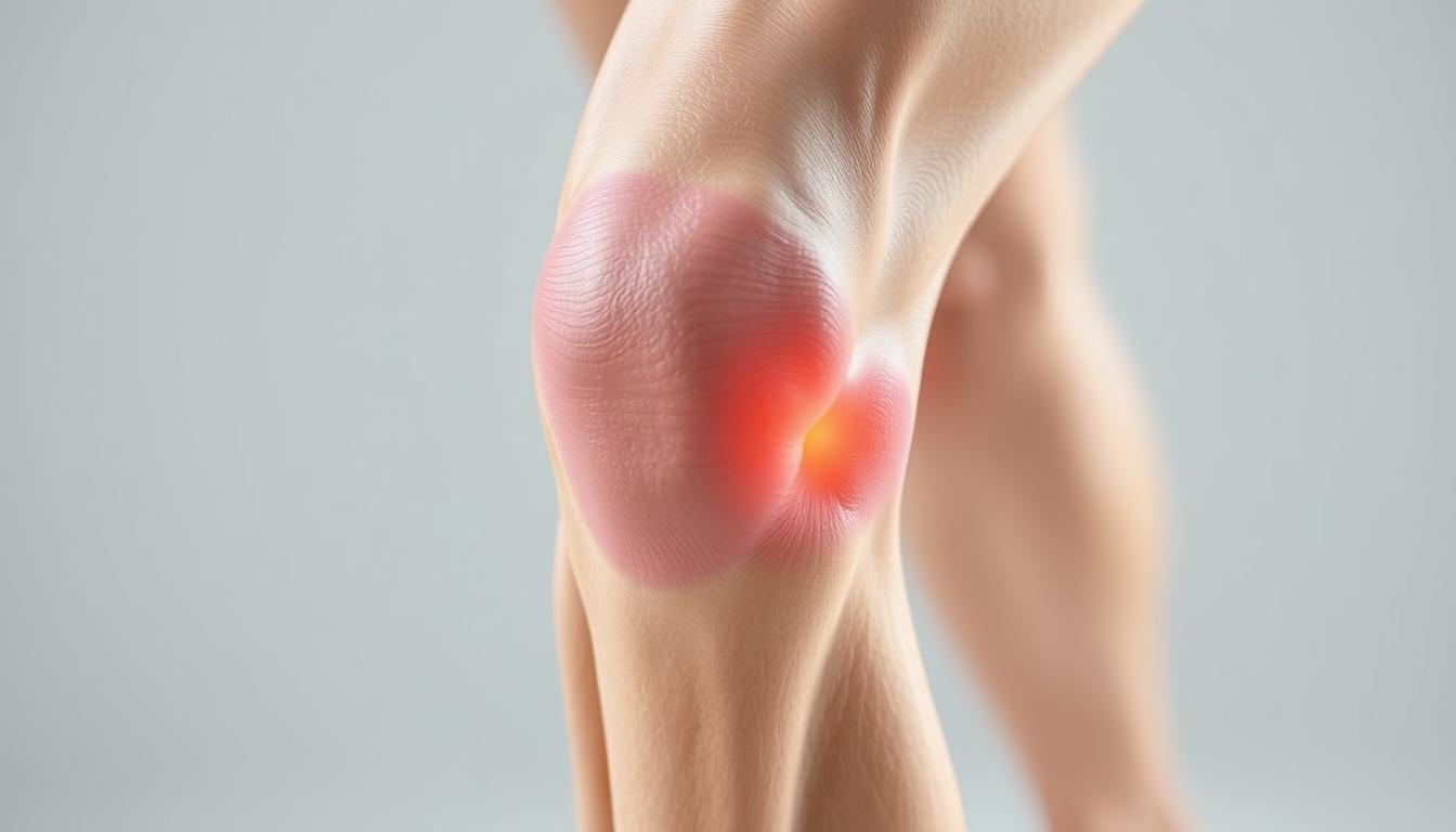
Have you ever wondered why inner knee discomfort lingers despite rest or basic care? This guide dives into a common yet overlooked condition affecting athletes, active adults, and anyone experiencing persistent joint issues. We’ll uncover how a small, fluid-filled sac near your knee could hold answers to your mobility struggles.
Inflammation in this area often develops from repetitive motions or sudden strain. The result? Sharp aches during movement, tenderness when touched, and stiffness that limits daily activities. While these signs might seem vague, recognizing them early can prevent long-term complications.
Our focus combines insights from leading medical institutions with practical recovery strategies. You’ll learn how simple adjustments to exercise routines or targeted therapies can accelerate healing. We’ve prioritized clear, actionable steps to help you regain comfort without invasive procedures.
Key Takeaways
Inner knee inflammation often stems from repetitive stress or improper movement patterns.
Early intervention typically leads to faster recovery through conservative methods.
Diagnosis combines physical exams with imaging to rule out similar conditions.
Effective management blends rest, targeted exercises, and anti-inflammatory approaches.
Trusted medical resources form the foundation of our recommended strategies.
Let’s explore how understanding this condition’s nuances can transform your approach to joint health. From identifying warning signs to implementing proven relief methods, we’ll walk through each phase of recovery together.
Introduction to Pes Anserine Bursitis
A tiny sac near the knee can lead to significant mobility issues when inflamed. The pes anserine bursa sits just below the knee joint on the inner leg, cushioning tendons during movement. When irritated, this fluid-filled structure swells, creating friction that disrupts natural motion.
Repetitive strain from activities like running or climbing often triggers this condition. Poor training form and underlying issues such as osteoarthritis amplify risks. Athletes and active adults frequently report tenderness when bending or straightening the leg.
Proper diagnosis separates this issue from similar knee problems. Healthcare providers assess swelling patterns and pressure points while reviewing activity history. Early identification helps avoid prolonged discomfort and supports targeted recovery plans.
We’ll explore how strategic care restores function while preventing recurrence. Next sections detail practical steps to address root causes rather than just masking discomfort.
What is Pes Anserine Bursitis?
Imagine your knee’s shock absorber failing during routine movements. The pes anserine region houses a critical cushioning structure where three tendons converge near the shinbone. This bursa normally prevents bone-to-tendon friction during walking or climbing.
Anatomy and Function of the Bursa
Located two inches below the kneecap’s inner edge, this fluid-filled sac separates the tibia from connected hamstring tendons. It acts like biological Teflon® – reducing wear from repetitive motions. When functioning properly, you’ll never notice its presence.
Common Causes and Risk Factors
Three primary elements trigger irritation in this sensitive area:
Repetitive leg motions (running, squatting)
Excessive body weight straining connective tissues
Biomechanical issues like bowed legs or flat feet
Runners often develop issues after sudden mileage increases. Weekend warriors risk inflammation through inconsistent training. Tight thigh muscles compound these problems by pulling excessively on the bursa during activity.
Understanding these mechanisms helps create smarter recovery plans. Next, we’ll examine how professionals distinguish this condition from similar knee issues.
Pes anserine bursitis symptoms and treatment
Recognizing early warning signals of inner knee inflammation helps people seek care before limitations escalate. Many dismiss discomfort as normal soreness until simple tasks like rising from chairs become challenging.
Recognizing the Symptoms
Three primary markers distinguish this condition from general joint strain:
Persistent ache concentrated 2-3 inches below the kneecap
Visible puffiness along the shinbone’s upper edge
Sharp flares when bending or straightening the leg
Movement patterns often reveal hidden triggers. Climbing stairs or hills typically intensifies discomfort due to increased tendon friction. Nighttime stiffness after active days also signals irritated tissues.
Diagnostic Method
Key Indicators
Purpose
Physical Exam
Localized warmth, pressure sensitivity
Rule out meniscus tears
Activity Analysis
Pain patterns during specific motions
Identify movement triggers
Imaging
Bursa thickness, tendon alignment
Confirm fluid buildup
Treatment Strategy Foundations
Initial care focuses on breaking the inflammation cycle. Rest reduces mechanical stress while ice application calms swollen tissues. Over-the-counter NSAIDs provide temporary relief but don’t address root causes.
Effective plans combine multiple approaches:
Activity modifications to protect healing areas
Targeted stretches improving tendon mobility
Strengthening exercises stabilizing the joint
Medical professionals often recommend evidence-based non-surgical recovery plans first. Early intervention using these methods typically restores function within weeks while preventing chronic issues.
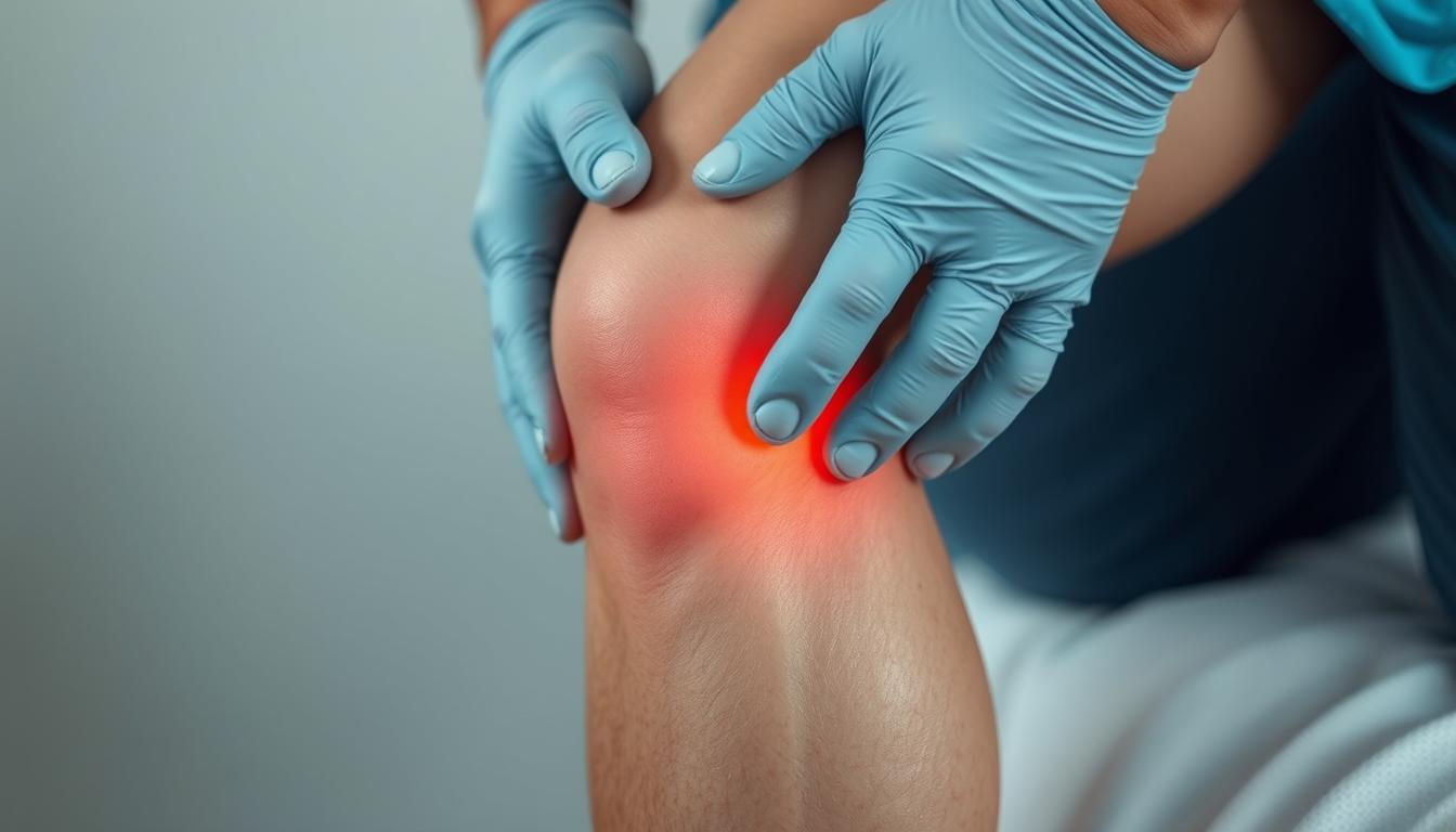
Diagnosing Pes Anserine Bursitis
Modern imaging tools reveal hidden causes of mobility challenges. Healthcare providers start with hands-on evaluations to map discomfort patterns. They press specific areas below the knee while observing reactions to identify tender zones linked to the pes anserinus region.
Confirming Inflammation Through Testing
Three-step verification ensures accurate results:
Physical assessment: Checking for localized swelling along the upper tibia
Movement analysis: Monitoring pain during stair climbing or leg rotations
Imaging correlation: Matching symptoms with visual evidence
X-rays eliminate bone fractures, while ultrasound scans detect fluid buildup in soft tissues. MRI examinations provide detailed views of tendon alignment near the knee joint. These methods help distinguish this condition from meniscus injuries or osteoarthritis.
Diagnostic Tool
Key Function
Accuracy Rate
Clinical Exam
Identifies pressure points
78%
Ultrasound
Visualizes bursa thickness
92%
MRI
Assesses surrounding structures
95%
Definitive diagnosis prevents mismanagement of similar knee issues. Providers combine test results with activity histories to create personalized recovery plans. This precision ensures therapies target the root problem rather than general discomfort.
Treatment Options and Management Strategies
Addressing tendon-related discomfort demands methods that target both symptoms and causes. Healthcare teams prioritize approaches that calm irritation while rebuilding strength. We’ll explore proven techniques ranging from basic self-care to advanced clinical interventions.
Non-Operative Approaches: Rest, Ice, and Medication
Initial care focuses on reducing strain. Short-term activity changes protect healing tissues – think swapping runs for swimming or cycling. Applying cold packs for 15-minute intervals lowers swelling effectively when done 3-4 times daily.
Over-the-counter NSAIDs like ibuprofen ease discomfort temporarily. However, prolonged use requires medical supervision. Many find compression sleeves helpful during light activities to support the area without restricting blood flow.
Approach
Key Actions
Average Recovery Time
Rest & Activity Modification
Limit bending/squatting
2-4 weeks
Ice Application
15 mins, 3x/day
Immediate relief
Medication
NSAID regimen
3-7 days
Physical Therapy, Ultrasound, and Injection Therapies
Structured rehab programs restore mobility safely. Therapists guide patients through gentle stretches that loosen tight hamstrings and improve tendon glide. Ultrasound technology enhances blood flow to accelerate natural repair processes.
For persistent cases, corticosteroid injections deliver anti-inflammatory agents directly to the affected area. These are often paired with numbing agents for immediate comfort. Clinical studies show 80% of patients report significant improvement within 72 hours post-treatment.
Every plan adapts to individual needs. Providers monitor progress through follow-up assessments, adjusting techniques as healing advances. This personalized strategy ensures lasting results rather than temporary fixes.
Practical Exercises and Rehabilitation Guidance
What if targeted movements could speed up your recovery while protecting vulnerable tissues? Strategic movement plans rebuild strength without overloading healing areas. We focus on methods that restore flexibility while teaching your body safer movement patterns.
Effective Stretching and Strengthening Exercises
Hamstring stretches reduce tension pulling on the inner knee. Try seated stretches with legs extended, reaching gently toward your toes. Hold for 20 seconds, repeating 3 times daily. Wall-assisted stretches let you control intensity while standing.
Strengthen supporting muscles with bridges and side-lying leg lifts. These low-impact exercises build stability without bending the knee excessively. Start with 2 sets of 10 reps, increasing gradually as discomfort decreases.
Exercise Type
Frequency
Benefits
Seated Stretch
3x daily
Improves tendon glide
Wall Push Stretch
2x daily
Reduces muscle tightness
Bridging
4x weekly
Strengthens glutes
Recovery Tips and Activity Modifications
Modify daily activities to avoid reinjury. Use handrails on stairs and limit squatting motions during household chores. Swap high-impact workouts for swimming or cycling until symptoms improve.
Track progress with a simple journal. Note pain levels during specific movements and adjust your program accordingly. Many find compression sleeves helpful during light activity, providing support without restricting circulation.
Lifestyle Adjustments and Preventive Measures
Protecting joint health requires smart daily choices that outpace wear and tear. For those recovering from or prone to pes anserine issues, small habit shifts create lasting protection. We’ll explore practical ways to maintain mobility while reducing strain on vulnerable areas.
Building Sustainable Routines
Three adjustments significantly lower recurrence risks:
Footwear upgrades: Choose shoes with arch support and shock absorption
Movement pacing: Alternate high-impact sports with low-stress activities
“Gradual progression in training intensity allows tissues to adapt without overload,” notes sports physical therapist Dr. Elena Martinez.
Focus Area
Action Steps
Benefits
Footwear Selection
Replace worn shoes every 300-500 miles
Reduces knee torque by 18%
Training Modifications
Mix running with swimming or cycling
Cuts repetitive stress by 40%
Weight Management
Combine balanced nutrition with strength training
Lowers joint pressure 5x per pound lost
Individuals with osteoarthritis management strategies should prioritize consistent strength programs. Focus on quadriceps and hip stabilizers during workouts – these muscles absorb impact before it reaches the knee.
Weekly activity plans balance challenge and recovery. Sample schedules might include two days of strength training, three days of moderate cardio, and dedicated flexibility sessions. Tracking progress helps identify patterns that trigger discomfort early.
Conclusion
Effective management of knee discomfort begins with understanding its origins. Early recognition of pes anserine bursitis allows for swift action, combining rest with targeted therapies to reduce inflammation. Diagnostic tools like ultrasound help confirm fluid buildup while ruling out other joint issues.
Successful recovery hinges on tailored plans addressing both symptoms and causes. Physical therapy strengthens surrounding muscles, while activity modifications prevent reinjury. Studies show structured exercise programs improve mobility in 89% of cases within six weeks.
Consult healthcare providers if inner-leg tenderness persists during daily movements. Accurate imaging and professional guidance create roadmaps for lasting relief. Preventive strategies like supportive footwear and gradual training progressions further protect vulnerable areas.
With proper care, most individuals regain full function without invasive procedures. Small, consistent changes in movement patterns and self-care routines make recovery achievable. Reach out to specialists to design a plan matching your unique needs and lifestyle.
FAQ
How does pes anserine bursitis differ from other knee conditions?
Unlike arthritis or ligament injuries, this condition specifically involves inflammation of the bursa near the hamstring tendons. Pain typically occurs 2–3 inches below the knee joint and worsens with activities like climbing stairs or prolonged sitting.
Can physical therapy exercises worsen the pain?
When guided by a licensed therapist, targeted stretches and strengthening routines often reduce discomfort. We recommend avoiding high-impact movements initially and focusing on low-stress exercises like seated leg lifts or gentle hamstring stretches to avoid aggravating the area.
Are corticosteroid injections safe for long-term use?
While effective for short-term relief, repeated injections may weaken nearby tissues. We prioritize combining them with rest, ice therapy, and anti-inflammatory medications to minimize risks. Always discuss treatment plans with your healthcare provider.
What daily habits contribute to flare-ups?
Repetitive motions like squatting, sudden increases in exercise intensity, or poor footwear choices often trigger inflammation. We suggest modifying workouts, using supportive shoes, and incorporating rest days to manage stress on the knee.
How long does recovery typically take?
Most people see improvement within 4–6 weeks with consistent treatment. Chronic cases linked to osteoarthritis or obesity may require longer rehab. Early diagnosis and a structured therapy program improve outcomes significantly.
Is ultrasound imaging necessary for diagnosis?
While MRI or ultrasound can confirm inflammation, many providers diagnose based on physical exams and symptom history. Imaging is usually reserved for unclear cases or to rule out tears in the tendons or meniscus.
Can ice packs replace prescription medications?
Ice reduces swelling effectively but doesn’t address underlying inflammation. We combine cryotherapy with NSAIDs like ibuprofen for comprehensive management. Always consult a doctor before starting new medications.
Are there sports to avoid during recovery?
High-impact activities like basketball or running often strain the knee. We recommend switching to swimming, cycling, or yoga until tenderness subsides. Gradually reintroduce sports under a therapist’s supervision.
What if your knee pain isn’t just from overuse? Millions of Americans struggle with discomfort during daily activities or workouts, but pinpointing the cause can feel overwhelming. Two common culprits—plica syndrome and runner’s knee—are often confused, even though their treatments differ significantly.
Both conditions affect the joint but stem from distinct issues. One involves inflamed tissue folds, while the other arises from repetitive stress or alignment problems. Misdiagnosis can delay recovery, leaving you stuck in a cycle of frustration.
We’ll break down the key differences in symptoms, causes, and diagnostic methods. You’ll learn how medical professionals distinguish these injuries using physical exams and imaging tools. We’ve also included insights from recent studies to ensure you get accurate, up-to-date information.
Key Takeaways
Plica syndrome often involves sharp pain and swelling near the kneecap
Runner’s knee typically develops gradually due to overuse or muscle imbalances
Diagnostic tests like MRI scans help confirm the specific condition
Treatment plans vary, with rest and therapy working for most cases
Severe instances might require specialized care or surgical options
Early intervention prevents long-term joint damage
Introduction
Many assume knee discomfort is straightforward, but underlying causes vary widely. Over 25% of adults experience joint issues annually, with misdiagnosis delaying recovery for countless individuals. Recognizing patterns in symptoms helps separate temporary strain from chronic conditions requiring targeted care.
Sharp twinges during stair climbing or persistent swelling after activity often signal deeper problems. Medical professionals emphasize reviewing injury history and movement habits during evaluations. “The timeline of discomfort matters as much as its location,” notes a Cleveland Clinic orthopedic specialist.
Early intervention prevents minor irritations from becoming long-term limitations. Rest and ice work for simple strains, but recurring issues demand proper assessment. We explore effective relief strategies backed by Harvard Medical School research, including strength exercises that stabilize the joint.
Our analysis combines anatomical insights with practical recovery approaches. You’ll discover how specific tests identify tissue inflammation versus cartilage wear. Trustworthy diagnosis methods empower patients to make informed decisions about therapy options.
Understanding Knee Pain and Common Conditions
The human knee is a marvel of engineering, combining bones, cartilage, and soft tissues to handle daily stress. Its complex structure includes three main bones—femur, tibia, and patella—connected by ligaments and cushioned by shock-absorbing cartilage. Synovial folds, thin tissue layers within the joint, help reduce friction during movement.
Breaking Down the Joint’s Components
Healthy cartilage acts like a natural shock absorber between bones. When worn down, it leads to stiffness and discomfort during activities like climbing stairs. Research shows anterior knee pain affects 1 in 4 adults annually, often limiting workouts or even simple tasks.
Over 40% of athletes report activity-limiting knee problems each year. Even non-athletes face challenges—studies link prolonged sitting to weakened joint support. People experiencing knee pain during stair use often show early signs of cartilage wear or tissue inflammation.
Understanding this anatomy helps explain why similar symptoms can stem from different causes. Proper diagnosis relies on recognizing how specific structures contribute to discomfort—a foundation we’ll use to explore treatment paths next.
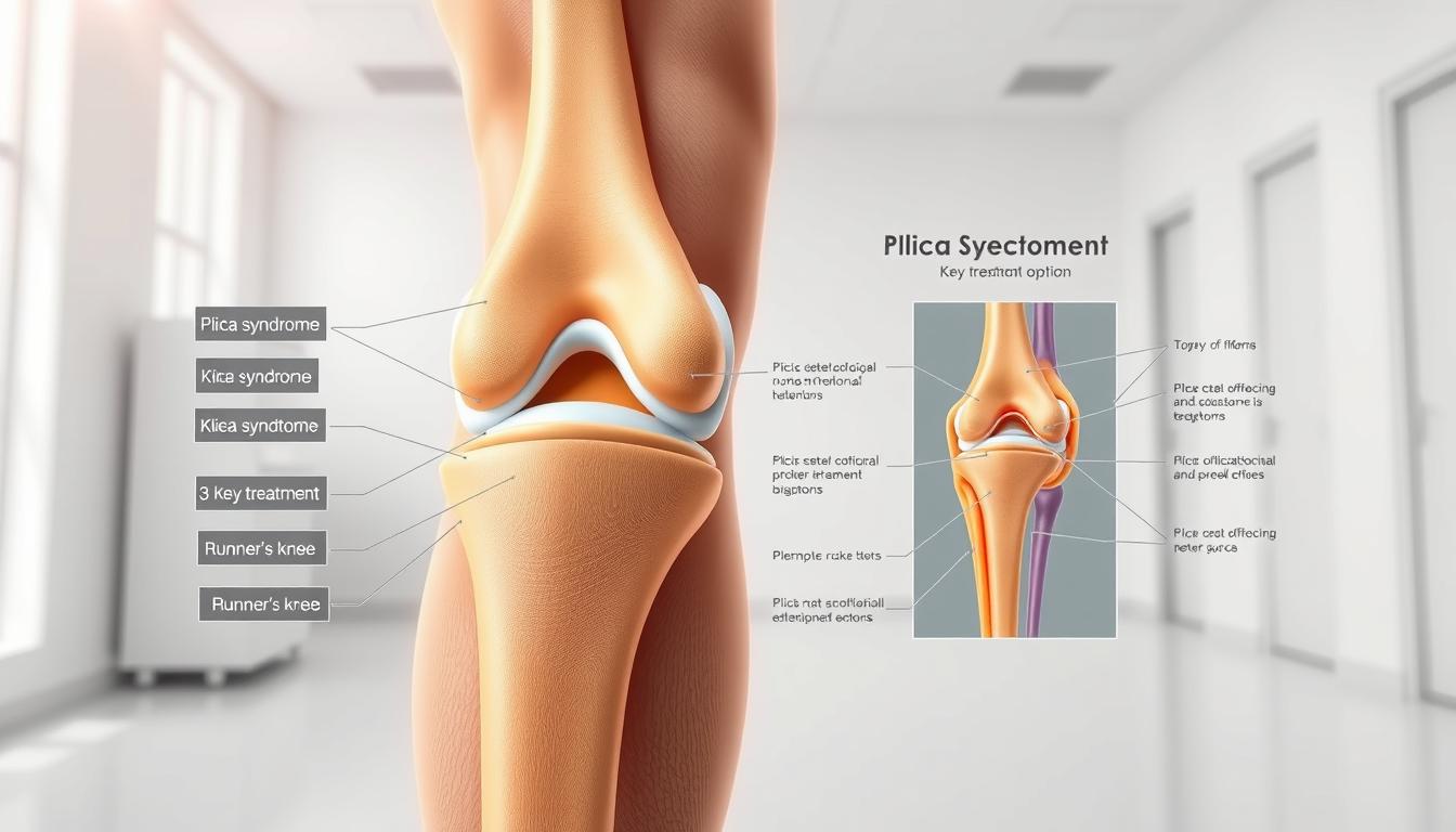
What is Plica Syndrome?
Hidden within your knee lies a potential troublemaker—a synovial fold that most people never notice until it becomes irritated. When this thin tissue layer thickens or scars, it transforms from a helpful joint lubricant to a source of persistent discomfort.
Definition and Underlying Causes
Plica syndrome occurs when repetitive motions or injuries inflame these natural tissue folds. Athletes who perform frequent knee bends—like cyclists or volleyball players—face higher risks. Even minor trauma from a fall can trigger thickening that leads to friction during movement.
Common culprits include:
Sudden increases in workout intensity
Improper warm-up routines
Direct impacts during sports
Clinical Presentation and Symptoms
Patients often report sharp pinching sensations when straightening the leg, accompanied by audible clicks. Swelling typically appears above the kneecap, worsening after activity. “The catching feeling distinguishes it from general wear-and-tear issues,” notes a 2023 Johns Hopkins study on knee mechanics.
Key indicators include:
Localized tenderness along the inner knee
Episodes of temporary joint locking
Pain patterns that fluctuate with activity levels
Advanced imaging reveals fibrotic tissue changes in chronic cases, confirming why rest alone often fails to resolve symptoms. Early intervention with targeted therapy prevents permanent damage to surrounding cartilage.
What is Runner’s Knee?
Millions feel that familiar ache after a long run—but this condition strikes more than just athletes. Runner’s knee describes patellofemoral pain syndrome, a cluster of issues causing discomfort around the kneecap. Unlike sudden injuries, it often creeps in gradually as cartilage wears down from repetitive stress.
Root Causes and Risk Factors
Overuse tops the list of culprits. Marathon training, excessive stair climbing, or sudden activity spikes strain the joint. Weak thigh muscles and flat feet also contribute by altering knee alignment. Women face higher risks due to wider pelvic structures, while excess weight amplifies pressure on the patella.
Contributing Factor
Effect on Knee
Prevention Tip
High-Impact Sports
Repeated patella stress
Cross-train with swimming
Muscle Imbalances
Patella tracking issues
Strengthen quadriceps
Improper Footwear
Increased joint torsion
Get gait analysis
Recognizing the Warning Signs
Dull, throbbing pain beneath the kneecap worsens during squats or downhill walks. Some hear occasional pops when bending, though swelling stays mild compared to inflammatory conditions. “The pain pattern helps distinguish it from acute injuries,” states a Harvard Medical School review on overuse injuries.
Treatment starts with rest and ice packs. Physical therapy focuses on rebuilding muscle support around the joint. Supportive braces and orthotic inserts often complement recovery plans. Severe cartilage damage might require surgery, but most find relief through conservative measures.
Differentiating plica syndrome from runner’s knee
Medical professionals rely on specific clues to tell apart these frequently confused joint issues. While both conditions cause anterior discomfort, their origins and progression patterns differ substantially. Accurate identification directly impacts treatment success rates and recovery timelines.
Key Clinical Differences
Patient histories often reveal distinct triggers. Those with irritated synovial folds typically report sudden pain after direct trauma or intense activity spikes. In contrast, patellofemoral cases usually develop gradually from repetitive motions like running or squatting.
Physical exams provide critical evidence. Clinicians check for a thickened plica band through specialized manipulation tests. A positive result involves localized tenderness and audible clicking when straightening the leg. Assessments for alignment-related stress focus on cartilage response to pressure.
Diagnostic Marker
Synovial Fold Irritation
Patellofemoral Stress
Primary Pain Location
Medial joint line
Under kneecap
Swelling Pattern
Localized above patella
Diffuse around joint
Treatment Response
Anti-inflammatory protocols
Quadriceps strengthening
Imaging studies further clarify uncertainties. MRI scans detect inflamed tissue bands in persistent cases, while X-rays rule out cartilage degeneration. “Targeted therapy based on precise diagnosis prevents unnecessary interventions,” states a recent Johns Hopkins orthopedic review. Early intervention tailored to each condition’s mechanics reduces long-term joint damage risks.
Comparing Symptoms and Physical Signs
Not all knee pain tells the same story. While plica irritation and patellofemoral stress share some surface-level similarities, their distinct symptom patterns help clinicians separate these conditions during evaluations.
Pain Patterns and Onset
Sharp, stabbing sensations during knee extension often point to synovial fold inflammation. This discomfort typically flares suddenly after specific movements like squatting. In contrast, cartilage-related issues develop gradually, with dull aches worsening during prolonged sitting or stair descent.
Swelling and Inflammation
Localized puffiness above the kneecap suggests irritated tissue folds. Runner’s knee usually shows minimal swelling unless cartilage damage progresses. A 2022 clinical review notes inflammatory markers appear earlier in synovial conditions than in mechanical wear cases.
Symptom
Synovial Fold Issue
Cartilage Stress
Pain Onset
Sudden after activity
Gradual over weeks
Swelling Location
Above patella
Around joint line
Response to Rest
Partial relief
Temporary improvement
Mechanical Sensations and Function
Patients often describe “catching” feelings when bending knees with plica involvement. Joint instability dominates in alignment-related cases.
“Mechanical symptoms act like breadcrumbs leading to the root issue,”
explains a Johns Hopkins sports medicine specialist.
Physical tests reveal further clues. Medial joint line tenderness accompanies synovial irritation, while patellar grind tests provoke cartilage-related pain. These distinctions guide treatment plans before imaging confirmation.
Diagnostic Approaches and Examination
Accurate diagnosis forms the cornerstone of effective knee pain management. Doctors combine patient histories, hands-on assessments, and advanced imaging to pinpoint issues. This multi-step process reduces guesswork and tailors treatment plans.
Clinical History and Physical Tests
Providers first ask about pain patterns and activity triggers. Recent injuries or repetitive motions often surface during these discussions. Physical exams check for swelling, tenderness, and joint mobility.
Common tests include:
Medial plica test: Detects thickened tissue folds through specific knee bends
Patellar grind assessment: Evaluates cartilage wear under the kneecap
Gait analysis to spot alignment issues
Imaging Techniques and MRI Use
When physical exams suggest structural issues, imaging provides confirmation. X-rays reveal bone alignment problems, while MRIs excel at showing soft tissue damage. Recent guidelines recommend MRI for persistent swelling or suspected ligament injuries.
Method
Best For
Limitations
Use Cases
Physical Exam
Initial assessment
Limited to surface findings
Early-stage discomfort
X-ray
Bone alignment
Misses soft tissue issues
Trauma evaluation
MRI
Cartilage/ligaments
Higher cost
Unexplained joint locking
Blood tests help rule out infections or autoimmune conditions. A 2023 Johns Hopkins study found “combined diagnostic approaches increase accuracy by 40% compared to single-method evaluations.” Most patients receive clear answers within 2-3 clinical visits when providers follow these protocols.
Treatment and Management Options
When joint discomfort strikes, effective treatment begins with understanding your options. We prioritize approaches that address root causes while minimizing disruption to daily life. Most plans combine short-term relief with long-term joint protection strategies.
Conservative Management and Therapy
Initial care focuses on reducing inflammation and restoring mobility. The RICE method—rest, ice, compression, elevation—remains foundational for acute flare-ups. Clinical guidelines from the Cleveland Clinic show 78% of patients improve within 2-4 weeks using this approach combined with activity modification.
Targeted physical therapy builds crucial support around the joint. Strengthening the quadriceps muscles improves patellar tracking and reduces pressure on sensitive tissues. A 2023 study found patients completing 8-week exercise programs reported 62% less pain during daily activities compared to rest-only groups.
When to Consider Surgical Intervention
Surgery becomes necessary when conservative measures fail after 3-6 months. Arthroscopic procedures remove scarred tissue folds or repair damaged cartilage in severe cases. Research indicates surgical success rates exceed 85% for properly selected candidates.
Key factors influencing this decision include:
Persistent locking or catching sensations
Progressive cartilage deterioration visible on MRI
Limited response to NSAIDs and therapeutic exercises
Individualized plans account for activity levels and recovery goals. As one orthopedic surgeon notes,
“The best outcomes occur when patients actively participate in choosing their treatment path.”
Regular progress evaluations ensure therapies remain aligned with healing milestones.
Prevention and Rehabilitation Strategies
Strong knees begin long before discomfort appears. Proactive care combines targeted exercises with smart activity choices to maintain joint health. Research shows consistent prevention strategies reduce injury risks by 65% compared to reactive approaches.
Exercise and Strengthening Programs
Quadriceps strength forms the foundation of joint stability. Focus on low-impact movements like wall sits and step-ups to build muscle without strain. A 2023 Mayo Clinic study found patients who completed 12 weeks of these exercises reported 54% fewer pain episodes during daily activities.
Exercise
Frequency
Muscle Focus
Straight Leg Raises
3x weekly
Quadriceps
Clamshells
Daily
Hip stabilizers
Resistance Band Walks
2x weekly
Gluteal muscles
Physical therapy programs often incorporate balance training using foam pads or wobble boards. These tools improve proprioception – your body’s ability to sense joint position during movement.
Activity Modification and Lifestyle Changes
Gradual intensity increases prevent overuse injuries. Follow the 10% rule: never boost workout duration or weight by more than 10% weekly. Supportive knee bands during high-impact activities help distribute pressure evenly.
Swap concrete running paths for rubberized tracks
Use orthotic inserts if flat feet contribute to alignment issues
Schedule rest days between intense training sessions
“Consistency beats intensity when rebuilding joint resilience,”
notes a recent Harvard Health Publishing analysis. Pair these changes with dynamic stretching before activities to prepare tissues for stress. Monthly progress checks ensure your prevention plan evolves with your fitness level.
Research and Expert Insights
Recent breakthroughs in orthopedic research are reshaping how we approach joint care. Studies now reveal critical connections between tissue health and long-term mobility. These findings help refine diagnostic accuracy while guiding personalized treatment plans.
Evidence-Based Findings
New data sources highlight quadriceps strength as the cornerstone of knee stability. A 2024 Mayo Clinic trial showed targeted strength training reduces reinjury risk by 38% compared to general exercise. Supportive bands during activity also minimize strain on vulnerable tissues.
Current Approaches
Emerging Methods
Success Rate
Manual therapy
Biologic injections
72% vs 84%
Standard MRI
AI-enhanced imaging
89% accuracy
Generic exercise
DNA-based programs
41% improvement
Future Directions in Knee Health
Researchers now explore cellular therapies to repair damaged cartilage. Wearable sensors that track joint stress during daily activities may soon prevent overuse injuries. “We’re moving from reactive care to predictive models,” notes Dr. Ellen Torres from Johns Hopkins.
Key areas of focus include:
Genetic markers for chronic conditions
3D-printed support bands
Activity-specific risk assessments
These innovations could transform how people manage joint health. Early adoption of evidence-backed strategies helps avoid invasive treatments later.
Conclusion
Navigating knee discomfort requires precision. While both conditions affect the joint, their origins and management differ sharply. Thickened tissue folds demand targeted anti-inflammatory care, while cartilage stress responds best to muscle strengthening.
Accurate diagnosis remains critical. Clinical exams paired with imaging tools like MRI scans help pinpoint the source. We base our recommendations on Mayo Clinic protocols and Johns Hopkins research to ensure reliable guidance.
Most cases improve with rest and therapy. For persistent issues, surgical options show high success rates when conservative methods stall. Individualized plans prove essential – no two injuries follow identical recovery paths.
If discomfort lingers beyond 3-4 weeks, consult a specialist. Proper support bands and patellofemoral alignment strategies often prevent recurring issues. Remember: early intervention protects long-term joint function better than delayed care.
Our analysis combines clinical expertise with real-world recovery data. Whether addressing sudden inflammation or gradual wear, tailored approaches yield optimal results. Trust professional evaluations to guide your path back to pain-free movement.
FAQ
How can I tell if my knee pain is from plica syndrome or runner’s knee?
We identify plica syndrome by localized tenderness along the inner knee, often with a “snapping” sensation. Runner’s knee typically causes dull pain around the kneecap, worsening during activities like squatting or climbing stairs. A physical exam and imaging help confirm the diagnosis.
Does swelling always occur with these conditions?
Swelling is more common in plica syndrome due to synovial tissue irritation. Runner’s knee may involve mild inflammation but rarely significant fluid buildup. Persistent swelling warrants evaluation to rule out cartilage damage or other injuries.
Can physical therapy resolve both issues?
Yes, therapy often helps. For plica syndrome, we focus on reducing inflammation and improving quadriceps flexibility. For runner’s knee, strengthening the hips and correcting patellar alignment are prioritized. Severe cases might require corticosteroid injections or surgery.
Are MRIs necessary for diagnosis?
While MRIs detect thickened plica or cartilage wear, many diagnoses rely on clinical history and physical tests like the “mediopatellar plica test.” Imaging is reserved for atypical presentations or when conservative treatments fail.
What activities increase risk for these injuries?
Repetitive bending or sudden increases in running mileage raise risks. Plica syndrome is linked to overuse in cyclists or gymnasts, while runner’s knee often stems from weak glutes or improper footwear. Cross-training and gradual progression lower recurrence rates.
How long does recovery typically take?
With rest and therapy, most see improvement in 4–6 weeks. Chronic cases may take 3–6 months. Surgery for persistent plica or cartilage damage requires 6–8 weeks of rehab. Consistency with strengthening exercises speeds recovery.
Can these conditions affect both knees simultaneously?
While uncommon, bilateral involvement happens with systemic overuse or biomechanical imbalances. We assess gait, footwear, and training habits to address root causes and prevent future strain on the knee joint.
Osteoporosis, often called the “silent disease,” is a condition characterized by decreased bone density and deterioration of bone tissue, leading to increased fragility and risk of fractures. The condition affects approximately 1 in 3 women and 1 in 5 men over the age of 50, making it a significant public health concern worldwide. The impact of osteoporosis extends beyond bone health – hip fractures, in particular, are associated with a 3-4 times greater risk of dying within 12 months compared to the general population of the same age.
Throughout our lives, our bones undergo a continuous process of remodeling, with old bone being removed (resorption) and new bone being formed. In healthy individuals, this process maintains a balance. However, in osteoporosis, bone resorption outpaces bone formation, resulting in a net loss of bone mass and structural deterioration.
The good news is that osteoporosis is both preventable and treatable. While conventional medical treatments play a crucial role in managing the disease, especially for those at high fracture risk, natural approaches can complement these treatments and provide additional benefits for bone health. This article explores the full spectrum of osteoporosis management, from medical interventions to evidence-based natural therapies that can help strengthen bones and reduce fracture risk.
By understanding both conventional treatments and natural approaches, individuals can work with their healthcare providers to develop a comprehensive strategy tailored to their specific needs and risk factors.
Understanding Osteoporosis
To effectively address osteoporosis, it’s important to understand the disease process and the factors that contribute to its development. At its core, osteoporosis occurs when the body loses too much bone, makes too little bone, or both. This results in weakened bones that can break from minor falls or, in serious cases, even from simple actions like sneezing or bumping into furniture.
Several risk factors contribute to the development of osteoporosis:
Age and Gender: Risk increases with age, with women at significantly higher risk than men. The rapid bone loss that occurs in the 5-7 years following menopause makes women particularly vulnerable.
Genetic Factors: Family history of osteoporosis increases risk, as does being of Caucasian or Asian descent.
Hormonal Changes: The decline in estrogen during menopause in women and testosterone in men accelerates bone loss. Other hormonal disorders, such as hyperthyroidism or hyperparathyroidism, can also affect bone health.
Body Size: Small-framed individuals and those with low body weight have less bone mass to draw from as they age.
Lifestyle Factors: Inadequate calcium and vitamin D intake, sedentary lifestyle, smoking, and excessive alcohol consumption all contribute to bone loss.
Medications: Long-term use of certain medications, including corticosteroids, anticonvulsants, and some cancer treatments, can adversely affect bone health.
Osteoporosis is typically categorized as either primary or secondary. Primary osteoporosis is related to aging and hormonal changes, while secondary osteoporosis results from specific medical conditions or medications that affect bone metabolism.
Diagnosis typically involves dual-energy X-ray absorptiometry (DEXA scan), which measures bone mineral density (BMD). The results are presented as a T-score, comparing an individual’s bone density to that of a healthy 30-year-old of the same sex. A T-score of -1.0 or above is considered normal, while scores between -1.0 and -2.5 indicate osteopenia (low bone mass), and scores below -2.5 indicate osteoporosis.
The Fracture Risk Assessment Tool (FRAX) may also be used to predict the 10-year probability of a major osteoporotic fracture based on individual risk factors, with or without BMD measurements.
One of the challenges of osteoporosis is that it progresses silently, often without symptoms until a fracture occurs. This makes screening and preventive measures crucial, especially for those with known risk factors.
Conventional Medical Treatments
Modern medicine offers several effective treatments for osteoporosis, designed to reduce fracture risk by slowing bone loss, increasing bone formation, or both. Treatment recommendations typically consider factors such as age, sex, fracture history, bone density measurements, and overall fracture risk.
First-Line Medications
Bisphosphonates remain the most commonly prescribed first-line treatment for osteoporosis. These medications slow bone resorption by inhibiting the activity of osteoclasts, the cells responsible for breaking down bone. The American College of Physicians (ACP) recommends bisphosphonates as the initial pharmacologic treatment for reducing fracture risk in postmenopausal women with osteoporosis.
Common bisphosphonates include:
Alendronate (Fosamax) – taken weekly or monthly
Risedronate (Actonel) – taken weekly or monthly
Ibandronate (Boniva) – taken monthly or as quarterly injections
Zoledronic acid (Reclast) – administered as a yearly intravenous infusion
These medications have been shown to reduce the risk of vertebral fractures by 40-70% and non-vertebral fractures, including hip fractures, by 20-40%. Side effects can include gastrointestinal issues with oral formulations and flu-like symptoms with intravenous formulations. Rare but serious side effects include osteonecrosis of the jaw and atypical femur fractures, particularly with long-term use.
Second-Line Treatments
When bisphosphonates are not appropriate or effective, several second-line treatments are available:
Denosumab (Prolia) is a RANK ligand inhibitor that blocks the development and activity of osteoclasts. Given as a subcutaneous injection every six months, it has been shown to reduce vertebral, non-vertebral, and hip fractures. Unlike bisphosphonates, denosumab does not accumulate in the bone, so its effects reverse quickly if treatment is stopped, potentially leading to rapid bone loss and increased fracture risk if not properly managed.
Selective Estrogen Receptor Modulators (SERMs) such as raloxifene (Evista) mimic estrogen’s beneficial effects on bone without some of the risks associated with estrogen. They can reduce vertebral fracture risk but have not been shown to reduce non-vertebral or hip fracture risk.
Hormone Replacement Therapy (HRT) was once widely used for osteoporosis prevention but is now primarily recommended for managing menopausal symptoms in women at high risk for osteoporosis, and only for the shortest duration possible due to potential risks of breast cancer, heart disease, and stroke.
Advanced Treatments for Severe Cases
For patients with severe osteoporosis or those who have experienced fractures despite other treatments, more potent options are available:
Anabolic Therapies stimulate bone formation rather than simply slowing bone loss. These include:
Teriparatide and abaloparatide – synthetic forms of parathyroid hormone that stimulate bone formation when given intermittently. They are administered as daily injections for up to two years.
Romosozumab (Evenity) – a sclerostin inhibitor that both increases bone formation and decreases bone resorption. It is given as monthly injections for one year.
These medications can increase bone density more substantially than antiresorptive drugs, especially at the spine. However, they are typically reserved for those at very high fracture risk due to their cost, route of administration, and limited treatment duration.
Treatment Duration and Management
The optimal duration of osteoporosis treatment continues to be studied. Many experts recommend reassessing after 3-5 years of bisphosphonate therapy, with consideration of a “drug holiday” for patients whose fracture risk has decreased. For high-risk patients, sequential therapy (starting with an anabolic agent followed by an antiresorptive) may provide optimal fracture protection.
Regular monitoring of bone mineral density and, in some cases, biochemical markers of bone turnover, can help assess treatment response and guide decisions about continuing or modifying therapy.
It’s important to remember that medication is just one component of osteoporosis management. All treatment approaches should be accompanied by adequate calcium and vitamin D intake, appropriate exercise, and measures to prevent falls.
Natural Therapy #1: Nutrition for Bone Health
Nutrition plays a fundamental role in both preventing and managing osteoporosis. While medications can effectively slow bone loss or stimulate bone formation, they cannot compensate for nutritional deficiencies that compromise bone health. A bone-healthy diet provides the essential building blocks needed for optimal bone remodeling and maintenance.
Calcium: The Foundation of Bone Health
Calcium is the primary mineral found in bone, making adequate intake essential throughout life. The recommended daily intake varies by age and gender:
Leafy green vegetables (kale, collard greens, bok choy)
Calcium-set tofu
Canned fish with bones (sardines, salmon)
Fortified cereals and juices
While food sources are preferred, calcium supplements can help bridge dietary gaps. Two main types are available:
Calcium carbonate: Higher concentration of elemental calcium (40%), best absorbed with food
Calcium citrate: Lower concentration (21%), but better absorbed on an empty stomach and by those with reduced stomach acid
For optimal absorption, calcium supplements should be taken in doses of 500-600 mg or less at a time, spaced throughout the day. Taking them with meals can reduce the risk of kidney stones and improve absorption, particularly for calcium carbonate.
Vitamin D: The Essential Partner
Vitamin D is crucial for calcium absorption and proper bone mineralization. Without adequate vitamin D, the body cannot effectively utilize calcium, regardless of intake. Current recommendations include:
Adults up to age 70: 600-800 IU daily
Adults over 70: 800-1,000 IU daily
Higher doses may be needed for those with vitamin D deficiency or limited sun exposure
Vitamin D sources include:
Sunlight (the body produces vitamin D when skin is exposed to UVB rays)
Fatty fish (salmon, mackerel, tuna)
Fortified foods (milk, orange juice, cereals)
Egg yolks
Supplements (D3 is generally preferred over D2)
Many healthcare providers recommend checking vitamin D levels through a blood test (25-hydroxyvitamin D) to determine if supplementation is needed. Optimal levels are generally considered to be 30-60 ng/mL.
Beyond Calcium and Vitamin D
While calcium and vitamin D receive the most attention, other nutrients also contribute to bone health:
Protein: Provides the structural matrix for bone and stimulates insulin-like growth factor I, which promotes bone formation. Aim for 0.8-1.0 g/kg of body weight daily, from both animal and plant sources.
Vitamin K: Important for bone protein synthesis. Found in leafy greens, broccoli, and fermented foods.
Magnesium: Influences crystal formation in bone and calcium metabolism. Found in nuts, seeds, whole grains, and leafy greens.
Potassium: Helps maintain acid-base balance, reducing calcium loss from bone. Abundant in fruits and vegetables.
Zinc and Manganese: Essential for bone formation enzymes. Found in whole grains, nuts, and seeds.
Certain dietary patterns may also impact bone health. The Mediterranean diet, rich in fruits, vegetables, whole grains, fish, olive oil, and nuts, has been associated with higher bone density and lower fracture risk.
Conversely, some dietary factors may negatively affect bone health:
High sodium intake increases calcium excretion
Excessive caffeine may interfere with calcium absorption
Very high protein diets can increase calcium excretion
Carbonated beverages, particularly colas, have been associated with lower bone density in some studies
Figure 2: Bone-building nutrients and their food sources. A balanced diet rich in these nutrients supports optimal bone health and complements medical treatments for osteoporosis.
Natural Therapy #2: Exercise for Osteoporosis
Exercise is a powerful tool for building and maintaining bone strength. Unlike medication, which primarily works to slow bone loss, appropriate physical activity can actually stimulate bone formation, improve balance and coordination (reducing fall risk), and enhance overall functional capacity.
How Exercise Strengthens Bones
Bones respond to mechanical loading much like muscles respond to resistance – they adapt and strengthen. This phenomenon, known as Wolff’s Law, explains why weight-bearing activities and resistance training are particularly beneficial for bone health. When muscles pull on bones during these activities, they create stress that stimulates osteoblasts (bone-building cells) to lay down new bone tissue.
Different types of exercise affect bone health in different ways:
Weight-Bearing Exercises
Weight-bearing exercises force you to work against gravity while staying upright. These activities are particularly effective for strengthening the bones of the hips, legs, and lower spine. They include:
High-impact weight-bearing exercises: Running, jumping, high-impact aerobics, dancing, tennis, and basketball can build bone mass effectively in those without osteoporosis or previous fractures.
Low-impact weight-bearing exercises: Walking, elliptical training, stair climbing, and low-impact aerobics provide less bone stimulation but are safer options for those with osteoporosis, previous fractures, or other health limitations.
A general recommendation is to engage in weight-bearing aerobic activities for 30 minutes on most days of the week.
Resistance Training
Resistance or strength training involves working against resistance, whether from weights, bands, water, or body weight. These exercises target specific muscle groups and the bones they attach to. Effective resistance training for bone health includes:
Free weights (dumbbells, barbells)
Weight machines
Resistance bands
Body weight exercises (push-ups, squats)
Resistance training should be performed 2-3 times per week, targeting all major muscle groups. For bone health benefits, moderate intensity (8-12 repetitions with a weight that creates fatigue by the final repetition) is generally recommended.
Balance and Posture Exercises
While these exercises don’t directly build bone, they help prevent falls – a critical consideration for those with osteoporosis:
Tai chi
Yoga (with modifications for osteoporosis)
Posture training
Stability ball exercises
Single-leg standing
Balance exercises should be incorporated into daily routines, even if just for a few minutes each day.
Exercise Precautions for Osteoporosis
For those already diagnosed with osteoporosis, exercise remains beneficial but requires certain precautions:
Avoid high-impact activities if you have severe osteoporosis or previous fractures
Avoid rapid, forceful movements that might increase fracture risk
Avoid extreme forward bending and twisting of the spine
Focus on proper form rather than amount of weight lifted
Start slowly and progress gradually
Consider working with a physical therapist to develop a safe, effective program
Developing an Exercise Program
An ideal exercise program for bone health includes:
30 minutes of weight-bearing aerobic activity on most days
Resistance training for all major muscle groups 2-3 times weekly
Balance exercises daily
Posture and core strengthening exercises regularly
For those new to exercise or with health concerns, starting with a physical therapist or qualified fitness professional experienced in working with osteoporosis is recommended. They can design a program tailored to individual needs, limitations, and goals.
Remember that consistency is key – the bone benefits of exercise are lost when activity stops, so finding enjoyable activities that can be maintained long-term is essential for ongoing bone health.
Natural Therapy #3: Vitamin K for Bone Health
Vitamin K has emerged as an important nutrient for bone health that often doesn’t receive the same attention as calcium and vitamin D. Research increasingly suggests that adequate vitamin K intake is essential for optimal bone metabolism and strength.
The Role of Vitamin K in Bone Metabolism
Vitamin K serves as a cofactor for the enzyme that activates osteocalcin, a protein that binds calcium to the bone matrix. Without sufficient vitamin K, osteocalcin remains inactive, leading to reduced bone mineralization and potentially increased fracture risk.
There are two main forms of vitamin K:
Vitamin K1 (Phylloquinone): The primary dietary form, found mainly in green leafy vegetables
Vitamin K2 (Menaquinones): Found in fermented foods and produced by intestinal bacteria; appears to be more effective for bone health than K1
Studies have found that higher vitamin K intake is associated with higher bone mineral density and lower fracture risk. Low circulating levels of vitamin K have been linked to lower bone mass and increased fracture risk, particularly hip fractures.
However, optimal amounts for bone health may be higher than these basic recommendations.
Vitamin K Supplementation
Research on vitamin K supplementation for osteoporosis has shown mixed results. Some studies, particularly those using vitamin K2 (MK-4 form) at doses of 45mg daily, have shown reduced fracture risk in Japanese populations. Other studies using different forms or lower doses have shown more modest or inconsistent effects.
When considering vitamin K supplementation:
Form matters: Vitamin K2, particularly the MK-4 and MK-7 forms, may be more beneficial for bone health than K1
Dosage varies: Therapeutic doses used in studies range from 45-180 micrograms for MK-7 and up to 45mg for MK-4
Medication interactions: Vitamin K can interfere with certain blood-thinning medications, particularly warfarin. Those taking such medications should consult their healthcare provider before supplementing
Current Evidence and Recommendations
While the evidence for vitamin K supplementation is promising, it’s not yet conclusive enough for most major medical organizations to recommend routine supplementation specifically for osteoporosis. Current approaches include:
Ensuring adequate vitamin K intake through diet, particularly green leafy vegetables
Considering supplementation under healthcare provider guidance, especially for those with low dietary intake or at high fracture risk
Using vitamin K as part of a comprehensive bone health strategy that includes calcium, vitamin D, and other nutrients
For those interested in supplementation, consulting with a healthcare provider is essential, particularly for those on medications that might interact with vitamin K.
Natural Therapy #4: Magnesium and Bone Health
Magnesium is an essential mineral that plays multiple roles in bone health yet is often overlooked in discussions about osteoporosis prevention and treatment. Approximately 60% of the body’s magnesium is stored in bone tissue, highlighting its importance to skeletal structure.
Magnesium’s Role in Bone Metabolism
Magnesium contributes to bone health through several mechanisms:
It influences the activity of osteoblasts and osteoclasts, the cells responsible for bone formation and resorption
It affects crystal formation in bone, influencing bone quality and strength
It regulates calcium transport and metabolism
It is required for the conversion of vitamin D to its active form, which is necessary for calcium absorption
It helps maintain appropriate calcium levels in the blood and tissues
Research has found that magnesium deficiency is associated with reduced bone mineral density, altered bone and mineral metabolism, and increased fracture risk. One study found that 40% of women with osteoporosis or low bone density had low circulating magnesium levels.
Dietary Sources of Magnesium
The recommended dietary allowance (RDA) for magnesium is:
Adult women ages 19-30: 310 mg daily
Adult women ages 31+: 320 mg daily
Adult men ages 19-30: 400 mg daily
Adult men ages 31+: 420 mg daily
Excellent food sources of magnesium include:
Dark leafy greens (spinach, chard)
Nuts and seeds (almonds, pumpkin seeds)
Whole grains (brown rice, quinoa)
Legumes (black beans, chickpeas)
Dark chocolate
Avocados
Bananas
Despite its abundance in whole foods, many people don’t consume enough magnesium due to dietary patterns high in processed foods, which typically contain little magnesium. Soil depletion of minerals may also reduce the magnesium content of foods compared to historical levels.
Magnesium Supplementation
For those who cannot meet their magnesium needs through diet alone, supplements can be beneficial. Several forms are available, each with different properties:
Magnesium citrate: Well-absorbed, may have a mild laxative effect
Magnesium glycinate: Well-absorbed with minimal digestive side effects
Magnesium malate: Well-tolerated and may help with muscle pain
Magnesium oxide: Lower absorption rate but higher elemental magnesium content
Magnesium chloride: Good absorption and often available as a topical oil
When supplementing, it’s generally recommended to start with a lower dose and gradually increase to avoid digestive discomfort. Taking magnesium supplements with food can also improve tolerance.
Safety Considerations
While magnesium is generally safe, excessive intake from supplements (not food) can cause diarrhea, nausea, and abdominal cramping. In people with reduced kidney function, high doses of magnesium supplements can lead to magnesium toxicity, characterized by low blood pressure, confusion, and cardiac complications.
Those with kidney disease, heart problems, or on certain medications should consult their healthcare provider before supplementing with magnesium.
Evidence and Recommendations
While more research is needed on the specific effects of magnesium supplementation on fracture risk, ensuring adequate magnesium intake is a sensible strategy for supporting bone health. The greatest benefits are likely to be seen in those who are magnesium deficient.
Current recommendations include:
Prioritizing magnesium-rich whole foods in the diet
Considering supplementation if dietary intake is insufficient or if deficiency is suspected
Integrating magnesium into a comprehensive bone health approach alongside calcium, vitamin D, and other supportive nutrients
Figure 3: The six natural therapies work synergistically to support bone health, complementing conventional medical treatments for osteoporosis.
Natural Therapy #5: Soy Isoflavones and Phytoestrogens
The dramatic increase in osteoporosis risk that follows menopause highlights the crucial role of estrogen in maintaining bone health. As estrogen levels decline, bone resorption accelerates, often leading to significant bone loss in the first 5-7 years after menopause. This connection between estrogen and bone health has led researchers to investigate plant compounds with estrogen-like properties, known as phytoestrogens, as potential natural therapies for osteoporosis.
Understanding Isoflavones and Phytoestrogens
Isoflavones are a class of phytoestrogens – plant compounds that have a structure similar to human estrogen and can bind to estrogen receptors in the body, though their effects are typically much weaker than those of human estrogen. The most well-studied isoflavones include:
Genistein
Daidzein
Glycitein
These compounds are found primarily in soybeans and soy products, but also in smaller amounts in other legumes such as chickpeas, lentils, and beans.
Mechanisms of Action
Isoflavones may support bone health through several mechanisms:
Binding to estrogen receptors in bone tissue, potentially slowing bone resorption
Inhibiting the activity of osteoclasts (cells that break down bone)
Supporting the activity of osteoblasts (cells that build bone)
Providing antioxidant effects that may protect bone cells from oxidative stress
Research on Isoflavones and Bone Health
Research on soy isoflavones for bone health has shown promising but mixed results:
A meta-analysis of 19 studies in postmenopausal women found that soy isoflavone supplementation significantly increased bone mineral density by 54% and reduced bone resorption markers by 23% compared to baseline values. The most significant benefits were seen in studies lasting at least one year and using higher doses (80-90 mg of isoflavones daily).
However, not all studies have shown positive results, and the effects may vary based on factors such as:
Individual metabolism of isoflavones (some people convert daidzein to equol, a more potent compound, while others do not)
Age and years since menopause
Baseline bone density
Dose and type of isoflavones used
Study duration
Dietary Sources of Isoflavones
The richest food sources of isoflavones include:
Soybeans and whole soy foods (tofu, tempeh, edamame)
Soy flour and soy protein
Soy milk and other soy beverages
Other legumes (chickpeas, lentils, beans)
Red clover (used in some supplements)
The isoflavone content varies widely among soy foods, with whole and minimally processed soy foods generally providing higher amounts than highly processed soy ingredients.
Supplementation Considerations
For those considering isoflavone supplements for bone health:
Dosage: Studies showing benefits typically used 40-110 mg of isoflavones daily
Duration: Longer-term use (at least one year) appears necessary for significant effects on bone
Form: Supplements may contain isolated isoflavones or whole soy extracts
Quality: Look for standardized products from reputable manufacturers
Safety and Concerns
While moderate consumption of dietary soy is generally considered safe for most people, questions have been raised about the long-term safety of isolated isoflavone supplements, particularly for women with a history of hormone-sensitive conditions such as breast cancer.
Current evidence does not indicate that moderate soy consumption increases breast cancer risk, and some studies suggest it may even be protective. However, the effects of high-dose isoflavone supplements taken for extended periods are less well understood.
Potential side effects of isoflavone supplements may include:
Digestive discomfort
Menstrual changes in premenopausal women
Theoretical interactions with thyroid hormones
Recommendations for Use
Given the current evidence:
Including whole soy foods in the diet is a reasonable approach for supporting bone health
Those considering supplements should discuss them with their healthcare provider
Women with a history of hormone-sensitive conditions should be particularly cautious and seek medical guidance
Isoflavones should be viewed as one component of a comprehensive bone health program, not as a standalone treatment for osteoporosis
As research continues, our understanding of the optimal use of isoflavones for bone health will likely evolve.
Natural Therapy #6: Lifestyle Modifications
Beyond nutrition and targeted supplements, several lifestyle modifications can significantly impact bone health and fracture risk. These changes, while sometimes overlooked, can be powerful components of a comprehensive approach to osteoporosis prevention and management.
Smoking Cessation
Smoking has multiple detrimental effects on bone health:
It reduces blood supply to bones
It impairs the function of osteoblasts (bone-building cells)
It interferes with calcium absorption
It alters hormonal balance, including estrogen levels
It may accelerate the breakdown of exogenous estrogen
Studies have consistently shown that smokers have lower bone density and higher fracture risk compared to non-smokers. The longer one smokes, the greater the impact on bone health.
The good news is that quitting smoking can help slow the rate of bone loss, though it may not fully reverse existing damage. Former smokers gradually see their fracture risk decrease after quitting, though it may take years to approach the risk level of people who never smoked.
Resources for quitting smoking include nicotine replacement therapies, prescription medications, counseling programs, and support groups. Healthcare providers can help develop a personalized smoking cessation plan.
Alcohol Moderation
Excessive alcohol consumption negatively impacts bone health through multiple mechanisms:
Direct toxic effects on osteoblasts
Interference with vitamin D metabolism and calcium absorption
Disruption of hormone production and metabolism
Increased fall risk
Malnutrition associated with heavy drinking
Moderate alcohol consumption (up to one drink daily for women and up to two drinks daily for men) has not been clearly associated with increased osteoporosis risk. However, heavy drinking significantly increases the risk of bone loss and fractures.
For those who drink heavily, reducing alcohol consumption or abstaining completely can help preserve bone mass and reduce fracture risk. Support is available through healthcare providers, counseling, and programs like Alcoholics Anonymous.
Fall Prevention Strategies
For those with osteoporosis, preventing falls is crucial for avoiding fractures. Comprehensive fall prevention includes:
Home Safety Modifications:
Remove tripping hazards (loose rugs, clutter)
Improve lighting, especially in stairways and at night
Install grab bars in bathrooms and on stairs
Use non-slip mats in bathtubs and showers
Consider placing frequently used items within easy reach
Personal Safety Measures:
Wear properly fitting, supportive shoes with non-slip soles
Use assistive devices (cane, walker) if needed
Get up slowly from sitting or lying positions to avoid dizziness
Use caution when walking on wet, icy, or uneven surfaces
Consider wearing hip protectors if at very high fall risk
Health Management:
Review medications with healthcare providers to identify those that might cause dizziness or affect balance
Get regular vision and hearing checks
Address foot problems promptly
Manage conditions that might affect balance, such as Parkinson’s disease or arthritis
Stress Management
Emerging research suggests that chronic stress may contribute to bone loss through several mechanisms:
Increased production of cortisol, which can directly inhibit bone formation
Disruption of calcium absorption and metabolism
Inflammation, which can accelerate bone resorption
Indirect effects through poor diet, reduced physical activity, and increased smoking or alcohol consumption associated with stress
Effective stress management techniques include:
Mindfulness meditation
Progressive muscle relaxation
Regular physical activity
Adequate sleep
Social connection
Cognitive-behavioral techniques
Time in nature
Incorporating stress reduction into daily routines can support overall health, including bone health.
Weight Management
Maintaining a healthy weight is important for bone health:
Being underweight (BMI < 18.5) is a significant risk factor for low bone density and fractures
Very high body weight increases stress on bones and risk of falls
Weight cycling (repeated weight loss and regain) may be detrimental to bone density
Achieving and maintaining a healthy weight through nutritious eating and regular physical activity supports optimal bone health and reduces fracture risk.
Integrating Conventional and Natural Approaches
The most effective approach to osteoporosis prevention and treatment often combines conventional medical treatments with natural therapies. This integrated strategy addresses the condition from multiple angles, potentially providing more comprehensive protection against bone loss and fractures than either approach alone.
Building a Comprehensive Treatment Plan
An optimal osteoporosis management plan typically includes:
Proper medical assessment and diagnosis, including bone density testing, fracture risk assessment, and evaluation for secondary causes of osteoporosis
Appropriate medication based on individual risk factors, with higher-risk individuals typically benefiting most from pharmacologic intervention
Nutritional optimization with adequate calcium, vitamin D, protein, and other bone-supporting nutrients
Regular weight-bearing and resistance exercise tailored to individual fitness level and fracture risk
Targeted supplementation based on individual needs, potentially including vitamin K, magnesium, and isoflavones for appropriate candidates
Lifestyle modifications such as smoking cessation, alcohol moderation, and fall prevention strategies
Regular monitoring of bone density, biochemical markers, and overall health status to assess progress and adjust the plan as needed
Personalizing Your Approach
The optimal combination of treatments varies based on individual factors:
Age and gender: Younger individuals may focus more on prevention through nutrition and exercise, while those at higher risk due to age or menopause may need more aggressive intervention
Fracture history and risk level: Those with previous fractures or very low bone density typically need medical treatment alongside natural approaches
Personal preferences and values: Some individuals may prefer to emphasize lifestyle and nutritional approaches, while others may be more comfortable with medication
Comorbidities: Other health conditions and medications may influence which treatments are most appropriate
Response to treatment: The approach may need adjustment based on how bone density and other markers respond over time
Working with Healthcare Providers
Effective osteoporosis management requires collaboration with healthcare providers, potentially including:
Primary care physician
Endocrinologist or rheumatologist
Registered dietitian
Physical therapist
Pharmacist
Open communication about all therapies being used, including supplements and exercise programs, is essential to avoid potential interactions and ensure complementary rather than conflicting approaches.
Potential Interactions to Consider
Some natural therapies may interact with osteoporosis medications or other treatments:
Calcium supplements can interfere with the absorption of certain medications, including some antibiotics and thyroid medications, and should be taken at different times
Vitamin K supplements can interfere with warfarin and other blood-thinning medications
High-dose vitamin A supplements may counteract the beneficial effects of vitamin D and potentially harm bone health
Some herbal supplements may interact with osteoporosis medications or affect bone metabolism through unknown mechanisms
Discussing all supplements with healthcare providers helps minimize the risk of adverse interactions.
Monitoring Progress
Regular assessment helps determine whether the treatment plan is working:
Bone density testing typically every 1-2 years while establishing treatment efficacy, then potentially less frequently
Blood and urine tests to assess bone turnover markers and vitamin D levels
Height measurements to detect potential vertebral fractures
Fall risk assessments
Review of any pain or functional limitations
Based on these assessments, the treatment plan can be adjusted to optimize outcomes.
Conclusion
Osteoporosis is a complex condition that requires a multifaceted approach to prevention and treatment. While conventional medical treatments play a crucial role, especially for those at high fracture risk, natural therapies can significantly complement these approaches and provide additional benefits for bone health.
The six natural therapies discussed – nutrition, exercise, vitamin K, magnesium, soy isoflavones, and lifestyle modifications – each address different aspects of bone health. When combined appropriately and personalized to individual needs, they create a comprehensive strategy that supports both bone quantity (density) and quality (structure).
The most effective approach is typically one that integrates conventional and natural strategies based on individual risk factors, preferences, and needs. This may mean using medications for those at high fracture risk while simultaneously optimizing nutrition, incorporating appropriate exercise, and addressing lifestyle factors that affect bone health.
For those at lower risk, focusing primarily on natural approaches may be appropriate, with regular monitoring to ensure bone health is maintained. The key is early intervention – whether through natural or conventional means – as preventing bone loss is easier than reversing it once significant deterioration has occurred.
Working collaboratively with healthcare providers to develop and adjust your bone health strategy over time ensures that you receive the most appropriate combination of treatments for your specific situation. With this comprehensive approach, many individuals can maintain bone strength, reduce fracture risk, and continue to lead active, independent lives despite osteoporosis.
References
American College of Physicians. (2023). Pharmacologic Treatment of Primary Osteoporosis or Low Bone Mass to Prevent Fractures in Adults: A Living Clinical Guideline.
Bone Health and Osteoporosis Foundation. (2024). Calcium/Vitamin D Requirements, Recommended Foods & Supplements.
International Osteoporosis Foundation. (2024). New evidence-based guideline for the management of osteoporosis in men.
National Institutes of Health. (2021). Vitamin D Fact Sheet for Health Professionals.
National Osteoporosis Foundation. (2023). Clinician’s Guide to Prevention and Treatment of Osteoporosis.
Palermo, A., et al. (2017). Vitamin K and osteoporosis: Myth or reality? Metabolism, 70, 57-71.
Rodríguez-Olleros Rodríguez, C., & Díaz Curiel, M. (2019). Vitamin K and Bone Health. Journal of Osteoporosis.
Taku, K., et al. (2010). Effect of soy isoflavone extract supplements on bone mineral density in menopausal women. Asia Pacific Journal of Clinical Nutrition.
Watson, R.R., & Preedy, V.R. (2020). Bioactive Food as Dietary Interventions for the Aging Population.
Weaver, C.M., et al. (2016). Calcium plus vitamin D supplementation and risk of fractures. Osteoporosis International.
Are you experiencing persistent knee pain that doesn’t seem to subside? The knee is a complex structure made up of various soft tissues, and issues like Synovial Plica Syndrome can be a source of discomfort.
This condition occurs when the plica, a fold in the membrane surrounding the knee joint, becomes irritated and inflamed. Understanding the symptoms is crucial for proper diagnosis and treatment, as they can be similar to those of other knee problems.
Key Takeaways
Synovial Plica Syndrome affects the knee, causing pain and discomfort.
The condition is characterized by irritation and inflammation of the plica.
Symptoms can be similar to other knee issues, making diagnosis challenging.
Understanding the symptoms is key to proper diagnosis and treatment.
Accurate diagnosis is crucial for effective treatment.
Understanding Synovial Plica Syndrome
Understanding the intricacies of Synovial Plica Syndrome is crucial for diagnosing and treating knee discomfort. Synovial Plica Syndrome involves the irritation of the plica, which are folds in the synovial tissue surrounding the knee joint.
What is Synovial Plica Syndrome?
Synovial Plica Syndrome occurs when the plica becomes inflamed, typically due to overuse or direct trauma to the knee. This inflammation can lead to plica irritation, causing pain and discomfort, particularly in the center of the kneecap, known as medial patellar plica syndrome.
Research indicates that a significant percentage of patients undergoing arthroscopic surgery have remnants of their synovial plicae, highlighting the prevalence of this condition.
Causes of Synovial Plica Syndrome
The primary causes of Synovial Plica Syndrome include repetitive knee movements and direct trauma to the knee. Athletes and individuals engaged in activities that involve frequent knee bending are more susceptible to this condition.
Overuse or repetitive strain on the knee
Direct trauma or injury to the knee
Poor knee alignment or tracking
Who is Affected?
Synovial Plica Syndrome commonly affects athletes and individuals who participate in sports or activities that involve repetitive knee movements. This includes runners, cyclists, and those involved in jumping sports.
Understanding who is at risk can help in prevention and early diagnosis, potentially reducing the severity of symptoms and improving treatment outcomes.
Common Symptoms
Knee problems are a hallmark of Synovial Plica Syndrome, with several symptoms being commonly reported. The condition can manifest in different ways, but there are key indicators that healthcare professionals look for when diagnosing Synovial Plica Syndrome.
Pain Location
Pain associated with Synovial Plica Syndrome is typically located around the kneecap. This pain is often described as achy rather than sharp or shooting. Activities that stress the knee, such as squatting, bending, or using stairs, can exacerbate the pain.
Swelling Around the Knee
Swelling is another common symptom of Synovial Plica Syndrome. The swelling occurs due to inflammation of the plica, which can be irritated by repetitive knee motions or direct trauma to the knee. This swelling can lead to discomfort and may affect the appearance of the knee.
Stiffness and Reduced Mobility
Stiffness and reduced mobility are also characteristic symptoms. The inflammation and irritation of the synovial plica can lead to a decrease in the knee’s range of motion, making it difficult to perform daily activities or engage in physical exercises without discomfort.
The symptoms of Synovial Plica Syndrome can vary in severity and impact daily life to different extents. Understanding these symptoms is crucial for seeking appropriate medical care.
Symptom
Description
Common Triggers
Pain Around Kneecap
Achy pain rather than sharp
Squatting, bending, using stairs
Swelling
Inflammation causing knee swelling
Repetitive knee motions, direct trauma
Stiffness and Reduced Mobility
Decreased range of motion
Inflammation and irritation of the plica
How Symptoms Manifest
The manifestation of Synovial Plica Syndrome symptoms can vary significantly among individuals. Symptoms can be influenced by the level of physical activity, the specific irritation of the plica, and individual tolerance to pain.
Symptoms During Physical Activity
During physical activity, symptoms of Synovial Plica Syndrome can worsen. Activities that involve bending, jumping, or repetitive knee movements can irritate the plica, leading to pain and discomfort. Some individuals may experience a catching or locking sensation in the knee, particularly when getting up from a seated position after sitting for an extended period.
Common symptoms during physical activity include:
Pain or tenderness around the knee
Joint clicking or cracking noises when bending or straightening the knee
Swelling or inflammation around the affected area
Stiffness or reduced mobility in the knee
Symptoms at Rest
Even at rest, some individuals with Synovial Plica Syndrome may experience symptoms. The condition can cause discomfort or pain when sitting for long periods or when the knee is in a bent position for an extended time.
Night-time Pain
Night-time pain is another symptom that some individuals with Synovial Plica Syndrome may experience. The pain can be severe enough to disrupt sleep, particularly if the individual sleeps with their knee bent. Understanding the pattern of night-time pain can help in diagnosing and managing the condition.
Key factors that contribute to night-time pain include:
The severity of the plica irritation
The individual’s overall knee health
Sleeping position and habits
Diagnosis of Synovial Plica Syndrome
Diagnosing Synovial Plica Syndrome requires a comprehensive approach that involves evaluating the patient’s medical history, conducting a thorough physical examination, and sometimes utilizing imaging tests. This multi-faceted diagnostic strategy is crucial for accurately identifying the condition and ruling out other potential causes of knee problems.
Medical History Evaluation
The first step in diagnosing Synovial Plica Syndrome is a thorough medical history evaluation. This involves assessing the patient’s symptoms, including the onset and duration of Synovial plica syndrome symptoms, to understand the condition’s progression. Healthcare professionals will also inquire about any previous knee injuries or conditions that could be contributing to the current symptoms.
Physical Exam Details
A physical examination is a critical component of the diagnostic process. During the exam, the healthcare provider will assess the knee’s range of motion, check for tenderness, and perform specific maneuvers to reproduce the patient’s symptoms. This hands-on evaluation helps to identify the presence of a synovial plica and assess its impact on knee function. For more information on when to seek medical help for knee pain, visit https://kneehurt.com/when-to-seek-medical-help-for-knee-pain/.
Imaging Tests Utilized
While a diagnosis can often be made based on medical history and physical examination, imaging tests may be employed to confirm the diagnosis or rule out other causes of knee pain. Common imaging tests include X-rays, MRI (Magnetic Resonance Imaging), and ultrasound. These tests provide detailed images of the knee’s internal structures, helping healthcare professionals to visualize the synovial plica and assess any associated inflammation or irritation.
In conclusion, diagnosing Synovial Plica Syndrome involves a combination of medical history evaluation, physical examination, and sometimes imaging tests. By understanding the diagnostic process, patients can better appreciate the complexity of identifying this condition and the importance of seeking professional medical advice for accurate diagnosis and effective treatment.
Treatment Options
Synovial Plica Syndrome treatment encompasses a range of strategies from conservative management to surgical interventions, depending on the severity and persistence of symptoms.
Conservative Treatments
Most instances of medial patellar plica syndrome respond well to alternative treatment options. These include chiropractic care, physical therapy, or even a tailored exercise plan at home. Such conservative approaches aim to alleviate knee pain and knee discomfort by improving knee function and reducing inflammation.
Surgical Interventions
In cases where conservative treatments fail to provide relief, surgical options may be considered. Surgical interventions for Synovial Plica Syndrome typically involve arthroscopic procedures to remove or repair the problematic plica. For more information on Synovial Plica Syndrome, you can visit Healthline’s resource on the condition.
Rehabilitation Strategies
Regardless of whether the treatment involves conservative methods or surgery, rehabilitation plays a crucial role in recovery. Rehabilitation strategies often include a structured physical therapy program aimed at restoring knee strength and mobility, thereby reducing the risk of future complications.
Treatment Approach
Description
Benefits
Conservative Treatments
Includes chiropractic care, physical therapy, and home exercise plans.
Non-invasive, reduces knee pain and discomfort.
Surgical Interventions
Arthroscopic procedures to remove or repair the plica.
Effective for severe cases not responding to conservative treatments.
Rehabilitation Strategies
Structured physical therapy programs.
Restores knee strength and mobility, prevents future issues.
Managing Pain from Symptoms
Effective management of Synovial Plica Syndrome symptoms involves a multi-faceted approach to pain relief. This condition can cause significant discomfort due to plica irritation and swelling around the knee. Managing pain is crucial to improve the quality of life for individuals affected by this condition.
Over-the-Counter Medications
Over-the-counter (OTC) medications are often the first line of defense against pain associated with Synovial Plica Syndrome. Nonsteroidal anti-inflammatory drugs (NSAIDs) like ibuprofen can help reduce pain and inflammation. It’s essential to follow the recommended dosage and consult with a healthcare professional before starting any medication regimen.
Some individuals may find relief with topical creams or gels that contain NSAIDs or capsaicin. These products can be applied directly to the skin around the affected area, providing localized pain relief.
Alternative Pain Management Techniques
In addition to OTC medications, alternative pain management techniques can be beneficial. Physical therapy is a valuable approach, as it can help improve knee function and reduce pain. A physical therapist can design a personalized exercise program to strengthen the muscles around the knee and improve flexibility.
Applying ice packs to reduce swelling and ease pain
Using compression bandages to provide support
Elevating the knee to minimize swelling
In some cases, healthcare professionals may recommend corticosteroid injections to reduce inflammation and relieve pain. These injections can provide significant relief, especially when the pain is severe and interferes with daily activities.
It’s crucial for individuals with Synovial Plica Syndrome to work closely with their healthcare provider to develop a comprehensive pain management plan. By combining different strategies, individuals can better manage their symptoms and improve their overall quality of life.
Lifestyle Adjustments
To manage Synovial Plica Syndrome effectively, it’s essential to make certain lifestyle adjustments. These modifications can help alleviate symptoms such as stiffness and joint clicking, improving overall knee function.
Exercises to Alleviate Symptoms
Engaging in specific exercises can help strengthen the muscles around the knee, thereby reducing the severity of symptoms. Activities such as biking, swimming, walking, or using an elliptical machine are beneficial as they promote knee mobility without excessive strain.
Strengthening the quadriceps through specific stretches and exercises is particularly helpful. This can be achieved through:
Straight leg raises to strengthen the quadriceps
Wall squats to improve knee stability
Leg press exercises to enhance muscle strength around the knee
Importance of Rest and Recovery
Adequate rest and recovery are crucial for managing Synovial Plica Syndrome. Avoiding activities that exacerbate the condition and ensuring sufficient rest can help alleviate symptoms.
It’s also important to listen to your body and take regular breaks during activities to prevent overuse. By balancing activity with rest, individuals can better manage their symptoms and improve their quality of life.
When to See a Doctor
Recognizing the signs that necessitate a doctor’s visit is vital for individuals suffering from Synovial Plica Syndrome. It’s essential to be aware of the symptoms that indicate a need for immediate medical evaluation.
Warning Signs of Severe Symptoms
Severe symptoms of Synovial Plica Syndrome can manifest as significant pain or locking of the knee, which are indicative of potential complications. If you experience any of the following, seek medical attention promptly:
Intense pain that interferes with daily activities
Knee locking or catching sensations
Significant swelling or redness around the knee
Instability or feeling of the knee giving way
If chiropractic care, physical therapy, or other treatments do not alleviate knee plica syndrome symptoms, a procedure known as arthroscopic resection may be necessary. Understanding these warning signs can help prevent further knee problems.
Regular Check-Ups and Monitoring
Regular check-ups with a healthcare provider are crucial for monitoring the condition and adjusting treatment as necessary. These visits can help in early detection of any worsening of symptoms or development of new issues.
Monitoring Aspect
Importance
Action
Symptom Severity
Understanding the progression of the syndrome
Adjust treatment plan
Treatment Effectiveness
Evaluating the success of current treatments
Modify or continue treatment
Knee Functionality
Assessing the impact on knee mobility and strength
Implement rehabilitation strategies
Living with Synovial Plica Syndrome
Living with Synovial Plica Syndrome can be challenging, but there are several strategies that can help alleviate its symptoms. Effective management involves a combination of medical treatments, lifestyle adjustments, and self-care practices.
Coping Strategies
Coping with Synovial Plica Syndrome requires a multifaceted approach. Physical therapy is often recommended to improve knee function and reduce pain. Exercises tailored to strengthen the muscles around the knee can help stabilize the joint and alleviate stress on the plica.
In addition to physical therapy, lifestyle modifications play a crucial role. Avoiding activities that aggravate the condition, maintaining a healthy weight, and using proper footwear can significantly impact symptom management.
Engage in low-impact activities such as cycling or swimming to maintain fitness without exacerbating the condition.
Use knee supports or braces if recommended by a healthcare provider to provide additional stability.
Practice stress-reducing techniques, such as meditation or deep breathing, to manage stress that can exacerbate pain.
Supporting Resources and Communities
Connecting with others who understand the challenges of living with Synovial Plica Syndrome can be incredibly beneficial. Support groups, either online or in-person, offer a platform to share experiences, gain insights, and find emotional support.
Furthermore, educational resources from reputable organizations can provide valuable information on managing the condition. Staying informed about the latest treatment options and management strategies is empowering.
By adopting a proactive approach to managing Synovial Plica Syndrome and leveraging available resources, individuals can lead active and fulfilling lives despite the challenges posed by the condition.
Conclusion
Understanding the symptoms and treatment options is crucial for managing Synovial Plica Syndrome. Early diagnosis and appropriate treatment can significantly alleviate knee discomfort associated with plica irritation.
Personalized Management Plans
Developing a personalized management plan with healthcare professionals is essential. This plan may include conservative treatments, lifestyle adjustments, and rehabilitation strategies to address the condition effectively.
By adopting a comprehensive approach to managing Synovial Plica Syndrome, individuals can reduce the risk of complications and improve their overall quality of life. This involves being aware of the symptoms, making necessary lifestyle changes, and adhering to the recommended treatment plan to mitigate knee discomfort and plica irritation.
FAQ
What are the common symptoms of Synovial Plica Syndrome?
The common symptoms include pain around the kneecap, swelling, stiffness, and reduced mobility. Some individuals may also experience joint clicking, locking, or knee discomfort.
What causes Synovial Plica Syndrome?
Synovial Plica Syndrome is often caused by overuse or direct trauma to the knee, leading to irritation and inflammation of the plica. Activities that involve repetitive knee movements can exacerbate the condition.
How is Synovial Plica Syndrome diagnosed?
Diagnosis involves a comprehensive approach, including medical history evaluation, physical examination, and sometimes imaging tests, to rule out other possible causes of knee pain and accurately diagnose the condition.
What are the treatment options for Synovial Plica Syndrome?
Treatment can range from conservative management, including physical therapy, chiropractic care, and activity modifications, to surgical interventions, depending on the severity and persistence of symptoms.
How can pain from Synovial Plica Syndrome be managed?
Pain management can involve over-the-counter medications, alternative pain management techniques, and in some cases, corticosteroid injections to alleviate symptoms and improve quality of life.
What lifestyle adjustments can help alleviate Synovial Plica Syndrome symptoms?
Engaging in exercises that strengthen the muscles around the knee, avoiding activities that exacerbate the condition, and ensuring adequate rest and recovery can help alleviate symptoms and improve function.
When should I seek medical attention for Synovial Plica Syndrome?
Severe symptoms, such as significant pain or locking of the knee, warrant immediate medical evaluation. Regular check-ups with a healthcare provider can also help monitor the condition and adjust treatment as necessary.
Can Synovial Plica Syndrome be managed effectively?
Yes, with proper diagnosis and treatment, Synovial Plica Syndrome is a manageable condition. By understanding the symptoms, treatment options, and lifestyle adjustments, individuals can effectively manage their condition and improve their quality of life.
What are some coping strategies for living with Synovial Plica Syndrome?
Coping strategies include making lifestyle adjustments, seeking support from resources and communities, and working closely with healthcare professionals to develop a personalized management plan.
Are you experiencing pain and swelling around your knee and wondering what could be the cause? Knee bursitis is a common condition that affects many individuals, particularly athletes and those with certain occupations.
Inflammation of the bursae around the knee joint can lead to this condition, which manifests in different forms, including anserine and prepatellar bursitis. Understanding the differences between these two types is crucial for effective diagnosis and treatment.
The impact of knee bursitis on the knee joint can be significant, affecting mobility and causing discomfort. By exploring the distinct characteristics of anserine and prepatellar bursitis, individuals can better understand their condition and seek appropriate medical care.
Key Takeaways
Understanding the differences between anserine and prepatellar bursitis is crucial for effective diagnosis and treatment.
Knee bursitis is a common condition affecting athletes and individuals with certain occupations.
Inflammation of the bursae around the knee joint leads to knee bursitis.
Anserine and prepatellar bursitis have distinct characteristics.
Proper diagnosis is essential for appropriate medical care.
Definition of Knee Bursitis
Understanding knee bursitis requires a look into the inflammation of bursae, the fluid-filled sacs that cushion joints. Knee bursitis is a condition that affects these sacs, leading to pain and discomfort in the knee area.
What is Bursitis?
Bursitis refers to the inflammation of a bursa, which is a fluid-filled sac that reduces friction between bones, tendons, and ligaments. When a bursa becomes inflamed, it can cause significant pain and limit the mobility of the affected joint.
Types of Knee Bursitis
There are several types of knee bursitis, each affecting different bursae around the knee. The most common types include:
Prepatellar Bursitis: Inflammation of the bursa in front of the kneecap.
Anserine Bursitis: Inflammation of the bursa on the lower inner aspect of the knee.
Other less common types that may affect different bursae around the knee.
Understanding the specific type of knee bursitis is crucial for determining the appropriate treatment.
Importance of Understanding Knee Bursitis
Recognizing the causes, symptoms of knee bursitis, and the process for knee bursitis diagnosis is vital for effective management. Knee bursitis can result from knee bursitis causes such as overuse, direct trauma, or conditions like arthritis. Proper understanding helps in seeking the right medical attention and treatment, thereby reducing the risk of complications.
By comprehending the nature of knee bursitis, individuals can take proactive steps towards prevention and treatment, ultimately improving their quality of life.
Overview of Anserine Bursitis
The anserine bursa, located on the inner aspect of the knee, can become inflamed, leading to a condition known as anserine bursitis. This condition is characterized by pain and swelling in the lower inner part of the knee.
Anserine bursitis is common among athletes, particularly runners, and individuals with knee osteoarthritis. Activities that involve repetitive knee movements or direct pressure on the knee can exacerbate the condition.
Causes of Anserine Bursitis
The primary cause of anserine bursitis is the inflammation of the anserine bursa due to friction or direct trauma. Repetitive stress from activities such as running or cycling can irritate the bursa, leading to inflammation.
Overuse or repetitive stress on the knee
Direct trauma to the knee
Knee osteoarthritis
Poor training habits or improper footwear
Symptoms to Look For
Individuals with anserine bursitis often experience pain on the lower inner aspect of the knee, which can be accompanied by swelling and tenderness. The pain can be particularly noticeable when climbing stairs, standing up from a seated position, or during activities that involve knee flexion.
Common symptoms include:
Pain or tenderness on the inner knee
Swelling or redness in the affected area
Warmth or tenderness to the touch
Treatment Options for Anserine Bursitis
Treatment for anserine bursitis typically involves a combination of conservative measures aimed at reducing inflammation and alleviating pain. Initial steps may include rest, ice, compression, and elevation (RICE) to help reduce swelling.
Anti-inflammatory medications to reduce pain and swelling
Corticosteroid injections to decrease inflammation
Overview of Prepatellar Bursitis
The prepatellar bursa, located at the front of the kneecap, is susceptible to inflammation, leading to prepatellar bursitis. This condition is often associated with individuals who frequently kneel, hence the common name “housemaid’s knee.”
Prepatellar bursitis is characterized by swelling and tenderness at the front of the knee. Understanding its causes, symptoms, and treatment options is crucial for effective management.
Causes of Prepatellar Bursitis
Prepatellar bursitis is primarily caused by repetitive kneeling, which leads to friction and irritation of the prepatellar bursa. Other causes include:
Direct blow to the knee
Infection
Rheumatoid arthritis
Gout
Occupations or activities that involve frequent kneeling, such as plumbing, gardening, or carpet laying, increase the risk of developing prepatellar bursitis.
Symptoms of Prepatellar Bursitis
The symptoms of prepatellar bursitis include:
Swelling in front of the kneecap
Pain or tenderness when kneeling or pressing on the knee
Redness and warmth around the affected area
Limited range of motion due to swelling
In cases where the bursitis is caused by infection, additional symptoms such as fever and increased redness may be present.
Treatment Options for Prepatellar Bursitis
Treatment for prepatellar bursitis focuses on reducing inflammation, relieving symptoms, and preventing recurrence. Common treatment options include:
Rest and Ice: Avoiding activities that aggravate the condition and applying ice to reduce swelling.
Compression and Elevation: Using compression bandages and elevating the knee to reduce swelling.
Anti-inflammatory Medications: Over-the-counter medications such as ibuprofen to reduce pain and inflammation.
Aspiration and Injection: In some cases, draining the bursa or injecting corticosteroids may be necessary.
In recurrent or severe cases, surgical removal of the bursa may be considered.
Comparing Symptoms: Anserine vs Prepatellar
The symptoms of anserine and prepatellar bursitis can be similar, yet distinct differences exist. Both conditions involve inflammation of the bursae around the knee, leading to pain and discomfort. However, the location and nature of the pain can vary significantly between the two conditions.
Similar Symptoms
Both anserine and prepatellar bursitis present with knee pain, swelling, and limited mobility. The pain can be exacerbated by movement and relieved by rest. In some cases, the affected area may be warm to the touch and tender. These similarities can make diagnosis challenging without a thorough examination.
Distinguishing Factors
The primary distinguishing factor between anserine and prepatellar bursitis lies in the location of the pain. Anserine bursitis typically affects the lower inner aspect of the knee, about 2-3 inches below the joint line, causing pain when climbing stairs or getting up from a seated position. On the other hand, prepatellar bursitis affects the front of the kneecap, leading to swelling and pain directly over the kneecap, often associated with kneeling.
Understanding these differences is crucial for developing an effective treatment plan. While both conditions may benefit from rest, ice, and anti-inflammatory medications, the specific location and nature of the pain can dictate additional treatments, such as physical therapy for anserine bursitis or aspiration of the bursa for prepatellar bursitis.
Diagnosis of Knee Bursitis
Knee bursitis diagnosis involves a comprehensive medical evaluation. This process is crucial for determining the presence and type of bursitis, which in turn guides the treatment plan.
Medical Evaluation Process
The medical evaluation for knee bursitis begins with a thorough medical history and physical examination. During the history taking, the healthcare provider will ask questions about the onset of symptoms, the nature of the pain, and any activities that may have contributed to the condition. The physical examination involves assessing the knee for tenderness, swelling, and range of motion.
A healthcare professional may perform specific tests to differentiate between types of knee bursitis, such as anserine and prepatellar bursitis. For instance, tenderness in the lower inner aspect of the knee may indicate anserine bursitis, while swelling at the front of the knee could suggest prepatellar bursitis. More information on the diagnosis and treatment can be found on Mayo Clinic.
Imaging Techniques Used
In some cases, imaging studies are necessary to confirm the diagnosis or rule out other conditions. Common imaging techniques used include:
X-rays: To rule out bone-related issues such as fractures.
Ultrasound: Useful for visualizing soft tissue and detecting fluid accumulation in the bursa.
MRI (Magnetic Resonance Imaging): Provides detailed images of soft tissues, including bursae, tendons, and ligaments.
For a detailed understanding of how these imaging techniques aid in diagnosis, consider visiting KneeHurt.com for additional insights.
Imaging Technique
Use in Knee Bursitis Diagnosis
X-ray
Rules out bone-related issues
Ultrasound
Detects fluid accumulation in the bursa
MRI
Provides detailed images of soft tissues
Risk Factors for Bursitis
Understanding the risk factors associated with knee bursitis is crucial for prevention and effective management. Certain factors can increase the likelihood of developing this condition, and being aware of these can help in taking preventive measures.
Common Risk Factors for Anserine Bursitis
Anserine bursitis is often associated with specific risk factors, including obesity and knee arthritis. These conditions can put additional stress on the knee, increasing the risk of bursitis. Activities that involve repetitive knee movements, such as cycling or running, can also contribute to the development of anserine bursitis.
Risk Factor
Description
Impact on Anserine Bursitis
Obesity
Increased weight puts additional stress on the knee.
Higher risk due to increased pressure.
Knee Arthritis
Inflammation and degeneration of the knee joint.
Increased risk due to joint instability.
Repetitive Knee Movements
Activities like cycling or running.
Increased friction and irritation.
Common Risk Factors for Prepatellar Bursitis
Prepatellar bursitis is commonly associated with activities that involve frequent kneeling, such as carpet laying or gardening. This condition is also known as “housemaid’s knee.” Other risk factors include direct blows to the knee and infections.
Frequent kneeling
Direct blows to the knee
Infections
Lifestyle Considerations
Lifestyle factors play a significant role in the development of knee bursitis. Maintaining a healthy weight can reduce the risk, as can avoiding activities that put repetitive stress on the knee. Proper knee protection during sports and activities can also help prevent bursitis.
By understanding these risk factors and making appropriate lifestyle adjustments, individuals can reduce their likelihood of developing knee bursitis. Prevention strategies, including proper knee care and protection, are essential for maintaining knee health.
Prevention Strategies
Effective prevention of knee bursitis requires a combination of maintaining a healthy weight, minimizing repetitive knee stress, and employing proper activity techniques. By adopting these strategies, individuals can significantly reduce their risk of developing both anserine and prepatellar bursitis.
Preventing Anserine Bursitis
Anserine bursitis is often associated with activities that involve repetitive knee flexion and stress on the medial (inner) aspect of the knee. To prevent this condition, it’s essential to strengthen the muscles around the knee, particularly the hamstring muscles, through targeted exercises. Additionally, proper training techniques during sports or activities that involve running, cycling, or repetitive knee movements can help mitigate the risk.
Maintaining a healthy weight is also crucial, as excess weight can put additional stress on the knee joint, increasing the risk of developing anserine bursitis. For more information on managing knee pain, visit Understanding and Managing Knee Pain When.
Preventing Prepatellar Bursitis
Prepatellar bursitis, commonly known as “housemaid’s knee,” is often caused by repetitive pressure or friction on the front of the knee. Prevention strategies include avoiding prolonged kneeling or using knee pads to cushion the knee during activities that require kneeling. It’s also beneficial to take regular breaks to stand up, stretch, and move around, reducing prolonged pressure on the knee.
Furthermore, strengthening the quadriceps muscles through exercises like squats and lunges can help stabilize the knee and reduce the risk of prepatellar bursitis. Ensuring proper technique during activities and avoiding direct blows to the knee can also contribute to prevention.
By understanding and implementing these prevention strategies, individuals can effectively manage and reduce their risk of knee bursitis, maintaining healthier knees and overall mobility.
Rehabilitation and Recovery
Recovery from knee bursitis requires a comprehensive approach that includes rest, rehabilitation, and specific exercises. This multifaceted strategy is crucial for healing, strengthening the knee, and preventing future occurrences.
Importance of Rest
Giving your knee adequate rest is the first step towards recovery. This means avoiding activities that aggravate the condition, such as repetitive kneeling or direct pressure on the knee. Rest allows the inflamed bursa to heal, reducing pain and swelling.
During the initial rest period, it’s also beneficial to apply ice to the affected area to reduce inflammation and pain. This should be done for 15-20 minutes, several times a day. Elevating the knee above the level of the heart can also help reduce swelling.
Rehabilitation Strategies
Once the initial pain and inflammation have subsided, rehabilitation exercises can begin. These exercises are designed to improve knee mobility, strengthen the surrounding muscles, and enhance overall knee function.
A physical therapist can tailor a rehabilitation program to the individual’s needs, which may include:
Range of motion exercises to improve flexibility
Strengthening exercises for the quadriceps and hamstring muscles
Functional training to improve knee function during daily activities
Sample Rehabilitation Exercises
Exercise
Description
Repetitions
Straight Leg Raise
Lift your leg straight out in front of you, keeping it straight, then lower it back down.
3 sets of 10
Quad Sets
Tighten your quadriceps muscles by straightening your knee, hold for 5 seconds.
3 sets of 10
Hamstring Curls
Bend your knee, bringing your heel towards your buttocks, then straighten your knee.
3 sets of 10
It’s essential to progress through these exercises gradually, under the guidance of a healthcare professional, to avoid re-injury. Consistency and patience are key to a successful rehabilitation program.
By understanding the importance of rest and rehabilitation, and by incorporating specific exercises into your recovery plan, you can effectively manage knee bursitis and work towards a full recovery.
When to Seek Medical Help
Understanding when to seek medical attention is crucial for effective treatment for knee bursitis. If left untreated, bursitis can lead to chronic pain and potentially serious complications.
Recognizing the Signs
It’s essential to be aware of the signs that indicate a need for professional care. Persistent pain, swelling, or redness around the knee are indicators that medical evaluation is necessary. Additionally, if the knee is warm to the touch or if there’s a significant decrease in mobility, seeking medical help is advisable.
Potential Complications
Untreated bursitis can result in infection or chronic inflammation, significantly impacting quality of life. Prompt medical attention can help prevent these complications and improve outcomes. Knee bursitis prevention strategies, such as maintaining a healthy weight and avoiding repetitive knee motions, can also play a crucial role in reducing the risk of developing bursitis.
By being proactive about knee health and seeking timely medical intervention, individuals can effectively manage knee bursitis and prevent long-term damage.
FAQ
What is knee bursitis, and how does it affect the knee joint?
Knee bursitis is a condition characterized by inflammation of the bursae in the knee joint, leading to pain, swelling, and limited mobility. It can significantly impact an individual’s quality of life, making everyday activities challenging.
What are the main differences between anserine and prepatellar bursitis?
Anserine bursitis affects the bursa located on the lower inner aspect of the knee, often associated with activities involving running or repetitive knee stress. Prepatellar bursitis, on the other hand, affects the bursa in front of the kneecap, commonly seen in individuals who frequently kneel.
What are the common causes of anserine bursitis?
Anserine bursitis is often caused by overuse or repetitive stress on the knee, commonly seen in athletes or individuals who participate in activities that involve running or sudden changes of direction.
How is prepatellar bursitis typically diagnosed?
Prepatellar bursitis is usually diagnosed through a combination of medical evaluation, physical examination, and sometimes imaging techniques such as X-rays or ultrasound to rule out other conditions.
What are the treatment options for knee bursitis?
Treatment for knee bursitis typically involves a combination of rest, ice, compression, and elevation (RICE), along with physical therapy, anti-inflammatory medications, and in some cases, corticosteroid injections or aspiration of the bursa.
How can I prevent anserine and prepatellar bursitis?
Preventing knee bursitis involves avoiding repetitive stress or overuse, maintaining a healthy weight, wearing proper footwear, and incorporating exercises that strengthen the muscles around the knee.
What are the signs that indicate I should seek medical help for knee bursitis?
If you experience severe pain, swelling, redness, or warmth around the knee, or if you have difficulty moving the knee or bearing weight on it, you should seek medical attention.
Can knee bursitis be managed without medical treatment?
While mild cases of knee bursitis may be managed with rest, ice, and over-the-counter pain relievers, it’s essential to seek medical evaluation to rule out other conditions and prevent potential complications.
What are the potential complications of untreated knee bursitis?
Untreated knee bursitis can lead to chronic pain, limited mobility, and potentially, infection or damage to surrounding tissues, emphasizing the importance of seeking medical care if symptoms persist.
Are there any exercises that can help in the recovery from knee bursitis?
Yes, exercises that strengthen the muscles around the knee, improve flexibility, and promote healing can aid in recovery. A healthcare professional or physical therapist can recommend appropriate exercises.
Chronic pain is a long-lasting pain that affects millions of people. It can make daily life really hard and can come from many different causes. Finding ways to manage this pain is important for improving the quality of life. This article will explore different types of chronic pain, what causes it, and how it impacts daily life. We’ll also look at various treatments, both with and without medicine, and lifestyle changes that can help manage pain.
Key Takeaways
Chronic pain can come from many sources and affects daily life in many ways.
There are both medical and non-medical treatments available to help manage chronic pain.
Making lifestyle changes, like eating healthy and managing stress, can also help with pain relief.
Understanding Chronic Pain
Types of Chronic Pain
Chronic pain can come in many forms. Some people feel it in their back, while others might have it in their joints. Pain that lasts for more than three months is considered chronic. It can be constant or come and go. Some common types include back pain, arthritis, and nerve pain.
Causes and Risk Factors
There are many reasons why someone might have chronic pain. Injuries, surgeries, and illnesses can all lead to long-term pain. Sometimes, the cause is not clear. Age, genetics, and lifestyle can also play a role. For example, older people are more likely to have chronic pain. People who smoke or are overweight are also at higher risk.
Impact on Daily Life
Living with chronic pain can be very hard. It can make it difficult to do everyday things like working, exercising, or even sleeping. Many people with chronic pain also feel sad or anxious. They might have trouble focusing or remembering things. This can make it hard to enjoy life.
Chronic pain affects not just the body, but also the mind and spirit. It can change how you see the world and yourself.
Non-Pharmacological Treatments
Physical Therapy
Physical therapy is a common method to manage chronic pain. It involves exercises and techniques that help improve movement and strength. Regular sessions can reduce pain and improve quality of life. Therapists may use heat, cold, or electrical stimulation to aid recovery.
Cognitive Behavioral Therapy
Cognitive Behavioral Therapy (CBT) helps people change how they think about pain. This therapy teaches coping skills and ways to manage stress. Learning to think differently about pain can make it easier to handle.
Alternative Therapies
Alternative therapies include methods like acupuncture, massage, and yoga. These treatments can provide relief and improve well-being. Some people find these methods helpful when combined with other treatments.
Non-pharmacological treatments offer various ways to manage chronic pain without relying on medications. They can be effective and have fewer side effects.
Pharmacological Treatments
Over-the-Counter Medications
Over-the-counter (OTC) medications are often the first line of defense against chronic pain. These include common pain relievers like acetaminophen and nonsteroidal anti-inflammatory drugs (NSAIDs) such as ibuprofen. These medications can help reduce inflammation and alleviate pain. However, it’s important to follow the recommended dosages to avoid potential side effects.
Prescription Medications
When OTC medications are not enough, doctors may prescribe stronger medications. These can include opioids, antidepressants, and anticonvulsants. Opioids are powerful pain relievers but come with a risk of addiction. Antidepressants and anticonvulsants can help manage pain by altering the way the brain processes pain signals. It’s crucial to use these medications under strict medical supervision.
Potential Side Effects
All medications come with potential side effects. Common side effects of OTC medications include stomach upset and dizziness. Prescription medications can have more serious side effects, such as dependency, liver damage, or increased risk of heart problems. Always discuss potential side effects with your doctor before starting any new medication.
Managing chronic pain often requires a combination of treatments. Medications can be effective, but they should be part of a broader pain management plan that includes lifestyle changes and non-pharmacological treatments.
Lifestyle Changes for Pain Management
Diet and Nutrition
Eating a balanced diet can help manage chronic pain. Foods rich in omega-3 fatty acids, like fish and flaxseeds, can reduce inflammation. Staying hydrated is also important, as dehydration can worsen pain. Avoiding processed foods and sugars can help keep inflammation in check.
Exercise and Physical Activity
Regular exercise can be a powerful tool for pain relief. Activities like walking, swimming, and yoga can improve flexibility and strength. Start with low-impact exercises and gradually increase intensity. Consistency is key to seeing benefits over time.
Stress Management Techniques
Managing stress is crucial for those dealing with chronic pain. Techniques such as deep breathing, meditation, and mindfulness can help. Relaxation exercises can reduce tension and improve overall well-being. Consider setting aside time each day for these practices to help manage pain effectively.
Making small, consistent changes in your lifestyle can have a big impact on managing chronic pain. It’s about finding what works best for you and sticking with it.
Conclusion
In the end, finding relief from chronic pain is a journey that requires patience and persistence. There are many ways to manage pain, from medications and physical therapy to lifestyle changes and alternative treatments. It’s important to work closely with healthcare providers to find the best plan for you. Remember, what works for one person might not work for another. Keep trying different methods until you find what helps you the most. Stay hopeful and keep pushing forward. Relief is possible, and you deserve to live a life with less pain.
Chronic pain affects millions of people around the world. It is a pain that lasts for months or even years. There are many reasons why someone might have chronic pain. In this article, we will look at some of the main causes of chronic pain.
Key Takeaways
Chronic pain can be caused by problems with the nerves, such as nerve damage or central sensitization.
Inflammation from autoimmune diseases, infections, or chronic inflammatory conditions can lead to long-term pain.
Musculoskeletal issues like arthritis, fibromyalgia, and muscle strain are common causes of chronic pain.
Neurological Factors
Nerve Damage
Nerve damage can lead to chronic pain. When nerves are injured, they might send incorrect signals to the brain, causing pain even without a clear reason. This can happen due to accidents, surgeries, or diseases like diabetes.
Central Sensitization
Central sensitization is when the nervous system becomes overly sensitive. This heightened sensitivity makes even normal touch or movement feel painful. It can result from ongoing pain or injury, making the body react more strongly to pain signals.
Neuropathic Pain
Neuropathic pain is a type of pain caused by nerve problems. It often feels like burning, tingling, or shooting pain. People with conditions like shingles or multiple sclerosis might experience this. Treating neuropathic pain can be challenging, as it doesn’t always respond to regular painkillers.
Chronic pain from neurological factors can be hard to manage, but understanding the causes can help in finding the right treatment.
Inflammatory Causes
Autoimmune Diseases
Autoimmune diseases occur when the body’s immune system mistakenly attacks its own tissues. This can lead to chronic pain as the inflammation persists. Conditions like rheumatoid arthritis and lupus are common examples. Managing these diseases often requires long-term treatment.
Infections
Infections can also cause chronic pain. When the body fights off an infection, inflammation can linger, causing ongoing discomfort. For instance, some bacterial or viral infections can lead to conditions like post-infectious arthritis. It’s important to treat infections promptly to avoid long-term issues.
Chronic Inflammatory Conditions
Chronic inflammatory conditions, such as Crohn’s disease or ulcerative colitis, involve ongoing inflammation in the body. This persistent inflammation can cause significant pain and discomfort. Effective management of these conditions often includes medication, lifestyle changes, and sometimes surgery.
Chronic inflammation can severely impact quality of life, making daily activities challenging.
Musculoskeletal Issues
Arthritis
Arthritis is a common cause of chronic pain. It involves inflammation of the joints, which can lead to stiffness, swelling, and pain. Osteoarthritis and rheumatoid arthritis are the most common types. Managing arthritis often requires a combination of medication, physical therapy, and lifestyle changes.
Fibromyalgia
Fibromyalgia is a condition characterized by widespread musculoskeletal pain. People with fibromyalgia often experience fatigue, sleep disturbances, and memory issues. The exact cause is unknown, but it is believed to involve a combination of genetic and environmental factors. Treatment typically includes medication, exercise, and stress management techniques.
Muscle Strain
Muscle strain occurs when muscles are overstretched or torn. This can happen due to overuse, improper use, or injury. Symptoms include pain, swelling, and limited movement. Rest, ice, compression, and elevation (RICE) are common treatments for muscle strain. In severe cases, physical therapy may be needed to regain strength and flexibility.
Chronic musculoskeletal issues can significantly impact daily life, making it essential to seek appropriate treatment and management strategies.
Psychological Influences
Stress and Anxiety
Stress and anxiety can make chronic pain worse. When you’re stressed, your body releases chemicals that can increase pain. Managing stress is important for people with chronic pain.
Depression
Depression and chronic pain often go hand in hand. Feeling sad or hopeless can make pain feel worse. It’s important to treat both the pain and the depression to feel better.
Trauma and PTSD
People who have been through trauma or have PTSD might feel more pain. The brain and body remember the trauma, which can make pain last longer. Getting help for trauma can also help with pain.
It’s important to understand that the mind and body are connected. Taking care of your mental health can help reduce chronic pain.
Conclusion
Chronic pain can come from many different causes. It might be from an old injury, a disease, or even stress. Understanding what causes chronic pain is important. It helps doctors find the best ways to treat it. If you or someone you know has chronic pain, it’s important to talk to a doctor. They can help find the cause and suggest ways to feel better. Remember, you don’t have to live with pain. There are many treatments that can help.