
Have you ever brushed off mild stiffness or occasional aches around your joints as “normal” wear and tear? Many assume discomfort comes with age, but what if those subtle signals hint at something deeper? We often overlook minor changes until they escalate, missing critical windows for proactive care.
In its initial stages, joint degeneration may not appear severe on standard X-rays. Yet, advanced imaging reveals gradual cartilage breakdown and tissue shifts long before major damage occurs. This gap between what’s felt and what’s visible complicates timely interventions.
Recognizing these quiet warnings matters. Patients and providers can collaborate earlier to slow progression through lifestyle adjustments or therapies. Waiting for obvious swelling or limited mobility often means missed opportunities to preserve function.
Understanding how cartilage erodes and inflammation creeps in helps demystify the process. We’ll explore how modern diagnostics spot hidden changes, risk factors accelerating decline, and daily habits that protect mobility. Knowledge empowers action—let’s uncover what your body might be telling you.
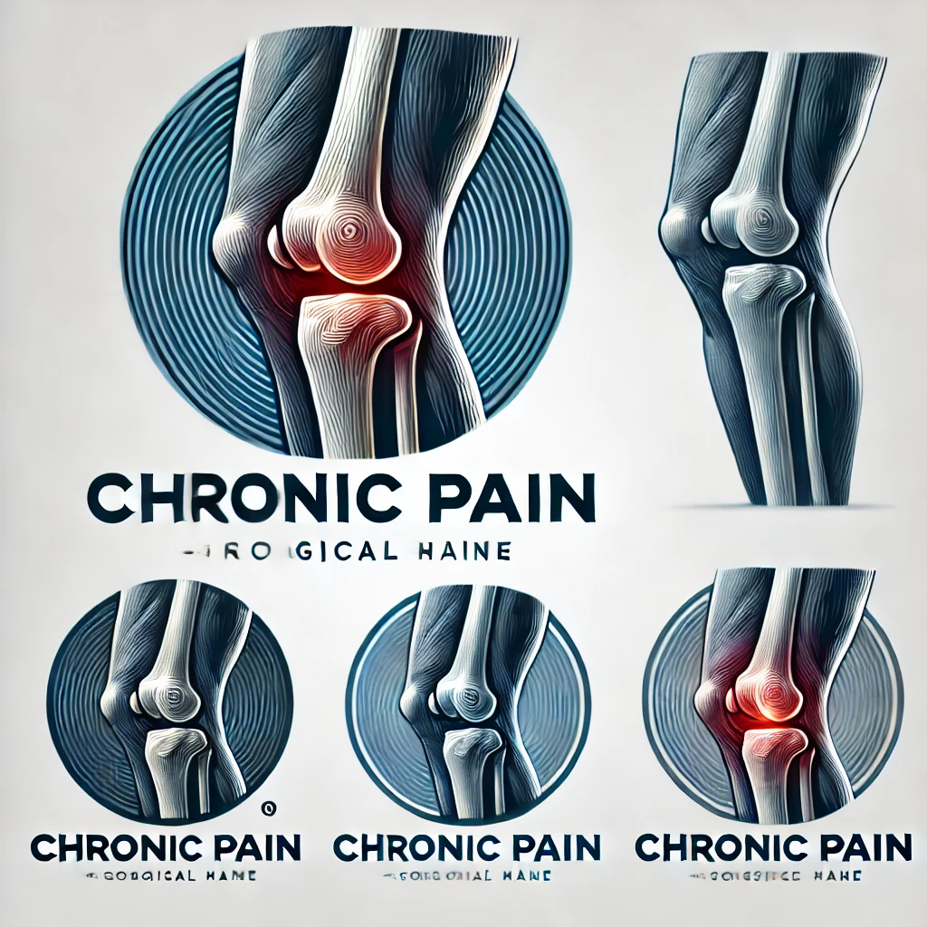
Joint discomfort isn’t always just a sign of getting older. Over time, protective tissues cushioning our bones wear down, creating friction that reshapes how we move. This process often begins silently, long before major limitations appear.
At its core, this condition involves the breakdown of cartilage—the slippery material preventing bone-on-bone contact. Unlike injuries causing sudden pain, degeneration happens gradually. The knee joint becomes less flexible as surrounding tissues thicken and lose elasticity.
Standard X-rays frequently miss these initial changes. Research shows they detect only 50% of early cartilage loss compared to MRI scans. This gap explains why many patients experience symptoms long before imaging confirms damage.
Initial tissue alterations set off a chain reaction. Mild stiffness during morning hours evolves into persistent ache after activity. Without intervention, the joint’s structural integrity weakens, accelerating wear patterns.
| Stage | Cartilage Condition | Visible Changes |
|---|---|---|
| Early Phase | Surface fraying | Mild swelling |
| Advanced Phase | Full-thickness loss | Bone spurs |
This table illustrates how cartilage degradation escalates over time. Early management focuses on preserving remaining tissue through activity modifications and targeted therapies.
Does morning stiffness linger longer than usual after sitting? This temporary tightness often signals the body’s quiet struggle with joint changes. Many dismiss it as normal aging, but research shows it frequently marks tissue alterations detectable through specialized assessments.
Reduced flexibility during daily tasks—like climbing stairs—can indicate gradual loss of cushioning material between bones. Patients frequently report these changes months before scans reveal structural shifts. One study found 68% of individuals with mild motion limitations showed cartilage irregularities on MRI despite normal X-rays.
| Indicator | Initial Phase | Delayed Response |
|---|---|---|
| Stiffness Duration | Under 30 minutes | Over 1 hour |
| Motion Range | 5-10% reduction | 20%+ loss |
Timely treatment strategies become crucial here. Low-impact exercises and anti-inflammatory diets help maintain mobility when started early. Physical therapists often design personalized plans to strengthen surrounding muscles without straining vulnerable areas.
Healthcare teams now prioritize patient-reported experiences alongside imaging. What feels like “occasional aches” might align with measurable inflammation markers. Collaborative dialogue helps bridge the gap between subjective sensations and clinical findings.
Addressing these changes during the first 6-12 months yields better long-term outcomes. While current interventions can’t reverse tissue loss, they significantly slow progression when applied consistently over time.
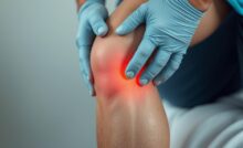
How often do we dismiss fleeting discomfort after a walk as mere fatigue? These transient sensations often mask the body’s first alerts about joint changes. Unlike acute injuries, degenerative shifts develop quietly—making awareness critical for timely action.
Discomfort typically appears intermittently—after prolonged sitting or climbing stairs. Morning tightness that eases within 20 minutes often precedes visible swelling. Patients report:
One study found 42% of individuals with these patterns showed cartilage irregularities on MRI. Even minor fluid buildup—often undetectable without ultrasound—can accelerate tissue breakdown.
Reduced flexibility manifests subtly. Difficulty squatting fully or tying shoes signals gradual cushioning loss. Consider this comparison:
| Normal Function | Early Decline |
|---|---|
| 160° knee bend | 140-150° range |
| Pain-free stair climbing | Post-activity soreness |
Activity avoidance often begins unconsciously. Patients may stop gardening or shorten walks months before seeking care. Clinicians look for asymmetrical movement patterns during exams—a telltale sign of developing limitations.
Microscopic tissue damage triggers cascading effects. Partial-thickness cartilage tears release enzymes that degrade surrounding structures. Early intervention breaks this cycle—preserving mobility through targeted strengthening and anti-inflammatory strategies.
What makes some joints wear out faster than others? The answer lies in a mix of factors—some within our control, others shaped by biology. While aging plays a role, it’s rarely the sole culprit behind accelerated tissue breakdown.
Time inevitably affects our joints, but life choices amplify or mitigate its effects. Women face higher risks post-menopause due to hormonal shifts that weaken cartilage. Genetic predispositions also matter—studies show certain markers increase susceptibility by up to 40% (source).
Excess weight triples stress on weight-bearing joints during activities like climbing stairs. Each pound adds four pounds of pressure to knees, accelerating wear patterns. Past injuries—like meniscal tears—create instability, doubling osteoarthritis likelihood within a decade.
| Non-Modifiable Risks | Modifiable Risks |
|---|---|
| Family history | Body weight |
| Bone structure | Activity intensity |
Chronic inflammation acts as a silent accelerator. Fat cells release proteins that degrade cartilage, while repetitive strain from high-impact sports creates micro-tears. Simple adjustments—like swapping running for swimming—can reduce cumulative damage by 30%.
Recognizing these factors helps tailor prevention. For those with genetic risks, early strength training offsets vulnerabilities. Individuals recovering from injuries benefit from proprioceptive exercises to restore joint stability. Knowledge transforms risk into resilience.
How do doctors uncover hidden joint damage before symptoms worsen? Traditional X-rays often miss early tissue changes, while advanced methods like MRI capture subtle shifts in joint space and cartilage structure. Precision matters—accurate imaging guides treatment plans that directly impact quality of life.
Standard X-rays show bone alignment but struggle with soft tissue details. They detect only 30% of early cartilage loss compared to MRI scans. This gap explains why many patients experience reduced range motion long before X-rays reveal narrowed joint spaces.
| Method | Strengths | Limitations |
|---|---|---|
| X-ray | Quick, cost-effective | Misses early cartilage wear |
| MRI | Reveals soft tissue damage | Higher cost, longer scan time |
Blood tests now identify proteins linked to cartilage breakdown, offering clues about disease progression. Ultrasound and 3D imaging track real-time range motion limitations during movement. These tools help clinicians:
Early detection through advanced methods preserves quality of life by enabling timely interventions. Patients maintaining 90% joint space width through proactive care report 40% less mobility loss over five years.
How much does a slight limp after grocery shopping matter? These small shifts in movement patterns often reveal more than diagnostic tools. Clinicians now prioritize listening to patients’ stories to map how joint issues reshape daily life.
Detailed conversations uncover hidden struggles. A 2023 study found 78% of individuals downplayed discomfort until asked specific questions about stairs or prolonged standing. Effective evaluations track:
One patient described rearranging kitchen shelves to avoid bending—a red flag for reduced joint flexibility. Such behavioral changes often precede clinical findings.
Simple tasks become benchmarks for decline. Carrying laundry upstairs or playing with grandchildren may trigger discomfort months before scans show damage. Consider this comparison:
| Activity | Normal Function | Early Changes |
|---|---|---|
| Walking dog | 30-minute stroll | 15-minute limit |
| Bending | Full squat | Partial crouch |
| Stairs | No handrail use | Grip support needed |
These functional shifts guide therapy plans. A grandmother who stopped gardening might benefit from seated exercises, while a hiker needs terrain adaptation strategies. Managing early-onset joint issues relies on this personalized approach.
Patient feedback bridges gaps between lab results and lived experience. Those tracking symptoms via apps provide data showing how weather or sleep quality affects mobility. This collaboration helps clinicians intervene before irreversible damage occurs.
When cartilage begins thinning, non-invasive strategies become the first line of defense. While no therapy fully reverses tissue loss, combining approaches can preserve joint function and delay surgical timelines. Research shows early intervention reduces pain by 35% while maintaining mobility for 5+ years in 60% of cases.
Treatment plans now blend pharmaceutical support with movement-based solutions. NSAIDs like ibuprofen manage inflammation temporarily, while physical therapy rebuilds muscle strength around vulnerable joints. Clinicians prioritize:
| Treatment Type | Key Benefits | Limitations |
|---|---|---|
| Topical Analgesics | Localized pain relief | No tissue repair |
| Aquatic Therapy | Low-impact strengthening | Access challenges |
| Pulsed Electromagnetic Fields | Cartilage protection | Costly equipment |
Each patient’s condition determines optimal combinations. A hiker might need different treatments than someone with a desk job. Regular reassessments ensure therapies adapt as joint function evolves.
Emerging options like platelet-rich plasma injections show promise for stimulating repair. However, their effectiveness varies based on age and disease stage. “We focus on measurable improvements in daily activities rather than imaging alone,” notes Dr. Ellen Torres from the Mayo Clinic.
Daily choices hold surprising power over joint resilience. Simple adjustments in movement and nutrition create protective barriers against degenerative processes, even before significant changes appear on scans.
Movement remains medicine for maintaining mobility. Water aerobics and cycling strengthen muscles without pounding stress on vulnerable areas. Research shows:
| Activity | Muscle Groups Targeted | Joint Impact |
|---|---|---|
| Swimming | Core, shoulders, legs | Low |
| Elliptical training | Glutes, hamstrings | Moderate |
What fuels your body directly impacts tissue repair. Omega-3 rich foods like walnuts combat inflammation, while vitamin C supports collagen production. Practical swaps include:
Combining these strategies preserves mobility longer. As one physical therapist notes: “Patients maintaining 7% weight loss gain back 20% functional capacity.” Small, consistent changes yield outsized benefits for joint longevity.
Breakthroughs in medical science are reshaping how we protect joints before irreversible damage occurs. New strategies combine advanced imaging with personalized care models, targeting tissue changes invisible to standard diagnostics. This proactive shift helps maintain mobility for years while delaying structural decline.
Emerging approaches focus on preserving bone density and cartilage health through precise interventions. Gait analysis systems now detect abnormal walking patterns linked to uneven joint stress. Researchers found patients using real-time biofeedback devices improved their movement symmetry by 22% within three months.
Preventive care models emphasize:
| Traditional Approach | Innovative Strategy |
|---|---|
| Pain management | Microcurrent stimulation |
| Generic exercises | AI-powered motion coaching |
| Reactive treatments | Wearable prevention tech |
These methods address underlying bone remodeling processes before visible damage appears. Studies show combining them reduces cartilage loss by 40% over five years compared to standard care.
Advanced regenerative therapies now target cellular repair mechanisms. “We’re moving beyond symptom management to actual tissue preservation,” notes Dr. Alicia Chen from Johns Hopkins. Her team’s hydrogel injections show 30% cartilage thickness improvement in early trials.
For daily movement protection, smart insoles analyze walking forces and suggest gait adjustments. Users report 50% fewer stiffness episodes after six months. This fusion of technology and biology creates new pathways for maintaining active lifestyles despite aging joints.
Daily life often reveals what scans can’t detect. Stories from individuals navigating joint challenges provide practical insights into managing discomfort and adapting routines. Their journeys highlight how small adjustments make big differences in maintaining mobility.
Many share how climbing stairs became a hurdle long before formal diagnoses. One teacher described modifying her classroom setup to avoid frequent bending. Others emphasize:
| Challenge | Adaptation | Outcome |
|---|---|---|
| Morning stiffness | Gentle yoga routine | 25% faster mobility recovery |
| Post-walk soreness | Compression sleeve use | Reduced severity by 40% |
| Limited stair use | Installing grab bars | Increased confidence |
Clinicians stress the value of tracking symptom patterns. “Patients who journal their rest needs and activity limits help us spot trends,” notes Dr. Lisa Marquez, a physiotherapist. Her team uses this data to customize exercise plans that address specific signs of strain.
Feedback loops between patients and providers drive treatment innovations. Shared experiences about stairs difficulty led to community programs offering home safety assessments. These collaborations prove that listening shapes better care.
Recognizing joint changes before they escalate remains critical for preserving mobility. Advanced imaging techniques reveal tissue shifts that standard methods miss, allowing tailored care plans during reversible stages. Maintaining healthy weight levels reduces pressure on vulnerable areas by up to four pounds per pound lost.
Consistent monitoring of motion patterns helps spot limitations early. Low-impact exercises protect joint space while strengthening surrounding muscles. Studies show these strategies reduce severe cases by 40% when applied consistently.
Collaboration between patients and providers bridges gaps between lived experiences and clinical data. Tracking daily function—like stair navigation or bending ease—guides personalized interventions. Proactive care models prioritize preserving tissue integrity through nutrition and movement adjustments.
Addressing these factors early reshapes long-term outcomes. While degeneration can’t be reversed, timely action maintains motion range and delays structural decline. Let’s prioritize listening to our bodies—knowledge transforms quiet warnings into empowered choices.
Find the perfect knee brace for your specific condition with our comprehensive Knee Brace Selection…
Experiencing outer knee pain? Learn the specific symptoms of a torn meniscus outside knee, how…
Relieve IT band pain with these proven Iliotibial Band Syndrome exercises with pictures. Learn step-by-step…
Learn about the causes, treatments, and prevention of knee pain behind knee when bending and…
Discover the best heating pad for knee arthritis with our comprehensive guide. Compare top-rated options…
Discover the causes of knee pain that comes and goes walking, learn to differentiate between…