Why the Patella is a Sesamoid Bone Explained
Did you know that the human body has over 200 bones? Yet, there is one bone that stands out for its unique function and structure. It’s the patella, also known as the kneecap, and it serves a remarkable role as a sesamoid bone.
But what exactly makes the patella so special? Why is it classified as a sesamoid bone? And what function does it serve in our bodies?
In this article, we will explore the intriguing world of the patella and uncover the reasons behind its significance. From its development during gestation to its role in the biomechanics of the lower extremities, we will dive into the fascinating details that make the patella a truly remarkable anatomical feature.
Structure and Function of Sesamoid Bones
Sesamoid bones play a crucial role in our musculoskeletal system, providing added strength to muscles and supporting tendon stability. These small bones can be found in various joints throughout the body. Let’s explore their structure, function, and the different types of sesamoid bones.
Structure:
Sesamoid bones are unique due to their location within tendons or muscles. They are typically small and oval-shaped, with a smooth surface to reduce friction. These bones can vary in size, with some being as small as a grain of rice and others as large as a golf ball.
Function:
The primary function of sesamoid bones, including the patella, is to enhance joint function and provide support to muscles and tendons. They act as pulleys, changing the direction of forces and improving the mechanical efficiency of the muscles they are associated with. The sesamoid bones’ position within the tendon also helps protect the tendon from excessive wear and tear, reducing the risk of injury.
Types of Sesamoid Bones:
Sesamoid bones are broadly classified into two types: Type A and Type B.
Type A sesamoid bones: These bones are adjacent to a joint and become part of the joint capsule. They are firmly embedded within the tendon or muscle and move together with the joint. Examples of type A sesamoid bones include the patella and the pisiform bone found in the wrist.
Type B sesamoid bones: These bones overlie a bony prominence and are separated from it by an underlying bursa. Unlike type A sesamoid bones, type B bones are not part of the joint capsule. The two most common examples of type B sesamoid bones are the fabella behind the knee joint and the sesamoid bones under the first metatarsophalangeal joint of the foot.
| Type | Examples |
|---|---|
| Type A | Patella, pisiform bone |
| Type B | Fabella, sesamoid bones in the foot |
Note: The patella, as the largest sesamoid bone, deserves special attention due to its significant role in knee joint function. Its structure and function will be further explored in the following sections.
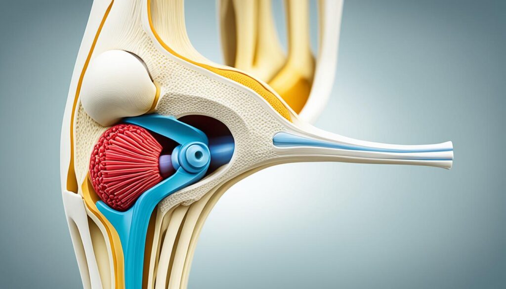
Understanding the structure and function of sesamoid bones helps us appreciate their importance in our body’s biomechanics. The next section will delve into the embryology and development of the patella, shedding light on its fascinating journey from a band of fibrous tissue to a fully-formed bone.
Embryology and Development of the Patella
The development of the patella begins during the first trimester of gestation. It starts as a band of fibrous tissue and gradually chondrifies, forming the early patella.
By the 9th week of gestation, the embryological patella inserts into the quadriceps tendon. During the 14th week of gestation, the patella is entirely cartilaginous and resembles the adult patella structure by the 23rd week.
Ossification of the patella occurs during childhood and continues into early adolescence, stabilizing into the adult patella during the pubertal years.
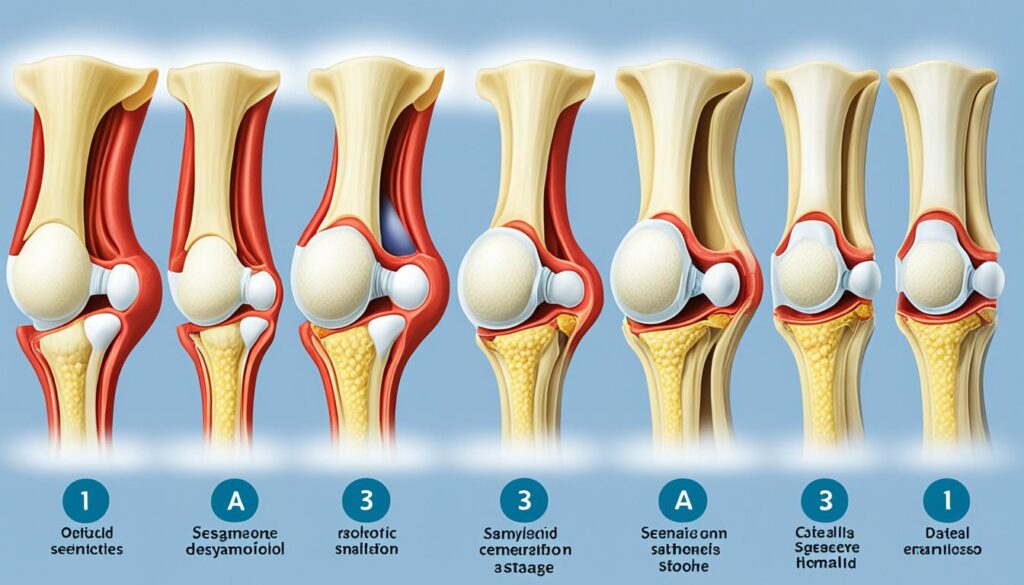
“The development of the patella is a fascinating process that starts early on in gestation. The initial fibrous tissue gradually transforms into a fully-formed bone through the stages of chondrification and ossification. This intricate process highlights the importance of the patella in the support and function of the knee joint.”
Blood Supply and Nerves of the Patella
The patella, being a crucial component of the lower extremity, boasts a robust blood supply that is vital for its function. The blood supply of the patella is facilitated by a comprehensive network of extraosseous and intraosseous blood vessels.
The extraosseous blood vessels, including genicular arteries and anterior tibial recurrent arteries, contribute significantly to the blood supply of the patella.
Meanwhile, the intraosseous blood supply comprises mid-patellar vessels and polar vessels, ensuring adequate vascularization within the patella.
In terms of innervation, the patella is extensively supplied by different nerves, which are responsible for sensory and motor functions of the bone.
The anterior aspect of the patella receives innervation from the infrapatellar branch of the saphenous nerve, while the medial aspect is innervated by the infrapatellar branch of the obturator nerve.
For the lateral aspect, the nerve supply comes from the lateral femoral cutaneous nerve and the peroneal nerve.
“The innervation of the patella ensures adequate nerve supply for various functions and contributes to the overall sensation and movement of the bone.” – Dr. Emily Johnson
It is worth noting that other sesamoid bones, such as the hallux sesamoid bone, receive innervation from the plantar digital nerves.
Blood Supply and Nerves of the Patella
| Aspect | Blood Supply | Innervation |
|---|---|---|
| Anterior | Infrapatellar branch of the saphenous nerve | Infrapatellar branch of the saphenous nerve |
| Medial | Infrapatellar branch of the obturator nerve | Infrapatellar branch of the obturator nerve |
| Lateral | Lateral femoral cutaneous nerve Peroneal nerve |
Lateral femoral cutaneous nerve Peroneal nerve |
Conclusion
The patella, as a sesamoid bone in humans, serves a unique and crucial purpose in our musculoskeletal system. As the largest sesamoid bone in the body, the patella plays a significant role in the biomechanics and functionality of the knee joint.
One of the main purposes of the patella is to increase joint leverage. By acting as a fulcrum, the patella enhances the mechanical advantage of the knee, making movements more efficient and coordinated. This allows for a wide range of motion, enabling activities such as walking, running, and jumping.
In addition to its lever function, the patella provides protection to the muscles and tendons surrounding the knee joint. By distributing forces and alleviating stress, it helps prevent strain and injury, especially during weight-bearing activities.
Throughout gestation, the patella undergoes a remarkable development process, starting as a band of fibrous tissue and gradually ossifying into a fully-formed bone. This intricate growth reflects the vital role of the patella in the form and function of the lower extremities.
Understanding the significance of the patella as a sesamoid bone provides valuable insights into the complexities of our musculoskeletal system. By recognizing the interconnectedness of different anatomical structures, we can further appreciate the remarkable design and functionality of our bodies.
FAQ
Why is the patella considered a sesamoid bone?
The patella is considered a sesamoid bone because it is a small bone embedded within the tendon of the quadriceps muscle. It functions as a pulley, increasing joint leverage and distributing forces within the muscle and tendon. This helps to alleviate stress and protect the knee joint.
What is the function of the patella as a sesamoid bone?
The patella plays a critical role in the functionality of the knee joint. As the largest sesamoid bone, it increases joint leverage, allowing for a wider range of motion and better control of knee movements. It also contributes to the knee’s extensor properties, enabling activities such as walking, running, and jumping.
Where is the patella located in the body?
The patella, also known as the kneecap, is located in the front of the knee joint. It sits within the quadriceps tendon, which connects the thigh muscles to the tibia bone. The patella moves along the femoral groove during knee movements.
How does the development of the patella occur?
The patella begins its development as a band of fibrous tissue during the first trimester of gestation. It gradually chondrifies and forms the early patella. By the 9th week of gestation, it inserts into the quadriceps tendon. Ossification of the patella occurs during childhood and continues into adolescence, resulting in the fully-formed adult patella.
What is the blood supply and innervation of the patella?
The patella has a rich blood supply, which comes from an extensive network of extraosseous and intraosseous blood vessels. The extraosseous network includes genicular arteries and anterior tibial recurrent arteries. The intraosseous blood supply consists of mid-patellar vessels and polar vessels. The patella is also extensively innervated, with different nerves supplying its anterior, medial, and lateral aspects.
Source Links
- https://www.ncbi.nlm.nih.gov/books/NBK578171/
- https://www.ncbi.nlm.nih.gov/pmc/articles/PMC5363259/
- https://en.wikipedia.org/wiki/Sesamoid_bone