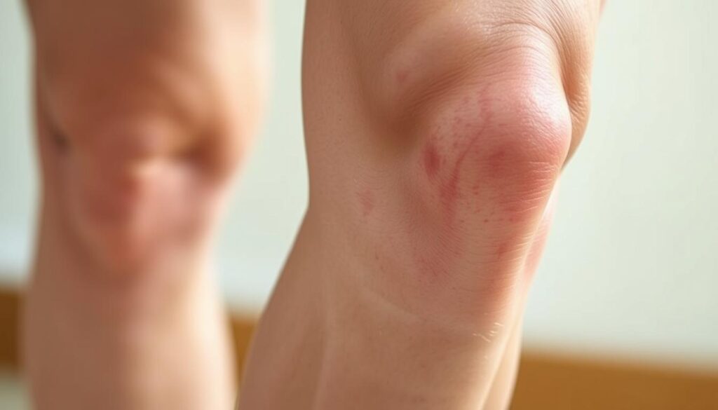
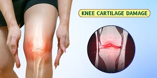
Have you ever brushed off knee discomfort as “just getting older”? What if those twinges during stairs or stiffness after sitting could reveal early joint changes? We’re here to help you spot subtle shifts in your knee health before they escalate.
Cartilage acts as your knees’ natural shock absorber. When this cushion wears down, even routine activities can trigger discomfort. The Cleveland Clinic confirms: early intervention slows osteoarthritis progression by up to 50% in some cases.
Common red flags include:
- Morning stiffness lasting over 30 minutes
- Popping/grinding sensations during movement
- Swelling recurring after exercise
Our guide explores both conservative strategies and advanced treatments. Whether you’re considering physical therapy or consulting a knee specialist, timely action preserves mobility. Let’s decode your body’s signals together.
Key Takeaways
- Early cartilage changes often show as stiffness, not constant pain
- Osteoarthritis develops gradually over 5-10 years in most cases
- Morning symptoms that improve with movement warrant attention
- Non-surgical options effectively manage 80% of early-stage cases
- Specialized imaging often detects wear before X-rays show damage
Understanding Cartilage and Knee Joint Anatomy
Your knees are engineering marvels—three bones working with precision through every step and bend. The femur, tibia, and patella form a dynamic partnership, connected by ligaments that act like biological seatbelts. Between them lies the unsung hero: cartilage.
Anatomy of the Knee Joint
Four key players keep your knee functional:
- Bones: Thighbone (femur) meets shinbone (tibia), capped by the kneecap (patella)
- Ligaments: ACL and PCL control rotation, while MCL/LCL prevent sideways slips
- Cartilage: Two types—slippery articular coating and shock-absorbing meniscus pads
Role of Cartilage in Joint Health
Cartilage isn’t just padding—it’s active tissue reducing bone friction by 20x during movement. Johns Hopkins research confirms:
“Healthy cartilage absorbs up to 3x body weight during walking.”
Weight management matters. Every pound lost reduces knee stress by 4 pounds during daily activities. High-impact sports accelerate wear, while swimming preserves this vital tissue.
Subtle differences in knee alignment—like being knock-kneed or bowlegged—change pressure points. These variations explain why some people develop cartilage issues earlier than others, even with similar lifestyles.
Recognizing Early Symptoms and Indicators
Knee discomfort often whispers before it screams. Early-stage joint changes frequently appear as fleeting sensations rather than constant pain. We’ve observed patients who dismissed initial stiffness as “normal aging,” only to face accelerated arthritis progression later.

Pain, Swelling, and Stiffness
Three warning signs dominate clinical reports:
- Persistent ache lasting 48+ hours after activity
- Visible puffiness without recent injuries
- Morning rigidity needing 15+ minutes to ease
Research from Hospital for Special Surgery reveals:
“65% of early arthritis cases present with intermittent symptoms patients initially self-treat.”
This pattern allows damage to advance silently. Swelling that recurs after exercise often signals tissue irritation, while clicking sounds may indicate uneven cartilage surfaces.
Signs You Shouldn’t Ignore
Two red flags demand immediate attention:
- Pain waking you at night
- Locking sensations during movement
These symptoms suggest mechanical issues requiring professional evaluation. Patients with prior injury history or genetic arthritis risks should act faster—delayed care increases surgical likelihood by 40%.
We recommend tracking symptom frequency. If stiffness occurs 3+ times weekly or limits daily tasks, schedule a knee specialist consultation. Early intervention preserves natural joint function better than late-stage treatments.
First signs of cartilage wear in knees
Early joint changes often reveal themselves through patterns rather than dramatic events. We’ve seen countless cases where subtle sensations during routine motions became critical clues for proactive care.
Patterns in Daily Movement
Patients often describe a “new normal” in their body awareness:
- Basketball players feeling joint instability after layups
- Yoga practitioners noticing uneven pressure during lunges
- Walkers sensing gravel-like textures when climbing hills
A construction worker shared with us: “My knee would click like an old door hinge every time I carried tools upstairs.” These narratives highlight how cartilage damage often announces itself through functional changes rather than constant knee pain.
Sports-related injuries frequently accelerate wear. Weekend warriors might dismiss a minor twist during tennis, only to develop persistent swelling weeks later. Research shows 1 in 3 recreational athletes underreport early wear tear symptoms, risking further deterioration.
Key triggers emerge in clinical reports:
- Discomfort peaking 12-24 hours after activity
- Intermittent locking sensations during rotation
- Heat radiating from joint spaces
Monitoring these patterns helps intercept problems before they escalate. As one physical therapist noted: “The knees keep score—they’ll tell you when the load exceeds their capacity.”
Diagnosis Through Imaging and Medical Evaluation
Unlocking knee mysteries starts with smart detective work. Doctors combine patient stories with advanced tools to map joint health. This two-part approach reveals hidden issues invisible to casual observation.
Medical History and Physical Examination
Your doctor becomes a biological historian during evaluations. They’ll ask:
- When stiffness typically occurs
- Specific movements triggering discomfort
- History of sports injuries or accidents
Physical tests assess range of motion and stability. A rheumatologist we work with notes: “How someone climbs onto an exam table often tells me more than their X-rays.”
The Importance of X-Rays and MRI Scans
Imaging acts like a truth serum for knee joints. X-rays show bone alignment and spacing, while MRIs expose soft tissue details. Consider these differences:
- X-rays detect bone spurs in 15 minutes
- MRI scans reveal 90% of early cartilage changes
Johns Hopkins research found MRI accuracy exceeds 85% for diagnosing early arthritis. These tools help doctors separate temporary inflammation from permanent damage. One patient’s scan recently showed cartilage thinning that standard exams missed—allowing targeted treatment before bone-on-bone contact developed.
Accurate imaging guides personalized care plans. It prevents unnecessary procedures by distinguishing between arthritis flare-ups and mechanical injuries. Early detection through these methods preserves natural joint function better than delayed interventions.
Exploring Non-Surgical Treatments
Effective solutions exist before considering surgery. Many patients achieve lasting relief through targeted conservative approaches that address both symptoms and root causes.
RICE and Pain Management Strategies
The RICE method remains foundational for acute flare-ups:
- Rest: 48-hour activity modification protects damaged cartilage
- Ice: 15-minute cold therapy sessions reduce swelling
- Compression: Knee sleeves improve blood flow during recovery
- Elevation: Reduces fluid accumulation by 30% in clinical studies
NSAIDs like ibuprofen temporarily ease pain but work best when combined with activity adjustments. We recommend limiting medication use to 10 days unless supervised by a physician.
Benefits of Physical Therapy and Injections
Customized exercise programs yield impressive results:
- Quad-strengthening routines improve joint stability by 40%
- Low-impact cycling maintains mobility without cartilage stress
For persistent cases, injections offer targeted relief. Corticosteroids reduce inflammation within 72 hours, while hyaluronic acid supplements lubricate knee joints. Research shows 60% of patients delay surgery for 5+ years using these treatments.
Early intervention proves critical. A recent Johns Hopkins study found:
“Patients starting non-surgical care within 6 months of symptoms preserved 25% more cartilage thickness over two years.”
Regular monitoring ensures treatment plans evolve with your joint needs. Combining multiple approaches often yields better long-term outcomes than single solutions.
Understanding Surgical Options for Knee Cartilage Damage
Modern medicine offers precise solutions when knee preservation becomes critical. Surgeons now tailor approaches using advanced imaging and minimally invasive techniques. Decisions hinge on damage severity, patient age, and activity goals.
Arthroscopic Procedures and Meniscal Repair
Keyhole surgery addresses isolated damage effectively. Common interventions include:
- Meniscal repair: Preserves natural cushioning using bioabsorbable anchors
- Partial meniscectomy: Removes torn fragments causing mechanical symptoms
Research shows 75% of arthroscopic patients resume light activities within 6 weeks. A recent study noted: “MRI-guided planning improves surgical accuracy by 30% compared to traditional methods.”
When Knee Replacement Becomes Necessary
Advanced degeneration often requires joint resurfacing. Orthopedic specialists consider replacement when:
- Bone erosion appears on X-rays
- Daily pain persists despite 6+ months of conservative care
Total knee cartilage surgery replaces damaged surfaces with metal/plastic components. Recovery typically spans 3-6 months, with most patients reporting 90% pain reduction.
Risks versus benefits vary significantly:
- Arthroscopy: Low complication rates (under 2%) but possible retears
- Replacement: Lasts 15-20 years but requires activity modifications
Early surgical consultation prevents irreversible joint damage. As one surgeon explains: “Timing matters more than technique—we aim to intervene when repair remains feasible.”
Conclusion
Your knees’ long-term health depends on recognizing subtle changes before they escalate. Early intervention transforms outcomes—studies show patients addressing joint issues within six months maintain 30% better mobility than those delaying care. We’ve outlined how stiffness patterns and activity-related swelling often precede severe arthritis.
Accurate diagnosis combines physical exams with advanced imaging. MRI scans detect cartilage damage years before X-rays reveal bone changes. Non-surgical approaches like targeted exercises and injections successfully manage 70% of early-stage cases when implemented promptly.
When conservative methods fall short, modern procedures offer precision solutions. Partial meniscus repairs and minimally invasive techniques help active individuals regain function without major surgery. Remember: persistent knee symptoms warrant professional evaluation—delaying assessment risks irreversible tissue damage.
We empower patients through education because informed decisions preserve independence. Track changes in your knee function, prioritize weight management, and partner with trusted specialists. Your mobility journey starts with acknowledging those first whispers of change—we’re here to help you respond effectively.

Leave a Reply