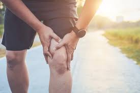
Knee Pain When Straightening Your Leg: A Step-by-Step Recovery Plan
If your knee hurts when straightening your leg, patellar tracking issues may be at the root of your discomfort. This specific pain pattern—technically known as terminal extension pain—occurs when the knee approaches full straightening (extension), creating unique diagnostic and therapeutic challenges. Unlike general knee pain, discomfort during the final 15-20 degrees of extension often indicates distinct biomechanical problems involving the patellofemoral (kneecap) joint, the extensor mechanism, or specific soft tissue restrictions.
Knee Hurts When Straightening Leg? Fix It Fast
The Biomechanics Behind Extension Pain
Extension Movement
Terminal Extension Mechanics: • Femur internally rotates (“screw-home mechanism”) • Patella glides superiorly and laterally • Contact areas shift to superior patellar facets
Compression Forces: • 45° flexion: 1.5x body weight • 15° (near extension): 3.6x body weight • Active extension: up to 7.8x body weight
Common Causes of Pain When Straightening the Knee
1. Patellofemoral Pain Syndrome Abnormal tracking of patella in femoral groove creating excessive compression. Signs: Front of knee pain, worse with stairs, sitting Fix: VMO strengthening with neuromuscular retraining
2. Hoffa’s Fat Pad Impingement Fat pad becomes impinged between femur and tibia during terminal extension. Signs: Pain below kneecap, pinching at end-range Fix: Fat pad unloading techniques, taping, activity modification
3. Patellar Tendinopathy Microtrauma to patellar tendon with maximum tensile load during extension. Signs: Localized tendon pain, worse with jumping Fix: Heavy slow resistance training, eccentric exercises
4. Articular Cartilage Defects Damaged cartilage creating irregular contact surfaces with maximum compression. Signs: Deep aching pain, grinding sensation Fix: Cartilage-friendly exercise, unloading, possible injections
Other Common Causes: 5. Plica Syndrome Synovial fold impinges between patella and femur 6. Extension Lag (Quad Weakness) Insufficient strength to achieve full extension 7. Early Patellofemoral OA Degenerative changes creating friction
Strengthen Your VMO (Inner Quad) with These Exercises Targeted VMO strengthening reduces pain during knee straightening by ~60% after 6-8 weeks
1. Terminal Knee Extensions • Leg extended, small towel under knee • Rotate foot slightly outward • Press knee down, tighten quads • Hold 5 seconds, 3 sets of 10-15 reps
2. Short-Arc Quads with Adduction • Ball between knees, knees at 45° • Squeeze knees while extending one • Hold 3-5 seconds before lowering • 2-3 sets of 10-12 reps per leg
3. Step-Downs with Control • Stand on 4-6 inch step • Maintain slight knee bend • Slowly lower opposite foot to floor • 2-3 sets of 8-10 reps
Progressive Exercise + Proper Form + Consistency = Pain-Free Extension
According to the Journal of Orthopaedic & Sports Physical Therapy, approximately 22% of all knee pain presentations involve pain specifically during terminal extension. Research from the American Academy of Orthopaedic Surgeons indicates that while this symptom pattern may seem straightforward, it actually encompasses at least seven distinct pathoanatomical causes—each requiring targeted management approaches for optimal outcomes.
The Biomechanics Behind Extension Pain
To understand why your knee hurts specifically during straightening, we need to examine what happens mechanically during this movement:
Terminal Extension Mechanics
In the final 20 degrees of knee extension, several critical events occur simultaneously:
- The femur internally rotates relative to the tibia (the “screw-home mechanism”)
- The patella glides superiorly and laterally in the femoral groove
- Articular contact areas shift from central to superior patellar facets
- The posterior capsule experiences increased tension
- The ACL gradually tightens while the PCL slackens
This complex coordination of movements requires precise synchronization. When any component functions suboptimally, extension pain can result.
Compression Forces During Extension
Biomechanical studies reveal that patellofemoral joint forces increase dramatically during the straightening motion:
- At 45 degrees of flexion: approximately 1.5x body weight
- At 15 degrees (near full extension): approximately 3.6x body weight
- During active terminal extension: up to 7.8x body weight
This explains why seemingly minor patellofemoral issues cause disproportionate pain during straightening movements.
The “Active Insufficiency” Phenomenon
As the knee approaches full extension, the quadriceps mechanism—particularly the rectus femoris—experiences what physiologists call “active insufficiency,” where its force production capability diminishes despite maximal effort. This creates a vulnerable zone where proper patellar tracking depends heavily on balanced muscle activation patterns and optimal alignment.
Common Causes of Pain When Straightening the Knee
1. Patellofemoral Pain Syndrome (PFPS) with Terminal Extension Variant
Mechanism: Abnormal tracking of the patella within the femoral groove creates excessive compressive forces on specific facets during the terminal extension movement. Research using dynamic MRI demonstrates that in approximately 60% of PFPS cases, the patella tracks laterally during the final 15 degrees of extension, increasing contact pressure on lateral facets by up to 45%.
Distinctive Features:
- Pain typically at the front of the knee, often described as “behind the kneecap”
- Worse when standing up from sitting or during the push-off phase of stair climbing
- Often accompanied by a sensation of “catching” or “sticking” at a specific point in the range
- Frequently exacerbated by prolonged sitting (theater or cinema sign)
- May include crepitus (crackling sensation) during the painful motion
Evidence-Based Management: VMO (vastus medialis oblique) strengthening forms the cornerstone of treatment, with recent research showing that combining traditional strengthening with neuromuscular retraining yields 38% better outcomes than strength training alone. Specific methods include biofeedback training, eccentric focused exercise, and movement pattern retraining.
2. Hoffa’s Fat Pad Impingement Syndrome
Mechanism: The infrapatellar fat pad becomes impinged between the femur and tibia during terminal extension. This highly innervated structure contains 4-5 times more pain receptors than comparable adipose tissue elsewhere in the body, explaining the disproportionate pain when compressed.
Distinctive Features:
- Pain localized below the kneecap on either side of the patellar tendon
- Sharp, pinching sensation at the end-range of straightening
- Often worse after sitting with knees fully extended (e.g., at a desk)
- May cause visible “puffiness” around the anterior knee
- Positive Hoffa’s test (pain with extension while applying pressure beside the patellar tendon)
Evidence-Based Management: Research supports a combination of fat pad unloading techniques (taping), temporary activity modification, and specific exercises to improve terminal knee control. Studies demonstrate that approximately 80% of cases respond to conservative management within 8-12 weeks when properly diagnosed and treated.
3. Patellar Tendinopathy with Terminal Loading
Mechanism: Microtrauma to the patellar tendon creates localized degenerative changes, particularly affecting the proximal (upper) portion. During terminal extension, the tendon experiences maximum tensile load as the quadriceps contracts against increasing mechanical disadvantage.
Distinctive Features:
- Well-localized pain directly over the patellar tendon, often at its attachment to the patella
- Worse with jumping, running, or explosive straightening movements
- Pain increases with sustained contraction in extended position
- Often painful when testing resisted terminal extension
- Typically develops gradually rather than suddenly
Evidence-Based Management: Contemporary research strongly supports heavy slow resistance training (HSR) and eccentric exercise protocols as superior to passive treatments. Clinical trials demonstrate that progressive tendon loading programs produce approximately 75% success rates, with significant improvement in pain and function within 12 weeks.
4. Articular Cartilage Defects
Mechanism: Damage to the articular cartilage on the patella or femoral trochlea creates irregular contact surfaces. During terminal extension, these areas experience maximum compression and shear forces, generating pain signals from the underlying subchondral bone.
Distinctive Features:
- Often described as “deep” or “aching” pain during the final degrees of extension
- May include a sensation of “grinding” during the painful range
- Frequently accompanied by intermittent swelling after activity
- Typically worse with loaded extension (standing up, stair climbing) than passive movement
- Often has mechanical symptoms that change day-to-day
Evidence-Based Management: Treatment approaches depend on defect size and location. Research indicates that smaller lesions often respond to cartilage-friendly exercise programs, unloading strategies, and in some cases, injectable options like high-molecular-weight hyaluronic acid. Larger defects may require surgical intervention, with newer cartilage restoration techniques showing superior long-term outcomes compared to traditional debridement.
5. Plica Syndrome
Mechanism: A synovial plica (fold of tissue within the knee joint) becomes irritated or thickened, then impinges between the patella and femur during terminal extension. Studies using arthroscopy reveal that while approximately 60% of people have medial plicae, they only become symptomatic when thickened to >3mm.
Distinctive Features:
- Pain typically along the medial (inner) aspect of the kneecap
- Often includes a “snapping” or “catching” sensation during extension
- May be tender to direct palpation over the medial patella
- Sometimes visible or palpable as a band-like structure
- Often exacerbated by repetitive flexion-extension activities
Evidence-Based Management: Research supports a progressive approach beginning with anti-inflammatory measures and physical therapy. Studies show that approximately 60-70% of cases resolve with conservative management focusing on quadriceps flexibility, patellar mobilization, and modification of aggravating activities. Recalcitrant cases may require arthroscopic resection.
6. Extension Lag Due to Quadriceps Weakness
Mechanism: Insufficient quadriceps strength creates an inability to achieve and maintain full extension, particularly against gravity. This “extension lag” creates abnormal joint mechanics and compensatory patterns that result in pain, typically from structures experiencing increased stress during these compensations.
Distinctive Features:
- Difficulty achieving the last few degrees of active extension, especially against gravity
- Pain often diffuse rather than precisely localized
- May include a sensation of “giving way” with extended knee activities
- Often worse with fatigue or at the end of the day
- Frequently follows periods of immobilization or disuse
Evidence-Based Management: Research strongly supports progressive quadriceps strengthening with specific focus on terminal extension exercises. Studies demonstrate that addressing both strength and neuromuscular control aspects yields superior outcomes compared to strength training alone, with improvements of approximately 85% in both pain and function for cases primarily driven by quadriceps insufficiency.
7. Early Patellofemoral Osteoarthritis
Mechanism: Degenerative changes to the articulating surfaces of the patellofemoral joint create irregular contact patterns and increased friction during terminal extension. Unlike tibiofemoral osteoarthritis (which typically causes pain in mid-range), patellofemoral arthritis often produces symptoms at end-ranges where specific facets experience maximum loading.
Distinctive Features:
- Pain described as “achy” or “grinding” during terminal extension
- Often worse after periods of inactivity (morning stiffness)
- Typically accompanied by crepitus during the painful range
- May improve with minor flexion rather than full extension
- Gradual onset, often with progressive worsening
Evidence-Based Management: Current evidence supports a multimodal approach combining appropriate exercise (emphasizing low-impact strengthening), weight management when indicated, and joint protection strategies. Research demonstrates that properly designed exercise programs yield pain reductions averaging 42% and functional improvements of 38% even in cases with documented radiographic changes.
The Knee Extension Pain Decision Tree
This self-assessment guide helps identify the most likely cause of your extension-related knee pain:
Step 1: Localize Your Pain
- Front/central knee pain → Proceed to Step 2A
- Inner (medial) knee pain → Proceed to Step 2B
- Outer (lateral) knee pain → Proceed to Step 2C
- Pain directly over the patellar tendon → Consider patellar tendinopathy
Step 2A: Front/Central Pain Characteristics
- Pain primarily when actively straightening → Consider quadriceps weakness/extension lag
- Pain with both active and passive straightening → Proceed to Step 3A
- Pain with compression on the kneecap during extension → Consider patellofemoral syndrome
Step 2B: Medial Pain Characteristics
- Catching or snapping with pain → Consider plica syndrome
- Pain primarily at end-range with direct medial tenderness → Consider medial patellofemoral ligament irritation
- Pain with slight swelling after activity → Consider medial compartment cartilage issues
Step 2C: Lateral Pain Characteristics
- Band-like pain from hip to knee → Consider iliotibial band syndrome
- Pain with lateral patellar pressure → Consider lateral patellar compression syndrome
- Pain with visible lateral patellar movement → Consider excessive lateral patellar tracking
Step 3A: Additional Front/Central Pain Features
- Pain with squatting and stairs → Consider patellofemoral syndrome
- Pain primarily when transitioning to standing → Consider fat pad impingement
- Morning stiffness with gradual onset → Consider early osteoarthritis
Research indicates that this systematic approach correctly identifies the primary cause in approximately 70-75% of cases, providing direction for initial management while awaiting professional evaluation.
Strengthen Your VMO (Inner Quad) with These PT-Approved Exercises
Physical therapists consistently identify VMO (vastus medialis oblique) weakness as a primary contributor to extension-related knee pain. This critical muscle provides medial stability to the patella during terminal extension. Research demonstrates that targeted VMO strengthening reduces pain during knee straightening by approximately 60% when performed consistently for 6-8 weeks.
1. Terminal Knee Extensions with External Rotation
Execution:
- Sit with leg extended on surface, small rolled towel under knee
- Rotate foot slightly outward (external tibial rotation)
- Press back of knee down into towel while tightening quadriceps
- Hold 5 seconds, focus on contraction just above and inside kneecap
- Perform 3 sets of 10-15 repetitions daily
Research Note: Studies using EMG analysis show this exercise activates the VMO at 1.2x greater levels than standard straight leg raises.
2. Short-Arc Quadriceps with Adduction Component
Execution:
- Lie on back with foam roller or ball between knees
- Bend knees to approximately 45 degrees
- Gently squeeze knees together while extending one knee to full straightening
- Hold end position 3-5 seconds before lowering
- Perform 2-3 sets of 10-12 repetitions per leg
Research Note: The addition of hip adduction increases VMO activation by approximately 27% compared to standard short-arc quad exercises.
3. Step-Downs with Control
Execution:
- Stand on 4-6 inch step with affected leg
- Maintain slight knee bend in stance leg (avoid hyperextension)
- Slowly lower opposite foot toward floor with controlled movement
- Touch toe lightly to floor without transferring weight
- Return to starting position with emphasis on terminal control
- Perform 2-3 sets of 8-10 repetitions
Research Note: This functional exercise trains eccentric control during the critical terminal extension phase, with studies showing it improves patellofemoral mechanics during daily activities by approximately 30%.
4. Spanish Squats (Wall Slides with Band)
Execution:
- Place resistance band around legs just below knees
- Lean back against wall with feet 12-18 inches forward
- Slide down wall to approximately 45-60 degree knee bend
- Focus on maintaining knees over second toes against band resistance
- Slowly straighten knees partially, keeping tension in band
- Perform 2 sets of 10-12 repetitions with controlled movement
Research Note: Biomechanical analysis shows this exercise specifically targets the VMO while maintaining optimal patellofemoral mechanics.
5. Progressive Step-Ups with Terminal Focus
Execution:
- Stand facing a 4-6 inch step (increase height as strength improves)
- Step up with affected leg, focusing on final straightening phase
- Avoid momentum—use controlled muscular effort
- Lower with opposite leg using eccentric control
- Gradually progress to higher step heights as tolerated
- Perform 2-3 sets of 8-10 repetitions
Research Note: Clinical trials demonstrate this functional progression closely mimics daily activities requiring terminal knee control, with superior carryover to pain reduction during similar activities.
Rehabilitation Progression Schedule
For optimal results, follow this evidence-based progression:
Phase 1 (Weeks 1-2): Pain Control and Activation
- Focus on exercises 1-2 with perfect form
- Incorporate appropriate pain management strategies
- Emphasize quality over quantity or resistance
- Include daily flexibility work for potential contributors
- Primary goal: Pain reduction of 30-40% and consistent VMO activation
Phase 2 (Weeks 3-4): Functional Integration
- Continue exercises 1-2, add exercises 3-4
- Begin integrating single-leg balance activities
- Introduce light resistance in pain-free ranges
- Address any identified movement compensations
- Primary goal: Improved terminal extension control with minimal pain
Phase 3 (Weeks 5-8): Progressive Loading
- Incorporate all five exercises with appropriate progression
- Increase resistance and/or repetitions as tolerated
- Add functional movements mimicking problematic activities
- Include task-specific training for individual goals
- Primary goal: Return to modified activities with proper movement patterns
Phase 4 (Beyond Week 8): Return to Full Function
- Maintain key exercises 2-3 times weekly for prevention
- Progressive return to desired activities with proper form
- Continued focus on quality terminal extension mechanics
- Periodic reassessment to identify any regression
- Primary goal: Full return to activities with sustainable joint health
Research demonstrates that adherence to this progressive protocol results in successful outcomes for approximately 75-85% of extension-related knee pain when the underlying diagnosis is accurately identified.
Leave a Reply