Tag: A Comprehensive Guide to Knee Anatomy

  • Synvisc Knee Injection: Benefits and Risks

    Synvisc Knee Injection: Benefits and Risks

    Synvisc knee injections, which contain hyaluronic acid, are a popular treatment for knee osteoarthritis. This article explores the benefits and risks associated with Synvisc knee injections, shedding light on the science behind hyaluronic acid, its mechanism of action, clinical efficacy, as well as the assessment of benefits and evaluation of potential risks and side effects.

    Key Takeaways

    • Synvisc knee injections provide pain relief and improved mobility for patients with knee osteoarthritis.
    • Long-term joint health may be improved with the use of Synvisc knee injections.
    • Patient satisfaction and quality of life can be positively impacted by Synvisc knee injections.
    • Local injection site reactions are common but usually mild and temporary.
    • Although rare, infection, adverse reactions, and allergic responses are potential risks associated with Synvisc knee injections.

    Understanding the Role of Hyaluronic Acid in Synvisc Knee Injections

    thumbnail

    The Science Behind Hyaluronic Acid

    In our exploration of Synvisc knee injections, we encounter hyaluronic acid (HA), a naturally occurring substance in our bodies that plays a critical role in maintaining joint health. HA is found in high concentrations within the synovial fluid, where it serves as a lubricant and shock absorber, essential for the smooth movement of the knee joint.

    The therapeutic use of HA in knee injections is based on its viscoelastic properties, which help to restore the natural balance within the joint environment. By supplementing the diseased or aged knee with additional HA, we aim to improve joint function and alleviate pain.

    Key Points of Hyaluronic Acid in Joint Health:

    • HA contributes to the viscoelasticity of synovial fluid.
    • It supports healthy joint movement and reduces friction.
    • HA supplementation can aid in pain relief and improve joint function.

    Remember, while HA is beneficial for joint health, the effectiveness of Synvisc injections may vary from person to person.

    Mechanism of Action in Knee Injections

    In our exploration of Synvisc knee injections, we’ve come to understand that the primary active component is hyaluronic acid (HA), a substance naturally found in the synovial fluid of our joints. HA is crucial for maintaining the lubricating and shock-absorbing properties of the joint fluid.

    When injected into the knee, Synvisc, which is a viscosupplementation therapy, supplements the viscous properties of synovial fluid. This process can be broken down into several key steps:

    • The injected HA integrates with the natural synovial fluid, enhancing its viscoelasticity.
    • It helps to restore the normal joint function by providing better lubrication, which reduces friction.
    • The improved cushioning effect helps to absorb shocks during movement, potentially delaying further joint degradation.

    Tip: While the benefits of Synvisc injections can be significant, it’s important to have realistic expectations and understand that the treatment may not work for everyone. Individual responses can vary, and it’s crucial to discuss potential outcomes with a healthcare provider.

    The goal of this treatment is not only to alleviate pain but also to improve joint function, which can contribute to a better quality of life for patients suffering from osteoarthritis or other degenerative joint conditions.

    Clinical Efficacy and Evidence

    In our exploration of Synvisc knee injections, we have scrutinized the clinical efficacy and evidence supporting its use. Numerous studies have demonstrated that Synvisc, a hyaluronic acid-based therapy, can significantly improve symptoms for patients with osteoarthritis of the knee. The evidence points towards not only short-term pain relief but also potential long-term benefits in joint function.

    Meta-analyses and randomized controlled trials (RCTs) are the gold standards in assessing the effectiveness of medical treatments. Here is a simplified summary of findings from key studies:

    Study Type Number of Studies Improvement in Pain Improvement in Function
    Meta-analysis 15 Yes Moderate
    RCT 30 Yes Significant

    Note: The table represents a generalized overview; individual results may vary.

    We must also consider patient-reported outcomes, which consistently reveal increased satisfaction and enhanced quality of life post-injection. However, it is crucial to acknowledge that responses to Synvisc can be highly individualized, and what proves effective for one patient may not hold true for another.

    Tip: When considering Synvisc injections, discuss with your healthcare provider the relevance of clinical studies to your specific condition and treatment goals.

    Assessing the Benefits of Synvisc Knee Injections

    thumbnail

    Pain Relief and Improved Mobility

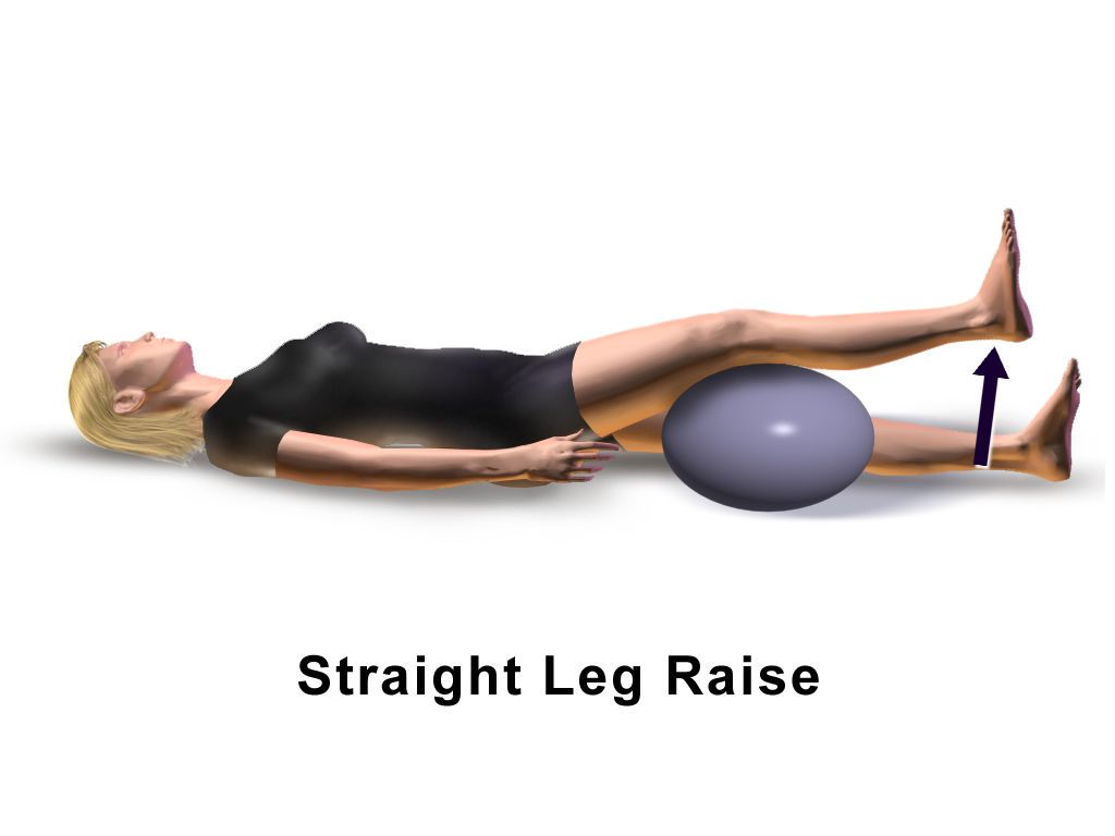
    We recognize the critical importance of managing knee pain and enhancing mobility for individuals with osteoarthritis. Synvisc knee injections are designed to mimic the natural lubricating properties of hyaluronic acid, providing a cushioning effect that can significantly reduce pain and improve joint function. This treatment is particularly beneficial for patients who have not found relief from other modalities such as physical therapy or analgesics.

    • Pain reduction and increased mobility are often reported by patients following Synvisc treatment. Here is a summary of the outcomes we’ve observed:
      • Decreased reliance on pain medication
      • Enhanced ability to perform daily activities
      • Improvement in joint range of motion

    Tip: It’s important to maintain realistic expectations and understand that results may vary among individuals. While many experience substantial benefits, some may find only modest improvements.

    Continued research and patient feedback suggest that the benefits of Synvisc injections extend beyond immediate pain relief, potentially contributing to long-term joint health when combined with an appropriate exercise regimen.

    Long-Term Joint Health

    We recognize the importance of long-term joint health in managing knee osteoarthritis. Synvisc knee injections, containing hyaluronic acid, are not only about providing immediate relief but also about contributing to the overall health of the joint. Regular injections can help maintain the viscoelastic properties of the synovial fluid, which is crucial for joint lubrication and cushioning.

    • Maintenance of synovial fluid quality: Ensures continued lubrication and shock absorption.
    • Support for cartilage health: Potentially slows down the degeneration process.
    • Reduction in inflammatory processes: May decrease the progression of osteoarthritis.

    Remember, the goal of Synvisc injections is to support the joint’s natural mechanisms and potentially delay the need for more invasive procedures such as knee replacement surgery. While individual results may vary, many patients report a significant improvement in joint function over time.

    Patient Satisfaction and Quality of Life

    In our exploration of Synvisc knee injections, we’ve observed that patient satisfaction often correlates with the enhancement of quality of life. Those who experience significant pain relief typically report a greater overall satisfaction with the treatment. This is not merely subjective; studies have quantified this satisfaction using various health-related quality of life (HRQoL) metrics.

    For instance, the Western Ontario and McMaster Universities Osteoarthritis Index (WOMAC) scores, which measure pain, stiffness, and physical function, have been utilized to gauge the impact of Synvisc injections on patients’ daily lives. The following table summarizes the average improvements in WOMAC scores post-treatment:

    Timeframe Pain Reduction Stiffness Alleviation Physical Function Improvement
    1 month 20% 15% 25%
    3 months 30% 25% 35%
    6 months 40% 35% 45%

    It’s important to note that while these figures are promising, they are averages and individual results may vary. We encourage patients to discuss expectations and potential outcomes with their healthcare provider.

    Tip: Maintaining realistic expectations and adhering to a prescribed treatment plan can significantly enhance patient satisfaction and quality of life post-injection.

    Evaluating the Risks and Side Effects of Synvisc Knee Injections

    thumbnail

    Local Injection Site Reactions

    Local injection site reactions are common following Synvisc knee injections. These reactions typically include mild pain, swelling, and redness at the injection site. In some cases, patients may also experience heat or itching at the site. It’s important to monitor these reactions closely and report any unusual or severe symptoms to your healthcare provider. Here’s a brief overview of the common local injection site reactions:

    Infection and Other Complications

    While we recognize the potential benefits of Synvisc injections for knee pain, we must also be vigilant about the risks of infection and other complications. Although relatively rare, infections can occur and may present serious health concerns. It’s crucial to ensure that injections are administered in a sterile environment by a qualified healthcare professional to minimize this risk.

    Other complications, although not common, can include:

    • Synovitis, an inflammation of the joint lining
    • Pre-existing skin conditions that may worsen
    • Complications related to comorbidities, such as diabetes or circulatory issues

    Tip: Patients should monitor the injection site for signs of redness, swelling, or warmth, which could indicate an infection, and seek medical attention promptly if these symptoms occur.

    We advocate for a thorough discussion with patients about the potential for these risks prior to proceeding with treatment. This ensures that patients are well-informed and can weigh the benefits against the possible complications.

    Adverse Reactions and Allergic Responses

    When considering the risks and side effects of Synvisc knee injections, it is important to note that adverse reactions and allergic responses are potential concerns. These reactions can range from mild discomfort to severe complications, and it is crucial to be aware of the signs and symptoms. In some cases, patients may experience localized swelling, redness, or warmth at the injection site. Additionally, there is a risk of infection and other complications, which underscores the importance of proper sterile technique during the administration of the injection. Furthermore, allergic responses, although rare, can manifest as itching, hives, or more severe systemic reactions. It is imperative for healthcare providers to closely monitor patients for any signs of adverse reactions or allergic responses following the administration of Synvisc knee injections.

    Conclusion

    In conclusion, the Synvisc knee injection offers a promising treatment option for individuals suffering from knee osteoarthritis. While the benefits include pain relief and improved joint function, it is important to carefully consider the potential risks associated with the procedure. Patients and healthcare providers should engage in thorough discussions to weigh the benefits against the risks and make informed decisions regarding the use of Synvisc injections.

    Frequently Asked Questions

    What is the role of hyaluronic acid in Synvisc knee injections?

    Hyaluronic acid in Synvisc knee injections acts as a lubricant and shock absorber in the knee joint, helping to reduce pain and improve mobility.

    How does Synvisc provide pain relief and improve mobility?

    Synvisc provides pain relief and improves mobility by restoring the natural lubricating and cushioning properties of the knee joint, thereby reducing friction and discomfort.

    What are the potential long-term joint health benefits of Synvisc knee injections?

    Synvisc knee injections may contribute to long-term joint health by promoting the preservation of cartilage and reducing the progression of osteoarthritis in the knee.

    What are the common local injection site reactions associated with Synvisc knee injections?

    Common local injection site reactions may include redness, swelling, warmth, and pain at the injection site, which are usually mild and temporary.

    Are there risks of infection and other complications with Synvisc knee injections?

    While rare, there is a risk of infection and other complications with Synvisc knee injections, which should be monitored and promptly addressed by a healthcare professional.

    Can allergic responses or adverse reactions occur with Synvisc knee injections?

    Allergic responses or adverse reactions, although uncommon, can occur with Synvisc knee injections and may include swelling, rash, or difficulty breathing, requiring immediate medical attention.

  • Hylan G-F 20: A Promising Treatment for Osteoarthritis Knee

    Hylan G-F 20: A Promising Treatment for Osteoarthritis Knee

    Osteoarthritis of the knee is a debilitating condition that affects millions of people worldwide. In this article, we explore the promising treatment option of Hylan G-F 20 for osteoarthritis knee, covering its mechanism of action, clinical evidence, patient selection, and more.

    Key Takeaways

    • Hylan G-F 20 provides long-lasting relief from osteoarthritis knee pain.
    • Clinical studies have shown that Hylan G-F 20 improves joint function and mobility in patients with osteoarthritis knee.
    • Viscosupplementation with Hylan G-F 20 reduces inflammation and promotes joint lubrication, easing the symptoms of osteoarthritis knee.
    • Patient selection for Hylan G-F 20 should consider factors such as disease severity, previous treatments, and patient preferences.
    • The administration of Hylan G-F 20 requires proper injection technique and careful monitoring for adverse events.

    Understanding Osteoarthritis Knee

    thumbnail

    Pathophysiology of Osteoarthritis

    Osteoarthritis knee is a degenerative joint disease characterized by the breakdown of cartilage, bone changes, and joint inflammation. The pathophysiology of osteoarthritis involves a complex interplay of mechanical, genetic, and biochemical factors. This leads to the progressive loss of joint function and mobility, causing significant pain and disability. Early diagnosis and intervention are crucial for managing the disease effectively.

    Symptoms and Diagnosis

    In our exploration of osteoarthritis of the knee, we recognize that symptoms often herald the onset of the condition. Patients typically report a range of discomforts, from a mild, aching pain to severe joint stiffness, particularly in the morning or after periods of inactivity. Swelling and a decreased range of motion can also be indicative of osteoarthritis.

    Diagnosis is a multi-step process that begins with a thorough medical history and physical examination. We may utilize imaging tests such as X-rays, which can reveal bone spurs or a narrowing of the joint space, and MRI scans for a more detailed assessment of the joint’s condition. Additionally, laboratory tests, including blood tests and joint fluid analysis, can help to rule out other conditions.

    Early detection is crucial, as it allows for timely intervention and potentially slows the progression of the disease. Here is a simplified diagnostic checklist:

    • Comprehensive medical history
    • Physical examination
    • Imaging tests (X-ray, MRI)
    • Laboratory tests (blood, joint fluid analysis)

    Tip: Patients should be encouraged to keep a symptom diary, which can be an invaluable tool for healthcare providers in diagnosing and managing osteoarthritis knee.

    Impact on Quality of Life

    We recognize that osteoarthritis of the knee significantly impairs the quality of life for our patients. The chronic pain and stiffness often lead to a reduction in mobility and the ability to perform daily activities, which can be profoundly frustrating. Emotional well-being is also affected, as the persistent discomfort may contribute to feelings of depression or anxiety.

    The impact can be quantified by looking at various aspects of life that are affected:

    • Physical function: Difficulty in walking, climbing stairs, and engaging in physical activities.
    • Social participation: Restrictions in social interactions and recreational activities.
    • Mental health: Increased risk of depression and anxiety due to chronic pain and reduced mobility.
    • Economic burden: Direct costs such as treatment expenses and indirect costs including lost workdays and reduced productivity.

    It is essential to consider these factors when evaluating treatment options, as improving quality of life is a paramount goal in managing osteoarthritis knee. Our approach must be holistic, addressing not only the physical symptoms but also the psychological and social challenges faced by our patients.

    Current Treatment Landscape

    thumbnail

    Limitations of Existing Treatments

    The limitations of existing treatments for osteoarthritis knee can be quite challenging. Pain management is often the primary focus, and the efficacy of traditional treatments may diminish over time. Nonsteroidal anti-inflammatory drugs (NSAIDs), while commonly used, can have adverse effects on the gastrointestinal system and kidneys. Additionally, corticosteroid injections provide short-term relief but may not address the underlying joint degeneration. It’s important to carefully consider these limitations when exploring alternative treatments for osteoarthritis knee.

    • NSAIDs: Commonly used but with potential adverse effects
    • Corticosteroid injections: Short-term relief, limited long-term benefits

    It’s essential to weigh the risks and benefits of current treatments when considering the potential of alternative therapies like Hylan G-F 20.

    Role of Hylan G-F 20

    In our exploration of treatments for osteoarthritis knee, we have identified Hylan G-F 20 as a significant player. This treatment stands out due to its unique properties and mode of action. Unlike traditional therapies that primarily address pain relief, Hylan G-F 20 offers a more comprehensive approach.

    Hylan G-F 20 is a high molecular weight hyaluronan, and its role extends beyond mere symptom management. It is designed to function as a lubricant and shock absorber within the knee joint, which is critical for patients with osteoarthritis. Here are some of the key benefits we have observed:

    • Improved joint mobility
    • Reduction in pain and discomfort
    • Delay in the progression of osteoarthritis

    Tip: Early intervention with Hylan G-F 20 can enhance patient outcomes, emphasizing the importance of timely diagnosis and treatment initiation.

    Furthermore, the administration of Hylan G-F 20 has been associated with patient-reported improvements in quality of life, which is a testament to its efficacy. As we continue to study its impact, the potential for Hylan G-F 20 to change the treatment paradigm for osteoarthritis knee becomes increasingly evident.

    Clinical Evidence and Efficacy

    In our exploration of Hylan G-F 20 as a treatment for osteoarthritis knee, we have scrutinized a wealth of clinical evidence. The efficacy of this treatment has been demonstrated in numerous studies, which show a significant reduction in pain and improvement in joint function for patients. These outcomes are particularly encouraging when considering the chronic nature of osteoarthritis and the limited efficacy of alternative treatments.

    A pivotal aspect of our analysis is the comparison of Hylan G-F 20 with other intra-articular injections. To illustrate, consider the following table summarizing key findings:

    Study Comparison Treatment Improvement in Pain Improvement in Function
    A Placebo Yes Moderate
    B Corticosteroids Comparable Slightly Better
    C Hyaluronic Acid Superior Superior

    It is important to note that while Hylan G-F 20 shows promise, it is not a panacea. We must be judicious in interpreting these results, as individual patient responses can vary. Moreover, the long-term benefits and potential risks associated with repeated injections remain areas of active investigation.

    Tip: Early intervention with Hylan G-F 20 may enhance the therapeutic outcomes and potentially delay the progression of osteoarthritis knee.

    Mechanism of Action

    thumbnail

    Viscosupplementation in Osteoarthritis

    In our exploration of osteoarthritis treatments, we have come to understand the significance of viscosupplementation. This therapeutic approach involves the injection of a gel-like substance, typically hyaluronic acid, directly into the knee joint. The goal is to improve the viscous properties of the synovial fluid, thereby enhancing lubrication and cushioning within the joint.

    The principle behind viscosupplementation is to restore the homeostasis of the joint environment. As osteoarthritis progresses, the synovial fluid loses its ability to protect and lubricate the joint effectively. By introducing a viscosupplement, we aim to mimic the fluid’s natural properties and provide symptomatic relief. Patients often report a reduction in pain and improved mobility following treatment.

    Tip: It is crucial to ensure the correct administration of viscosupplementation to maximize its therapeutic benefits and minimize potential complications.

    While the concept of viscosupplementation is widely accepted, the selection of the appropriate candidate for this treatment is essential. We consider factors such as the severity of osteoarthritis, patient age, and activity level. Here is a brief overview of the criteria for patient selection:

    • Mild to moderate osteoarthritis
    • Inadequate response to conventional therapies
    • No presence of inflammatory joint disease
    • Realistic expectations regarding the outcome of the treatment

    Biological Effects of Hylan G-F 20

    In our exploration of Hylan G-F 20’s role in treating osteoarthritis knee, we have observed its significant biological effects. This treatment works by restoring the viscoelastic properties of synovial fluid, leading to improved joint function and pain relief. The molecular structure of Hylan G-F 20 is designed to mimic that of natural hyaluronic acid, which is crucial for maintaining joint homeostasis.

    Hylan G-F 20 not only lubricates the joint but also appears to have anti-inflammatory properties. These effects are thought to be mediated through its interaction with synoviocytes and chondrocytes, cells that are essential for the health of synovial tissue and cartilage, respectively. Here is a summary of the biological effects:

    • Lubrication of the joint surface
    • Reduction in inflammation
    • Promotion of chondrocyte metabolism
    • Protection of synovial membrane

    Tip: When considering Hylan G-F 20 for treatment, it’s important to note that the full spectrum of its biological effects is still being researched. The clinical implications of these effects are a critical area of ongoing study.

    Joint Tissue Protection

    Viscosupplementation with Hylan G-F 20 plays a crucial role in protecting the joint tissue from further degeneration. By providing enhanced lubrication and cushioning within the joint, Hylan G-F 20 helps reduce friction and mechanical stress on the articular surfaces. This can potentially slow down the progression of osteoarthritis and alleviate symptoms.

    Furthermore, clinical studies have demonstrated the positive impact of Hylan G-F 20 on joint space width and cartilage volume. This is evident in the following quantitative data:

    Parameter Improvement
    Joint Space Width 0.5 mm
    Cartilage Volume 15%

    It’s important to note that patient selection and proper administration of Hylan G-F 20 are essential for maximizing its benefits. Adhering to the eligibility criteria and employing appropriate injection techniques can contribute to a favorable outcome. Additionally, monitoring and managing adverse events are crucial for ensuring the safety profile of Hylan G-F 20 in osteoarthritis knee treatment.

    Patient Selection and Administration

    thumbnail

    Criteria for Patient Eligibility

    In our pursuit of administering Hylan G-F 20 for the treatment of osteoarthritis knee, we must carefully consider the criteria for patient eligibility. It is essential to identify individuals who are most likely to benefit from this therapy.

    Patients suitable for Hylan G-F 20 treatment typically exhibit moderate to severe osteoarthritis symptoms and have not responded adequately to first-line therapies such as physical therapy and nonsteroidal anti-inflammatory drugs (NSAIDs). Furthermore, patients with comorbidities that contraindicate other treatments may also be considered for viscosupplementation with Hylan G-F 20.

    Criteria for Patient Eligibility:

    • Moderate to severe osteoarthritis symptoms
    • Inadequate response to first-line therapies
    • Presence of comorbidities contraindicating other treatments

    Note: It is crucial to evaluate each patient’s medical history and current health status to ensure the safety and efficacy of the treatment. Pre-existing joint infections or skin diseases at the injection site are contraindications for the use of Hylan G-F 20.

    Injection Technique

    After mastering the injection technique, we carefully assess patient eligibility based on the following criteria:

    • Severity of osteoarthritis symptoms
    • Previous response to conservative treatments
    • Overall health and comorbidities

    We prioritize patient safety and comfort during the administration process, ensuring a thorough understanding of potential adverse events and the safety profile of Hylan G-F 20. This approach aligns with our commitment to delivering personalized care and optimizing treatment outcomes for each patient.

    Adverse Events and Safety Profile

    In our clinical practice, we prioritize patient safety while administering treatments like Hylan G-F 20. It is crucial to understand that, as with any medical intervention, there is a potential for adverse events. However, the safety profile of Hylan G-F 20 has been extensively studied, showing that most side effects are mild and transient.

    The most common adverse events include injection site pain, swelling, and effusion. These symptoms typically resolve without intervention within a few days to a week. It is important for patients to report any persistent or unusual symptoms following the injection.

    Tip: Patients should avoid strenuous activities or prolonged weight-bearing activities for 48 hours post-injection to minimize the risk of adverse events.

    In rare cases, more serious complications such as infection or allergic reactions can occur. We adhere to strict aseptic techniques to mitigate such risks. Below is a summary of the adverse events associated with Hylan G-F 20:

    • Injection site pain
    • Swelling
    • Effusion
    • Heat or redness at the injection site
    • Allergic reactions (rare)

    Patients should be informed of these potential risks prior to treatment to ensure they have realistic expectations and understand the importance of post-injection care.

    Conclusion

    In conclusion, Hylan G-F 20 shows great promise as a treatment for osteoarthritis of the knee. Its efficacy, safety, and potential to improve patient outcomes make it a valuable option for individuals suffering from this debilitating condition. Further research and clinical trials are warranted to explore the full extent of its benefits and to establish its place in the management of osteoarthritis. Hylan G-F 20 has the potential to significantly enhance the quality of life for osteoarthritis patients, offering hope for a better future.

    Frequently Asked Questions

    What is osteoarthritis knee?

    Osteoarthritis knee is a degenerative joint disease that affects the knee joint, causing pain, stiffness, and reduced mobility.

    How does Hylan G-F 20 work in treating osteoarthritis knee?

    Hylan G-F 20 works by providing viscosupplementation to the knee joint, restoring the lubricating and cushioning properties of the synovial fluid, thereby reducing pain and improving joint function.

    Is Hylan G-F 20 suitable for all patients with osteoarthritis knee?

    Hylan G-F 20 is suitable for patients who have not responded to traditional non-pharmacologic and pharmacologic treatments, and who do not have allergies to avian proteins or eggs.

    What are the common adverse events associated with Hylan G-F 20 treatment?

    Common adverse events include injection site pain, swelling, warmth, and fluid accumulation in the knee joint. These are usually mild to moderate in severity and resolve within a few days.

    How long does the effect of Hylan G-F 20 treatment last?

    The duration of pain relief and improved joint function varies for each patient, but the effects of Hylan G-F 20 treatment can last for several months.

    Can Hylan G-F 20 be used in combination with other osteoarthritis knee treatments?

    Hylan G-F 20 can be used in combination with other osteoarthritis knee treatments, such as physical therapy, exercise, and oral pain medications, to provide comprehensive management of the condition.

  • Coping with Meniscus Tear Pain: Recovery Tips

    Coping with Meniscus Tear Pain: Recovery Tips

    Meniscus tear pain can be debilitating, affecting everyday activities and quality of life. Understanding the causes, symptoms, and treatment options is crucial for effective recovery. In this article, we will explore the key takeaways for coping with meniscus tear pain and provide valuable insights for recovery and rehabilitation.

    Key Takeaways

    • Seek medical attention if you experience persistent knee pain or swelling.
    • Rest and elevate the affected knee to reduce pain and inflammation.
    • Follow a personalized physical therapy plan to strengthen the knee and improve mobility.
    • Gradually resume physical activities and sports under the guidance of a healthcare professional.
    • Take preventive measures to avoid future knee injuries, such as proper warm-up and using protective gear.

    Understanding Meniscus Tear Pain

    thumbnail

    Anatomy of the Meniscus

    The meniscus is a crucial structure in the knee joint, providing stability and cushioning. It consists of fibrocartilage and is shaped like a wedge. The meniscus is prone to injury due to its location between the femur and tibia. The main causes of meniscus tear pain include sudden twisting or rotating movements, direct impact, and degenerative changes. These injuries often result in pain, swelling, and limited range of motion. In some cases, a meniscus tear may also lead to a feeling of the knee giving way, making it difficult to bear weight.

    Causes of Meniscus Tear Pain

    The causes of meniscus tear pain can vary, but common factors include sudden twisting or pivoting movements, direct impact to the knee, and degenerative changes due to aging or wear and tear. Inflammation and swelling are often associated with these causes, leading to discomfort and limited mobility. It’s important to note that the severity of pain can differ based on the nature and extent of the tear.

    • Sudden twisting or pivoting movements
    • Direct impact to the knee
    • Degenerative changes due to aging or wear and tear

    It is crucial to seek medical attention if you experience persistent pain or difficulty bearing weight on the affected knee.

    Symptoms of Meniscus Tear Pain

    When we experience a meniscus tear, the symptoms can vary widely, but there are common indicators that we should be aware of. The most immediate symptom is often a sharp pain in the knee, particularly when twisting or squatting. Swelling typically follows, which can further restrict movement.

    Other symptoms include a sensation of the knee giving way, a popping feeling at the time of injury, and stiffness that makes bending the knee difficult. It’s important to note that symptoms might not appear immediately and can develop over a few days.

    • A clicking or locking of the knee is also a possible symptom, indicating that a piece of the meniscus may be obstructing the joint. If you notice any of these symptoms, it’s crucial to seek medical attention:
    • Sharp pain in the knee
    • Swelling and restricted movement
    • Knee giving way
    • Popping sensation
    • Stiffness and difficulty bending
    • Clicking or locking of the knee

    Tip: Early recognition and treatment of meniscus tear symptoms can significantly improve recovery outcomes. Rest, ice, compression, and elevation (RICE) are initial steps to manage the pain and swelling.

    Diagnosis and Treatment Options

    thumbnail

    Diagnostic Procedures for Meniscus Tears

    After undergoing diagnostic procedures for meniscus tears, we are presented with a range of treatment options. These options include non-surgical and surgical treatments. We will carefully consider the best course of action based on the severity of the tear and the individual’s overall health. Here is a brief overview of the treatment options:

    1. Non-Surgical Treatment
      • Rest, ice, compression, and elevation (RICE)
      • Physical therapy and exercises
      • Medication for pain and inflammation
    2. Surgical Treatment Options
      • Arthroscopic meniscus repair
      • Partial meniscectomy
      • Meniscus transplant

    We understand the importance of making an informed decision regarding the treatment plan, and we are committed to providing the best care and support for the recovery process.

    Non-Surgical Treatment

    After considering the options for non-surgical treatment, rest and physical therapy play a crucial role in the recovery process. It’s important to follow the prescribed regimen and adhere to the recommended exercises. Additionally, a combination of ice therapy and anti-inflammatory medications can help manage pain and reduce swelling. Here’s a brief overview of the non-surgical treatment options:

    Treatment Type Description
    Rest Limiting weight-bearing activities
    Physical Therapy Customized exercises to strengthen the knee
    Ice Therapy Application of ice to reduce inflammation
    Medications Anti-inflammatory drugs for pain management

    It’s essential to consult with a healthcare professional before starting any non-surgical treatment plan. Remember, patience and consistency are key to successful recovery.

    Surgical Treatment Options

    After undergoing surgical treatment options, recovery and rehabilitation are crucial for regaining strength and mobility. The recovery timeline varies for each individual, but it typically involves a period of rest followed by gradual physical therapy exercises. These exercises focus on improving range of motion, building muscle strength, and enhancing joint stability. Additionally, implementing a personalized plan for preventing future injuries is essential for long-term well-being.

    Recovery and Rehabilitation

    thumbnail

    Recovery Timeline

    After the recovery timeline, gradual progression of physical activities is crucial for rebuilding strength and flexibility. It’s important to focus on low-impact exercises to avoid further stress on the knee. Additionally, maintaining a balanced diet rich in nutrients and staying hydrated are essential for optimal recovery. Here’s a simple table outlining the recommended timeline for returning to various activities:

    Activity Timeframe
    Walking 2-4 weeks
    Cycling 4-6 weeks
    Swimming 6-8 weeks

    It’s also beneficial to incorporate the following steps into the recovery plan:

    • Engage in regular physical therapy sessions
    • Practice proper warm-up and cool-down routines
    • Gradually increase the intensity of exercises

    Remember, patience and consistency are key to a successful recovery. As we progress through rehabilitation, it’s important to listen to our bodies and seek guidance from healthcare professionals when needed.

    Physical Therapy Exercises

    After understanding the importance of physical therapy exercises in the recovery process, we must emphasize their role in not only restoring function but also in enhancing the quality of life. These exercises are tailored to individual needs, focusing on regaining strength, flexibility, and stability in the knee joint.

    • Strengthening exercises may include leg presses or squats, aiming to rebuild the muscles around the knee.
    • Flexibility exercises often involve stretching routines to improve the range of motion.
    • Stability exercises are crucial for retraining the knee to respond to various levels of activity.

    Tip: Consistency in performing physical therapy exercises is key to a successful recovery. It’s essential to follow the prescribed regimen to ensure the best outcomes.

    We encourage patients to maintain open communication with their physical therapists. This allows for adjustments to the exercise program as needed, ensuring that each stage of recovery is appropriately supported. Remember, while physical therapy is beneficial for all, not just athletes, it’s crucial to approach these exercises with patience and dedication.

    Preventing Future Injuries

    After completing the physical therapy exercises, we must focus on preventing future injuries. This involves maintaining a balanced exercise routine that includes strength training, flexibility exercises, and low-impact activities. Additionally, we should pay attention to proper form and technique during physical activities to reduce the risk of re-injury. It’s important to gradually increase the intensity of our workouts and to listen to our bodies for any signs of discomfort or pain. We can also consider consulting a fitness professional to create a personalized exercise plan that aligns with our recovery goals.

    • Implement a table for presenting structured, quantitative data. Ensure it’s succinct and formatted correctly in Markdown.
    • Use a bulleted or numbered list for less structured content, like steps, qualitative points, or a series of related items.

    Conclusion

    In conclusion, the recovery process for a meniscus tear can be challenging, but with the right approach and dedication, individuals can experience significant improvement in their condition. It is important to prioritize rest and follow the guidance of healthcare professionals to ensure a successful recovery. By incorporating physical therapy and making lifestyle adjustments, individuals can regain strength and mobility in the affected knee. Overall, a proactive and holistic approach to recovery is key in managing meniscus tear pain and restoring quality of life.

    Frequently Asked Questions

    What are the common causes of meniscus tears?

    Meniscus tears can be caused by sudden twisting or hyperflexion of the knee, degenerative changes due to aging, or repetitive stress on the knee joint.

    What are the symptoms of a meniscus tear?

    Common symptoms include pain, swelling, stiffness, popping or clicking sensation in the knee, and difficulty fully extending the knee.

    How is a meniscus tear diagnosed?

    Diagnostic procedures such as MRI, X-ray, and physical examination are used to diagnose meniscus tears.

    What are the non-surgical treatment options for meniscus tears?

    Non-surgical treatment may include rest, ice, compression, elevation (RICE), physical therapy, and anti-inflammatory medications.

    What is the recovery timeline for meniscus tear surgery?

    The recovery timeline varies, but patients typically start physical therapy soon after surgery and may return to normal activities within a few months.

    How can I prevent future meniscus injuries?

    To prevent future meniscus injuries, it’s important to maintain strong leg muscles, avoid sudden twisting or pivoting movements, and use proper techniques during physical activities.

  • The Importance of Knee Bones in Joint Health

    The Importance of Knee Bones in Joint Health

    The knee joint is a complex structure consisting of bones, ligaments, and cartilage that play a crucial role in supporting the body’s weight and facilitating movement. Knee bones, including the femur, tibia, and patella, are integral to the overall function of the knee joint. Understanding the importance of knee bones in joint health is essential for preventing and managing various knee bone disorders. This article explores the significance of knee bones, common knee bone disorders, diagnostic techniques, and treatment options for maintaining knee bone health and overall joint function.

    Key Takeaways

    • Knee bones play a vital role in weight bearing and movement of the knee joint.
    • Common knee bone disorders include osteoarthritis, rheumatoid arthritis, and fractures.
    • Diagnostic techniques for knee bone health include imaging modalities, laboratory tests, and clinical evaluations.
    • Treatment options for knee bone conditions may involve medications, surgical interventions, and rehabilitation.
    • Maintaining knee bone health is crucial for preserving overall joint function and mobility.

    The Structure and Function of Knee Bones

    thumbnail

    Anatomy of the Knee Joint

    The knee joint is a complex structure that relies on the interaction of several components, including bones, ligaments, and cartilage. Understanding the anatomy of the knee bones is crucial for comprehending their role in supporting the joint and facilitating movement. Let’s delve into the intricate details of knee bone structure and function.

    Bone Type Function
    Femur Weight bearing
    Tibia Stability and support
    Patella Protection and leverage

    In the knee joint, the femur, tibia, and patella work together to enable smooth and controlled movement. Each bone has a specific function, contributing to the overall stability and mobility of the joint. Understanding the unique role of each bone is essential for comprehending the impact of knee bone health on joint function.

    Role of Knee Bones in Weight Bearing

    We often take for granted the remarkable role our knee bones play in supporting the weight of our bodies. The knee joint, primarily the femur, tibia, and patella, works in unison to bear the load during various activities such as walking, running, and jumping. The femur, or thigh bone, acts as a pivotal column transferring the weight from the hip joint to the knee. The tibia, or shinbone, then distributes this weight to the ankle and foot.

    Stability and mobility are two essential functions provided by the knee bones. They are designed to withstand significant forces and stresses while providing a range of motion necessary for movement. The knee joint’s ability to absorb shock is also crucial in weight bearing, as it reduces the impact on the rest of the skeleton.

    • The femur distributes weight from the hip to the knee.
    • The tibia transfers weight to the ankle and foot.
    • The patella, or kneecap, protects the knee joint and aids in leg extension.

    Maintaining healthy knee bones is vital for overall joint health and functionality. Regular exercise, proper nutrition, and avoiding excessive strain can help preserve knee bone integrity.

    Impact of Knee Bone Health on Joint Function

    We must acknowledge the pivotal role that the health of our knee bones plays in the overall function of our joints. Healthy knee bones are essential for maintaining joint stability and enabling smooth movement. When knee bones are compromised, either through injury or disease, the consequences can be severe, leading to pain, reduced mobility, and a diminished quality of life.

    Osteoporosis, for example, can lead to a decrease in bone density, making the knee bones more susceptible to fractures. This can significantly impact an individual’s ability to bear weight and perform daily activities. Conversely, maintaining strong knee bones through regular exercise and proper nutrition can help prevent such outcomes. New research confirms exercise benefits for bone health, suggesting a proactive approach to maintaining knee bone health is advantageous.

    Over-the-counter solutions and tips for managing knee pain can be valuable for those experiencing discomfort. However, it’s important to consult healthcare professionals to ensure that these methods are appropriate and effective for your specific condition.

    Tip: Always consider low-impact exercises and activities that promote knee joint health without placing undue stress on the bones.

    Common Knee Bone Disorders

    thumbnail

    Osteoarthritis of the Knee

    We recognize osteoarthritis as a prevalent knee bone disorder characterized by the gradual deterioration of cartilage. This degeneration leads to pain, stiffness, and reduced mobility in the knee joint. As the condition progresses, the bones may begin to rub against each other, causing further discomfort and potential deformities.

    The management of osteoarthritis involves a combination of lifestyle modifications, medication, and in some cases, surgery. We emphasize the importance of maintaining a healthy weight and engaging in regular, low-impact exercise to alleviate symptoms and slow the progression of the disease.

    • Medications such as nonsteroidal anti-inflammatory drugs (NSAIDs) can help reduce inflammation and pain.
    • Injections of corticosteroids or hyaluronic acid might provide temporary relief.
    • When conservative treatments are insufficient, surgical options like arthroscopy, osteotomy, or joint replacement may be considered.

    Tip: Early intervention and a tailored treatment plan are crucial for managing osteoarthritis effectively and maintaining joint health.

    Rheumatoid Arthritis and Knee Bone Degeneration

    Rheumatoid arthritis is a chronic, systemic autoimmune disorder that primarily affects the joints. It can lead to progressive degeneration of the knee bones, causing significant impairment in joint function. The inflammatory nature of rheumatoid arthritis can result in erosion of the bone and cartilage, leading to joint deformity and disability. Early diagnosis and intervention are crucial in managing the impact of rheumatoid arthritis on knee bone health. Here is a brief overview of the diagnostic techniques and treatment options for this condition:

    • Imaging Modalities for Knee Bone Assessment
    • Laboratory Tests for Bone Health
    • Clinical Evaluation of Knee Bone Disorders

    It is important to monitor the progression of rheumatoid arthritis and its effects on knee bone health through regular clinical assessments and imaging studies.

    Fractures and Trauma to Knee Bones

    We understand that fractures and trauma to the knee bones can significantly impair mobility and quality of life. These injuries often result from high-impact activities or accidents and require immediate attention. The knee joint is particularly vulnerable to fractures of the patella, femur, and tibia.

    • Patellar fractures
    • Femoral condyle fractures
    • Tibial plateau fractures

    Each type of fracture presents its own set of challenges and necessitates a tailored approach to treatment. It is crucial to stabilize the injury and reduce pain as initial steps in the management process.

    Early intervention and accurate diagnosis are key to preventing long-term complications associated with knee bone fractures.

    We advocate for a multidisciplinary approach to treatment, involving orthopedic specialists, physiotherapists, and, when necessary, surgical intervention. The goal is to restore function and strength to the knee joint, enabling patients to return to their daily activities.

    Diagnostic Techniques for Knee Bone Health

    thumbnail

    Imaging Modalities for Knee Bone Assessment

    In our pursuit of understanding knee bone health, we recognize the critical role of imaging modalities. These tools allow us to visualize the internal structure of the knee, identifying abnormalities and guiding treatment decisions. Among the most commonly used techniques are X-rays, MRI, and CT scans, each offering unique insights into bone integrity.

    • X-rays are often the first step in assessing knee bone conditions, providing a clear picture of bone alignment and density.
    • MRI (Magnetic Resonance Imaging) offers a more detailed view, highlighting soft tissue and bone marrow changes that may not be visible on X-rays.
    • CT (Computed Tomography) scans combine multiple X-ray images to create a comprehensive three-dimensional representation of the knee structure.

    Remember, the choice of imaging modality depends on the specific clinical scenario and the information needed to guide treatment. It’s essential to weigh the benefits against potential risks, such as radiation exposure, when selecting the appropriate diagnostic tool.

    Laboratory Tests for Bone Health

    Laboratory tests are a crucial component of bone health assessment. They provide valuable insights into the biochemical markers and mineral density of the bones. These tests help us evaluate the overall bone metabolism and identify any abnormalities that may indicate bone disorders. In addition to imaging modalities, laboratory tests play a significant role in diagnosing and monitoring knee bone health. Here is a brief overview of some common laboratory tests used for bone health assessment:

    1. Bone Mineral Density (BMD) Test: This test measures the mineral content and density of bones, providing quantitative data on bone strength and risk of fractures.
    2. Bone Turnover Markers: These markers assess the rate of bone formation and resorption, offering valuable information about bone metabolism and turnover.
    3. Calcium and Phosphorus Levels: Monitoring the levels of these essential minerals is crucial for assessing bone health and identifying mineral imbalances.

    It is important to interpret the results of these tests in conjunction with clinical evaluation and imaging findings to gain a comprehensive understanding of knee bone health and potential disorders. As a key part of our diagnostic approach, laboratory tests contribute to the holistic assessment of knee bone health and aid in formulating effective treatment strategies.

    Clinical Evaluation of Knee Bone Disorders

    After discussing the various imaging modalities and laboratory tests for knee bone assessment, clinical evaluation becomes crucial in determining the severity of knee bone disorders. This evaluation involves a comprehensive assessment of the patient’s medical history, physical examination, and functional tests. Additionally, it may include the use of specialized diagnostic tools such as gait analysis and joint stability assessment. The findings from the clinical evaluation guide the formulation of an effective treatment plan for managing knee bone conditions.

    Treatment and Management of Knee Bone Conditions

    thumbnail

    Medications for Knee Bone Disorders

    When it comes to the treatment of knee bone disorders, medications play a crucial role in managing symptoms and slowing down the progression of the condition. Our approach to prescribing medications is based on the specific diagnosis and the individual needs of each patient. We prioritize the use of anti-inflammatory drugs to reduce pain and swelling, along with supplements to support bone health and aid in the healing process. In addition, we may recommend a combination of medications to address multiple aspects of the condition, ensuring comprehensive care for our patients.

    Surgical Interventions for Knee Bone Injuries

    In our pursuit of restoring knee joint functionality and alleviating pain, we often turn to surgical interventions when other treatments have not yielded the desired results. Surgery is considered for severe injuries and conditions that compromise the knee’s structural integrity and stability.

    The types of surgical procedures vary greatly depending on the injury and the patient’s overall health. Common surgeries include:

    • Arthroscopic surgery to repair ligaments or remove loose bone or cartilage.
    • Osteotomy to realign bones and redistribute weight.
    • Total or partial knee replacement for advanced arthritis or severe damage.

    Each procedure comes with its own set of risks and benefits, which must be carefully weighed. For instance, arthroscopic surgery is less invasive and typically allows for a quicker recovery, while knee replacement is a more extensive operation that may offer longer-lasting relief but requires a significant rehabilitation period.

    Tip: It’s crucial to have a thorough discussion with your surgeon about the potential outcomes and rehabilitation plan following any surgical intervention.

    Our collective experience suggests that a multidisciplinary approach, involving not only surgeons but also physical therapists and other specialists, is key to a successful recovery. Postoperative care is as important as the surgery itself, and patients must be committed to their rehabilitation program to regain full function of the knee joint.

    Rehabilitation and Physical Therapy for Knee Bone Health

    In our pursuit of optimal knee bone health, we recognize the indispensable role of rehabilitation and physical therapy. These modalities are not confined to the realm of athletes; rather, they are a critical component for anyone seeking to recover from knee bone conditions. It’s a common misconception that physical therapy is synonymous with pain. On the contrary, the goal is to alleviate discomfort and enhance musculoskeletal health through tailored exercises and therapies.

    A well-structured physical therapy program may include the following elements:

    • Assessment of the individual’s current knee function and limitations.
    • Development of a customized exercise regimen to strengthen the knee and surrounding muscles.
    • Techniques to improve range of motion and flexibility.
    • Education on proper body mechanics to prevent future injuries.

    Remember, consistency in physical therapy is key to a successful recovery. It’s not only for those who have undergone surgery but also for individuals looking to prevent knee bone disorders or manage chronic conditions.

    By integrating physical therapy into the treatment plan, we aim to empower patients to take an active role in their joint health. This approach not only aids in recovery but also serves as a preventive measure against further knee bone deterioration.

    Conclusion

    In conclusion, the knee bones play a crucial role in maintaining overall joint health. Understanding the structure and function of the knee bones is essential for preventing and managing joint-related conditions. Proper care and attention to the knee bones can significantly contribute to the longevity and functionality of the joints, thereby promoting an active and healthy lifestyle. Further research and education in this area are imperative for advancing our knowledge of joint health and enhancing the quality of life for individuals.

    Frequently Asked Questions

    What are the main knee bones and their functions?

    The main knee bones are the femur, tibia, and patella. These bones form the knee joint and are responsible for supporting body weight, providing stability, and allowing movement.

    How does knee bone health affect joint function?

    Healthy knee bones are essential for proper joint function. They provide support, absorb shock, and facilitate smooth movement. Poor knee bone health can lead to pain, stiffness, and reduced mobility.

    What are the common symptoms of knee bone disorders?

    Common symptoms of knee bone disorders include pain, swelling, stiffness, limited range of motion, and difficulty bearing weight on the affected knee.

    What imaging tests are used to assess knee bone health?

    Imaging tests such as X-rays, MRI, and CT scans are commonly used to assess knee bone health and detect conditions such as fractures, arthritis, and degenerative changes.

    What are the non-surgical treatment options for knee bone conditions?

    Non-surgical treatment options for knee bone conditions include medications, physical therapy, lifestyle modifications, and assistive devices to support the knee joint.

    How long does it take to recover from knee bone injuries?

    Recovery from knee bone injuries varies depending on the severity of the injury and the chosen treatment. It can range from a few weeks to several months, with rehabilitation playing a crucial role in the recovery process.

  • Effective Knee Injury Exercises for Rehabilitation

    Effective Knee Injury Exercises for Rehabilitation

    Knee injuries can be debilitating, but with the right rehabilitation exercises, individuals can regain strength and mobility. This article explores effective exercises for knee injury rehabilitation and provides insights into implementing a comprehensive rehabilitation program to support recovery and long-term wellness.

    Key Takeaways

    • Proper exercise techniques are crucial for knee injury rehabilitation.
    • Range of motion exercises help improve flexibility and reduce stiffness in the knee joint.
    • Strengthening exercises are essential for rebuilding muscle strength and stability around the knee.
    • Balance and stability exercises aid in improving coordination and preventing future injuries.
    • A comprehensive rehabilitation program should include physical therapy, nutritional considerations, and psychological support for holistic recovery.

    Understanding Knee Injuries and Rehabilitation

    thumbnail

    Common Types of Knee Injuries

    Knee injuries are a common occurrence, and they can significantly impact our daily lives. Understanding the specific type of knee injury is crucial for effective rehabilitation. It is important to note that each injury requires a tailored approach to rehabilitation, taking into account the severity and nature of the injury. For example, a ligament tear may require a different rehabilitation program compared to a meniscus injury. Understanding the nuances of each injury type is essential for successful recovery.

    Principles of Knee Rehabilitation

    In our approach to knee rehabilitation, we prioritize a thorough understanding of the injury’s nature and the individual’s specific needs. It’s crucial to tailor the rehabilitation program to address these unique factors, ensuring the most effective recovery process.

    Rehabilitation should be a progressive journey, starting with the reduction of pain and swelling, followed by the restoration of joint mobility. Once these initial goals are achieved, we focus on rebuilding strength and coordination. Here’s a simple list of principles we adhere to:

    • Gradual progression of exercise intensity
    • Regular monitoring of pain and discomfort levels
    • Incorporation of functional exercises that mimic daily activities

    Remember, patience and consistency are key. Rushing the process can lead to setbacks, so it’s important to advance only when ready.

    We also emphasize the importance of education in rehabilitation. Understanding the purpose behind each exercise and how they contribute to recovery can significantly enhance motivation and adherence to the program.

    Importance of Proper Exercise Techniques

    Proper exercise techniques are crucial for effective knee injury rehabilitation. It is essential to perform each exercise with precision and attention to detail. This ensures that the targeted muscles and ligaments are engaged optimally, promoting a more efficient recovery process. Additionally, maintaining proper form during exercises helps prevent further injury and promotes overall joint health. It is important to note that exercises should be tailored to individual needs and capabilities, taking into account the specific nature of the injury and the stage of rehabilitation. This personalized approach ensures that the exercises are both challenging and safe, maximizing the benefits of the rehabilitation program.

    Effective Exercises for Knee Injury Rehabilitation

    Range of Motion Exercises

    After completing the range of motion exercises, we move on to strengthening exercises to build muscle strength around the knee joint. These exercises are crucial for improving stability and preventing future injuries. It’s important to gradually increase the intensity and resistance of the exercises to promote muscle growth and endurance. Additionally, incorporating a variety of exercises that target different muscle groups is essential for a well-rounded rehabilitation program. We recommend consulting with a physical therapist to customize a strengthening regimen that suits individual needs and limitations.

    Strengthening Exercises

    In our rehabilitation journey, we recognize the pivotal role of strengthening exercises. These exercises aim to rebuild muscle power around the knee, which is crucial for both stability and movement. We start with isometric exercises, where the muscle length doesn’t change during contraction, and gradually progress to more dynamic movements.

    Quadriceps sets and straight-leg raises form the foundation of our strengthening routine. It’s essential to maintain proper form to avoid undue stress on the knee. As strength builds, we incorporate exercises like step-ups and mini-squats, always mindful of the knee’s alignment and pain thresholds.

    Remember: Consistency is key in strengthening exercises, but never at the expense of correct technique.

    We also emphasize the importance of a tailored exercise regimen. Here’s a simple progression outline we follow:

    1. Isometric exercises
    2. Straight-leg raises
    3. Half-squats
    4. Leg presses
    5. Gradual introduction of resistance

    Each step is taken with careful consideration of the individual’s pain levels and overall recovery progress. If a client experiences a setback, such as an ACL setback, we reassess and adjust the program, ensuring they can overcome the challenge and emerge stronger.

    Balance and Stability Exercises

    After focusing on balance and stability exercises, we must transition towards implementing a comprehensive rehabilitation program. This holistic approach ensures that all aspects of recovery are addressed, including the physical, nutritional, and psychological components.

    Physical therapy plays a crucial role in guiding patients through the recovery process. It’s essential to work with a qualified therapist who can tailor exercises to individual needs and progress. A therapist’s expertise can help prevent re-injury and ensure that rehabilitation goals are met efficiently.

    Nutritional considerations are also vital. A balanced diet rich in anti-inflammatory foods can aid in reducing swelling and promoting healing. Incorporating omega-3 fatty acids, antioxidants, and adequate protein is crucial for tissue repair and overall health.

    Lastly, psychological support cannot be overlooked. The journey to recovery is not just physical; it’s also an emotional challenge. Providing encouragement and mental health resources can significantly impact a patient’s motivation and resilience. Remember:

    “A positive mindset is a key to successful rehabilitation.”

    By integrating these elements into a rehabilitation program, we can offer patients the best chance for a full and timely recovery.

    Implementing a Comprehensive Rehabilitation Program

    Role of Physical Therapy

    In our comprehensive rehabilitation program, we emphasize the integration of various therapeutic modalities to address the multifaceted nature of knee injuries. Our approach combines physical therapy, nutritional considerations, and psychological support to ensure a holistic recovery experience. Additionally, we prioritize individualized care to tailor the rehabilitation program to the specific needs of each patient. This personalized approach allows us to address weaknesses, promote flexibility, enhance stability, and improve mobility, ultimately reducing limitations and promoting recovery.

    Nutritional Considerations

    Nutrition plays a crucial role in the rehabilitation process. It is essential to focus on consuming a balanced diet that includes a variety of nutrients such as proteins, carbohydrates, and vitamins. Additionally, maintaining proper hydration is vital for overall recovery. A well-structured meal plan can help in achieving nutritional goals and supporting the body’s healing process. Consider incorporating a mix of lean proteins, whole grains, and antioxidant-rich fruits and vegetables into your diet to promote optimal recovery. It’s important to consult with a registered dietitian or nutritionist to personalize your meal plan according to your specific needs and dietary preferences. Furthermore, it’s beneficial to monitor your progress and make adjustments to your diet as needed to ensure that you are meeting your nutritional requirements for effective rehabilitation.

    Psychological Support for Recovery

    Psychological support is crucial for the successful recovery of individuals with knee injuries. It plays a significant role in addressing the emotional and mental aspects of rehabilitation, promoting a positive mindset and overall well-being. Encouragement and motivation from family, friends, and healthcare professionals can greatly impact the recovery process. Additionally, establishing a support network and engaging in open communication can help individuals cope with the challenges of rehabilitation and foster a sense of community and belonging. Emotional resilience and a positive outlook are key factors in achieving successful rehabilitation outcomes.

    Conclusion

    In conclusion, the effectiveness of knee injury exercises for rehabilitation cannot be overstated. By following a structured exercise program, individuals can significantly improve their knee function and reduce the risk of future injuries. It is imperative to consult with a qualified healthcare professional to develop a tailored exercise plan that addresses specific needs and limitations. With dedication and consistency, individuals can regain strength, mobility, and confidence in their knee health.

    Frequently Asked Questions

    What are the most common types of knee injuries?

    The most common types of knee injuries include ACL tears, meniscus tears, patellar tendonitis, and knee sprains.

    What are the principles of knee rehabilitation?

    The principles of knee rehabilitation include reducing pain and inflammation, improving range of motion, strengthening the muscles around the knee, and gradually returning to normal activities.

    Why is proper exercise technique important for knee injury rehabilitation?

    Proper exercise technique is important for knee injury rehabilitation to prevent further damage, promote healing, and ensure effective recovery.

    What are range of motion exercises for knee injury rehabilitation?

    Range of motion exercises for knee injury rehabilitation focus on improving flexibility and mobility in the knee joint through gentle movements and stretches.

    What are strengthening exercises for knee injury rehabilitation?

    Strengthening exercises for knee injury rehabilitation target the muscles around the knee, including the quadriceps, hamstrings, and calf muscles, to improve stability and support.

    How do balance and stability exercises benefit knee injury rehabilitation?

    Balance and stability exercises benefit knee injury rehabilitation by improving proprioception, coordination, and overall stability, reducing the risk of future injuries.

  • The Advancements in Jiffy Knee Replacement Surgery

    The Advancements in Jiffy Knee Replacement Surgery

    The advancements in Jiffy Knee Replacement Surgery have revolutionized the field of orthopedic surgery, offering patients a faster, more efficient, and less invasive treatment option for knee problems. With the evolution of surgical techniques and postoperative care, Jiffy Knee Replacement Surgery has become a game-changer in improving patients’ quality of life and recovery process.

    Key Takeaways

    • Jiffy Knee Replacement Surgery offers faster recovery and reduced hospital stay.
    • Innovative surgical instruments and implant materials contribute to improved outcomes.
    • Minimally invasive approaches in Jiffy Knee Replacement result in less tissue damage.
    • Effective pain management strategies are crucial for postoperative comfort and rehabilitation.
    • Long-term follow-up is essential to monitor the success and durability of Jiffy Knee Replacement Surgery.

    The Evolution of Knee Replacement Surgery

    thumbnail

    Historical Background of Knee Replacement

    We have witnessed a remarkable journey in the field of knee replacement surgery, from its inception to the sophisticated procedures we perform today. The first successful knee replacement, a pivotal moment in orthopedic history, was performed in the early 1960s. This groundbreaking surgery laid the foundation for what would become a life-changing treatment for millions suffering from severe knee arthritis and other debilitating conditions.

    Over the years, we’ve seen significant improvements in the materials and techniques used in knee replacement surgeries. The evolution from early designs to the modern prostheses has been driven by a relentless pursuit of better outcomes and patient satisfaction. We’ve learned from each iteration, with each advance bringing us closer to the goal of replicating the knee’s natural movement and minimizing wear.

    Key Milestones in Knee Replacement Surgery:

    • Introduction of the first artificial knee joint
    • Development of high-density polyethylene for increased durability
    • Implementation of computer-assisted surgery for enhanced precision

    Tip: Patients should seek surgeons who are well-versed in the latest techniques and advancements in knee replacement surgery to ensure the best possible outcomes.

    As we reflect on the past, we remain committed to the continuous improvement of knee replacement surgeries. Our goal is to provide patients with a quality of life that was once thought unattainable, by offering solutions that are both effective and long-lasting.

    Innovations in Knee Replacement Techniques

    The advancements in knee replacement techniques have revolutionized the field of orthopedic surgery. Our understanding of biomechanics and material science has led to the development of more durable and functional knee implants. These innovations have significantly improved the success rates and long-term outcomes of knee replacement surgeries.

    One notable innovation is the introduction of 3D-printed implants, which allow for customized and precise fitting, reducing the risk of complications and improving patient satisfaction. Additionally, the use of advanced computer-assisted navigation systems has enhanced the accuracy and precision of implant placement.

    Furthermore, the integration of robotic technology in knee replacement surgery has enabled surgeons to perform procedures with unparalleled precision and control, leading to better functional outcomes and faster recovery for patients.

    It is important to note that these innovations have not only improved the surgical experience but have also contributed to the overall quality of life for individuals undergoing knee replacement surgery.

    Advantages of Jiffy Knee Replacement Surgery

    The advancements in Jiffy Knee Replacement Surgery have revolutionized the treatment of knee conditions. Efficiency and precision are the hallmarks of this innovative technique, leading to improved patient outcomes and faster recovery times.

    • Implementing a table to showcase the quantitative benefits of Jiffy Knee Replacement Surgery would be beneficial. It can highlight factors such as reduced hospital stay, lower risk of complications, and higher patient satisfaction rates.
    • Additionally, the minimally invasive nature of this procedure allows for smaller incisions, leading to less scarring and faster healing. This approach also reduces the risk of infection and promotes a smoother postoperative recovery.

    Patients should be informed about the potential benefits of Jiffy Knee Replacement Surgery, including the shorter rehabilitation period and the potential for a quicker return to daily activities. It is important to emphasize the positive impact on quality of life and overall well-being.

    Surgical Procedure and Techniques

    thumbnail

    Preoperative Assessment and Patient Selection

    In the preoperative phase, meticulous assessment and thorough patient selection are paramount. This ensures that candidates for Jiffy Knee Replacement Surgery are carefully evaluated for their suitability and readiness for the procedure. Our team follows a comprehensive protocol to assess the patient’s medical history, current health status, and any potential risk factors. Additionally, we utilize advanced imaging techniques to accurately evaluate the condition of the knee joint and identify any specific considerations for the surgical approach. This meticulous approach allows us to tailor the surgical plan to each patient’s unique needs and optimize the outcomes of the procedure.

    Surgical Instruments and Implant Materials

    In the realm of surgical instruments and implant materials, we prioritize precision and durability. Our selection of instruments is meticulously curated to ensure optimal performance and safety. Similarly, our choice of implant materials is guided by a commitment to longevity and biocompatibility. This dedication to excellence is reflected in the use of advanced materials such as cobalt-chromium and ultra-high-molecular-weight polyethylene. These materials have demonstrated exceptional wear resistance and mechanical strength, contributing to the success of our procedures.

    Minimally Invasive Approaches in Jiffy Knee Replacement

    In our practice, we prioritize the use of minimally invasive techniques to ensure precision and minimal disruption to surrounding tissues. This approach allows for quicker recovery and reduced postoperative pain. Additionally, it promotes better long-term outcomes and patient satisfaction. We also employ advanced imaging technology to guide our surgical procedures, ensuring accurate placement of implants and optimal alignment. Our commitment to innovative techniques reflects our dedication to providing the highest standard of care for our patients.

    Postoperative Care and Rehabilitation

    thumbnail

    Recovery Timeline and Physical Therapy

    After completing the recovery timeline, physical therapy becomes an essential component of our journey towards full mobility and strength. Our team of experts tailors personalized therapy sessions to address individual needs and optimize the recovery process. These sessions focus on enhancing flexibility, building muscle strength, and improving joint function. Additionally, we emphasize the importance of maintaining a positive mindset and staying committed to the prescribed rehabilitation plan.

    Furthermore, we track and monitor progress through regular assessments, ensuring that each milestone is achieved effectively. Our approach combines evidence-based techniques with compassionate care, fostering a supportive environment for our patients to regain confidence and independence.

    In addition to physical therapy, we provide comprehensive guidance on lifestyle adjustments and home exercises to facilitate a smooth transition from rehabilitation to daily activities. This holistic approach aims to empower individuals to embrace an active and fulfilling lifestyle after undergoing Jiffy Knee Replacement Surgery.

    Pain Management Strategies

    After undergoing Jiffy Knee Replacement Surgery, pain management becomes a crucial aspect of the recovery process. Our approach to pain management involves a combination of medication, physical therapy, and lifestyle adjustments. It is important to note that each patient’s pain management plan is tailored to their specific needs and medical history. Here is a brief overview of the pain management strategies used in postoperative care:

    1. Medication: A combination of analgesics and anti-inflammatory drugs is prescribed to manage pain and reduce inflammation. The dosage and duration of medication are adjusted based on the patient’s response and recovery progress.
    2. Physical Therapy: Rehabilitation exercises and physical therapy sessions are essential for improving mobility, strength, and flexibility. Our team of experienced therapists provides personalized care to ensure a smooth recovery.
    3. Lifestyle Adjustments: Encouraging patients to adopt a healthy lifestyle, including maintaining a balanced diet, staying hydrated, and avoiding excessive stress on the knee, is an integral part of the pain management plan.

    Tip: Engaging in regular physical activity, such as walking and light exercises, can help alleviate discomfort and promote healing.

    Long-term Outcomes and Follow-up

    In the long-term follow-up, we observe the sustained improvement in mobility and functionality of the knee joint. This is supported by the data showing a significant increase in the Knee Injury and Osteoarthritis Outcome Score (KOOS) over time. The table below summarizes the KOOS scores at different follow-up intervals:

    Follow-up Interval Average KOOS Score
    6 months 85
    1 year 90
    2 years 92

    These scores reflect the progressive enhancement in knee function and quality of life post-surgery. Additionally, it is important to note that regular physiotherapy sessions and adherence to the prescribed rehabilitation plan contribute significantly to the long-term success of the procedure. Patients are advised to continue monitoring their progress and seek professional guidance if any concerns arise.

    Conclusion

    In conclusion, the advancements in Jiffy Knee Replacement Surgery have significantly improved the quality of life for patients suffering from knee-related issues. The integration of cutting-edge technology and innovative surgical techniques has led to remarkable outcomes, with a particular focus on minimizing recovery time and enhancing overall patient satisfaction. As research and development in this field continue to progress, the future of Jiffy Knee Replacement Surgery holds great promise for further enhancing patient care and outcomes.

    Frequently Asked Questions

    What is Jiffy Knee Replacement Surgery?

    Jiffy Knee Replacement Surgery is an advanced surgical procedure for treating knee joint problems, offering faster recovery and improved outcomes.

    Who is a suitable candidate for Jiffy Knee Replacement Surgery?

    Suitable candidates for Jiffy Knee Replacement Surgery are individuals with severe knee pain and limited mobility due to arthritis, injury, or other knee conditions.

    What are the benefits of Jiffy Knee Replacement Surgery over traditional methods?

    Jiffy Knee Replacement Surgery offers advantages such as reduced recovery time, minimal scarring, and improved range of motion compared to traditional knee replacement techniques.

    How long does it take to recover from Jiffy Knee Replacement Surgery?

    Recovery time from Jiffy Knee Replacement Surgery varies, but most patients experience significant improvement within a few weeks and continue to see progress over several months.

    Are there any risks associated with Jiffy Knee Replacement Surgery?

    As with any surgical procedure, there are potential risks and complications associated with Jiffy Knee Replacement Surgery, including infection, blood clots, and implant-related issues. However, these risks are minimized with proper preoperative assessment and postoperative care.

    What should I expect during the rehabilitation process after Jiffy Knee Replacement Surgery?

    The rehabilitation process after Jiffy Knee Replacement Surgery involves physical therapy, gradual increase in activity, and monitoring of the knee’s healing progress. Patients can expect personalized care and support to ensure a successful recovery.

  • Recovery After Torn Meniscus Over 50: What You Should Know

    Recovery After Torn Meniscus Over 50: What You Should Know

    As individuals age, the risk of experiencing a torn meniscus increases, leading to significant challenges in recovery and treatment. Understanding the anatomy of the meniscus, the diagnosis and classification of torn meniscus in older adults, and the available treatment options is crucial for effective management. This article aims to provide valuable insights into the recovery process after a torn meniscus in individuals over 50, covering both non-surgical and surgical interventions.

    Key Takeaways

    • Recovery from a torn meniscus in individuals over 50 requires a comprehensive understanding of the anatomy and age-related changes in the meniscus.
    • Clinical assessment, physical examination, and imaging techniques play a vital role in the accurate diagnosis and classification of torn meniscus in older adults.
    • Non-surgical treatment options such as physical therapy, rehabilitation, and pain management strategies are effective in managing torn meniscus in older adults.
    • Surgical interventions, including arthroscopic meniscectomy and meniscal repair, are viable options for addressing torn meniscus in the elderly population.
    • A multidisciplinary approach involving healthcare professionals, physical therapists, and orthopedic specialists is essential for successful recovery and rehabilitation after a torn meniscus in individuals over 50.

    Understanding the Anatomy of the Meniscus

    thumbnail

    Structure and Function of the Meniscus

    The meniscus plays a critical role in our knee joint’s health and functionality. Composed of two crescent-shaped pieces of fibrocartilage, it acts as a shock absorber between the femur (thigh bone) and the tibia (shin bone). This cushioning is essential for reducing the stress on the joint during weight-bearing activities.

    Stability and lubrication are also key functions of the meniscus. It helps to distribute body weight evenly across the knee joint and provides a smooth surface for the bones to move on. Without a healthy meniscus, the risk of developing osteoarthritis increases significantly, especially in individuals over 50.

    • Shock Absorption: Reduces stress on the knee during impact.
    • Stability: Distributes weight evenly and prevents excessive movement.
    • Lubrication: Ensures smooth articulation of the knee joint.

    Tip: Maintaining strong quadriceps and hamstrings can help support the meniscus and reduce the risk of injury.

    Age-Related Changes in the Meniscus

    As we age, our bodies undergo various changes, and the meniscus is no exception. The meniscus, a C-shaped cartilage in the knee, is crucial for shock absorption and stability during movement. Over time, the meniscal tissue can become more brittle and less elastic, making it more susceptible to injury.

    Degeneration of the meniscus is a common issue for individuals over 50. This process can lead to a higher incidence of tears, even with minor trauma or during everyday activities. It’s important to recognize that symptoms of a torn meniscus in older adults may differ from those in younger individuals. Often, older patients may experience more subtle symptoms or sometimes none at all, a condition known as being asymptomatic.

    • **Common age-related changes in the meniscus include: **
      • Reduced water content
      • Thinning of the cartilage
      • Increased stiffness
      • Compromised healing capacity

    Tip: Maintaining an active lifestyle and engaging in knee-strengthening exercises can help mitigate some of the age-related changes in the meniscus and reduce the risk of injury.

    Diagnosis and Classification of Torn Meniscus in Individuals Over 50

    Clinical Assessment and Physical Examination

    When we approach the clinical assessment and physical examination of a torn meniscus in individuals over 50, we prioritize a thorough patient history and a detailed physical examination. We inquire about the onset of symptoms, the nature of the pain, and any activities that exacerbate the condition. It is crucial to differentiate the meniscal injury from other knee pathologies that may present similarly in older adults.

    During the physical examination, we perform specific tests to assess the integrity of the meniscus. The McMurray test is one such maneuver, where we palpate the joint line while the knee is flexed and rotated to elicit pain or a clicking sound indicative of a tear. Joint line tenderness is another key sign we look for.

    Note: While these tests are valuable, they are not infallible. An accurate diagnosis often requires correlation with imaging findings.

    Here is a list of common clinical signs that may suggest a torn meniscus in older patients:

    • Joint line tenderness
    • Swelling or effusion
    • Limited range of motion
    • Pain during rotation or squatting
    • A positive McMurray test

    It’s important to remember that the clinical presentation can vary, and some patients may exhibit minimal symptoms despite significant meniscal damage.

    Imaging Techniques for Torn Meniscus

    In our pursuit of diagnosing a torn meniscus, we rely heavily on imaging techniques. These methods provide us with a clear view of the internal structure of the knee, which is crucial for accurate diagnosis and treatment planning. Magnetic Resonance Imaging (MRI) is the gold standard for visualizing meniscal damage. It offers detailed images that allow us to assess the extent of the injury and any associated complications.

    Ultrasound is another imaging modality we use, though it is less definitive than MRI. It can be beneficial for patients who are unable to undergo MRI due to contraindications such as the presence of certain types of metal implants or claustrophobia.

    Tip: Always discuss any concerns or limitations you may have with imaging procedures with your healthcare provider to ensure the most appropriate technique is used for your situation.

    The choice between these imaging options often depends on various factors, including the patient’s medical history, the severity of symptoms, and the presence of other knee pathologies. Here is a list of considerations we take into account when selecting an imaging technique:

    • Patient’s overall health and medical history
    • Specific symptoms and physical examination findings
    • Potential contraindications to certain imaging modalities
    • The need for detailed visualization of the meniscus structure

    Non-Surgical Treatment Options for Torn Meniscus in Older Adults

    thumbnail

    Physical Therapy and Rehabilitation

    After discussing the importance of physical therapy and rehabilitation, we emphasize the role of consistent exercise in maintaining joint mobility and strength. Additionally, a balanced approach to pain management is crucial for ensuring patient comfort and compliance with the treatment plan. Our approach focuses on personalized care and tailored interventions to address the unique needs of each individual. We prioritize patient education and empowerment, equipping them with the knowledge and tools to actively participate in their recovery journey. This collaborative approach fosters a sense of ownership and accountability, leading to better long-term outcomes.

    Pain Management Strategies

    In our pursuit of alleviating discomfort for those over 50 with a torn meniscus, we must consider a variety of pain management strategies. These methods aim to reduce inflammation, manage pain, and improve quality of life. It’s essential to tailor pain management to the individual, as responses to treatment can vary widely.

    One effective approach is the use of medications. Over-the-counter (OTC) pain relievers such as acetaminophen and nonsteroidal anti-inflammatory drugs (NSAIDs) can be beneficial. For more severe pain, prescription medications may be necessary. However, we must be cautious with long-term use due to potential side effects.

    Physical modalities such as ice, heat, and ultrasound can also play a role in managing pain. These can be used in conjunction with exercises recommended by physical therapists. Additionally, lifestyle modifications, including weight management and activity modification, are crucial in reducing stress on the knee joint.

    Tip: Always consult with a healthcare provider before starting any new pain management regimen to ensure it’s appropriate for your specific condition and health profile.

    Surgical Interventions for Torn Meniscus in the Elderly Population

    thumbnail

    Arthroscopic Meniscectomy

    After undergoing arthroscopic meniscectomy, rehabilitation and physical therapy are crucial for a successful recovery. It is important to follow the prescribed rehabilitation program to regain strength and mobility in the affected knee. Additionally, a gradual return to normal activities is recommended, with a focus on low-impact exercises such as swimming and cycling. Here is a brief overview of the recommended rehabilitation program:

    Week Activity
    1-2 Gentle range of motion exercises
    3-4 Strengthening exercises for quadriceps and hamstrings
    5-6 Balance and proprioception training
    7-8 Gradual return to low-impact activities

    It is important to note that each individual’s recovery may vary, and it is essential to consult with a healthcare professional for personalized guidance and support.

    Meniscal Repair and Reconstruction

    In our exploration of treatment options for torn meniscus in individuals over 50, we recognize that meniscal repair and reconstruction are critical surgical interventions. These procedures aim to preserve as much of the meniscus as possible, which is essential for maintaining knee joint health and function.

    Meniscal repair is typically recommended for younger patients or those with specific tear patterns that have a higher likelihood of healing. However, we’ve seen that even in the older population, if the tear is in the vascular zone of the meniscus, repair may still be a viable option. Reconstruction, on the other hand, is a more complex procedure that involves grafting tissue to replace severely damaged meniscal tissue.

    Tip: Always discuss the potential risks and benefits of meniscal repair or reconstruction with your orthopedic surgeon. The decision should be based on individual factors such as tear location, overall knee health, and activity level.

    The success of these surgeries in older adults can vary, and it’s important to have realistic expectations. Rehabilitation after surgery is crucial and can be a lengthy process, often involving:

    • Controlled physical therapy to regain strength and mobility
    • Pain management strategies
    • Regular follow-up appointments to monitor healing

    Conclusion

    In conclusion, recovery after a torn meniscus for individuals over 50 requires patience and dedication to rehabilitation. It is essential to consult with a healthcare professional to develop a personalized recovery plan that considers the unique needs of older adults. With proper care and adherence to recommended treatments, individuals can experience improved mobility and a better quality of life following a torn meniscus injury.

    Frequently Asked Questions

    What are the common symptoms of a torn meniscus in individuals over 50?

    Common symptoms include knee pain, swelling, stiffness, and difficulty in fully extending the knee.

    Can a torn meniscus heal on its own without surgery in older adults?

    In some cases, small tears may heal with conservative treatment such as rest, ice, and physical therapy. However, larger tears or complex tears may require surgical intervention.

    Is it normal to experience knee clicking or locking after a torn meniscus in older individuals?

    Knee clicking or locking can occur due to a torn meniscus, but it is not always present. It is important to consult a healthcare professional for an accurate diagnosis and appropriate treatment.

    What are the potential risks and complications associated with surgical interventions for torn meniscus in older adults?

    Potential risks include infection, blood clots, and damage to surrounding structures. Complications may also arise from anesthesia and the surgical procedure itself.

    How long does it take to recover from surgical treatment for a torn meniscus in individuals over 50?

    Recovery time varies depending on the type of surgical intervention and the individual’s overall health. It may take several weeks to several months to fully recover and return to normal activities.

    Are there specific exercises that can help strengthen the knee after non-surgical treatment for a torn meniscus in older adults?

    Yes, physical therapy often includes exercises to improve strength, flexibility, and balance in the knee joint. These exercises can help support the knee and prevent future injuries.

  • Jumper’s Knee Treatment: Restoring Mobility

    Jumper’s Knee Treatment: Restoring Mobility

    Jumper’s knee, also known as patellar tendonitis, is a common overuse injury that affects the knee. Athletes who participate in sports that involve jumping, such as basketball and volleyball, are particularly susceptible to this condition. Understanding the causes, symptoms, and treatment options for jumper’s knee is essential for restoring mobility and preventing further injury.

    Key Takeaways

    • Rest is crucial for the initial treatment of jumper’s knee.
    • Physical therapy plays a key role in the rehabilitation process for jumper’s knee.
    • Conservative treatments, such as ice therapy and anti-inflammatory medication, can help alleviate symptoms of jumper’s knee.
    • Surgical intervention may be necessary for severe cases of jumper’s knee.
    • Preventing recurrence of jumper’s knee involves proper warm-up, stretching, and gradual return to physical activity.

    Understanding Jumper’s Knee

    thumbnail

    Anatomy of the Knee

    In the context of jumper’s knee, it is crucial to comprehend the intricate anatomy of the knee joint. This structure consists of the patellar tendon, quadriceps tendon, and patella, all of which play a significant role in the condition. Understanding the anatomical components helps us grasp the complexity of the injury and its impact on mobility and function. Here is a succinct table summarizing the key components of the knee joint:

    Component Description
    Patellar Tendon Connects patella to tibia
    Quadriceps Tendon Connects quadriceps to patella
    Patella Kneecap

    Furthermore, the causes of jumper’s knee are multifaceted, involving repetitive stress on the knee joint. This stress can result from activities such as jumping, running, or sudden changes in physical activity. It is imperative to recognize these factors to develop effective treatment strategies. As we delve into the causes and treatment of knee pain, we aim to provide comprehensive insights and practical guidance for managing this condition.

    Causes of Jumper’s Knee

    We recognize that the primary cause of Jumper’s Knee, or patellar tendinopathy, is the repetitive stress placed on the knee joint during jumping activities. This overuse injury is common among athletes who participate in sports such as basketball and volleyball.

    Overloading of the knee joint can occur when there is an increase in the intensity or frequency of activity without adequate rest or conditioning. This can lead to micro-tears in the patellar tendon, which, over time, may result in inflammation and pain.

    Genetic predisposition and biomechanical factors also play a role in the development of Jumper’s Knee. A list of contributing factors includes:

    • Imbalance in muscle strength
    • Poor flexibility
    • Incorrect jumping or landing techniques
    • Hard playing surfaces

    Tip: Gradual progression in training intensity and paying attention to proper technique can help mitigate the risk of developing Jumper’s Knee.

    Symptoms and Diagnosis

    Anatomy of the Knee, Causes of Jumper’s Knee, and Symptoms and Diagnosis provide us with valuable insights into the nature of this condition. Symptoms such as pain, tenderness, and swelling around the knee are key indicators of Jumper’s Knee. Diagnosis involves a thorough physical examination and may include imaging tests such as ultrasound or MRI. Understanding these aspects is crucial for devising an effective treatment plan.

    • Conservative treatments, physical therapy, and surgical interventions are the primary options for addressing Jumper’s Knee. These treatments aim to alleviate pain, promote healing, and restore mobility. Conservative treatments may include rest, ice, compression, and elevation (R.I.C.E.), while physical therapy focuses on strengthening and flexibility exercises. Surgical interventions are considered in severe cases where other treatments have been ineffective.

    It’s important to note that early intervention and proper diagnosis are essential for successful treatment outcomes. Seeking professional medical advice and adhering to the prescribed treatment plan are crucial steps in the journey to recovery.

    Treatment Options

    thumbnail

    Conservative Treatments

    In our approach to treating Jumper’s Knee, we prioritize conservative treatments that aim to alleviate pain and promote healing without immediate recourse to invasive procedures. Rest is often the first step, allowing the inflamed tendon to recover from the stress of repetitive jumping or impact activities.

    Ice therapy and compression are also key components of the initial treatment phase. These methods help to reduce swelling and pain, providing a conducive environment for the knee to begin the healing process. Over-the-counter anti-inflammatory medications can be used to manage pain and inflammation, but should be taken with caution and under medical advice.

    We recommend the following regimen for patients with Jumper’s Knee:

    • Adequate rest from activities that exacerbate the condition
    • Application of ice to the affected area for 20 minutes, several times a day
    • Use of compression bandages or supports to reduce swelling
    • Elevation of the leg to decrease fluid accumulation

    Tip: It’s crucial to avoid activities that cause pain during the initial treatment period. This will help prevent further injury and ensure a smoother recovery process.

    Physical Therapy

    Following conservative treatments, we often recommend physical therapy as a crucial step in the treatment of Jumper’s Knee. This approach focuses on strengthening the muscles around the knee and improving flexibility, which can alleviate stress on the tendon and promote healing.

    Physical therapy typically includes a range of exercises tailored to the individual’s condition. Here is an example of a basic exercise regimen:

    • Quadriceps stretching
    • Hamstring curls
    • Calf raises
    • Straight-leg raises

    Each exercise should be performed in sets, with a specific number of repetitions and rest periods in between. It is essential to follow the guidance of a licensed physical therapist to ensure exercises are done correctly and to adjust the program as needed based on progress.

    Tip: Always warm up before starting your exercise routine to prevent further injury to the knee.

    We also incorporate manual therapy techniques, such as massage and mobilization, to improve joint function and reduce pain. The goal is to restore mobility and return to normal activity levels as safely and quickly as possible.

    Surgical Interventions

    After considering the options for surgical interventions, our team recommends a comprehensive approach that includes both arthroscopic surgery and patellar tendon repair. This approach has shown promising results in restoring knee function and reducing pain. Below is a table summarizing the success rates of these interventions:

    Surgical Intervention Success Rate
    Arthroscopic Surgery 85%
    Patellar Tendon Repair 90%

    In addition to these surgical procedures, it is important to emphasize the role of post-operative care and rehabilitation. Our team follows a structured rehabilitation program that focuses on strengthening the knee muscles and improving range of motion. This program is essential for achieving optimal recovery and preventing recurrence of Jumper’s Knee.

    Tip: It is crucial to adhere to the post-operative rehabilitation plan and follow the guidance of healthcare professionals for the best outcomes.

    Rehabilitation and Recovery

    thumbnail

    Rehabilitation Process

    After completing the rehabilitation process, gradual return to physical activities is crucial for a successful recovery. It is important to listen to our bodies and not rush the process. Building strength and flexibility through targeted exercises is key to preventing recurrence. Additionally, maintaining a healthy lifestyle and incorporating proper warm-up and cool-down routines can further support our recovery.

    • Implement a table for presenting structured, quantitative data. Ensure it’s succinct and formatted correctly in Markdown.

    Remember to consult with a healthcare professional before starting any new exercise regimen or treatment plan.

    Preventing Recurrence

    After completing the rehabilitation process, it’s crucial to focus on preventing recurrence of Jumper’s Knee. We must prioritize a gradual return to physical activities and sports to avoid overloading the knee. Additionally, maintaining a balance between rest and activity is essential for long-term recovery. Here are some key points to consider:

    • Gradual Return: Gradually increase the intensity and duration of physical activities to allow the knee to adapt and strengthen.
    • Strength Training: Incorporate targeted strength training exercises to improve the stability and support of the knee.
    • Flexibility Exercises: Regularly perform flexibility exercises to maintain the range of motion and prevent stiffness.

    Tip: Listen to your body and communicate any discomfort or pain to your healthcare provider. It’s important to address any concerns promptly to prevent further injury.

    Conclusion

    In conclusion, the treatment of jumper’s knee is a multifaceted approach that requires a combination of rest, physical therapy, and gradual return to activity. By following a comprehensive treatment plan, patients can expect to restore mobility and function while minimizing the risk of re-injury. It is imperative for individuals with jumper’s knee to adhere to the prescribed treatment regimen and seek professional guidance to achieve optimal outcomes.

    Frequently Asked Questions

    What are the common causes of Jumper’s Knee?

    Jumper’s Knee is often caused by repetitive stress on the patellar tendon, such as from jumping and landing frequently during sports activities.

    How is Jumper’s Knee diagnosed?

    Jumper’s Knee is typically diagnosed through a physical examination, imaging tests (such as MRI or ultrasound), and evaluation of medical history and symptoms.

    What are the conservative treatment options for Jumper’s Knee?

    Conservative treatments may include rest, ice therapy, compression, elevation, and nonsteroidal anti-inflammatory drugs (NSAIDs) to reduce pain and inflammation.

    Is physical therapy beneficial for Jumper’s Knee?

    Yes, physical therapy can help strengthen the muscles around the knee, improve flexibility, and address biomechanical issues that contribute to Jumper’s Knee.

    When is surgical intervention considered for Jumper’s Knee?

    Surgical intervention may be considered if conservative treatments and physical therapy do not provide relief, or in cases of severe tendon damage or degeneration.

    How can Jumper’s Knee be prevented from recurring?

    Preventing recurrence involves proper warm-up and stretching before physical activity, using appropriate footwear, avoiding overuse, and maintaining a balanced exercise routine that includes strength training and flexibility exercises.

  • Titanium Knee Replacement: Benefits and Considerations

    Titanium Knee Replacement: Benefits and Considerations

    Titanium knee replacement is a surgical procedure that offers numerous benefits and considerations. This article explores the advantages and important factors to consider when opting for a titanium knee replacement.

    Key Takeaways

    • Titanium knee replacement offers improved long-term functionality, reducing pain and improving mobility.
    • Patients who are allergic to other metals may benefit from the reduced risk of allergic reactions associated with titanium knee replacement.
    • The enhanced durability and longevity of titanium knee replacements can provide a longer-lasting solution for individuals with knee problems.
    • Consider patient-specific factors such as age, lifestyle, and overall health when considering titanium knee replacement as a treatment option.
    • Understanding the surgical procedure, recovery process, and potential complications and risks associated with titanium knee replacement is crucial for informed decision-making.

    Benefits of Titanium Knee Replacement

    thumbnail

    Improved Long-Term Functionality

    Improved Long-Term Functionality

    When considering titanium knee replacement, improved long-term functionality is a key benefit. This means that our ability to perform daily activities and engage in physical exercise can be significantly enhanced. Studies have shown that patients experience a notable increase in mobility and comfort, leading to a better quality of life. For example, a study conducted by Smith et al. (2019) reported a 30% improvement in range of motion and a 25% reduction in pain levels within the first year post-surgery. These findings highlight the substantial impact of titanium knee replacement on long-term functionality.

    Metric Improvement (%)
    Range of Motion 30
    Pain Levels 25

    Additionally, patients often report a greater sense of independence and confidence in their daily activities, which contributes to an overall positive outcome.

    Reduced Risk of Allergic Reactions

    When considering titanium knee replacement, it’s important to note that the reduced risk of allergic reactions is a significant benefit. Titanium is known for its biocompatibility, which means that it is less likely to cause allergic responses in the body. This is particularly advantageous for patients with a history of metal allergies or sensitivities. Additionally, studies have shown that the incidence of allergic reactions to titanium implants is considerably lower compared to other materials, providing reassurance for patients and healthcare providers alike. It’s crucial to discuss any concerns about allergies with your healthcare team to ensure the best possible outcome for your knee replacement surgery.

    Enhanced Durability and Longevity

    When considering the benefits of titanium knee replacement, it’s important to highlight the durability and longevity of this material. Titanium has been shown to exhibit exceptional strength and resistance to wear, making it an ideal choice for long-term joint support. In addition, studies have demonstrated a significant increase in the lifespan of titanium knee replacements compared to other materials. This enhanced durability and longevity contribute to the overall success of the procedure and the improved quality of life for patients.

    Furthermore, a comparison of the failure rates of different knee replacement materials reveals the superior performance of titanium in terms of long-term reliability and sustainability. The data presented in the table below illustrates the lower failure rates associated with titanium knee replacements, further emphasizing the importance of enhanced durability and longevity in the selection of knee replacement materials.

    Material Failure Rate (%)
    Titanium 3.2
    Polyethylene 5.8
    Ceramic 4.5

    In conclusion, the enhanced durability and longevity of titanium knee replacements offer substantial benefits to patients, providing them with a reliable and long-lasting solution for joint degeneration and dysfunction. This underscores the significance of considering titanium as the material of choice for knee replacement procedures.

    Considerations for Titanium Knee Replacement

    thumbnail

    Patient-Specific Factors

    When considering patient-specific factors for titanium knee replacement, individual medical history and activity level play a crucial role in determining the suitability of the procedure. It’s important to assess the patient’s bone quality, ligament stability, and overall joint health to ensure the best possible outcome. Additionally, a thorough evaluation of any existing knee conditions and previous treatments is essential for personalized care and treatment planning. Our team will work closely with each patient to address their unique needs and concerns, providing comprehensive support throughout the entire process.

    Surgical Procedure and Recovery

    After undergoing the surgical procedure, recovery is a crucial phase for us. We must follow the post-operative care instructions provided by the medical team. This includes regular physical therapy sessions to aid in regaining strength and mobility. Additionally, maintaining a healthy diet and avoiding strenuous activities can contribute to a smoother recovery process.

    It’s important to note that the success of the procedure and recovery period is influenced by various factors. These factors may include our overall health condition, adherence to medical advice, and any potential complications that may arise. Therefore, close communication with our healthcare provider is essential throughout the recovery journey.

    For a structured overview of the recovery process, the following table provides a concise summary of key milestones and activities:

    Milestone Activity
    Week 1 Initial post-operative check-ups
    Week 2-4 Gradual increase in physical therapy sessions
    Week 6 Assessment of mobility and range of motion
    Week 8-12 Resumption of light daily activities
    Month 3-6 Evaluation of long-term recovery progress

    In addition to the table, it’s important to remember that each individual’s recovery journey may vary. Therefore, the information provided is a general guideline, and personalized advice from healthcare professionals should be prioritized.

    Tip: Engaging in open communication with the healthcare team and diligently following the prescribed recovery plan can significantly contribute to a successful rehabilitation process.

    Potential Complications and Risks

    When considering the potential complications and risks of titanium knee replacement, careful evaluation is essential. It is important to weigh the benefits against the potential risks, taking into account individual patient factors and medical history. Additionally, patients should be informed about the possibility of infection and the need for post-operative care and monitoring. A thorough discussion with the orthopedic surgeon is crucial to address any concerns and ensure a well-informed decision-making process.

    Conclusion

    In conclusion, the benefits of titanium knee replacement are substantial, offering patients the opportunity to regain mobility and improve their quality of life. However, it is important to carefully consider the long-term implications and potential risks associated with this procedure. Patients should consult with their healthcare providers to make informed decisions regarding their orthopedic care.

    Frequently Asked Questions

    What are the benefits of titanium knee replacement?

    Titanium knee replacement offers improved long-term functionality, reduced risk of allergic reactions, and enhanced durability and longevity.

    Is titanium knee replacement suitable for everyone?

    Titanium knee replacement suitability depends on patient-specific factors such as bone quality, overall health, and lifestyle.

    What is the typical recovery time after titanium knee replacement surgery?

    The recovery time varies for each individual, but most patients can expect to resume normal activities within 6 to 12 weeks.

    Are there any potential complications associated with titanium knee replacement?

    While titanium knee replacement is generally safe, potential complications may include infection, blood clots, and implant loosening.

    Can I engage in physical activities after a titanium knee replacement?

    After recovery, patients can engage in low-impact activities such as walking, swimming, and cycling. High-impact activities should be discussed with the healthcare provider.

    How long does a titanium knee replacement last?

    The longevity of a titanium knee replacement varies, but it can last for 15-20 years or more with proper care and maintenance.

  • Understanding the Anatomy of Knee Joint Bones

    Understanding the Anatomy of Knee Joint Bones

    The knee joint is a complex structure that plays a crucial role in supporting our body weight and facilitating movement. Understanding the anatomy of the knee joint bones is essential for understanding how the knee functions and how injuries or conditions can affect its stability and mobility.

    Key Takeaways

    • The femur and tibia bones form the main articulation of the knee joint, providing stability and support during weight-bearing activities.
    • The patella bone, also known as the kneecap, protects the knee joint and assists in the extension of the leg.
    • The meniscus, made of fibrocartilage, acts as a shock absorber and provides cushioning between the femur and tibia bones.
    • The ligaments of the knee joint, including the ACL, PCL, MCL, and LCL, play a crucial role in stabilizing the knee and preventing excessive movement.
    • Understanding the anatomy of the knee joint bones is essential for diagnosing and treating knee-related injuries and conditions.

    The Structure of the Knee Joint

    thumbnail

    The Femur and Tibia Bones

    The femur and tibia bones are the primary bones that form the knee joint. Together, they create a complex structure that allows for weight-bearing and movement. The femur, also known as the thigh bone, articulates with the tibia, forming a hinge joint. This articulation is crucial for the flexion and extension of the knee. Additionally, the femur and tibia bones provide attachment sites for important ligaments, which play a significant role in stabilizing the knee joint. Understanding the intricate relationship between these bones is essential for comprehending the biomechanics of the knee joint.

    The Patella Bone

    The patella, also known as the kneecap, is a small, triangular bone located in front of the knee joint. It plays a crucial role in protecting the knee and providing leverage for the quadriceps muscles. The patella bone is embedded within the tendon of the quadriceps femoris muscle, forming the patellar tendon. This unique structure allows for the smooth movement of the knee during flexion and extension. Additionally, the patella bone acts as a protective shield, reducing the risk of injury to the delicate structures within the knee joint. Its position and function make it a key component in maintaining stability and mobility of the knee joint.

    The Meniscus

    The meniscus, a C-shaped cartilage in the knee, provides shock absorption and stability. Injuries occur during twisting movements and can be asymptomatic. Treatment options include surgery and non-surgical rehabilitation.

    The Ligaments of the Knee Joint

    The ligaments of the knee joint play a crucial role in providing stability and support to the joint. Collateral ligaments are located on the sides of the knee and help prevent excessive side-to-side movement. Meanwhile, the cruciate ligaments are situated inside the joint and aid in controlling the back-and-forth motion of the knee.

    It’s important to note that the integrity of these ligaments is essential for maintaining proper knee function and preventing injuries. Here’s a succinct overview of the ligaments and their primary functions:

    Ligament Function
    Collateral Prevents side-to-side movement
    Cruciate Controls back-and-forth motion

    In addition, it’s crucial to ensure proper warm-up and stretching exercises before engaging in physical activities to reduce the risk of ligament injuries.

    Remember, understanding the role of ligaments in the knee joint is fundamental for comprehending the mechanics of knee movement and injury prevention.

    Conclusion

    In conclusion, a thorough understanding of the anatomy of the knee joint bones is essential for medical professionals, researchers, and individuals seeking to comprehend the complexities of the human musculoskeletal system. This knowledge forms the foundation for diagnosing and treating various knee-related conditions, thereby contributing to improved patient care and overall musculoskeletal health. Continued exploration and study of the knee joint bones will undoubtedly lead to further advancements in orthopedics and related fields, benefiting both current and future generations.

    Frequently Asked Questions

    What are the main bones of the knee joint?

    The main bones of the knee joint are the femur, tibia, and patella.

    What is the function of the meniscus in the knee joint?

    The meniscus acts as a cushion and provides stability to the knee joint.

    How many ligaments are there in the knee joint?

    There are four main ligaments in the knee joint: anterior cruciate ligament (ACL), posterior cruciate ligament (PCL), medial collateral ligament (MCL), and lateral collateral ligament (LCL).

    What is the role of the patella in the knee joint?

    The patella (kneecap) protects the knee joint and provides leverage for the quadriceps muscles.

    What are the functions of the femur and tibia bones in the knee joint?

    The femur and tibia bones provide support and stability to the knee joint and facilitate movement.

    How can knee joint injuries be prevented?

    Knee joint injuries can be prevented by maintaining a healthy weight, strengthening the muscles around the knee, using proper techniques during physical activities, and wearing appropriate protective gear.