Medial and lateral meniscus injuries are common orthopedic conditions that can significantly impact an individual’s mobility and quality of life. Understanding the anatomy, causes, symptoms, diagnosis, and treatment options for these injuries is crucial for effective management and recovery. This comprehensive guide provides insights into the structure, function, and management of medial and lateral meniscus injuries, offering valuable information for both patients and healthcare professionals.
Key Takeaways
- Medial and lateral meniscus injuries can result from traumatic events, degenerative changes, and sports-related activities.
- Common symptoms of meniscus injuries include pain, swelling, stiffness, and limited range of motion in the knee joint.
- Physical examination and diagnostic imaging, such as MRI, are essential for accurate diagnosis of meniscus injuries.
- Conservative treatment methods, including rest, ice, compression, and elevation (RICE), are often effective for managing mild meniscus injuries.
- Surgical interventions, such as meniscectomy or meniscus repair, may be necessary for severe meniscus injuries, followed by rehabilitation and recovery programs.
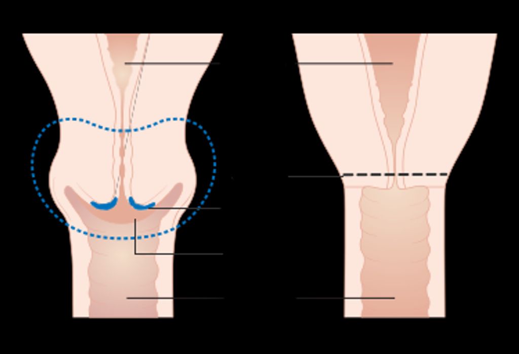
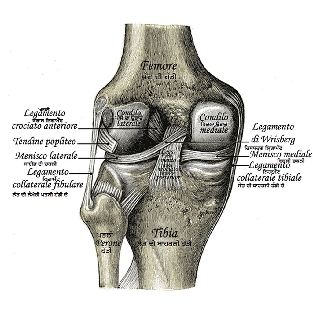
Anatomy of the Medial and Lateral Meniscus

Structure of the Medial Meniscus
The medial meniscus is a C-shaped structure that provides stability to the knee joint. It is composed of fibrocartilage and is thicker along the outer edge, tapering towards the inner edge. The main function of the medial meniscus is to distribute load and absorb shock within the knee joint. Here is a brief overview of the structural differences between the medial and lateral meniscus:
| Property | Medial Meniscus | Lateral Meniscus |
|---|---|---|
| Shape | C-shaped | O-shaped |
| Attachment | Strongly attached to the joint capsule and medial collateral ligament | Loosely attached to the joint capsule and lateral collateral ligament |
| Blood Supply | Poor blood supply | Rich blood supply |
| Mobility | Less mobile | More mobile |
These structural variances contribute to the differential vulnerability of the medial and lateral meniscus to injuries. It is important to consider these differences when evaluating and treating meniscus injuries.
Structure of the Lateral Meniscus
We turn our attention to the lateral meniscus, which, unlike its medial counterpart, is more circular in shape and covers a larger portion of the tibial plateau. This unique shape allows for greater mobility and a wider range of motion, which is crucial given its role in the complex mechanics of the knee joint.
The lateral meniscus is also distinct in its attachment to the joint capsule and the popliteus tendon, which provides stability during knee movements. It’s important to note that the lateral meniscus is less firmly attached to the tibial plateau compared to the medial meniscus, making it somewhat more flexible but also potentially more susceptible to certain types of injuries.
Key differences between the medial and lateral meniscus:
- Shape: The lateral meniscus is more circular.
- Coverage: It covers a larger portion of the tibial plateau.
- Mobility: Offers greater mobility and a wider range of motion.
- Attachments: Connected to the joint capsule and the popliteus tendon.
Tip: When assessing for meniscus injuries, it’s crucial to consider the unique anatomical features of the lateral meniscus to accurately diagnose and treat the condition.
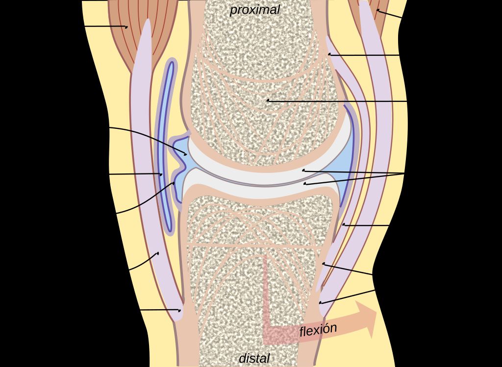
Function of the Medial and Lateral Meniscus
We understand the menisci to be crucial for the proper functioning of the knee joint. The primary role of both the medial and lateral meniscus is to distribute load across the knee, providing stability and reducing the stress on the articular cartilage. They act as shock absorbers, evenly dispersing the weight of the body during activities such as walking, running, and jumping.
The menisci also contribute to joint lubrication and nutrition of the articular cartilage. They facilitate the smooth movement of the femur over the tibia and play a role in proprioception, helping us sense the position and movement of the knee. This sensory feedback is essential for maintaining balance and coordinating complex movements.
- Load Distribution: The menisci spread out compressive forces over a larger area, minimizing peak stress on articular surfaces.
- Shock Absorption: They absorb and dissipate the forces generated during impact activities.
- Joint Lubrication: Menisci aid in the distribution of synovial fluid, which lubricates the knee joint.
- Proprioception: They provide sensory feedback for joint position and movement.
Tip: Preserving the integrity of the menisci is vital for long-term knee health. Avoiding activities that put excessive strain on the knee can help prevent meniscus injuries.
Causes of Medial and Lateral Meniscus Injuries
Traumatic Injuries
Traumatic injuries to the meniscus are often the result of sudden, forceful twisting or hyperextension of the knee joint. These injuries commonly occur during activities that involve rapid changes in direction, such as pivoting or sudden stops. Additionally, direct impact to the knee, especially when the foot is planted, can also lead to traumatic meniscus injuries. It’s important to note that athletes involved in sports that require quick, dynamic movements are particularly susceptible to these types of injuries.
When considering the causes of traumatic meniscus injuries, it’s essential to recognize the potential risk factors associated with certain activities. For instance, sports like basketball, soccer, and tennis, which involve frequent pivoting and cutting maneuvers, pose a higher risk for traumatic meniscus injuries. Understanding these risk factors can aid in injury prevention and the development of targeted rehabilitation programs.
- It’s crucial to seek prompt medical attention if a traumatic meniscus injury is suspected, as early diagnosis and intervention can significantly impact the prognosis and recovery process.
Tip: Proper warm-up exercises and conditioning routines can help reduce the risk of traumatic meniscus injuries during physical activities.
Degenerative Changes
Degenerative changes in the meniscus are often associated with aging and long-term wear and tear on the knee joint. These changes can lead to a gradual breakdown of the meniscus tissue, resulting in increased susceptibility to injury. One important study found that individuals with degenerative meniscal tears had a higher risk of developing osteoarthritis in the affected knee. This underscores the significance of early detection and appropriate management of degenerative changes in the meniscus.
- Implement a table for presenting structured, quantitative data. Ensure it’s succinct and formatted correctly in Markdown.
- Use a bulleted or numbered list for less structured content, like steps, qualitative points, or a series of related items.
It is crucial to emphasize the role of preventive measures and lifestyle modifications in minimizing the progression of degenerative changes in the meniscus. Regular exercise, maintaining a healthy weight, and avoiding activities that place excessive stress on the knee joint can significantly reduce the risk of degenerative meniscus injuries.
Sports-Related Injuries
In our exploration of meniscus injuries, we recognize that athletes frequently encounter these issues due to the high demands placed on their knees. Activities that involve twisting, pivoting, or sudden stops and starts are particularly notorious for causing meniscus tears. For instance, sports such as basketball, football, and soccer see a higher incidence of these injuries.
The following list outlines common sports that may lead to meniscus injuries:
- Basketball
- Football
- Soccer
- Tennis
- Skiing
- Volleyball
We must emphasize the importance of proper technique and preventive measures in sports to mitigate the risk of meniscus injuries. Prevention strategies include strength training, flexibility exercises, and using proper equipment. It’s also crucial for athletes to be aware of their body’s signals and to seek timely medical advice when experiencing knee pain or discomfort.
Tip: Always warm up before engaging in sports and cool down afterwards to reduce the risk of meniscus and other types of injuries.
Symptoms and Diagnosis of Meniscus Injuries

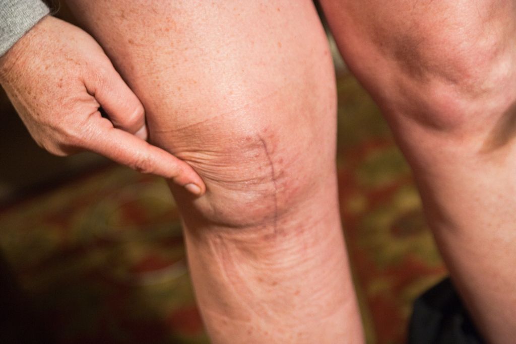
Common Symptoms
After experiencing discomfort or pain in the knee, we may notice swelling and tenderness in the joint. Additionally, we might feel a sensation of locking or catching in the knee during movement. It is important to pay attention to any changes in the knee’s range of motion, as this can indicate a potential meniscus injury. We should also be aware of any clicking or popping sounds that occur during knee movement. These symptoms, when present, may warrant further examination and diagnostic testing to confirm the diagnosis of a meniscus injury.
Physical Examination
During the physical examination, we assess the knee’s range of motion, stability, and any signs of tenderness or swelling. Specific maneuvers, such as the McMurray test, are performed to evaluate the integrity of the menisci. This test involves bending, straightening, and rotating the knee to elicit pain or a clicking sound, which may indicate a meniscus tear.
We also look for joint line tenderness, which is pain along the joint where the meniscus is located. The assessment of the knee might include comparing it to the uninjured knee to identify discrepancies in structure or function. It’s crucial to note that while these tests can be highly indicative of a meniscus injury, they are not definitive without corroborating diagnostic imaging.
Tip: Always report any discomfort experienced during the examination to the clinician, as this can provide valuable information regarding the location and severity of the injury.
Diagnostic Imaging
After the diagnostic imaging, we carefully analyze the results to identify any abnormalities or damage to the meniscus. This may involve assessing the size, shape, and location of any tears or lesions. In some cases, a MRI scan may be used to provide detailed images of the meniscus and surrounding structures. Additionally, we consider the patient’s reported symptoms and medical history to form a comprehensive diagnosis.
- Diagnostic Imaging Results:
| Type of Imaging | Findings |
|---|---|
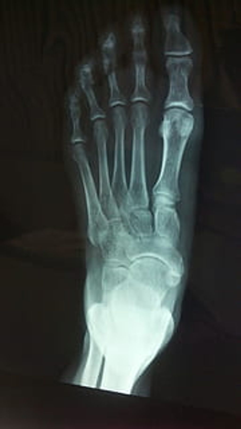
| X-ray | Normal |
| MRI | Tear in medial meniscus |
It is important to note that accurate diagnosis is crucial for determining the most effective treatment plan. Therefore, thorough evaluation of both the symptoms and diagnostic imaging results is essential for guiding the next steps in the management of meniscus injuries.
Treatment Options for Meniscus Injuries

Conservative Treatment
In our approach to treating meniscus injuries, we often begin with conservative treatment options. These are designed to alleviate pain, reduce inflammation, and promote healing without the need for surgical intervention. The cornerstone of conservative treatment is rest, which allows the injured meniscus time to recover. Patients are advised to avoid activities that exacerbate the injury, particularly those that involve twisting or over-flexing the knee.
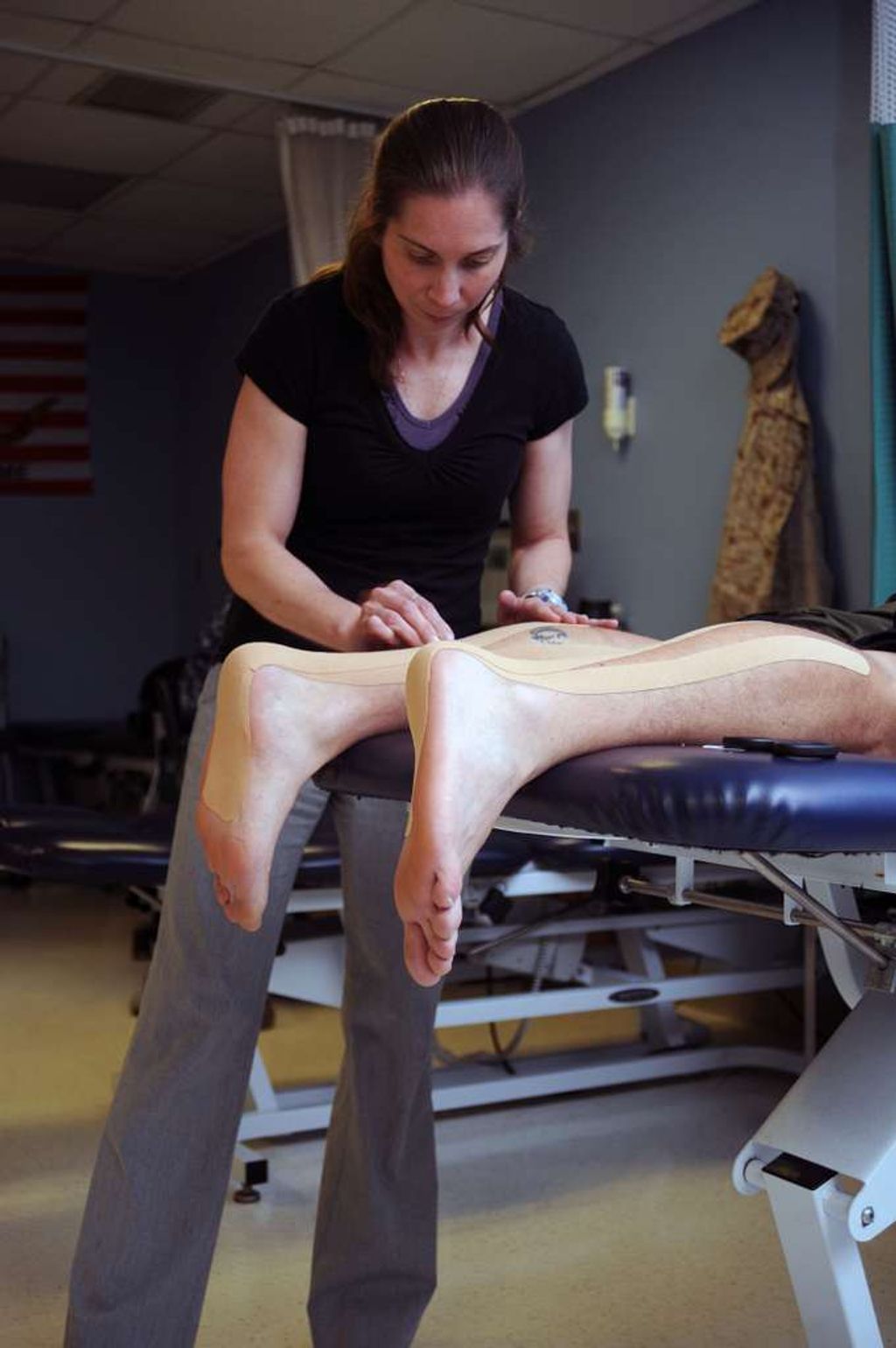
Physical therapy plays a pivotal role in conservative treatment, focusing on exercises that strengthen the muscles around the knee, thereby providing better support and stability to the joint. It’s important to follow a structured therapy program tailored to the individual’s specific condition and recovery goals.
In addition to rest and physical therapy, other conservative measures include:
- The use of nonsteroidal anti-inflammatory drugs (NSAIDs) to manage pain and inflammation
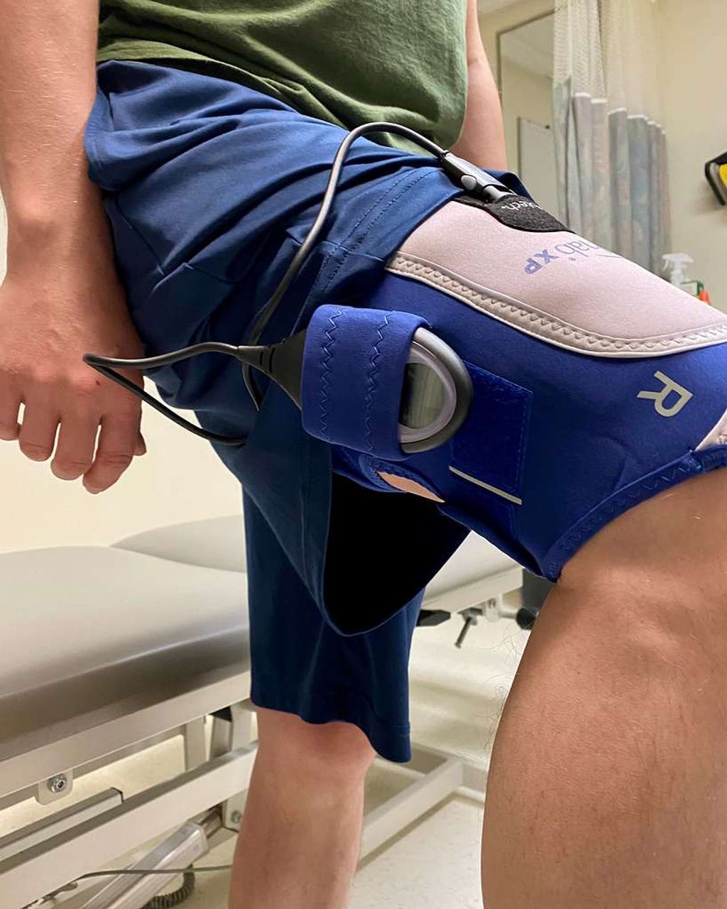
- Application of ice to reduce swelling
- Compression bandages to provide support
- Elevation of the leg to decrease fluid accumulation
Tip: Consistent application of conservative treatments is crucial for effective recovery. Patients should adhere to the prescribed regimen and communicate regularly with their healthcare provider to monitor progress and make necessary adjustments.
Surgical Interventions
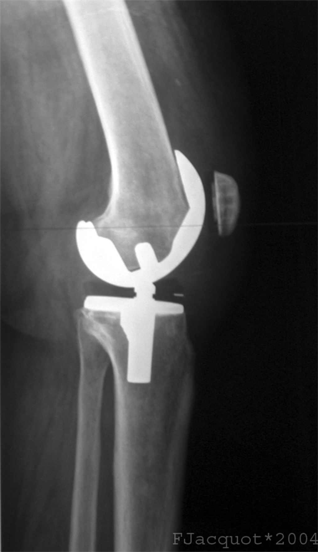
When conservative treatments for meniscus injuries fail to provide relief, we often consider surgical interventions. The primary goal is to preserve as much of the meniscus as possible, promoting long-term joint health and function. There are two main types of surgical procedures: meniscectomy, where damaged meniscal tissue is removed, and meniscus repair, where the tear is sutured.
- Meniscectomy is often performed arthroscopically and is less invasive, with quicker recovery times. However, it may lead to an increased risk of osteoarthritis in the long term.
- Meniscus repair, on the other hand, aims to maintain meniscal integrity and is preferred when the tear is repairable. Recovery from meniscus repair is typically longer, as the meniscus needs time to heal.
It is crucial to tailor the surgical approach to the individual patient, considering factors such as the type of tear, the patient’s age, activity level, and overall knee health.
We must also be mindful of the rising surgery rates and the limitations of current treatments, especially in the context of early osteoarthritis post-knee trauma. This underscores the importance of ongoing research and development in this field.
Rehabilitation and Recovery
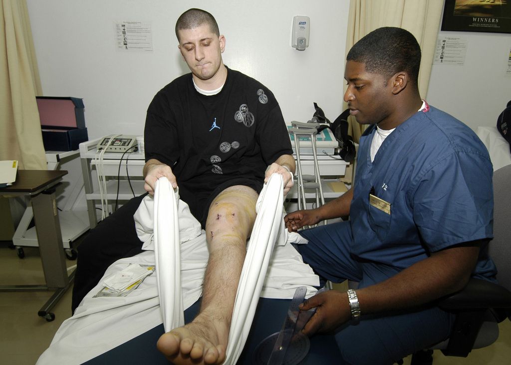
Following surgical intervention for meniscus injuries, we emphasize the importance of a structured rehabilitation and recovery process. This phase is crucial for restoring knee function, improving strength, and ensuring a safe return to daily activities or sports. Our approach to rehabilitation includes a variety of exercises tailored to each patient’s specific needs and recovery goals.
The initial stages of rehabilitation focus on reducing inflammation and pain, followed by gradual progression to exercises that enhance knee stability and mobility. We advocate for a multidisciplinary approach, often involving physical therapists, to guide patients through their recovery journey. The use of technology, such as the Curovate physiotherapy app, can complement traditional therapy by providing additional support with strength exercises, knee range measurement, and access to educational resources.
Tip: Always adhere to the prescribed rehabilitation protocol and communicate regularly with your healthcare provider to monitor progress and adjust the plan as necessary.
It’s imperative to understand that recovery timelines can vary significantly among individuals. Factors such as the extent of the injury, the type of surgical intervention, and the patient’s adherence to the rehabilitation program all play a role in determining the duration and success of the recovery.
Conclusion
In conclusion, the complexities of medial and lateral meniscus injuries necessitate a thorough understanding of their anatomical structure, biomechanical function, and diagnostic approaches. The management of these injuries requires a multidisciplinary approach, involving orthopedic specialists, physical therapists, and sports medicine professionals. Early diagnosis and intervention are crucial in preventing long-term complications and promoting optimal recovery. Further research and advancements in treatment modalities are essential to enhance the outcomes for individuals affected by these debilitating injuries.
Frequently Asked Questions
What are the common causes of medial and lateral meniscus injuries?
Common causes include traumatic injuries, degenerative changes, and sports-related injuries.
How are meniscus injuries diagnosed?
Meniscus injuries are diagnosed through physical examination and diagnostic imaging such as MRI or X-ray.
What are the conservative treatment options for meniscus injuries?
Conservative treatment options include rest, ice, compression, elevation (RICE), physical therapy, and non-steroidal anti-inflammatory drugs (NSAIDs).
When is surgical intervention necessary for meniscus injuries?
Surgical intervention is necessary for severe meniscus tears that do not respond to conservative treatment or for cases where the meniscus is causing mechanical symptoms.
How long does it take to recover from meniscus surgery?
Recovery time varies depending on the type of surgery and the individual patient, but it generally takes several weeks to months for full recovery.
Can meniscus injuries lead to long-term complications?
Yes, untreated meniscus injuries can lead to long-term complications such as chronic pain, instability, and increased risk of developing osteoarthritis.