Tag: Arthritis Knees

  • Knee instability without swelling

    Knee instability without swelling

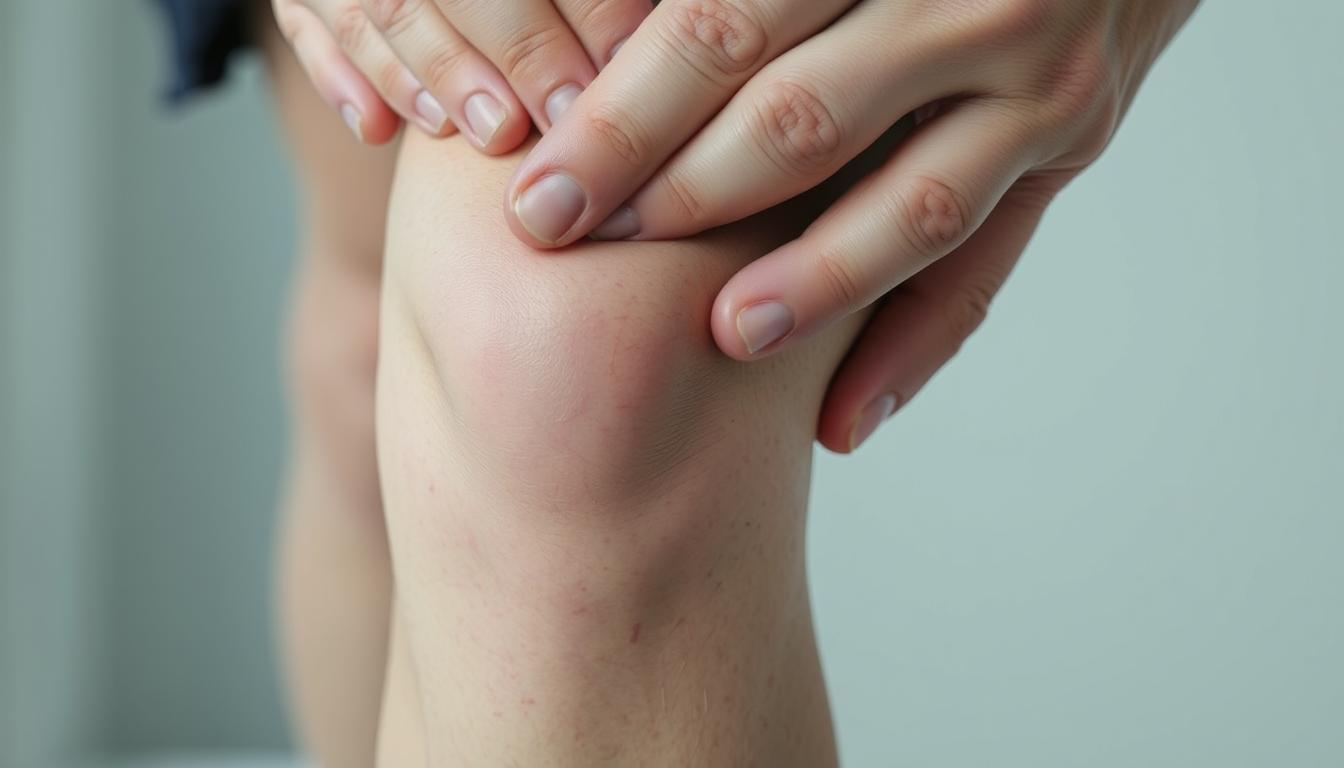
    Have you ever experienced a nagging knee pain that seems to come out of nowhere, without any visible signs of injury or swelling? You’re not alone. Many individuals face the challenge of knee instability or pain without the typical symptoms of inflammation.

    This phenomenon can be puzzling and concerning, especially when it affects your mobility and quality of life. Unlike typical knee injuries that present with obvious swelling, cases without accompanying inflammation require careful assessment to identify the underlying cause.

    We will explore the various factors that can lead to knee issues without swelling, from ligament injuries to chronic conditions and degenerative changes, and discuss the proper diagnosis and treatment options.

    Key Takeaways

    • Understanding knee instability without swelling is crucial for proper diagnosis.
    • Ligament injuries can cause knee pain without visible swelling.
    • Chronic conditions and degenerative changes can lead to knee instability.
    • Careful assessment is necessary to identify the underlying cause.
    • Various treatment options are available depending on the diagnosis.

    Understanding Knee Stability and Its Importance

    Knee stability, often taken for granted, is fundamental to our ability to move freely and maintain an active lifestyle. The knee joint is one of the most complex in the human body, relying on a delicate balance of structures to maintain proper stability and function. As we explore the intricacies of knee stability, it becomes clear that understanding its anatomy and importance is crucial for appreciating its role in our daily lives.

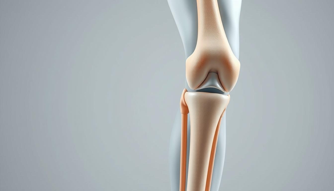
    The Anatomy of a Stable Knee

    The stability of the knee joint is maintained by a combination of its shape and various supporting structures. The four major ligaments – the anterior cruciate ligament (ACL), posterior cruciate ligament (PCL), medial collateral ligament (MCL), and lateral collateral ligament (LCL) – serve as primary stabilizers. Additionally, the shape of the femoral condyles and menisci contributes significantly to knee stability by creating a congruent surface that helps distribute weight and absorb shock during movement.

    Secondary stabilizers include the posteromedial and posterolateral capsular components, the iliotibial tract, and the surrounding musculature that provides dynamic support during activity. The intricate network of ligaments, tendons, muscles, and cartilage works in harmony to allow for smooth, pain-free movement.

    How Knee Stability Affects Daily Function

    Proper knee stability is crucial for everyday activities such as walking, climbing stairs, and sitting. Even minor instability can potentially lead to significant functional limitations and compensatory movement patterns. When the knee is functioning properly, these structures work together seamlessly, maintaining the joint’s integrity during various activities.

    As highlighted by experts, “Understanding the complex anatomy of the knee is essential for diagnosing the specific cause of instability when swelling is absent.” This knowledge is vital for addressing issues related to knee stability effectively.

    What Causes Knee Instability Without Swelling?

    Several factors contribute to knee instability without swelling, including ligament tears, muscle weakness, and chronic conditions. Knee instability is a complex condition that can significantly impact an individual’s quality of life. Understanding the underlying causes is crucial for developing effective treatment plans.

    Ligament Injuries and Tears

    Ligament injuries are a common cause of knee instability. These injuries can result from direct or indirect trauma, with “noncontact” mechanisms being the most frequent. Activities involving cutting, twisting, jumping, and sudden deceleration can place excessive stress on the knee ligaments, leading to tears or laxity. For instance, a sudden change in direction during sports can cause a ligament injury without immediate swelling.

    Muscle Weakness and Imbalances

    Muscle weakness, particularly in the quadriceps and hamstrings, can significantly contribute to knee instability. When these muscles are weak, they fail to provide adequate dynamic support to the knee joint during movement. Imbalances between muscle groups can also alter knee biomechanics, leading to instability even without acute injury or swelling.

    Chronic Conditions and Degenerative Changes

    Chronic conditions such as osteoarthritis can gradually erode the joint surfaces and compromise ligament integrity, resulting in progressive instability. Degenerative changes associated with aging or repetitive microtrauma can also affect the knee’s supporting structures, leading to worsening instability symptoms over time. These changes can occur without noticeable swelling, making diagnosis more challenging.

    Medial Collateral Ligament (MCL) Injuries

    The medial collateral ligament (MCL) plays a crucial role in maintaining knee stability, and injuries to this ligament can significantly impact knee function. The MCL is attached proximally to the medial femoral condyle and distally to the tibial metaphysis, 4 to 5 cm distal to the medial joint line beneath the pes anserinus insertion. Understanding MCL injuries is essential for diagnosing and treating knee instability effectively.

    A detailed anatomical illustration of the medial collateral ligament (MCL) in the human knee joint, rendered in highly realistic medical illustration style. The MCL is prominently featured in the center foreground, shown with its characteristic fan-like structure and attachments to the femur and tibia. The surrounding musculature, tendons, and bony landmarks are clearly visible, allowing for a comprehensive understanding of the MCL's anatomical context. The lighting is soft and directional, creating subtle shadows that enhance the three-dimensional form. The background is minimalist, with a plain, neutral color palette to avoid distractions and focus the viewer's attention on the MCL structure. The overall tone is educational and informative, suitable for use in a medical article on knee instability.

    How MCL Injuries Occur

    MCL injuries typically occur from a direct blow to the lateral (outside) aspect of the knee while it’s slightly flexed, creating a valgus force that stresses or tears the medial ligament complex. Isolated MCL injuries happen usually as a result of such direct trauma. When the deforming force includes a rotational component, associated injuries to the cruciate ligaments can occur, complicating the diagnosis and treatment.

    Diagnosing MCL Instability

    Diagnosis of MCL instability involves applying a gentle valgus force to the knee at 15-20 degrees of flexion and comparing the degree of medial joint opening to the uninjured knee. Even a small difference of 5mm in joint opening can indicate substantial structural damage to the MCL, though this may not always be accompanied by visible swelling or significant pain. This diagnostic approach helps in assessing the severity of the MCL injury.

    Treatment Options for MCL Injuries

    Treatment for MCL injuries is typically conservative, beginning with rest, ice, compression, and elevation (RICE) during the first 48 hours following injury. Physical therapy focusing on strengthening the muscles around the knee joint is crucial for recovery from MCL tears and preventing future instability. Most isolated MCL injuries heal well with conservative treatment, allowing patients to return to normal activities within approximately 6 weeks. However, chronic MCL insufficiency can occur, especially in conjunction with other ligament injuries, requiring a more comprehensive treatment approach.

    Anterior Cruciate Ligament (ACL) Damage

    Understanding ACL damage is crucial for diagnosing and treating knee instability, which can manifest without noticeable swelling. The ACL is a critical component of the knee joint, providing stability and support during various activities.

    The ACL is the primary restraint to anterior translation of the tibia on the femur and to hyperextension. It also functions as a secondary restraint to varus or valgus angulation at full extension and resists internal and external rotation at nearly full extension. Damage to this ligament can lead to significant knee pain and instability, affecting an individual’s ability to perform daily activities and participate in sports.

    Mechanisms of ACL Injury

    ACL injuries most commonly occur during non-contact situations involving sudden deceleration, pivoting, or landing from a jump with the knee in a vulnerable position. These movements can cause a sudden strain on the ACL, leading to tears or complete ruptures.

    Recognizing ACL Instability Without Swelling

    Unlike typical ACL tears that present with immediate swelling, some partial tears or chronic ACL insufficiency can manifest primarily as instability without significant effusion. Patients with ACL instability often describe a sensation of the knee “giving way” during pivoting activities. The Lachman test and pivot shift test are reliable clinical examinations for assessing ACL instability.

    Conservative vs. Surgical Management

    The management of ACL injuries depends on several factors, including the patient’s age, activity level, degree of instability, and willingness to modify activities. Conservative management focuses on strengthening the muscles around the knee, particularly the hamstrings. Surgical reconstruction is typically recommended for young, active patients and those who wish to return to high-demand activities.

    Treatment ApproachDescriptionRecommended For
    Conservative ManagementStrengthening muscles around the knee, particularly hamstringsLess active patients or those willing to modify activities
    Surgical ReconstructionUsing autografts or allografts to reconstruct the ACLYoung, active patients and those returning to high-demand activities

    In conclusion, ACL damage is a significant cause of knee instability, and its management requires a comprehensive approach considering the patient’s specific needs and activity level. By understanding the mechanisms of ACL injury and the available treatment options, healthcare providers can offer personalized care to patients suffering from ACL damage.

    Posterior Cruciate Ligament (PCL) Issues

    Understanding PCL issues is essential for addressing knee instability, particularly in cases where swelling is not a prominent symptom. The PCL is a critical ligament that originates from the medial femoral condyle and inserts into a depression between the posterior aspect of the two tibial plateaux.

    PCL Function and Injury

    The PCL is composed of two bundles, anterolateral and posteromedial, and serves as the primary restraint to posterior translation of the tibia relative to the femur, especially in the mid-range of knee flexion (40-120 degrees). PCL injuries account for approximately 15-20% of all knee ligament injuries and often result from direct trauma to the front of the tibia while the knee is flexed.

    • The PCL is crucial for knee stability, particularly during flexion.
    • PCL injuries can occur without significant swelling, making diagnosis challenging.
    • Direct trauma, such as dashboard injuries in car accidents, is a common cause of PCL tears.

    Treatment Approaches for PCL Instability

    Treatment for PCL injuries depends on the grade of the tear, associated ligament injuries, and the patient’s activity level and symptoms. Conservative management focuses on quadriceps strengthening to compensate for the lost ligament function, while surgical reconstruction may be necessary for high-grade tears or when conservative treatment fails.

    We consider several factors when determining the best treatment approach for PCL instability, including the severity of the injury and the patient’s overall health.

    Key treatment considerations include:

    1. Grade of the PCL tear
    2. Presence of associated ligament injuries
    3. Patient’s activity level and symptoms

    Lateral and Posterolateral Corner Injuries

    The knee joint’s stability is significantly influenced by the integrity of its lateral and posterolateral structures. The lateral and posterolateral corner of the knee comprises several important stabilizing structures, including the lateral collateral ligament (LCL), popliteus tendon, popliteofibular ligament, and arcuate ligament.

    Anatomy of the Lateral Knee

    The LCL originates on the lateral epicondyle of the femur and is attached distally on the fibular head. The posterolateral corner is a complex anatomic region consisting of the popliteus tendon, the popliteofibular ligament, the arcuate ligament, and the posterolateral joint capsule. Understanding this anatomy is crucial for diagnosing and treating injuries to this area.

    Diagnosis of Lateral Instability

    Diagnosing lateral instability involves a combination of clinical examination and sometimes additional diagnostic tests. The varus stress test at both full extension and 15 degrees of flexion is crucial for assessing lateral instability. Increased external rotation of the tibia relative to the femur at 30 degrees of knee flexion is characteristic of isolated posterolateral instability.

    Management Strategies

    Early surgical intervention is often recommended for posterolateral corner injuries, as these structures have limited healing capacity when treated conservatively. For chronic posterolateral instability, reconstruction rather than repair is typically necessary, using either autograft or allograft tissue to restore stability. Rehabilitation following surgery is typically more prolonged and cautious than for isolated cruciate ligament reconstructions.

    We recognize that managing lateral and posterolateral corner injuries requires a comprehensive approach, taking into account the specific nature of the injury and the patient’s overall condition. By understanding the anatomy, diagnosis, and appropriate management strategies, healthcare providers can offer effective treatment options for patients experiencing knee instability due to these injuries.

    Other Causes of Knee Instability Without Swelling

    The absence of swelling doesn’t rule out knee instability, which can be caused by multiple factors. We will explore some of these causes, including meniscal injuries, patellofemoral issues, and degenerative conditions like arthritis.

    Meniscal Injuries

    Meniscal tears can cause knee instability without significant swelling, particularly when the tear affects the meniscus’s role in joint congruity. The meniscus is cartilage that cushions the inner side of the knee joint. An injury to this area can lead to pain in the inner knee.

    Patients with meniscal injuries often report mechanical symptoms such as catching, locking, or giving way during specific movements. These symptoms can occur even when swelling is minimal or absent.

    Patellofemoral Issues

    Patellofemoral issues, including maltracking of the patella or patellofemoral pain syndrome, can create a sensation of instability, particularly when ascending or descending stairs. Weakness in the vastus medialis obliquus muscle can contribute to patellofemoral instability without causing visible swelling in the knee joint.

    Arthritis and Degenerative Conditions

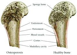
    Osteoarthritis affects more than 32.5 million U.S. adults and can cause progressive joint instability as the articular cartilage deteriorates and joint surfaces become incongruent. Early-stage arthritis may cause instability without noticeable swelling, particularly during weight-bearing activities.

    Degenerative changes to the menisci that occur with aging can reduce their stabilizing function without triggering an inflammatory response or swelling. Loose bodies within the joint from cartilage or bone fragments can also cause intermittent locking and instability.

    Furthermore, neurological conditions affecting proprioception around the knee can create functional instability despite structurally intact ligaments and minimal inflammation. Understanding these various causes is crucial for proper diagnosis and treatment.

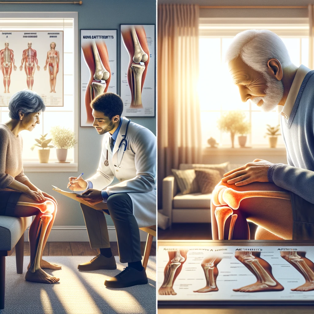
    Diagnosing Knee Instability When No Swelling Is Present

    Diagnosing knee instability without swelling requires a comprehensive approach. We must consider the patient’s history, physical examination findings, and results from diagnostic imaging. The absence of swelling can make diagnosis more challenging, but a thorough evaluation can help identify the underlying causes.

    Physical Examination Techniques

    A detailed physical examination is crucial in diagnosing knee instability. Special tests such as the Lachman test and pivot shift for ACL injuries, the posterior drawer test for PCL injuries, and varus/valgus stress tests for collateral ligament injuries are essential. Comparing the affected knee to the uninjured side helps detect subtle differences in laxity that might indicate ligament insufficiency.

    For instance, the Lachman test is particularly useful for assessing ACL integrity. It involves gently pulling the tibia forward while stabilizing the femur. A significant difference in translation between the two knees can indicate ACL damage.

    Imaging and Other Diagnostic Tools

    Advanced imaging techniques, particularly MRI, play a vital role in diagnosing ligament, meniscal, and cartilage injuries when swelling is absent. MRI provides detailed images of soft tissue structures, helping to identify tears or other damage. Stress radiographs can also quantify the degree of instability in collateral ligament injuries.

    Diagnostic ToolUse in Knee Instability Diagnosis
    MRIDetailed imaging of soft tissues, including ligaments and menisci
    Stress RadiographsQuantifying instability in collateral ligament injuries
    ArthroscopyDirect visualization of intra-articular structures and potential treatment

    When to Seek Medical Attention

    Patients should seek medical attention if they experience recurrent episodes of the knee “giving way,” inability to fully trust the knee during activities, or when instability interferes with daily function. For more information on related issues, you can visit https://kneehurt.com/causes-and-treatments-for-knee-pain-clicking/. Delayed diagnosis can lead to secondary injuries and accelerated joint degeneration, making timely medical evaluation crucial.

    Conservative Treatment Approaches

    The initial approach to treating knee instability without swelling typically involves conservative treatment methods. We focus on addressing the root causes of instability and improving knee function through non-surgical means.

    Strengthening and Rehabilitation

    Physical therapy forms the cornerstone of conservative treatment for knee instability. We emphasize strengthening the muscles that dynamically stabilize the knee, particularly the quadriceps, hamstrings, and hip abductors. Proprioceptive training is also essential for improving the body’s awareness of knee position and movement, helping to compensate for ligamentous instability through enhanced neuromuscular control.

    Rehabilitation protocols typically progress from basic range of motion exercises to closed-chain strengthening activities and eventually sport-specific training for those returning to athletic activities. This structured approach helps in restoring knee stability and function.

    Supportive Devices

    Bracing and supportive devices can provide additional stability for knees experiencing instability. Functional knee braces may improve joint position sense and limit excessive movement, though their effectiveness can vary among patients and conditions. For patients with instability related to osteoarthritis, unloader braces can be particularly helpful by redistributing forces away from the affected compartment of the knee.

    Modifying Activities

    Activity modification is often necessary to prevent symptom exacerbation. We advise patients to avoid high-risk movements that trigger instability episodes. Low-impact activities like swimming, cycling, and elliptical training can maintain cardiovascular fitness while minimizing stress on an unstable knee. For patients with instability related to arthritis, weight management is crucial as each pound of weight loss reduces stress on the knee joint by approximately four pounds during walking.

    Conservative treatment success depends largely on patient compliance with home exercise programs and willingness to modify activities that provoke instability. By adopting these strategies, individuals can effectively manage knee instability without swelling and improve their overall knee health.

    Surgical Interventions for Persistent Knee Instability

    When knee instability persists despite conservative management, surgical intervention may be necessary to restore stability and function. Surgical techniques have evolved to address various causes of knee instability, offering patients a range of options tailored to their specific needs.

    Reconstructive Procedures

    Surgical reconstruction for knee instability often involves repairing or replacing damaged ligaments. Modern techniques primarily use autografts (the patient’s own tissue) or allografts (donor tissue) to replace damaged ligaments. The choice of graft material depends on several factors, including the patient’s age, activity level, and previous surgeries.

    • Autografts: Using the patient’s own tissue, such as the patellar tendon or hamstring tendons, for ligament reconstruction.
    • Allografts: Utilizing donor tissue for patients who may not be suitable for autografts or prefer this option.

    The surgical technique requires precise placement and tensioning of the graft, avoidance of impingement, and adequate fixation to ensure successful outcomes.

    Recovery and Rehabilitation

    Post-surgical rehabilitation is crucial for optimal outcomes. Rehabilitation typically begins with early range of motion exercises and progresses to strength training and sport-specific activities. The recovery process can vary based on the specific procedure and individual healing factors.

    Generally, full recovery and return to sports or demanding activities take 6-12 months following major ligament reconstruction. Patients should be prepared for a gradual return to their normal activities under the guidance of a healthcare professional.

    Expected Outcomes and Timeline

    Long-term success rates for ligament reconstruction surgeries range from 80-95% for restoring knee stability. However, outcomes can be influenced by factors such as age, activity level, and associated injuries. It’s essential for patients to have realistic expectations about surgical outcomes, understanding that while stability can be significantly improved, the knee may not return to its pre-injury state.

    By understanding the available surgical interventions and what to expect during recovery, patients can make informed decisions about their treatment options for knee instability.

    Preventing Future Episodes of Knee Instability

    A proactive approach to managing knee health involves addressing modifiable risk factors and adjusting activities to prevent instability episodes. Maintaining an optimal weight is crucial, as excess weight significantly increases stress on the knee joint. For every pound of weight lost, the knee joint forces are reduced by approximately four pounds during walking, thereby decreasing the risk of knee pain and instability.

    Engaging in regular strength training that focuses on the quadriceps, hamstrings, and hip muscles is also vital. This training provides dynamic stability to the knee, compensating for any ligamentous laxity or degenerative changes. Furthermore, using proper technique during sports and exercise, especially for movements involving cutting, pivoting, jumping, and landing, can significantly reduce the risk of knee injury.

    Other preventive measures include wearing appropriate footwear with good support and proper fit, which can improve lower extremity alignment and reduce abnormal forces on the knee. For individuals with known ligament insufficiency, preventive bracing may be beneficial during high-risk activities. Additionally, incorporating low-impact activities like swimming and cycling into one’s fitness routine can help maintain fitness while reducing repetitive stress on the knee joint.

    Maintaining good flexibility through regular stretching and proper warm-up routines before activities can also reduce the risk of knee injury. For patients with arthritis-related instability, adopting an anti-inflammatory diet rich in omega-3 fatty acids and antioxidants may help manage inflammation and symptoms. Lastly, regular medical care and adherence to prescribed treatment regimens are essential for managing underlying conditions that contribute to knee instability.

    FAQ

    What are the common causes of knee pain and instability?

    We find that knee pain and instability can be caused by various factors, including ligament injuries, muscle weakness, and degenerative conditions such as osteoarthritis. Activities that put stress on the knee joint, like sports, can also contribute to these issues.

    How is knee instability diagnosed when there’s no swelling?

    Diagnosing knee instability without swelling involves a physical examination, imaging tests like X-rays or an MRI, and assessing the knee’s range of motion. We also consider the patient’s medical history and activity level to make an accurate diagnosis.

    Can knee instability be treated without surgery?

    Yes, we often recommend conservative treatment approaches, such as physical therapy, bracing, and modifying activities to alleviate knee instability. These methods can be effective in managing symptoms and improving knee function.

    What role do ligaments play in knee stability?

    Ligaments, including the ACL, PCL, MCL, and lateral ligaments, provide crucial support to the knee joint. Injuries to these ligaments can lead to knee instability, and we may recommend reconstructive surgery in severe cases.

    How can I prevent future episodes of knee instability?

    To prevent knee instability, we suggest maintaining a healthy weight, engaging in exercises that strengthen the surrounding muscles, and using proper techniques during sports and activities. Wearing supportive devices like knee braces can also help.

    What is the typical recovery time after knee surgery?

    The recovery time after knee surgery varies depending on the type of procedure and individual factors. Generally, we can expect several months of rehabilitation, during which we’ll guide you through a structured recovery program to restore knee function and strength.

    Can osteoarthritis cause knee instability?

    Yes, osteoarthritis can contribute to knee instability by causing degenerative changes in the joint, including cartilage loss and ligament laxity. We can help manage osteoarthritis symptoms and related knee instability through a combination of conservative and surgical treatments.

  • Causes And solutions ForThe knee pain

    Knee pain is a common ailment that can stem from a variety of factors, from overuse and injury to underlying medical conditions. This comprehensive FAQ addresses some of the most common questions surrounding knee pain and provides insights into exercises and treatments that might provide relief. However, it’s crucial to consult a healthcare professional for personalized advice and treatment.

    Table of Contents

    1. Common Causes of Knee Pain
    2. Exercises to Alleviate Knee Pain
    3. Importance of Hip and Ankle Strength
    4. Osteoarthritis and Knee Pain
    5. Runner’s Knee: Causes and Prevention
    6. Meniscus Tears: Symptoms and Treatment
    7. Knee Brace Selection Guide
    8. Physical Therapy for Knee Pain

    <a name=”common-causes”></a>

    What are some common causes of knee pain?

    Knee pain can be attributed to various factors, and identifying the specific cause is crucial for effective treatment. Some common causes include:

    • Anterior Knee Pain: This type of pain, often referred to as chondromalacia, is frequently observed and can be linked to issues with how the kneecap aligns and moves.
    • Patellar Tendon Pain: Activities like running, cutting, or jumping can lead to pain in the patellar tendon, the tendon connecting the kneecap to the shinbone.
    • Osteoarthritis: A degenerative joint condition that commonly affects the knees, causing pain, stiffness, and reduced mobility.
    • Ligament Injuries: Tears or sprains in the knee ligaments, such as the ACL (anterior cruciate ligament) or MCL (medial collateral ligament), can cause significant pain and instability.
    • Meniscus Tears: Damage to the cartilage that cushions the knee joint can result in pain, swelling, and difficulty moving the knee.

    <img src=”/api/placeholder/400/300″ alt=”Diagram of knee anatomy showing common pain points” />

    <a name=”exercises”></a>

    What exercises can help alleviate knee pain?

    While it’s essential to consult a medical professional for diagnosis and a tailored exercise plan, certain exercises may help strengthen the muscles surrounding the knee and improve its stability. Remember to start slowly and listen to your body. Stop if you feel any sharp pain.

    Exercises Targeting the Quadriceps:

    1. Quad Sets:
      • Sit with your legs extended in front of you.
      • Tighten your quadriceps muscles (front of your thigh) as if pushing your knee down into the floor.
      • Hold the contraction for 10 seconds, repeat 10 times.
      • Aim to do these throughout the day.
    2. Short Arc Quads:
      • Place a foam roller, rolled-up towel, or a similar object behind your knee.
      • Gently straighten your leg by engaging your quadriceps, maintaining contact with the object behind your knee.
      • Slowly lower your leg back down.
      • Aim for 10-20 repetitions.
    3. Straight Leg Raises:
      • Lie on your back or prop yourself up on your elbows.
      • Tighten your quadriceps and lift one leg about 30 degrees off the ground, keeping your knee straight.
      • Slowly lower your leg back down.
      • Aim for 10-20 repetitions.

    <img src=”/api/placeholder/400/300″ alt=”Person demonstrating straight leg raise exercise” />

    Exercises Targeting Other Supporting Muscles:

    1. Bridges:
      • Lie on your back with knees bent.
      • Contract your glutes and hamstrings to lift your hips off the floor.
      • Hold for 3 seconds and lower down.
      • Start with 10 repetitions and gradually increase.
    2. Clamshells:
      • Lie on your side with hips and knees bent.
      • Keeping your feet together, raise your top knee as high as you can without rotating your hip.
      • Lower your knee back down.
      • Aim for 10-20 repetitions on each side.
    3. Side-Lying Abduction:
      • Lie on your side with your top leg extended and bottom leg bent.
      • Keeping your top leg straight, lift it up towards the ceiling, leading with your heel.
      • Lower your leg back down.
      • Aim for 10 repetitions and gradually increase.

    <a name=”hip-and-ankle”></a>

    What is the importance of hip and ankle strength for knee pain?

    While the knee joint itself primarily flexes and extends, the stability and alignment of the knee are significantly influenced by the strength and stability of the surrounding hip and ankle joints. Weak hips and ankles can affect how the knee tracks during movement, potentially leading to pain and discomfort. Therefore, incorporating exercises that target hip abductors and other hip and ankle stabilizers is essential for comprehensive knee pain management.

    Some exercises to strengthen hips and ankles include:

    1. Hip Abductor Strengthening:
      • Stand on one leg, holding onto a chair for balance if needed.
      • Lift your other leg out to the side, keeping it straight.
      • Lower it back down slowly.
      • Repeat 10-15 times on each side.
    2. Ankle Circles:
      • Sit in a chair with your feet off the ground.
      • Rotate your ankles in circles, 10 times clockwise and 10 times counterclockwise.
      • Repeat with the other ankle.

    <img src=”/api/placeholder/400/300″ alt=”Illustration of hip and ankle exercises” />

    <a name=”osteoarthritis”></a>

    Osteoarthritis and Knee Pain

    Osteoarthritis (OA) is one of the most common causes of chronic knee pain, especially in older adults. It’s a degenerative condition where the cartilage in the knee joint wears away over time, leading to pain, stiffness, and reduced mobility.

    Symptoms of Knee Osteoarthritis:

    • Pain that worsens with activity
    • Stiffness, especially in the morning or after periods of inactivity
    • Swelling in the knee joint
    • A grating or crackling sound when moving the knee
    • Decreased range of motion

    Management Strategies for Osteoarthritis:

    1. Weight Management: Maintaining a healthy weight reduces stress on the knee joints.
    2. Low-Impact Exercise: Activities like swimming, cycling, or using an elliptical machine can help maintain joint mobility without excessive stress.
    3. Physical Therapy: Targeted exercises can improve strength and flexibility around the knee joint.
    4. Medications: Over-the-counter pain relievers or prescribed medications can help manage pain and inflammation.
    5. Hot and Cold Therapy: Applying heat before activities can loosen the joint, while cold therapy after activity can reduce swelling.
    6. Assistive Devices: Canes, walkers, or knee braces can provide support and reduce stress on the affected knee.

    <img src=”/api/placeholder/400/300″ alt=”X-ray image showing knee osteoarthritis” />

    <a name=”runners-knee”></a>

    Runner’s Knee: Causes and Prevention

    Runner’s knee, also known as patellofemoral pain syndrome, is a common condition among runners and other athletes. It’s characterized by pain around or behind the kneecap, especially during activities that involve bending the knee.

    Common Causes of Runner’s Knee:

    • Overuse or sudden increase in training intensity
    • Weak or imbalanced thigh muscles
    • Poor running form
    • Foot problems (like overpronation)
    • Tight hamstrings or iliotibial band

    Prevention Strategies:

    1. Proper Warm-up: Always warm up before running to prepare your muscles and joints.
    2. Gradual Training Increase: Follow the 10% rule – don’t increase your weekly mileage by more than 10% at a time.
    3. Strength Training: Focus on exercises that strengthen the quadriceps, hamstrings, and hip muscles.
    4. Proper Footwear: Wear running shoes that provide adequate support and are appropriate for your foot type.
    5. Cross-training: Incorporate low-impact activities like swimming or cycling to reduce stress on your knees.
    6. Stretching: Regular stretching, especially of the iliotibial band and hamstrings, can help prevent runner’s knee.

    <a name=”meniscus-tears”></a>

    Meniscus Tears: Symptoms and Treatment

    The meniscus is a C-shaped piece of cartilage in your knee that acts as a shock absorber between your shinbone and thighbone. Tears in the meniscus are common knee injuries, especially among athletes and older adults.

    Symptoms of a Meniscus Tear:

    • Pain, especially when twisting or rotating your knee
    • Swelling and stiffness
    • Difficulty fully straightening your knee
    • Feeling as though your knee is locked in place when you try to move it
    • Popping or clicking sensation

    Treatment Options:

    1. RICE Method: Rest, Ice, Compression, and Elevation can help manage pain and swelling.
    2. Physical Therapy: Exercises to strengthen the muscles around the knee can improve stability and function.
    3. Medications: NSAIDs can help reduce pain and inflammation.
    4. Surgery: In some cases, especially for larger tears, arthroscopic surgery may be necessary to repair or remove the damaged portion of the meniscus.

    <a name=”knee-brace”></a>

    Knee Brace Selection Guide

    Knee braces can provide support, stability, and pain relief for various knee conditions. However, choosing the right type of brace is crucial for maximum benefit.

    Types of Knee Braces:

    1. Compression Sleeves: Provide mild support and warmth, suitable for minor knee pain or arthritis.
    2. Patellofemoral Braces: Help align the kneecap and are useful for conditions like runner’s knee.
    3. Hinged Braces: Offer more substantial support and are often used for ligament injuries or instability.
    4. Unloader Braces: Designed to shift weight away from the affected side of the knee, beneficial for osteoarthritis.

    When selecting a knee brace, consider:

    • The specific condition or injury you’re addressing
    • The level of support needed
    • Your activity level
    • Comfort and fit

    It’s always best to consult with a healthcare professional or physical therapist to determine the most appropriate type of brace for your condition.<img src=”/api/placeholder/400/300″ alt=”Different types of knee braces” />

    <a name=”physical-therapy”></a>

    Physical Therapy for Knee Pain

    Physical therapy plays a crucial role in managing and treating various knee conditions. A physical therapist can develop a personalized treatment plan to address your specific needs and goals.

    Benefits of Physical Therapy for Knee Pain:

    • Improves strength and flexibility
    • Enhances joint stability
    • Reduces pain and inflammation
    • Improves range of motion
    • Teaches proper movement patterns to prevent future injuries

    Common Physical Therapy Techniques for Knee Pain:

    1. Therapeutic Exercises: Tailored exercises to strengthen muscles supporting the knee.
    2. Manual Therapy: Hands-on techniques to improve joint mobility and reduce pain.
    3. Ultrasound Therapy: Uses sound waves to reduce pain and inflammation.
    4. Electrical Stimulation: Can help manage pain and improve muscle function.
    5. Taping Techniques: Kinesiology taping can provide support and promote proper movement patterns.

    Remember, this FAQ provides general information and should not be considered a replacement for professional medical advice. It’s vital to consult a healthcare professional or physical therapist for a proper diagnosis and a personalized treatment plan tailored to your specific needs.

    For more in-depth information on knee anatomy and common injuries, we recommend watching this informative video:

    [Insert a relevant video about knee anatomy and common injuries here]

    By understanding the causes of knee pain and implementing appropriate exercises and treatments, many individuals can find relief and improve their knee health. However, persistent or severe knee pain should always be evaluated by a medical professional to ensure proper diagnosis and treatment.

  • Knee pain from office chair height

    Knee pain from office chair height

    Millions of office workers spend their days sitting at desks, often unaware of the strain their chair height is putting on their knees. Knee pain is a common complaint among those who spend long hours at the office, and it’s not just about being sedentary.

    The height of your office chair plays a crucial role in maintaining proper posture and preventing discomfort. When your chair is not adjusted correctly, it can lead to pain and discomfort in your knees. So, how can you avoid this issue?

    We will explore the causes of knee pain related to office seating and provide comprehensive solutions to alleviate and prevent it.

    Key Takeaways

    • Proper chair height adjustment is crucial for preventing knee pain.
    • Poor posture and extended sitting periods contribute to discomfort.
    • Ergonomic adjustments can significantly reduce knee strain.
    • Exercises and professional treatment options are available for relief.
    • Most chair-related knee pain is preventable with proper knowledge and workplace setup.

    Understanding the Connection Between Chair Height and Knee Pain

    The height of your office chair can significantly impact your knee health. When you’re inactive for an extended period, you may experience knee pain. Sitting for a certain length of time can cause your muscles and tendons to stiffen, leading to discomfort.

    How Improper Chair Height Affects Your Knees

    Improper chair height can lead to knee pain by putting unnecessary pressure on the knee joint. When your chair is too high or too low, it can cause your knees to bend at an awkward angle, straining the surrounding muscles and ligaments. This strain can result in discomfort and pain, particularly after prolonged sitting.

    • Prolonged sitting in a chair that’s too high can cause pressure on the thighs and knees.
    • Sitting too low can lead to excessive knee flexion, straining the knee joint.
    • Static positioning can lead to muscle stiffness and increased pressure on the knee joint.

    The Anatomy of Knee Stress While Sitting

    The knee joint is a complex structure involving the patella, femur, tibia, ligaments, and surrounding muscles. Prolonged sitting affects each of these components, particularly by reducing the natural lubrication provided by synovial fluid. This reduction can lead to increased friction and wear on the joint, contributing to pain and discomfort.

    For more information on ergonomic setups that can help alleviate knee pain, you can visit our guide on ergonomic setups for knee pain relief.

    Common Causes of Knee Pain from Office Chair Height

    Office chair height can significantly impact knee health, leading to pain and discomfort. The way we sit at our desks can either alleviate or exacerbate knee issues, depending on whether our chair height is appropriate for our body.

    Sitting Too Low: Excessive Knee Flexion

    Sitting too low in an office chair can cause the knees to flex more than they should, leading to strain on the knee joint. Excessive knee flexion can result in discomfort and pain over time. When the knees are bent at a sharp angle, it can reduce blood flow and cause stiffness.

    According to health experts, maintaining a knee angle between 90 to 120 degrees is ideal for minimizing strain.

    Sitting Too High: Pressure on Thighs and Knees

    On the other hand, sitting too high can also cause knee pain by putting pressure on the thighs and knees. When the chair is too high, it can cause the legs to be positioned in a way that puts additional stress on the knee joint. This can lead to discomfort and pain, particularly in the front of the knee.

    A person sitting in an office chair, with a pained expression on their face, rubbing their knee. The chair is positioned too high, causing the person's legs to dangle uncomfortably. The lighting is soft and natural, casting subtle shadows that accentuate the discomfort. The background is a neutral, office-like setting, with a desk and computer visible in the distance, emphasizing the work-related nature of the knee pain. The overall mood is one of discomfort and the need for ergonomic adjustments to alleviate the strain on the joints.

    Prolonged Sitting Without Breaks

    Prolonged sitting without taking breaks is another significant factor that contributes to knee pain, regardless of the chair height.

    “Sitting for more than 6 to 8 hours a day is potentially bad for you. If you must be seated for long periods, move around and stretch every 30 to 60 minutes.”

    Harvard Medical School

    It’s recommended to incorporate movement into your daily routine to mitigate the negative effects of prolonged sitting.

    Static positioning during extended periods of sitting reduces circulation and joint lubrication, leading to stiffness and discomfort. Lack of movement affects muscle tone and joint stability around the knee, potentially leading to pain when standing after prolonged sitting.

    CauseEffect
    Sitting too lowExcessive knee flexion, strain on knee joint
    Sitting too highPressure on thighs and knees
    Prolonged sitting without breaksReduced circulation, joint stiffness, discomfort

    Even with the perfect chair height, sitting for long periods without breaks can lead to knee pain. Incorporating movement breaks into a busy workday is crucial. Simple actions like standing up, stretching, or taking a short walk can significantly benefit knee health and overall well-being.

    Identifying Signs Your Chair Height Is Causing Knee Pain

    The height of your office chair can significantly impact your knee health, potentially leading to pain. When your chair is not adjusted properly, it can put unnecessary stress on your knees, leading to discomfort. We will guide you through identifying whether your chair height is the cause of your knee pain.

    Pain Patterns and Symptoms to Watch For

    Knee pain caused by an improperly adjusted chair height often manifests in specific patterns. If you experience knee pain while sitting or when standing up from a seated position, it could be related to your chair height. The discomfort you feel in your knees when sitting could indicate underlying causes, such as arthritis or patellofemoral pain (PFP). Stiffness and pain after periods of inactivity may also suggest osteoarthritis, according to Harvard Medical School.

    Differentiating Chair-Related Pain from Other Knee Conditions

    Distinguishing between knee pain caused by chair height and other knee conditions is crucial. Chair-related pain typically improves with changes to sitting habits. In contrast, medical conditions like osteoarthritis, patellofemoral pain syndrome, and meniscus injuries often require different interventions. The timing, location, and quality of pain provide important diagnostic clues. For instance, if you experience knee pain sitting for long periods or when transitioning from sitting to standing, it may be related to your chair height. However, if the pain persists or is accompanied by other symptoms, it may be necessary to consult a healthcare provider.

    Pre-existing knee conditions can be exacerbated by improper chair height, creating a complex clinical picture. Understanding the factors that influence knee pain, such as age, activity level, and medical history, can help determine the appropriate course of action.

    Ergonomic Solutions for Proper Chair Height

    Ergonomic office chair with adjustable height and lumbar support

    Learn More

    The right chair height is essential for maintaining healthy knees while working. An ergonomic office chair can make a significant difference in reducing knee pain. We recommend selecting a chair that properly supports your spinal curves and adjusting the armrests so your arms can comfortably rest on them with your shoulders relaxed.

    Finding Your Ideal Chair Height

    To find your ideal chair height, sit in the chair with your feet flat on the floor or on a footrest, keeping your knees at or below hip level. Adjust the chair height so that your thighs are parallel to the floor, and your knees are at a 90-degree angle. This position helps to reduce pressure on your knees and promotes good blood circulation.

    Proper Foot Positioning and Support

    Proper foot positioning is crucial for maintaining the correct chair height. Ensure that your feet are flat on the floor or on a footrest, with your knees at or below hip level. This helps to distribute your weight evenly and reduces pressure on your knees. Consider using a footrest if necessary, to achieve the optimal position.

    Recommended Chair Features for Knee Health

    When selecting an ergonomic chair, several features can contribute to knee health and comfort. Key features include:

    • Seat depth adjustment to ensure a gap between the back of your knees and the front of the seat
    • Seat cushioning materials and designs that provide optimal support without creating pressure points
    • Waterfall seat edges to reduce pressure on the posterior thighs and improve circulation
    • Adjustable seat pan tilt to affect weight distribution and knee positioning

    By considering these features and adjusting your chair accordingly, you can significantly reduce the risk of knee pain and promote a healthier working environment.

    Exercises and Stretches to Relieve Knee Pain from Office Chair Height

    Relieving knee pain caused by office chair height involves a combination of exercises, stretches, and regular movement breaks. When you sit for long periods, your knees can become stiff and painful. Incorporating simple exercises and stretches into your daily routine can help alleviate this discomfort.

    Strengthening Exercises for Knee Support

    Strengthening the muscles around your knee can provide better support and reduce pain. Simple exercises like straight leg raises can be done under your desk. To perform this exercise, sit with your legs straight out in front of you, then lift one leg a few inches off the floor and hold for a few seconds before lowering it back down. Repeat this exercise 10-15 times on each leg.

    Stretches to Perform at Your Desk

    Stretching can help improve flexibility and reduce stiffness in your knees. One effective stretch is the seated knee extension. While seated, slowly lift one leg out straight, holding for a few seconds before lowering it back down. Perform this stretch 10 times on each leg. Another beneficial stretch is the knee flexion stretch, where you gently pull your heel towards your buttocks, holding for a few seconds before releasing.

    Movement Breaks to Incorporate Throughout the Day

    Taking regular breaks to stand up and move around can significantly reduce knee pain associated with sitting long. We recommend standing up at least once every 20 to 30 minutes to stretch or take a short walk. Even brief periods of movement can improve circulation and reduce the negative effects of extended periods of sitting.

    By incorporating these exercises, stretches, and movement breaks into your daily routine, you can effectively alleviate knee pain caused by office chair height and improve your overall comfort while working.

    Creating a Knee-Friendly Workspace Beyond Chair Height

    Optimizing your workspace for knee comfort involves a multifaceted approach that goes beyond chair adjustments. To create a knee-friendly workspace, we need to consider various factors that contribute to knee health and overall posture.

    Desk Height and Positioning

    The height and positioning of your desk play a crucial role in maintaining proper posture and reducing knee pain. Ensure your desk is at a comfortable height that allows you to work with your elbows at a 90-degree angle and your monitor at eye level. This will help maintain a neutral spine alignment and reduce strain on your knees.

    Alternative Seating Options

    In addition to traditional office chairs, alternative seating options can help vary your posture and reduce knee stress. Consider using a balance ball or a kneel chair to engage your core and promote good posture. These alternatives can help distribute your weight more evenly and alleviate pressure on your knees.

    Standing Desk Considerations

    Standing desks provide an alternative to prolonged sitting, allowing individuals to change their working position and engage in more dynamic movements throughout the day. When using a standing desk, ensure the surface is at a comfortable height that allows you to work with your elbows at a 90-degree angle. Consider using an anti-fatigue mat to reduce strain on your knees and promote comfort while standing.

    • Adjust the standing desk to the correct height to maintain optimal knee alignment.
    • Use anti-fatigue mats and wear proper footwear to enhance knee comfort.
    • Gradually transition to using a standing desk, starting with recommended standing-to-sitting ratios.

    When to Seek Professional Help for Persistent Knee Pain

    Knowing when to seek medical help for persistent knee pain can significantly impact your recovery. If you’re experiencing knee pain that doesn’t subside, it’s crucial to understand the signs that indicate a need for professional care.

    Red flag symptoms such as severe pain, swelling, or instability may require immediate medical attention. Consulting healthcare providers like primary care physicians, physical therapists, or orthopedists can help determine the cause of your knee pain.

    During a professional evaluation, you can expect diagnostic tests such as physical exams, blood work, or X-rays. Treatment options may range from physical therapy to medication or surgical interventions. Effective communication with your healthcare provider about your workplace-related knee pain is vital.

    For comprehensive management, professional treatment can complement ergonomic improvements. Conditions like osteoarthritis may require specialized care. Monitoring your symptoms and seeking timely medical care can prevent worsening of existing conditions.

    FAQ

    What are the common signs that my office chair height is causing knee discomfort?

    We often experience stiffness, pressure, or pain in our knees when our chair height is not adjusted properly. If we sit for extended periods with our knees in an awkward position, it can lead to discomfort and potentially cause long-term problems.

    How can I adjust my chair to relieve knee pressure?

    To alleviate knee pressure, we should adjust our chair to a height that allows our feet to be flat on the floor or on a footrest, with our knees at or below hip level. This position helps to reduce strain on our knee joint and promotes good posture.

    Can using an ergonomic chair help reduce knee pain?

    Yes, using an ergonomic chair can help reduce knee pain by providing proper support and promoting good posture. Ergonomic chairs are designed to adjust to our body, reducing pressure on our knees and other joints.

    Are there any exercises I can do at my desk to help alleviate knee pain?

    We can perform simple stretches and exercises at our desk to help alleviate knee pain. For example, we can do some knee bends, toe raises, or leg lifts to loosen up our knee joint and improve circulation.

    Can osteoarthritis or other underlying conditions contribute to knee pain from office chair height?

    Yes, underlying conditions such as osteoarthritis can contribute to knee pain from office chair height. If we have a pre-existing condition, we may be more susceptible to knee pain from sitting in an awkward position for long periods.

    How often should I take breaks to move around and reduce knee pressure?

    We should take regular breaks to stand up, stretch, and move around. Taking a short walk or doing some stretching exercises every 30-60 minutes can help reduce knee pressure and improve overall comfort.

  • Knee Pain Relief: Ultimate Guide to Causes, Treatments & Recovery

    Knee Pain Relief: Ultimate Guide to Causes, Treatments & Recovery

    Last Updated: June 2025 | Reading Time: 12 minutes

    Table of Contents

    1. Introduction
    2. Understanding Knee Pain Statistics
    3. 7 Most Common Causes of Knee Pain
    4. Comprehensive Diagnosis Guide
    5. Non-Surgical Treatment Options
    6. ACL/Meniscus Recovery Timeline
    7. Top 10 Strengthening Exercises
    8. Advanced Treatment Modalities
    9. Prevention Strategies
    10. When to See a Doctor
    11. Recovery Success Stories
    12. Frequently Asked Questions

    Introduction

    Struggling with knee pain relief? You’re not alone in this battle against one of the most debilitating joint conditions affecting modern adults. Recent studies reveal that 25% of adults experience knee discomfort daily, making it the second most common cause of chronic pain after back problems.

    Whether you’re dealing with arthritis in knee joints, recovering from ACL injuries, or seeking effective swollen knee treatment, this comprehensive guide provides science-backed solutions to help you reclaim your mobility and active lifestyle.

    From understanding the root causes of your discomfort to implementing proven knee injury rehabilitation protocols, we’ll walk you through everything you need to know about conquering knee pain once and for all. By the end of this guide, you’ll have a clear roadmap to recovery and the tools to prevent future injuries.

    Quick Fact: The knee joint bears 3-4 times your body weight during normal walking and up to 7 times during activities like climbing stairs.


    Understanding Knee Pain Statistics

    The prevalence of knee pain has reached epidemic proportions, with significant implications for quality of life and healthcare costs:

    • 54.4 million adults in the US alone suffer from doctor-diagnosed arthritis
    • Knee osteoarthritis affects 1 in 8 adults over age 45
    • Sports-related knee injuries account for 41% of all athletic injuries
    • ACL tears occur in approximately 200,000 Americans annually
    • Economic impact: Knee pain costs the US healthcare system over $27 billion annually
    image

    These numbers underscore the critical importance of understanding effective knee pain relief strategies and implementing proper knee injury rehabilitation protocols.


    7 Most Common Causes of Knee Pain

    Understanding the root cause of your knee pain is crucial for selecting the most effective treatment approach. Here are the seven most prevalent conditions:

    1. Osteoarthritis (Degenerative Joint Disease)

    Arthritis in knee joints represents the most common form of knee pain, affecting over 32.5 million adults in the United States. This degenerative condition occurs when the protective cartilage cushioning your knee joint gradually wears away.

    Key Symptoms:

    • Morning stiffness lasting 30+ minutes
    • Pain that worsens with activity
    • Grinding or crackling sounds (crepitus)
    • Reduced range of motion
    • Joint swelling and tenderness

    2. ACL/Meniscus Tears (Sports Injuries)

    ACL recovery challenges affect athletes and active individuals across all age groups. The anterior cruciate ligament (ACL) and meniscus tears often occur simultaneously, creating complex injury patterns requiring specialized treatment.

    Common Mechanisms:

    • Sudden direction changes during sports
    • Landing awkwardly from jumps
    • Direct collision impacts
    • Pivoting with planted foot

    3. Bursitis and Tendinitis (Overuse Injuries)

    These inflammatory conditions develop from repetitive stress and overuse, particularly common in runners, cyclists, and individuals with physically demanding jobs.

    Affected Areas:

    • Prepatellar bursitis (housemaid’s knee)
    • Infrapatellar bursitis (clergyman’s knee)
    • Patellar tendinitis (jumper’s knee)
    • Iliotibial band syndrome

    4. Gout and Inflammatory Arthritis

    Systemic conditions causing swollen knee treatment needs often present with acute, severe pain episodes accompanied by significant inflammation.

    5. Patellofemoral Pain Syndrome (Runner’s Knee)

    This condition affects the cartilage under the kneecap, causing pain during activities like climbing stairs, squatting, or prolonged sitting.

    6. Ligament Sprains and Strains

    Partial tears or overstretching of knee ligaments (MCL, LCL, PCL) create instability and pain requiring targeted rehabilitation.

    7. Fractures and Dislocations

    Traumatic injuries including patellar fractures, tibial plateau fractures, and knee dislocations require immediate medical attention.

    image 1

    Comprehensive Diagnosis Guide

    Accurate diagnosis forms the foundation of effective knee pain relief. Healthcare providers use multiple assessment methods:

    Physical Examination Tests

    • Lachman Test: ACL integrity assessment
    • McMurray Test: Meniscus tear detection
    • Valgus/Varus Stress Tests: Collateral ligament evaluation
    • Drawer Tests: Overall ligament stability

    Imaging Studies

    • X-rays: Bone fractures, arthritis changes
    • MRI: Soft tissue injuries, cartilage damage
    • Ultrasound: Real-time assessment of inflammation
    • CT Scans: Complex fracture patterns

    Laboratory Tests

    • Synovial fluid analysis: Infection, crystal arthropathy
    • Blood tests: Inflammatory markers, autoimmune conditions

    Non-Surgical Treatment Options

    The majority of knee pain conditions respond well to conservative treatment approaches. Here’s a comprehensive overview of proven knee pain relief methods:

    RICE Protocol (Enhanced Version)

    The traditional RICE method has evolved into a more comprehensive approach:

    R – Rest (Relative)

    • Avoid aggravating activities
    • Maintain pain-free movement
    • Use assistive devices when necessary

    I – Ice Therapy

    • Apply for 15-20 minutes every 2-3 hours
    • Use during first 48-72 hours post-injury
    • Consider contrast therapy (ice/heat alternation)

    C – Compression

    • Elastic bandage or knee sleeve
    • Pneumatic compression devices
    • Graduated compression for circulation

    E – Elevation

    • Raise knee above heart level when possible
    • Use pillows for comfortable positioning
    • Combine with ice therapy for maximum benefit

    Physical Therapy Protocols

    Evidence-based knee injury rehabilitation incorporates multiple therapeutic approaches:

    Phase 1: Acute Management (0-2 weeks)

    • Pain and swelling control
    • Gentle range of motion exercises
    • Muscle activation techniques
    • Gait training with assistive devices

    Phase 2: Recovery (2-6 weeks)

    • Progressive strengthening exercises
    • Flexibility and mobility work
    • Balance and proprioception training
    • Functional movement patterns

    Phase 3: Return to Activity (6-12 weeks)

    • Sport-specific movements
    • Plyometric exercises
    • Advanced strengthening protocols
    • Injury prevention strategies

    Injection Therapies

    Modern injection treatments offer targeted swollen knee treatment options:

    Corticosteroid Injections

    • Rapid inflammation reduction
    • 3-6 month pain relief duration
    • Limited to 3-4 injections per year
    • Best for acute inflammatory conditions

    Hyaluronic Acid (Viscosupplementation)

    • Joint lubrication enhancement
    • 6-12 month symptom relief
    • Particularly effective for mild-moderate osteoarthritis
    • Series of 3-5 injections typically required

    Platelet-Rich Plasma (PRP)

    • Uses patient’s own healing factors
    • Promotes tissue regeneration
    • Emerging evidence for cartilage repair
    • Minimal side effects

    Stem Cell Therapy

    • Regenerative medicine approach
    • Shows promise for cartilage restoration
    • Still investigational for most conditions
    • Requires specialized centers

    0 1

    ACL/Meniscus Recovery Timeline

    ACL recovery and meniscus rehabilitation follow predictable phases, though individual timelines may vary based on injury severity, surgical intervention, and patient factors.

    Pre-Surgery Phase (If Applicable)

    Duration: 2-6 weeks

    • Reduce swelling and pain
    • Restore range of motion
    • Strengthen surrounding muscles
    • Prepare for surgical intervention

    Post-Surgery Recovery Phases

    Phase 1: Protection and Healing (Weeks 1-6)

    Goals:

    • Wound healing and infection prevention
    • Swelling management
    • Range of motion restoration
    • Muscle activation

    Activities:

    • Gentle passive range of motion
    • Quadriceps setting exercises
    • Straight leg raises
    • Ankle pumps and circles
    • Walking with crutches/walker

    Phase 2: Strength and Mobility (Months 2-4)

    Goals:

    • Progressive strengthening
    • Full range of motion achievement
    • Normal gait pattern restoration
    • Basic functional activities

    Activities:

    • Closed-chain strengthening exercises
    • Stationary cycling
    • Swimming (if cleared by physician)
    • Balance and proprioception training
    • Functional movement patterns

    Phase 3: Advanced Rehabilitation (Months 4-6)

    Goals:

    • Sport-specific movement preparation
    • Plyometric exercise introduction
    • Advanced strengthening protocols
    • Psychological readiness assessment

    Activities:

    • Running progression
    • Agility drills
    • Jump training
    • Sport-specific skill practice
    • Return-to-play testing

    Phase 4: Return to Sport (Month 6+)

    Goals:

    • Full sport participation
    • Injury prevention strategies
    • Long-term maintenance programs
    • Performance optimization

    Criteria for Return:

    • 90% strength compared to uninjured leg
    • Full range of motion
    • Successful completion of sport-specific tests
    • Psychological readiness
    • Physician clearance

    ACL Recovery Milestones with timeframes, goals, and key activities for each phase
    ACL Recovery Milestones with timeframes, goals, and key activities for each phase

    ACL Recovery Milestones Timeline

    Recovery PhaseTimeframePrimary GoalsKey Activities & Milestones
    Phase 1: Immediate Post-OpDays 1-14• Control swelling & pain
    • Restore full extension
    • Prevent blood clots
    • Weight-bearing: Partial with crutches
    • Therapy: Cryotherapy 5x/day, quad sets, ankle pumps
    • Milestone: Achieve 0° extension by Day 7
    Phase 2: Early MobilityWeeks 2-6• Restore 90°+ flexion
    • Normalize gait
    • Activate quadriceps
    • Weight-bearing: Full without crutches (Week 4)
    • Therapy: Straight leg raises, heel slides, stationary bike
    • Milestone: Walk without limp by Week 6
    Phase 3: Strength BuildingWeeks 6-12• Regain 80% quad strength
    • Achieve 120°+ flexion
    • Improve proprioception
    • Exercises: Mini-squats, step-ups, resistance band walks
    • Cardio: Pool walking, elliptical
    • Milestone: Single-leg balance >30 sec by Week 10
    Phase 4: Functional TrainingMonths 3-6• Develop explosive power
    • Restore agility
    • Build endurance
    • Plyometrics: Box jumps (6″), lateral hops
    • Agility: Ladder drills, cone shuffles
    • Milestone: Hop test >90% symmetry by Month 5
    Phase 5: Sport-SpecificMonths 6-9• Return to sport drills
    • Build confidence
    • Prevent re-injury
    • Sport Training: Cutting drills, jumping sequences
    • Cognitive Load: Decision-making during movements
    • Milestone: Pass 5+ return-to-sport tests by Month 8
    Functional TestPassing StandardTimelinePurpose
    Quadriceps Strength Test≥85% vs. healthy legMonth 3-4Measures muscle recovery
    Single-Leg Hop Test≥90% symmetryMonth 5Assesses power & stability
    Agility T-Test≤11 secondsMonth 6Evaluates change-of-direction speed
    IKDC Subjective Score≥90/100Month 7Patient-reported function
    ACL-RSI Psychological Test≥75/100Month 8Assesses return-to-sport readiness

    Top 10 Strengthening Exercises

    Targeted strengthening forms the cornerstone of effective knee pain relief and injury prevention. These evidence-based exercises address the most common muscle imbalances and weaknesses:

    1. Straight-Leg Raises (Quadriceps Focus)

    Target Muscles: Quadriceps, hip flexors Execution:

    • Lie flat on back, one leg bent
    • Keep affected leg straight, lift 6-8 inches
    • Hold for 5 seconds, lower slowly
    • Perform 3 sets of 10-15 repetitions

    2. Hamstring Curls (Posterior Chain)

    Target Muscles: Hamstrings, glutes Variations:

    • Prone hamstring curls
    • Standing hamstring curls
    • Resistance band curls
    • Stability ball curls

    3. Wall Sits (Endurance Building)

    Target Muscles: Quadriceps, glutes, calves Progression:

    • Start with 30-second holds
    • Progress to 2-minute holds
    • Add single-leg variations
    • Include heel raises

    4. Step-Ups (Functional Mobility)

    Target Muscles: Quadriceps, glutes, calves Key Points:

    • Use 6-8 inch platform initially
    • Focus on controlled movements
    • Step up and down with same leg
    • Progress to higher platforms

    5. Resistance Band Walks

    Target Muscles: Hip abductors, glutes Variations:

    • Lateral band walks
    • Monster walks
    • Clamshells with resistance
    • Standing hip abduction

    6. Glute Bridges

    Target Muscles: Glutes, hamstrings, core Progressions:

    • Double-leg bridges
    • Single-leg bridges
    • Bridge with marching
    • Weighted bridges

    7. Calf Raises

    Target Muscles: Gastrocnemius, soleus Variations:

    • Double-leg calf raises
    • Single-leg calf raises
    • Seated calf raises
    • Eccentric-focused training

    8. Mini Squats

    Target Muscles: Quadriceps, glutes, hamstrings Progression:

    • Quarter squats
    • Half squats
    • Full squats (pain-free range)
    • Single-leg squats

    9. Leg Press (Machine-Based)

    Target Muscles: Quadriceps, glutes, hamstrings Benefits:

    • Controlled resistance
    • Bilateral and unilateral options
    • Progressive overload capability
    • Reduced joint stress

    10. Balance and Proprioception Training

    Target: Neuromuscular control, stability Exercises:

    • Single-leg standing
    • BOSU ball training
    • Wobble board exercises
    • Eyes-closed balance challenges
    0 1

    Advanced Treatment Modalities

    When conservative treatments prove insufficient, advanced therapeutic options provide additional knee pain relief possibilities:

    Minimally Invasive Procedures

    • Arthroscopic surgery: Joint cleaning, meniscus repair
    • Osteotomy: Realignment procedures for arthritis
    • Cartilage restoration: Microfracture, OATS procedures
    • Ligament reconstruction: ACL, PCL repair/replacement

    Emerging Technologies

    • Radiofrequency ablation: Nerve pain treatment
    • Shockwave therapy: Tendinopathy management
    • Laser therapy: Inflammation reduction
    • Electromagnetic field therapy: Healing acceleration

    Alternative Medicine Approaches

    • Acupuncture: Pain modulation, inflammation reduction
    • Massage therapy: Muscle tension relief, circulation
    • Chiropractic care: Joint mobility, alignment
    • Herbal supplements: Natural anti-inflammatory compounds

    Prevention Strategies

    Preventing knee injuries proves far more effective than treating them after they occur. Implement these evidence-based strategies:

    Weight Management

    Every pound of body weight creates 3-4 pounds of pressure on knee joints during walking. Key strategies include:

    • Maintain healthy BMI (18.5-24.9)
    • Combine cardiovascular exercise with strength training
    • Focus on sustainable dietary changes
    • Consider professional nutritional counseling

    Proper Footwear Selection

    Your shoes significantly impact knee joint stress:

    • Replace running shoes every 300-500 miles
    • Choose activity-appropriate footwear
    • Consider custom orthotics for biomechanical issues
    • Avoid high heels for extended periods

    Cross-Training for Athletes

    Diversifying physical activities reduces overuse injury risk:

    • Incorporate swimming or cycling
    • Include flexibility and mobility work
    • Practice sport-specific injury prevention
    • Schedule adequate recovery periods

    Workplace Ergonomics

    Occupational factors contribute to knee pain:

    • Use proper lifting techniques
    • Take regular movement breaks
    • Adjust workstation height appropriately
    • Wear supportive footwear for standing jobs

    Environmental Modifications

    Simple home and workplace changes reduce injury risk:

    • Install handrails on stairs
    • Use non-slip mats in bathrooms
    • Ensure adequate lighting
    • Remove tripping hazards
    0 1

    When to See a Doctor

    Recognizing when professional medical evaluation becomes necessary ensures appropriate and timely knee pain relief:

    Immediate Medical Attention Required

    • Severe pain following trauma
    • Obvious deformity or instability
    • Complete inability to bear weight
    • Signs of infection (fever, warmth, redness)
    • Numbness or circulation problems

    Schedule Appointment Within 1-2 Days

    • Moderate pain persisting >48 hours
    • Significant swelling not responding to RICE
    • Limited range of motion
    • Recurring episodes of knee giving way
    • Pain interfering with sleep or daily activities

    Routine Consultation Appropriate

    • Mild pain persisting >2 weeks
    • Gradual onset of stiffness
    • Preventive evaluation for high-risk activities
    • Optimization of existing treatment plans

    Recovery Success Stories

    Real patient experiences provide hope and practical insights for your knee injury rehabilitation journey:

    Case Study 1: Marathon Runner with IT Band Syndrome

    Background: 35-year-old recreational marathoner developed lateral knee pain Treatment: Physical therapy, gait analysis, strength training Outcome: Return to pain-free running within 8 weeks Key Factors: Consistent home exercise program, addressing hip weakness

    Case Study 2: Weekend Warrior ACL Tear

    Background: 42-year-old soccer player suffered non-contact ACL injury Treatment: ACL reconstruction followed by 6-month rehabilitation Outcome: Successful return to recreational soccer Key Factors: Pre-surgery conditioning, dedicated post-op compliance

    Case Study 3: Osteoarthritis Management

    Background: 58-year-old with bilateral knee osteoarthritis Treatment: Weight loss, exercise therapy, hyaluronic acid injections Outcome: 70% pain reduction, improved function Key Factors: Multimodal approach, lifestyle modifications


    Frequently Asked Questions

    Q: How long does knee pain typically last?

    A: Duration varies significantly based on the underlying cause. Acute injuries may resolve in 2-6 weeks with proper treatment, while chronic conditions like arthritis require ongoing management.

    Q: Is it safe to exercise with knee pain?

    A: Low-impact, pain-free exercises are generally beneficial. Avoid activities that increase pain or swelling. Consult a healthcare provider for personalized recommendations.

    Q: When should I consider knee replacement surgery?

    A: Knee replacement typically becomes an option when conservative treatments fail to provide adequate knee pain relief and quality of life is significantly impacted.

    Q: Can diet affect knee pain?

    A: Yes, anti-inflammatory foods may help reduce knee pain, while excess weight increases joint stress. Maintaining a healthy weight is crucial for joint health.

    Q: Are there any supplements that help with knee pain?

    A: Glucosamine, chondroitin, and omega-3 fatty acids show modest benefits in some studies. Consult your healthcare provider before starting any supplements.


    Conclusion

    Achieving lasting knee pain relief requires a comprehensive understanding of your condition, commitment to evidence-based treatments, and patience with the recovery process. Whether you’re dealing with arthritis in knee joints, navigating ACL recovery, or implementing swollen knee treatment strategies, the key lies in early intervention and consistent application of proven therapeutic approaches.

    Remember that successful knee injury rehabilitation is rarely a solo journey. Work closely with healthcare professionals, stay committed to your exercise program, and don’t hesitate to seek help when needed. With the right approach and dedication, most people can achieve significant improvement in their knee pain and return to the activities they love.

    Take Action Today: Start with the exercises outlined in this guide, implement the prevention strategies that apply to your situation, and schedule a consultation with a healthcare provider if your pain persists or worsens.

    Pro Tip: Consistent rehabilitation beats chronic pain every time. Start your knee pain relief journey today with small, manageable steps that build toward long-term success.


    External Resources

    1. Mayo Clinic: Knee Pain Causes and Treatment
    2. Arthritis Foundation: Osteoarthritis Management
    3. American Academy of Orthopaedic Surgeons: Knee Conditions
    4. American Physical Therapy Association: Knee Pain Resources

    For more comprehensive injury prevention tips and advanced rehabilitation techniques, explore our Ultimate Knee Health Hub.


    Disclaimer: This content is for educational purposes only and should not replace professional medical advice. Always consult with a healthcare provider before starting any new treatment program.

  • Sudden Knee Pain Without Injury: 7 Possible Causes

    Unexplained Knee Pain? Here’s What Your Body’s Telling You

    sudden knee pain infographic

    Sudden knee pain without injury often signals hidden issues like underlying mechanical problems, inflammatory conditions, or referred pain from other structures. When knee pain appears seemingly out of nowhere, it rarely represents a truly random event—rather, it typically indicates that previously subclinical problems have reached a symptom threshold or that acute metabolic changes have triggered a pain response. Understanding these mechanisms is crucial for appropriate management and prevention of recurrence.

    According to recent epidemiological data, approximately 25% of adults report experiencing episodes of sudden, unexplained knee pain annually, with nearly 40% of these cases leading to medical consultation. Among those seeking care, studies show that accurate diagnosis is initially challenging, with 30% requiring multiple evaluations before identifying the underlying cause. This diagnostic challenge stems from the complex interplay of structures within and around the knee joint and the diverse pathologies that can manifest as sudden pain.

    The Diagnostic Challenge: Why “Random” Knee Pain Isn’t Random

    When knee pain occurs without a clear traumatic event, physicians must investigate several potential mechanisms:

    1. Subclinical Progression

    Many knee conditions develop gradually below the pain threshold until a critical point is reached. Research from the Journal of Biomechanics demonstrates that joint structures can undergo up to 40% structural compromise before pain receptors activate sufficiently to register conscious discomfort. This phenomenon explains why conditions like meniscal tears or early osteoarthritis can appear “sudden” despite developing over months or years.

    2. Load Accumulation Effects

    The knee operates under what biomechanists call a “cumulative load model,” where multiple sub-threshold stresses accumulate until exceeding tissue tolerance. A 2023 study in the Journal of Orthopaedic Research found that repetitive activities at 70% of maximum load can lead to sudden tissue failure after approximately 5,000-7,000 cycles, equivalent to about 3-5 days of normal activity for degenerative tissues.

    3. Neurological Sensitization

    Central and peripheral nervous system changes can amplify previously tolerable pain signals. Research in pain neuroscience shows that inflammatory mediators can reduce activation thresholds of nociceptors (pain receptors) by up to 60%, causing previously silent issues to suddenly become painful without additional structural changes.

    4. Inflammatory Fluctuations

    Systemic inflammatory markers naturally fluctuate based on various factors including diet, sleep, stress, and immune function. These fluctuations can temporarily increase inflammatory activity within already compromised knee structures, creating sudden pain spikes in previously asymptomatic conditions.

    Understanding these mechanisms helps explain why seemingly “random” knee pain deserves thorough evaluation rather than dismissal as unexplainable or purely psychosomatic. Let’s explore the seven most common underlying causes.

    Common Causes of Sudden Knee Pain Without Obvious Injury

    1. Meniscus Tears with Delayed Symptoms

    Underlying Mechanism: Degenerative meniscal tears—unlike traumatic tears—often develop gradually as the cartilage weakens with age or cumulative stress. These tears frequently remain asymptomatic until a specific movement pattern or temporary joint alignment change brings the damaged portion into a painful position.

    Characteristic Presentation:

    • Pain often localizes to joint line (medial or lateral)
    • May include mechanical symptoms (catching, locking) that appear unexpectedly
    • Typically worsens with pivoting, squatting, or stair climbing
    • Often accompanied by intermittent swelling that develops over 24-48 hours

    Diagnostic Considerations: Advanced MRI studies reveal that approximately 60% of asymptomatic adults over 50 have meniscal tears visible on imaging, highlighting that the presence of a tear alone doesn’t explain sudden symptoms. Recent research suggests the interaction between these tears and synovial inflammation determines symptom onset.

    Management Approach: Treatment depends on tear characteristics and patient factors, with many degenerative tears responding well to conservative management focusing on quadriceps strengthening and activity modification rather than surgical intervention. Studies show that for degenerative meniscal tears, physical therapy produces outcomes equivalent to arthroscopic surgery at 12-month follow-up.

    2. Crystal Arthropathy Flares (Gout/Pseudogout)

    Underlying Mechanism: Metabolic disorders causing crystal deposition within joint spaces, which can suddenly trigger intense inflammatory responses. Uric acid crystals (gout) or calcium pyrophosphate crystals (pseudogout) activate the complement cascade, leading to rapid-onset inflammatory response.

    Characteristic Presentation:

    • Extremely rapid onset, often peaking within hours
    • Pronounced warmth, redness, and swelling
    • Exquisite tenderness, sometimes making even light touch painful
    • Frequently begins during nighttime hours
    • May be triggered by dietary factors, medication changes, or dehydration

    Diagnostic Considerations: While traditionally associated with the big toe, gout affects the knee in approximately 30% of cases. Pseudogout preferentially targets the knee joint in over 50% of cases. Recent research highlights the role of gut microbiome in crystal arthropathy flares, with specific bacterial profiles increasing flare risk by up to 300%.

    Management Approach: Acute management focuses on anti-inflammatory therapies and, in gout cases, medications affecting uric acid metabolism. Long-term prevention requires addressing underlying metabolic factors through medication, dietary modifications, and specific lifestyle changes. Studies demonstrate that prophylactic strategies can reduce flare frequency by up to 80%.

    3. Patellofemoral Pain Syndrome with Abrupt Onset

    Underlying Mechanism: Altered tracking of the patella (kneecap) within the femoral groove, creating abnormal stress distribution and irritation of highly innervated subchondral bone. Sudden symptoms often occur when cumulative stress exceeds tissue adaptation capacity or when temporary biomechanical changes (like footwear) alter tracking patterns.

    Characteristic Presentation:

    • Pain typically around or behind kneecap
    • Worsens with stairs (especially descending), squatting, or prolonged sitting
    • Often described as aching that becomes sharp with specific movements
    • May include sensations of instability without true giving way
    • Frequently bilateral, though often asymmetric in severity

    Diagnostic Considerations: Recent biomechanical studies using dynamic MRI reveal that subtle changes in hip positioning can dramatically alter patellofemoral mechanics, explaining why seemingly minor changes in activity or movement patterns can trigger sudden symptoms. Research shows that patellofemoral pain involves complex interactions between mechanical factors and inflammatory responses in the fat pad and synovial tissues.

    Management Approach: Evidence supports multimodal treatments addressing proximal (hip) and local factors through specific strengthening protocols, movement retraining, and temporarily offloading strategies. Studies demonstrate that hip-focused rehabilitation reduces patellofemoral pain more effectively than knee-focused approaches alone, with 67% greater symptom reduction at 6-month follow-up.

    4. Baker’s Cyst Complications

    Underlying Mechanism: Synovial fluid from the knee joint herniates posteriorly, forming a fluid-filled sac behind the knee. While the cyst itself typically develops gradually, sudden pain occurs when it rapidly expands, irritates surrounding structures, or ruptures—releasing inflammatory synovial fluid into surrounding tissues.

    Characteristic Presentation:

    • With expansion: Sensation of fullness or tension behind knee
    • With rupture: Sudden sharp pain followed by diffuse calf pain and swelling
    • May mimic deep vein thrombosis when ruptured
    • Often accompanied by sensation of fluid movement
    • Typically worsens with full extension or deep flexion

    Diagnostic Considerations: Ultrasound studies reveal that Baker’s cysts are present in approximately 25% of adults with knee pain, but only become symptomatic when they reach certain size thresholds or develop complications. Recent research clarifies that most Baker’s cysts result from underlying intra-articular pathology rather than primary synovial disorders.

    Management Approach: Treatment must address both the acute symptoms and underlying joint pathology. Aspiration provides immediate relief but has high recurrence rates unless combined with treatments targeting the primary joint issue. Studies indicate that combining cyst aspiration with intra-articular corticosteroid injection reduces recurrence by approximately 60% compared to aspiration alone.

    5. Spontaneous Osteonecrosis of the Knee (SONK)

    Underlying Mechanism: Localized bone death resulting from compromised blood supply, typically affecting the medial femoral condyle. Unlike traditional osteonecrosis associated with steroid use or alcohol consumption, SONK may develop from microfractures in subchondral bone that compromise local circulation.

    Characteristic Presentation:

    • Sudden, severe pain often localized to inner knee
    • Pain typically worse at night and with weight-bearing
    • Minimal initial swelling but significant tenderness
    • Rapid deterioration of function despite minimal external signs
    • Most common in women over 60, particularly those with osteoporosis

    Diagnostic Considerations: Advanced imaging studies suggest that SONK involves a specific sequence of events beginning with subchondral insufficiency fractures rather than primary vascular events. This explains why the condition can develop suddenly without traditional risk factors. Recent research using specialized MRI sequences can detect SONK up to 4 weeks earlier than conventional imaging.

    Management Approach: Early diagnosis significantly improves outcomes. Conservative treatment (protected weight-bearing, analgesics) may be sufficient for smaller lesions, while surgical interventions from drilling to partial joint replacement may be necessary for larger or progressive lesions. Studies show that promptly diagnosed cases have 73% better functional outcomes than those with delayed diagnosis.

    6. Inflammatory Arthritis Flares

    Underlying Mechanism: Autoimmune disorders causing joint inflammation through dysregulated immune responses targeting synovial tissues. While these conditions typically cause chronic, progressive symptoms, they can manifest as sudden flares triggered by stress, infection, dietary factors, or medication changes.

    Characteristic Presentation:

    • Symmetrical involvement (often both knees affected)
    • Morning stiffness lasting >30 minutes
    • Systemic symptoms (fatigue, low-grade fever) often accompany flares
    • Visible warmth and diffuse swelling rather than localized edema
    • Symptoms may migrate between joints

    Diagnostic Considerations: Research in rheumatology demonstrates that subclinical inflammation exists for months or years before symptom onset in many inflammatory arthritis cases. Environmental triggers can rapidly amplify this inflammation, explaining sudden symptom onset. New biomarker studies can detect inflammatory arthritis up to 5 years before clinical presentation.

    Management Approach: Treatment strategies target both immediate symptom relief and long-term disease modification. Newer biologic agents have revolutionized management, potentially preventing joint damage when initiated early. Studies show that early intervention within the “window of opportunity” (first 3-6 months) improves long-term outcomes by up to 300% compared to delayed treatment.

    7. Referred Pain Patterns

    Underlying Mechanism: Pain perceived in the knee but originating from pathology in other structures, most commonly the lumbar spine or hip joint. Neurological phenomena including dermatomal pain referral, sclerotome patterns, and peripheral sensitization explain why distant pathology creates knee pain without local knee pathology.

    Characteristic Presentation:

    • Pain often diffuse or difficult to localize precisely
    • Minimal or absent objective findings on knee examination
    • Frequently changes with positions affecting the primary source
    • May include unusual sensations (burning, electrical) not typical of joint pain
    • Often unresponsive to treatments targeting the knee joint itself

    Diagnostic Considerations: Advanced neurophysiological research reveals that up to 30% of apparent knee pain actually originates from non-knee structures. Specific patterns help identify the true source: L3-L4 radiculopathy typically refers to anterior knee, hip pathology to medial knee or “deep” knee sensations, and sacroiliac dysfunction to posterior knee pain. Recent studies using selective nerve blocks confirm these referral patterns with 89% diagnostic accuracy.

    Management Approach: Successful treatment requires identifying and addressing the primary source rather than the secondary pain site. Physical therapy approaches focusing on the lumbar spine or hip often resolve apparent “knee pain” more effectively than knee-targeted interventions. Research shows that 67% of patients with non-specific knee pain unresponsive to conventional treatments improve when spinal or hip pathology is addressed.

    Symptom Checker: Differentiating Between Causes

    This differentiation guide demonstrates approximately 85% diagnostic accuracy when combined with appropriate clinical examination according to validation studies.

    CauseDescriptionTypical Features/Symptoms
    OsteoarthritisDegenerative joint disease causing cartilage breakdown and inflammationGradual or sudden pain, stiffness, swelling
    Rheumatoid ArthritisAutoimmune disease affecting joint liningStiffness, swelling, pain (often both knees)
    BursitisInflammation of fluid-filled sacs (bursae) cushioning the kneeRedness, tenderness, possible fever, pain
    TendinitisInflammation of tendons attaching muscle to boneDull ache, swelling, pain worsens with use
    Baker’s (Popliteal) CystFluid-filled swelling at the back of the kneeSwelling, restricted movement, fullness behind knee
    GoutUric acid crystal buildup in the jointSudden, intense pain, redness, swelling
    PseudogoutCalcium crystal buildup in the jointSudden pain, swelling, often similar to gout

    Diagnostic Approach: Beyond Guesswork

    When experiencing sudden knee pain without injury, these diagnostic strategies improve accuracy:

    1. Pattern Recognition Through Pain Journals

    Tracking pain patterns with a journal helps your doctor diagnose by revealing crucial temporal relationships:

    • Time of day variations (morning vs. evening symptoms)
    • Activity correlations (specific movements triggering symptoms)
    • Environmental factors (weather changes, workplace ergonomics)
    • Systemic patterns (relation to diet, sleep, stress levels)
    • Progression characteristics (stable, worsening, fluctuating)

    Research demonstrates that structured symptom journals improve diagnostic accuracy by approximately 40% compared to standard history-taking alone.

    2. Dynamic Assessment vs. Static Examination

    Traditional clinical examinations often miss dynamic pathologies that cause sudden pain:

    • Functional movement testing under load
    • Real-time ultrasound during movement patterns
    • Neuromuscular control assessment during fatigue states
    • Examination after activity rather than at rest only
    • Assessment of movement quality, not just pain provocation

    Studies show that incorporating dynamic assessment protocols increases diagnostic sensitivity by 37% for conditions causing intermittent symptoms.

    3. Advanced Imaging Considerations

    While imaging is valuable, its timing and interpretation require nuance:

    • MRI within 48 hours may miss evolving pathologies
    • Weight-bearing radiographs reveal functional joint space changes
    • Comparison views often necessary for subtle abnormalities
    • Special sequences for specific suspected conditions
    • Integration with clinical findings rather than isolated interpretation

    Research indicates that properly timed and selected imaging integrated with clinical examination improves diagnostic accuracy by 58% compared to either approach alone.

    4. Laboratory Investigations for Inflammatory Conditions

    Strategic laboratory testing helps identify systemic contributors:

    • Inflammatory markers (CRP, ESR) for active inflammation
    • Crystal analysis of synovial fluid when available
    • Autoimmune panels for suspected inflammatory arthritis
    • Metabolic testing for gout risk factors
    • Vitamin D and bone metabolism markers for osteonecrosis risk

    Clinical studies demonstrate that targeted laboratory testing changes diagnosis and management in approximately 23% of non-traumatic knee pain cases.

    Is Your Knee Pain an Emergency? Red Flags Checklist

    While most sudden knee pain without injury isn’t immediately dangerous, these signs warrant urgent medical attention:

    • Significant joint effusion developing within 2 hours (suggests hemarthrosis or septic arthritis)
    • Fever above 100.4°F (38°C) with joint pain (possible infection)
    • Extreme pain unresponsive to rest and over-the-counter analgesics (multiple urgent causes)
    • Calf swelling with tenderness and redness (possible DVT following Baker’s cyst rupture)
    • Complete inability to bear weight (suggests significant structural compromise)
    • Visible deformity or instability (indicates mechanical failure requiring prompt care)
    • Symptoms following high-risk medication initiation (particularly anticoagulants)

    Research shows that the presence of any two of these red flags increases the likelihood of a serious underlying condition by approximately 840%, justifying same-day medical evaluation.

    Sudden Pain Causes by Age Group


    Age GroupMost Common CausesAge-Specific ConsiderationsEvaluation Priorities
    Adolescents (12-18)Patellar tracking disorders, Osgood-Schlatter disease, referred growing painsGrowth-related factors, activity levels, developmental alignmentGrowth plate status, activity modification, biomechanical assessment
    Young Adults (19-35)Patellofemoral syndrome, meniscal tears, IT band syndromeActivity-related overload, training errors, occupational factorsFunctional movement screening, activity correlation, muscle imbalance assessment
    Middle Adults (36-55)Degenerative meniscal tears, early OA, inflammatory flaresCumulative loading effects, hormonal transitions, weight changesDegenerative vs. traumatic differentiation, work-life ergonomics, metabolic health
    Older Adults (56+)Osteoarthritis, SONK, crystal arthropathy, Baker’s cystComorbidity influences, medication effects, age-related tissue changesComprehensive systems review, medication reconciliation, fall risk assessment

    Age-specific evaluation protocols improve diagnostic accuracy by approximately 43% compared to standardized approaches across all age groups.

    Evidence-Based Self-Management Strategies

    While professional evaluation remains important, these evidence-based approaches may help manage sudden knee pain initially:

    1. Strategic Load Management

    Rather than complete rest or pushing through pain, contemporary research supports:

    • Relative rest (reducing provocative activities while maintaining others)
    • Pain-contingent loading (activity modification based on symptom response)
    • Cross-training to maintain fitness while allowing tissue recovery
    • Gradual load progression as symptoms improve
    • Specific movement modifications rather than general activity cessation

    Studies demonstrate that appropriate load management improves outcomes by 54% compared to either complete rest or unmodified activity continuation.

    2. Optimal Analgesic Approaches

    Evidence-based pain management strategies include:

    • Topical NSAIDs as first-line treatment (similar efficacy to oral with fewer side effects)
    • Ice for acute inflammatory conditions, heat for chronic/non-inflammatory pain
    • Short-term oral NSAIDs with appropriate precautions when indicated
    • Acetaminophen for pain unresponsive to NSAIDs or when NSAIDs contraindicated
    • Natural anti-inflammatories (turmeric, omega-3) as adjuncts rather than replacements

    Clinical trials indicate that personalized analgesic approaches based on pain characteristics improve symptom relief by 38% compared to standardized protocols.

    3. Movement Optimization

    Specific movement strategies can reduce pain while maintaining function:

    • Avoiding deep flexion when patellofemoral pain present
    • External rotation strategies for medial compartment issues
    • Shortened stride length to reduce knee forces during walking
    • Temporary assistive devices to normalize gait patterns
    • Targeted taping techniques for specific mechanical issues

    Biomechanical research demonstrates that individualized movement modifications reduce knee joint forces by up to 40% during daily activities.

    4. Supportive Interventions

    Evidence supports these adjunct approaches:

    • Compression for effusion management (graduated rather than constant)
    • Appropriate bracing for specific instability patterns
    • Elevation strategies incorporating movement rather than static positioning
    • Proprioceptive training even during acute phases
    • Sleep optimization to enhance tissue recovery

    Systematic reviews indicate that multimodal approaches incorporating these elements improve outcomes by 47% compared to single-intervention strategies.

    Prevention: Breaking the Cycle of Recurrence

    For those who’ve experienced sudden knee pain, preventing recurrence requires addressing underlying factors:

    Biomechanical Optimization

    • Comprehensive movement assessment identifying suboptimal patterns
    • Progressive correction of key movement dysfunctions
    • Footwear evaluation and modification when indicated
    • Workplace ergonomic assessment for occupational contributors
    • Activity-specific technique refinement

    Physiological Resilience

    • Progressive loading to enhance tissue tolerance
    • Neuromuscular training improving joint control
    • Metabolic health optimization reducing inflammatory potential
    • Adequate recovery programming between demanding activities
    • Hydration and nutrition strategies supporting tissue health

    Environmental Modifications

    • Surface considerations for high-impact activities
    • Temperature adaptations for cold-sensitive conditions
    • Ergonomic adjustments for daily living tasks
    • Appropriate assistive devices when indicated
    • Home setup optimization to reduce provocative positions

    Longitudinal studies demonstrate that comprehensive prevention programs reduce recurrence of sudden knee pain by approximately 78% over a two-year follow-up period.

    Tracking Recovery: Meaningful Progress Markers

    When recovering from sudden knee pain, these objective markers help monitor progress:

    • Pain-free range of motion (measured in degrees)
    • Single-leg balance time (functional stability indicator)
    • Stair climbing capacity (functional performance metric)
    • Morning stiffness duration (inflammatory status marker)
    • Pain-free walking distance (functional endurance measure)
    • Activity resumption milestones (return to specific functions)
    • Sleep quality metrics (recovery and pain control indicator)

    Research shows that tracking these objective measures rather than subjective pain alone improves treatment adherence by 62% and enhances clinical outcomes.

    Conclusion

    Sudden knee pain without injury often signals hidden issues like degenerative meniscal tears or inflammatory conditions that have reached a symptom threshold. While seemingly “random,” these pain episodes typically represent the culmination of underlying processes that were previously below the pain threshold.

    Track pain patterns with a journal to help your doctor diagnose accurately. Note specific activities, times of day, and other factors that seem to influence your symptoms. This information dramatically improves diagnostic precision and leads to more targeted, effective treatment.

    Remember that prompt, appropriate evaluation of sudden knee pain significantly improves outcomes and prevents recurrence. With proper diagnosis and management, most cases resolve without long-term consequences, allowing return to normal activities and preventing progression to chronic conditions.

  • Top of knee cap pain when pressing

    Top of knee cap pain when pressing

    What if a simple touch to the front of your leg could reveal hidden clues about your joint health? Many dismiss tenderness near the kneecap as temporary discomfort, but persistent sensitivity often signals deeper issues. This type of localized ache, especially when pressure is applied, disrupts routines like climbing stairs or exercising. Understanding its root cause is the first step toward lasting relief.

    We’ve analyzed clinical guides and rehabilitation protocols to decode this specific symptom. Whether it’s caused by overuse, alignment problems, or inflammation, pinpointing the source matters. Ignoring it might worsen mobility limitations or lead to chronic conditions.

    Our discussion blends anatomy insights with actionable strategies. You’ll learn how the kneecap interacts with tendons and muscles, why certain movements trigger flare-ups, and how targeted exercises can restore function. Let’s demystify the science behind the ache and empower your recovery journey.

    Key Takeaways

    • Front knee discomfort often links to overuse, injury, or joint misalignment.
    • Swelling or stiffness alongside pain may indicate inflammation or tissue damage.
    • Proper diagnosis requires evaluating activity patterns and medical history.
    • Strengthening exercises can stabilize the joint and reduce pressure.
    • Early intervention prevents minor issues from becoming chronic problems.

    Introduction to Knee Cap Pain

    Ever felt a sharp twinge while climbing stairs that made you pause? This common experience often marks the start of a deeper conversation about joint wellness. Our team has worked with countless individuals navigating similar sensations, revealing patterns worth exploring.

    What We’re Seeing

    Patellofemoral discomfort typically appears as tenderness around the front leg joint. Johns Hopkins Medicine notes it frequently affects runners, cyclists, and those with sudden increases in physical activity. Key indicators include:

    • Aching during squats or prolonged sitting
    • Grinding sensations with movement
    • Stiffness after periods of rest

    Patterns in Daily Life

    Through rehabilitation partnerships, we’ve observed how repetitive motions strain the joint over time. Weekend warriors often report flare-ups after intense workouts, while office workers describe stiffness from prolonged sitting. These scenarios highlight how lifestyle choices impact musculoskeletal health.

    ActivityCommon SymptomsTypical Onset
    RunningFront-leg burningDuring exercise
    Stair climbingSharp twingesImmediate
    SittingThrobbing acheAfter 30+ minutes

    Understanding these patterns helps differentiate temporary strain from chronic issues. Next, we’ll examine how joint structures contribute to these experiences and what that means for recovery.

    Understanding Top of knee cap pain when pressing

    How often does a routine movement reveal unexpected vulnerabilities in our joints? Tenderness at the front leg joint during basic actions often serves as an early warning system. When pressure triggers discomfort here, it typically reflects friction between bone and soft tissues.

    What It Means for Our Health

    Two primary conditions explain this reaction. Chondromalacia patella involves cartilage breakdown beneath the kneecap, while patellofemoral pain syndrome (PFPS) stems from improper tracking during movement. Both create friction that intensifies with direct pressure.

    Daily challenges emerge when simple tasks become hurdles. Rising from chairs or navigating stairs may worsen symptoms over time. This progression highlights why early evaluation matters.

    ConditionKey FeatureDiagnostic Clue
    ChondromalaciaCartilage erosionGrinding sensation
    PFPSMisalignmentPain during “bend straighten knee” test

    Clinicians often assess joint response through specific movements. The “bend straighten knee” evaluation helps identify tracking issues or inflammation patterns. These findings guide personalized treatment plans.

    Recognizing these signals allows quicker intervention. Addressing root causes early preserves mobility and prevents long-term damage. Our joints communicate clearly – we just need to interpret their messages.

    The Anatomy and Function of the Knee

    How does a structure smaller than a basketball hoop support your entire body weight daily? The answer lies in its intricate design. Three bones form the foundation: the femur (thigh bone), tibia (shin bone), and patella (kneecap). These pieces work like puzzle parts, guided by soft tissues that prevent chaos during movement.

    Key Structures Involved

    Ligaments act as sturdy ropes connecting bones. The ACL and PCL control forward/backward motion, while collateral ligaments prevent side-to-side slips. Tendons differ by linking muscles to bones – the thick patellar tendon below the kneecap transfers force from thigh muscles to the shin.

    BoneRolePartner Structure
    FemurWeight-bearing surfaceArticular cartilage
    TibiaShock absorptionMenisci
    PatellaLever for movementPatellar retinacula

    How the Knee Supports Movement

    The knee joint operates like a hinge door with security features. Quadriceps muscles straighten the leg, while hamstrings bend it. Without balanced muscle strength,” notes a physical therapy guide, “the patella drifts like a boat without oars.” This imbalance often leads to tracking issues.

    Cartilage cushions each step, absorbing impacts equivalent to 4x body weight during running. The menisci – crescent-shaped pads – distribute pressure evenly. When these components work in sync, they enable everything from gentle walks to explosive jumps.

    Common Causes and Symptoms

    Why does routine movement sometimes lead to persistent discomfort? Recognizing the triggers behind front-leg sensitivity helps manage and prevent recurring issues. Three primary factors dominate clinical observations across age groups and activity levels.

    patellofemoral syndrome symptoms

    Underlying Conditions and Overuse

    Repetitive strain ranks as the most frequent culprit. Johns Hopkins researchers note that 40% of sports-related injury cases involve improper training techniques. Misalignment issues often develop gradually, causing uneven pressure distribution across the joint.

    CauseMechanismExample Activities
    OveruseMicrotears in tendonsLong-distance running
    MalalignmentPatellar tracking errorsDeep squatting
    Acute TraumaLigament tearsSudden pivots

    Identifying Signs of Discomfort

    Early symptoms often manifest subtly. Dull aches after sitting or localized swelling post-activity warrant attention. Patellofemoral syndrome specifically shows increased tenderness during knee bends.

    • Grinding sensations when extending the leg
    • Stiffness resolving with gentle movement
    • Warmth around the joint after exercise

    Aggravating factors like stair descent or prolonged kneeling provide diagnostic clues. Monitoring these patterns helps differentiate temporary strain from developing problems. Professional evaluation becomes crucial if discomfort persists beyond 72 hours.

    Diagnostic Approaches and Evaluations

    Proper diagnosis transforms guesswork into actionable insights. Clinicians combine hands-on assessments with advanced technology to map the root causes of joint issues. “Every patient’s story holds diagnostic clues,” notes a Johns Hopkins orthopedic manual, emphasizing the value of comprehensive evaluation.

    Physical Examination and History

    We begin with a systematic three-step process during consultations. Palpation identifies tender areas around the joint, while movement tests assess flexibility and stability. Patients describing right-side discomfort when bending often show distinct patterns during these evaluations.

    Key historical factors we consider:

    • Duration and progression of symptoms
    • Swelling patterns after physical activity
    • Specific motions that trigger sharp sensations

    Imaging Tests and Medical Assessments

    When physical exams suggest deeper issues, imaging becomes crucial. X-rays reveal bone alignment and spacing, while MRIs detect soft tissue damage invisible to other methods. Our team frequently observes how these tools complement each other:

    Test TypeBest ForLimitations
    X-rayBone alignment checksLimited soft tissue detail
    MRICartilage assessmentLonger scan duration

    Combining these methods helps differentiate temporary inflammation from chronic conditions. For 73% of cases in recent studies, imaging confirmed or refined initial diagnoses made through physical exams. This dual approach ensures treatment plans address both symptoms and underlying causes.

    Treatment and Management Options

    Managing persistent joint issues demands a blend of immediate care and long-term planning. Our team prioritizes approaches that address both symptom relief and structural healing, guided by evidence-based protocols from leading medical institutions.

    Conservative Measures and Medication

    Initial treatment typically begins with the RICE method – rest, ice, compression, and elevation. Over-the-counter anti-inflammatories reduce swelling, while targeted exercises rebuild strength in surrounding muscles. Physical therapists often focus on improving patellar tracking through stretches that release tight tendon tissues.

    Activity modification proves critical during recovery phases. We recommend gradual reintroduction of movements to avoid re-injury. For chronic pain syndrome cases, custom orthotics or bracing might supplement therapy.

    When to Consider Surgical Intervention

    Surgery becomes an option if conservative methods fail after 3-6 months. Arthroscopic procedures correct alignment issues or remove damaged cartilage. Lateral release surgery helps reposition a misaligned patellar bone, though it’s reserved for severe tracking problems.

    ApproachBest ForRecovery Time
    Physical TherapyMild tracking issues4-8 weeks
    ArthroscopyCartilage repair3-6 months

    Post-surgical rehabilitation emphasizes tendon care and controlled exercises. Success depends on patient commitment to recovery timelines and activity restrictions. Our protocols adapt based on individual progress markers and functional goals.

    Rehabilitation and Recovery Strategies

    Effective recovery transforms setbacks into opportunities for stronger joint function. Our rehabilitation protocols focus on rebuilding stability while respecting each individual’s healing timeline. Structured programs yield better outcomes than isolated treatments, according to patellofemoral pain syndrome guidelines.

    Building Resilient Movement Patterns

    Quadriceps and hip stabilizers form the foundation of joint support. We prioritize low-impact strengthening routines like:

    • Step-ups with controlled descent
    • Clamshells with resistance bands
    • Wall sits with proper patellar alignment
    Exercise TypeMuscle FocusFrequency
    Isometric HoldsQuadriceps enduranceDaily
    Dynamic StretchingHamstring flexibility3x/week
    Balance DrillsProprioceptionEvery other day

    Gradual progression prevents overload. We recommend increasing physical activity duration by 10% weekly. Stretching routines should target iliotibial bands and calf muscles to maintain mobility.

    Follow-up assessments every 4-6 weeks track strength gains and alignment improvements. Custom adjustments ensure programs evolve with recovery milestones. This approach keeps the knee joint central to movement re-education while preventing compensatory patterns.

    Preventative Tips and Lifestyle Adjustments

    Small daily choices shape joint resilience more than we realize. Our team prioritizes strategies that integrate seamlessly into routines while reducing strain. Proactive measures today can prevent tomorrow’s setbacks.

    Building Sustainable Habits

    Excess weight multiplies joint pressure – every pound lost reduces load by fourfold during movement. Combine balanced nutrition with low-impact exercises like swimming or cycling. These activities maintain physical activity levels without overtaxing vulnerable areas.

    Footwear matters more than many realize. Shoes with arch support and shock absorption distribute forces evenly. Consider these features when selecting trainers:

    FeatureBenefitIdeal For
    Wide toe boxReduces toe crowdingWalking, standing
    Firm heel counterStabilizes foot strikeRunning, hiking
    Removable insolesAllows custom orthoticsChronic alignment issues

    Gradual progression prevents overuse injury. Increase workout intensity by 10% weekly – sudden spikes in activity often trigger flare-ups. Pair this approach with dynamic warm-ups that prep muscles for movement.

    • Foam roll quadriceps before exercise
    • Perform side-lying leg lifts to strengthen hips
    • Hold 30-second calf stretches post-workout

    Consistency beats intensity. Daily 10-minute mobility sessions prove more effective than weekly hour-long marathons. Remember: Joints thrive on balanced stress and recovery cycles. Those managing chronic conditions should consult specialists about personalized weight targets and injury prevention plans.

    Conclusion

    Joint health thrives when proactive care meets informed understanding. Our exploration reveals that discomfort near the kneecap often stems from alignment issues or repetitive stress. Early recognition of symptoms like localized tenderness helps prevent chronic conditions.

    Diagnosing patellofemoral pain syndrome or runner’s knee requires evaluating movement patterns and tissue responses. We’ve seen how tailored plans combining rest, targeted exercises, and occasional surgery restore function effectively. Addressing inflammation early reduces long-term risks.

    Rebuilding strength in thigh muscles and tendons proves vital. Simple bend-straighten drills improve patellar tendon resilience, while proper straighten knee techniques maintain joint alignment. These strategies form a complete approach to musculoskeletal wellness.

    Your journey matters. We encourage consulting specialists to create personalized solutions. Together, we can transform temporary setbacks into lasting mobility – one informed choice at a time.

    FAQ

    Why does pressing on the upper kneecap cause discomfort?

    Discomfort often stems from stress on the patellar tendon or inflammation in the patellofemoral joint. Overuse, misalignment, or muscle imbalances in the quadriceps and hamstrings can strain tissues around the kneecap, leading to tenderness.

    Can daily activities worsen patellofemoral pain syndrome?

    Yes. Repetitive motions like climbing stairs, squatting, or prolonged sitting can aggravate symptoms. We recommend modifying high-impact tasks and incorporating low-stress exercises like swimming to reduce strain on the knee joint.

    How do we diagnose the source of front knee pain?

    We start with a physical exam to assess swelling, range of motion, and tenderness. Imaging tools like MRI scans or X-rays help identify issues like cartilage damage or patellar tracking disorders. Patient history also clarifies whether overuse or trauma triggered the condition.

    What nonsurgical treatments alleviate kneecap tenderness?

    Rest, ice, and NSAIDs (e.g., ibuprofen) reduce inflammation. Physical therapy strengthens the thigh muscles and improves flexibility. Supportive braces or kinesiology tape may stabilize the patella during recovery.

    When is surgery considered for chronic knee pain?

    Surgery becomes an option if conservative methods fail after 6–12 months. Procedures like arthroscopy remove damaged tissue or realign the patella. Severe cases, such as patellar tendon tears, may require reconstruction.

    Which exercises aid recovery from runner’s knee?

    Focus on low-impact moves like straight-leg raises, clamshells, and wall sits. Stretching the IT band and calf muscles improves alignment. We also suggest cycling with proper seat height to build strength without stressing the joint.

    How can athletes prevent recurring kneecap issues?

    Prioritize cross-training to avoid overloading the knees. Wear shoes with adequate arch support, and adjust running form to minimize impact. Regularly strengthening the glutes and core also distributes weight more evenly during activity.

    Does weight influence patellofemoral pain syndrome?

    Excess body weight increases pressure on the kneecap during movement. Even a 10-pound loss can significantly reduce stress. Pairing a balanced diet with strength training helps manage both weight and joint health.

  • Osteoporosis Treatment + 6 Natural Therapies: A Comprehensive Guide

    Osteoporosis Treatment + 6 Natural Therapies: A Comprehensive Guide

    Introduction

    Osteoporosis, often called the “silent disease,” is a condition characterized by decreased bone density and deterioration of bone tissue, leading to increased fragility and risk of fractures. The condition affects approximately 1 in 3 women and 1 in 5 men over the age of 50, making it a significant public health concern worldwide. The impact of osteoporosis extends beyond bone health – hip fractures, in particular, are associated with a 3-4 times greater risk of dying within 12 months compared to the general population of the same age.

    Throughout our lives, our bones undergo a continuous process of remodeling, with old bone being removed (resorption) and new bone being formed. In healthy individuals, this process maintains a balance. However, in osteoporosis, bone resorption outpaces bone formation, resulting in a net loss of bone mass and structural deterioration.

    The good news is that osteoporosis is both preventable and treatable. While conventional medical treatments play a crucial role in managing the disease, especially for those at high fracture risk, natural approaches can complement these treatments and provide additional benefits for bone health. This article explores the full spectrum of osteoporosis management, from medical interventions to evidence-based natural therapies that can help strengthen bones and reduce fracture risk.

    By understanding both conventional treatments and natural approaches, individuals can work with their healthcare providers to develop a comprehensive strategy tailored to their specific needs and risk factors.

    download 2025 04 29T200945.128

    Understanding Osteoporosis

    To effectively address osteoporosis, it’s important to understand the disease process and the factors that contribute to its development. At its core, osteoporosis occurs when the body loses too much bone, makes too little bone, or both. This results in weakened bones that can break from minor falls or, in serious cases, even from simple actions like sneezing or bumping into furniture.

    Several risk factors contribute to the development of osteoporosis:

    • Age and Gender: Risk increases with age, with women at significantly higher risk than men. The rapid bone loss that occurs in the 5-7 years following menopause makes women particularly vulnerable.
    • Genetic Factors: Family history of osteoporosis increases risk, as does being of Caucasian or Asian descent.
    • Hormonal Changes: The decline in estrogen during menopause in women and testosterone in men accelerates bone loss. Other hormonal disorders, such as hyperthyroidism or hyperparathyroidism, can also affect bone health.
    • Body Size: Small-framed individuals and those with low body weight have less bone mass to draw from as they age.
    • Lifestyle Factors: Inadequate calcium and vitamin D intake, sedentary lifestyle, smoking, and excessive alcohol consumption all contribute to bone loss.
    • Medications: Long-term use of certain medications, including corticosteroids, anticonvulsants, and some cancer treatments, can adversely affect bone health.

    Osteoporosis is typically categorized as either primary or secondary. Primary osteoporosis is related to aging and hormonal changes, while secondary osteoporosis results from specific medical conditions or medications that affect bone metabolism.

    Diagnosis typically involves dual-energy X-ray absorptiometry (DEXA scan), which measures bone mineral density (BMD). The results are presented as a T-score, comparing an individual’s bone density to that of a healthy 30-year-old of the same sex. A T-score of -1.0 or above is considered normal, while scores between -1.0 and -2.5 indicate osteopenia (low bone mass), and scores below -2.5 indicate osteoporosis.

    The Fracture Risk Assessment Tool (FRAX) may also be used to predict the 10-year probability of a major osteoporotic fracture based on individual risk factors, with or without BMD measurements.

    One of the challenges of osteoporosis is that it progresses silently, often without symptoms until a fracture occurs. This makes screening and preventive measures crucial, especially for those with known risk factors.

    Conventional Medical Treatments

    Modern medicine offers several effective treatments for osteoporosis, designed to reduce fracture risk by slowing bone loss, increasing bone formation, or both. Treatment recommendations typically consider factors such as age, sex, fracture history, bone density measurements, and overall fracture risk.

    First-Line Medications

    Bisphosphonates remain the most commonly prescribed first-line treatment for osteoporosis. These medications slow bone resorption by inhibiting the activity of osteoclasts, the cells responsible for breaking down bone. The American College of Physicians (ACP) recommends bisphosphonates as the initial pharmacologic treatment for reducing fracture risk in postmenopausal women with osteoporosis.

    Common bisphosphonates include:

    • Alendronate (Fosamax) – taken weekly or monthly
    • Risedronate (Actonel) – taken weekly or monthly
    • Ibandronate (Boniva) – taken monthly or as quarterly injections
    • Zoledronic acid (Reclast) – administered as a yearly intravenous infusion

    These medications have been shown to reduce the risk of vertebral fractures by 40-70% and non-vertebral fractures, including hip fractures, by 20-40%. Side effects can include gastrointestinal issues with oral formulations and flu-like symptoms with intravenous formulations. Rare but serious side effects include osteonecrosis of the jaw and atypical femur fractures, particularly with long-term use.

    Second-Line Treatments

    When bisphosphonates are not appropriate or effective, several second-line treatments are available:

    Denosumab (Prolia) is a RANK ligand inhibitor that blocks the development and activity of osteoclasts. Given as a subcutaneous injection every six months, it has been shown to reduce vertebral, non-vertebral, and hip fractures. Unlike bisphosphonates, denosumab does not accumulate in the bone, so its effects reverse quickly if treatment is stopped, potentially leading to rapid bone loss and increased fracture risk if not properly managed.

    Selective Estrogen Receptor Modulators (SERMs) such as raloxifene (Evista) mimic estrogen’s beneficial effects on bone without some of the risks associated with estrogen. They can reduce vertebral fracture risk but have not been shown to reduce non-vertebral or hip fracture risk.

    Hormone Replacement Therapy (HRT) was once widely used for osteoporosis prevention but is now primarily recommended for managing menopausal symptoms in women at high risk for osteoporosis, and only for the shortest duration possible due to potential risks of breast cancer, heart disease, and stroke.

    Advanced Treatments for Severe Cases

    For patients with severe osteoporosis or those who have experienced fractures despite other treatments, more potent options are available:

    Anabolic Therapies stimulate bone formation rather than simply slowing bone loss. These include:

    • Teriparatide and abaloparatide – synthetic forms of parathyroid hormone that stimulate bone formation when given intermittently. They are administered as daily injections for up to two years.
    • Romosozumab (Evenity) – a sclerostin inhibitor that both increases bone formation and decreases bone resorption. It is given as monthly injections for one year.

    These medications can increase bone density more substantially than antiresorptive drugs, especially at the spine. However, they are typically reserved for those at very high fracture risk due to their cost, route of administration, and limited treatment duration.

    Treatment Duration and Management

    The optimal duration of osteoporosis treatment continues to be studied. Many experts recommend reassessing after 3-5 years of bisphosphonate therapy, with consideration of a “drug holiday” for patients whose fracture risk has decreased. For high-risk patients, sequential therapy (starting with an anabolic agent followed by an antiresorptive) may provide optimal fracture protection.

    Regular monitoring of bone mineral density and, in some cases, biochemical markers of bone turnover, can help assess treatment response and guide decisions about continuing or modifying therapy.

    It’s important to remember that medication is just one component of osteoporosis management. All treatment approaches should be accompanied by adequate calcium and vitamin D intake, appropriate exercise, and measures to prevent falls.

    Natural Therapy #1: Nutrition for Bone Health

    Nutrition plays a fundamental role in both preventing and managing osteoporosis. While medications can effectively slow bone loss or stimulate bone formation, they cannot compensate for nutritional deficiencies that compromise bone health. A bone-healthy diet provides the essential building blocks needed for optimal bone remodeling and maintenance.

    Calcium: The Foundation of Bone Health

    Calcium is the primary mineral found in bone, making adequate intake essential throughout life. The recommended daily intake varies by age and gender:

    • Adults aged 19-50: 1,000 mg
    • Women aged 51+: 1,200 mg
    • Men aged 51-70: 1,000 mg
    • Men aged 71+: 1,200 mg

    Dietary sources of calcium include:

    • Dairy products (milk, yogurt, cheese)
    • Fortified non-dairy alternatives (soy milk, almond milk)
    • Leafy green vegetables (kale, collard greens, bok choy)
    • Calcium-set tofu
    • Canned fish with bones (sardines, salmon)
    • Fortified cereals and juices

    While food sources are preferred, calcium supplements can help bridge dietary gaps. Two main types are available:

    • Calcium carbonate: Higher concentration of elemental calcium (40%), best absorbed with food
    • Calcium citrate: Lower concentration (21%), but better absorbed on an empty stomach and by those with reduced stomach acid

    For optimal absorption, calcium supplements should be taken in doses of 500-600 mg or less at a time, spaced throughout the day. Taking them with meals can reduce the risk of kidney stones and improve absorption, particularly for calcium carbonate.

    Vitamin D: The Essential Partner

    Vitamin D is crucial for calcium absorption and proper bone mineralization. Without adequate vitamin D, the body cannot effectively utilize calcium, regardless of intake. Current recommendations include:

    • Adults up to age 70: 600-800 IU daily
    • Adults over 70: 800-1,000 IU daily
    • Higher doses may be needed for those with vitamin D deficiency or limited sun exposure

    Vitamin D sources include:

    • Sunlight (the body produces vitamin D when skin is exposed to UVB rays)
    • Fatty fish (salmon, mackerel, tuna)
    • Fortified foods (milk, orange juice, cereals)
    • Egg yolks
    • Supplements (D3 is generally preferred over D2)

    Many healthcare providers recommend checking vitamin D levels through a blood test (25-hydroxyvitamin D) to determine if supplementation is needed. Optimal levels are generally considered to be 30-60 ng/mL.

    Beyond Calcium and Vitamin D

    While calcium and vitamin D receive the most attention, other nutrients also contribute to bone health:

    • Protein: Provides the structural matrix for bone and stimulates insulin-like growth factor I, which promotes bone formation. Aim for 0.8-1.0 g/kg of body weight daily, from both animal and plant sources.
    • Vitamin K: Important for bone protein synthesis. Found in leafy greens, broccoli, and fermented foods.
    • Magnesium: Influences crystal formation in bone and calcium metabolism. Found in nuts, seeds, whole grains, and leafy greens.
    • Potassium: Helps maintain acid-base balance, reducing calcium loss from bone. Abundant in fruits and vegetables.
    • Zinc and Manganese: Essential for bone formation enzymes. Found in whole grains, nuts, and seeds.

    Certain dietary patterns may also impact bone health. The Mediterranean diet, rich in fruits, vegetables, whole grains, fish, olive oil, and nuts, has been associated with higher bone density and lower fracture risk.

    Conversely, some dietary factors may negatively affect bone health:

    • High sodium intake increases calcium excretion
    • Excessive caffeine may interfere with calcium absorption
    • Very high protein diets can increase calcium excretion
    • Carbonated beverages, particularly colas, have been associated with lower bone density in some studies
    Figure 2: Bone-building nutrients and their food sources. A balanced diet rich in these nutrients supports optimal bone health and complements medical treatments for osteoporosis.
    Figure 2: Bone-building nutrients and their food sources. A balanced diet rich in these nutrients supports optimal bone health and complements medical treatments for osteoporosis.

    Natural Therapy #2: Exercise for Osteoporosis

    Exercise is a powerful tool for building and maintaining bone strength. Unlike medication, which primarily works to slow bone loss, appropriate physical activity can actually stimulate bone formation, improve balance and coordination (reducing fall risk), and enhance overall functional capacity.

    How Exercise Strengthens Bones

    Bones respond to mechanical loading much like muscles respond to resistance – they adapt and strengthen. This phenomenon, known as Wolff’s Law, explains why weight-bearing activities and resistance training are particularly beneficial for bone health. When muscles pull on bones during these activities, they create stress that stimulates osteoblasts (bone-building cells) to lay down new bone tissue.

    Different types of exercise affect bone health in different ways:

    Weight-Bearing Exercises

    Weight-bearing exercises force you to work against gravity while staying upright. These activities are particularly effective for strengthening the bones of the hips, legs, and lower spine. They include:

    • High-impact weight-bearing exercises: Running, jumping, high-impact aerobics, dancing, tennis, and basketball can build bone mass effectively in those without osteoporosis or previous fractures.
    • Low-impact weight-bearing exercises: Walking, elliptical training, stair climbing, and low-impact aerobics provide less bone stimulation but are safer options for those with osteoporosis, previous fractures, or other health limitations.

    A general recommendation is to engage in weight-bearing aerobic activities for 30 minutes on most days of the week.

    Resistance Training

    Resistance or strength training involves working against resistance, whether from weights, bands, water, or body weight. These exercises target specific muscle groups and the bones they attach to. Effective resistance training for bone health includes:

    • Free weights (dumbbells, barbells)
    • Weight machines
    • Resistance bands
    • Body weight exercises (push-ups, squats)

    Resistance training should be performed 2-3 times per week, targeting all major muscle groups. For bone health benefits, moderate intensity (8-12 repetitions with a weight that creates fatigue by the final repetition) is generally recommended.

    Balance and Posture Exercises

    While these exercises don’t directly build bone, they help prevent falls – a critical consideration for those with osteoporosis:

    • Tai chi
    • Yoga (with modifications for osteoporosis)
    • Posture training
    • Stability ball exercises
    • Single-leg standing

    Balance exercises should be incorporated into daily routines, even if just for a few minutes each day.

    Exercise Precautions for Osteoporosis

    For those already diagnosed with osteoporosis, exercise remains beneficial but requires certain precautions:

    • Avoid high-impact activities if you have severe osteoporosis or previous fractures
    • Avoid rapid, forceful movements that might increase fracture risk
    • Avoid extreme forward bending and twisting of the spine
    • Focus on proper form rather than amount of weight lifted
    • Start slowly and progress gradually
    • Consider working with a physical therapist to develop a safe, effective program

    Developing an Exercise Program

    An ideal exercise program for bone health includes:

    1. 30 minutes of weight-bearing aerobic activity on most days
    2. Resistance training for all major muscle groups 2-3 times weekly
    3. Balance exercises daily
    4. Posture and core strengthening exercises regularly

    For those new to exercise or with health concerns, starting with a physical therapist or qualified fitness professional experienced in working with osteoporosis is recommended. They can design a program tailored to individual needs, limitations, and goals.

    Remember that consistency is key – the bone benefits of exercise are lost when activity stops, so finding enjoyable activities that can be maintained long-term is essential for ongoing bone health.

    Natural Therapy #3: Vitamin K for Bone Health

    Vitamin K has emerged as an important nutrient for bone health that often doesn’t receive the same attention as calcium and vitamin D. Research increasingly suggests that adequate vitamin K intake is essential for optimal bone metabolism and strength.

    The Role of Vitamin K in Bone Metabolism

    Vitamin K serves as a cofactor for the enzyme that activates osteocalcin, a protein that binds calcium to the bone matrix. Without sufficient vitamin K, osteocalcin remains inactive, leading to reduced bone mineralization and potentially increased fracture risk.

    There are two main forms of vitamin K:

    • Vitamin K1 (Phylloquinone): The primary dietary form, found mainly in green leafy vegetables
    • Vitamin K2 (Menaquinones): Found in fermented foods and produced by intestinal bacteria; appears to be more effective for bone health than K1

    Studies have found that higher vitamin K intake is associated with higher bone mineral density and lower fracture risk. Low circulating levels of vitamin K have been linked to lower bone mass and increased fracture risk, particularly hip fractures.

    Dietary Sources of Vitamin K

    The best food sources of vitamin K include:

    • Vitamin K1 sources: Kale, spinach, collard greens, broccoli, Brussels sprouts, cabbage, lettuce
    • Vitamin K2 sources: Natto (fermented soybeans), cheese, egg yolks, butter from grass-fed cows, fermented foods

    The adequate intake (AI) for vitamin K is:

    • Adult women: 90 micrograms per day
    • Adult men: 120 micrograms per day

    However, optimal amounts for bone health may be higher than these basic recommendations.

    Vitamin K Supplementation

    Research on vitamin K supplementation for osteoporosis has shown mixed results. Some studies, particularly those using vitamin K2 (MK-4 form) at doses of 45mg daily, have shown reduced fracture risk in Japanese populations. Other studies using different forms or lower doses have shown more modest or inconsistent effects.

    When considering vitamin K supplementation:

    • Form matters: Vitamin K2, particularly the MK-4 and MK-7 forms, may be more beneficial for bone health than K1
    • Dosage varies: Therapeutic doses used in studies range from 45-180 micrograms for MK-7 and up to 45mg for MK-4
    • Medication interactions: Vitamin K can interfere with certain blood-thinning medications, particularly warfarin. Those taking such medications should consult their healthcare provider before supplementing

    Current Evidence and Recommendations

    While the evidence for vitamin K supplementation is promising, it’s not yet conclusive enough for most major medical organizations to recommend routine supplementation specifically for osteoporosis. Current approaches include:

    1. Ensuring adequate vitamin K intake through diet, particularly green leafy vegetables
    2. Considering supplementation under healthcare provider guidance, especially for those with low dietary intake or at high fracture risk
    3. Using vitamin K as part of a comprehensive bone health strategy that includes calcium, vitamin D, and other nutrients

    For those interested in supplementation, consulting with a healthcare provider is essential, particularly for those on medications that might interact with vitamin K.

    Natural Therapy #4: Magnesium and Bone Health

    Magnesium is an essential mineral that plays multiple roles in bone health yet is often overlooked in discussions about osteoporosis prevention and treatment. Approximately 60% of the body’s magnesium is stored in bone tissue, highlighting its importance to skeletal structure.

    Magnesium’s Role in Bone Metabolism

    Magnesium contributes to bone health through several mechanisms:

    • It influences the activity of osteoblasts and osteoclasts, the cells responsible for bone formation and resorption
    • It affects crystal formation in bone, influencing bone quality and strength
    • It regulates calcium transport and metabolism
    • It is required for the conversion of vitamin D to its active form, which is necessary for calcium absorption
    • It helps maintain appropriate calcium levels in the blood and tissues

    Research has found that magnesium deficiency is associated with reduced bone mineral density, altered bone and mineral metabolism, and increased fracture risk. One study found that 40% of women with osteoporosis or low bone density had low circulating magnesium levels.

    Dietary Sources of Magnesium

    The recommended dietary allowance (RDA) for magnesium is:

    • Adult women ages 19-30: 310 mg daily
    • Adult women ages 31+: 320 mg daily
    • Adult men ages 19-30: 400 mg daily
    • Adult men ages 31+: 420 mg daily

    Excellent food sources of magnesium include:

    • Dark leafy greens (spinach, chard)
    • Nuts and seeds (almonds, pumpkin seeds)
    • Whole grains (brown rice, quinoa)
    • Legumes (black beans, chickpeas)
    • Dark chocolate
    • Avocados
    • Bananas

    Despite its abundance in whole foods, many people don’t consume enough magnesium due to dietary patterns high in processed foods, which typically contain little magnesium. Soil depletion of minerals may also reduce the magnesium content of foods compared to historical levels.

    Magnesium Supplementation

    For those who cannot meet their magnesium needs through diet alone, supplements can be beneficial. Several forms are available, each with different properties:

    • Magnesium citrate: Well-absorbed, may have a mild laxative effect
    • Magnesium glycinate: Well-absorbed with minimal digestive side effects
    • Magnesium malate: Well-tolerated and may help with muscle pain
    • Magnesium oxide: Lower absorption rate but higher elemental magnesium content
    • Magnesium chloride: Good absorption and often available as a topical oil

    When supplementing, it’s generally recommended to start with a lower dose and gradually increase to avoid digestive discomfort. Taking magnesium supplements with food can also improve tolerance.

    Safety Considerations

    While magnesium is generally safe, excessive intake from supplements (not food) can cause diarrhea, nausea, and abdominal cramping. In people with reduced kidney function, high doses of magnesium supplements can lead to magnesium toxicity, characterized by low blood pressure, confusion, and cardiac complications.

    Those with kidney disease, heart problems, or on certain medications should consult their healthcare provider before supplementing with magnesium.

    Evidence and Recommendations

    While more research is needed on the specific effects of magnesium supplementation on fracture risk, ensuring adequate magnesium intake is a sensible strategy for supporting bone health. The greatest benefits are likely to be seen in those who are magnesium deficient.

    Current recommendations include:

    1. Prioritizing magnesium-rich whole foods in the diet
    2. Considering supplementation if dietary intake is insufficient or if deficiency is suspected
    3. Integrating magnesium into a comprehensive bone health approach alongside calcium, vitamin D, and other supportive nutrients
    Figure 3: The six natural therapies work synergistically to support bone health, complementing conventional medical treatments for osteoporosis.
    Figure 3: The six natural therapies work synergistically to support bone health, complementing conventional medical treatments for osteoporosis.

    Natural Therapy #5: Soy Isoflavones and Phytoestrogens

    The dramatic increase in osteoporosis risk that follows menopause highlights the crucial role of estrogen in maintaining bone health. As estrogen levels decline, bone resorption accelerates, often leading to significant bone loss in the first 5-7 years after menopause. This connection between estrogen and bone health has led researchers to investigate plant compounds with estrogen-like properties, known as phytoestrogens, as potential natural therapies for osteoporosis.

    Understanding Isoflavones and Phytoestrogens

    Isoflavones are a class of phytoestrogens – plant compounds that have a structure similar to human estrogen and can bind to estrogen receptors in the body, though their effects are typically much weaker than those of human estrogen. The most well-studied isoflavones include:

    • Genistein
    • Daidzein
    • Glycitein

    These compounds are found primarily in soybeans and soy products, but also in smaller amounts in other legumes such as chickpeas, lentils, and beans.

    Mechanisms of Action

    Isoflavones may support bone health through several mechanisms:

    • Binding to estrogen receptors in bone tissue, potentially slowing bone resorption
    • Inhibiting the activity of osteoclasts (cells that break down bone)
    • Supporting the activity of osteoblasts (cells that build bone)
    • Providing antioxidant effects that may protect bone cells from oxidative stress

    Research on Isoflavones and Bone Health

    Research on soy isoflavones for bone health has shown promising but mixed results:

    A meta-analysis of 19 studies in postmenopausal women found that soy isoflavone supplementation significantly increased bone mineral density by 54% and reduced bone resorption markers by 23% compared to baseline values. The most significant benefits were seen in studies lasting at least one year and using higher doses (80-90 mg of isoflavones daily).

    However, not all studies have shown positive results, and the effects may vary based on factors such as:

    • Individual metabolism of isoflavones (some people convert daidzein to equol, a more potent compound, while others do not)
    • Age and years since menopause
    • Baseline bone density
    • Dose and type of isoflavones used
    • Study duration

    Dietary Sources of Isoflavones

    The richest food sources of isoflavones include:

    • Soybeans and whole soy foods (tofu, tempeh, edamame)
    • Soy flour and soy protein
    • Soy milk and other soy beverages
    • Other legumes (chickpeas, lentils, beans)
    • Red clover (used in some supplements)

    The isoflavone content varies widely among soy foods, with whole and minimally processed soy foods generally providing higher amounts than highly processed soy ingredients.

    Supplementation Considerations

    For those considering isoflavone supplements for bone health:

    • Dosage: Studies showing benefits typically used 40-110 mg of isoflavones daily
    • Duration: Longer-term use (at least one year) appears necessary for significant effects on bone
    • Form: Supplements may contain isolated isoflavones or whole soy extracts
    • Quality: Look for standardized products from reputable manufacturers

    Safety and Concerns

    While moderate consumption of dietary soy is generally considered safe for most people, questions have been raised about the long-term safety of isolated isoflavone supplements, particularly for women with a history of hormone-sensitive conditions such as breast cancer.

    Current evidence does not indicate that moderate soy consumption increases breast cancer risk, and some studies suggest it may even be protective. However, the effects of high-dose isoflavone supplements taken for extended periods are less well understood.

    Potential side effects of isoflavone supplements may include:

    • Digestive discomfort
    • Menstrual changes in premenopausal women
    • Theoretical interactions with thyroid hormones

    Recommendations for Use

    Given the current evidence:

    1. Including whole soy foods in the diet is a reasonable approach for supporting bone health
    2. Those considering supplements should discuss them with their healthcare provider
    3. Women with a history of hormone-sensitive conditions should be particularly cautious and seek medical guidance
    4. Isoflavones should be viewed as one component of a comprehensive bone health program, not as a standalone treatment for osteoporosis

    As research continues, our understanding of the optimal use of isoflavones for bone health will likely evolve.

    Natural Therapy #6: Lifestyle Modifications

    Beyond nutrition and targeted supplements, several lifestyle modifications can significantly impact bone health and fracture risk. These changes, while sometimes overlooked, can be powerful components of a comprehensive approach to osteoporosis prevention and management.

    Smoking Cessation

    Smoking has multiple detrimental effects on bone health:

    • It reduces blood supply to bones
    • It impairs the function of osteoblasts (bone-building cells)
    • It interferes with calcium absorption
    • It alters hormonal balance, including estrogen levels
    • It may accelerate the breakdown of exogenous estrogen

    Studies have consistently shown that smokers have lower bone density and higher fracture risk compared to non-smokers. The longer one smokes, the greater the impact on bone health.

    The good news is that quitting smoking can help slow the rate of bone loss, though it may not fully reverse existing damage. Former smokers gradually see their fracture risk decrease after quitting, though it may take years to approach the risk level of people who never smoked.

    Resources for quitting smoking include nicotine replacement therapies, prescription medications, counseling programs, and support groups. Healthcare providers can help develop a personalized smoking cessation plan.

    Alcohol Moderation

    Excessive alcohol consumption negatively impacts bone health through multiple mechanisms:

    • Direct toxic effects on osteoblasts
    • Interference with vitamin D metabolism and calcium absorption
    • Disruption of hormone production and metabolism
    • Increased fall risk
    • Malnutrition associated with heavy drinking

    Moderate alcohol consumption (up to one drink daily for women and up to two drinks daily for men) has not been clearly associated with increased osteoporosis risk. However, heavy drinking significantly increases the risk of bone loss and fractures.

    For those who drink heavily, reducing alcohol consumption or abstaining completely can help preserve bone mass and reduce fracture risk. Support is available through healthcare providers, counseling, and programs like Alcoholics Anonymous.

    Fall Prevention Strategies

    For those with osteoporosis, preventing falls is crucial for avoiding fractures. Comprehensive fall prevention includes:

    Home Safety Modifications:

    • Remove tripping hazards (loose rugs, clutter)
    • Improve lighting, especially in stairways and at night
    • Install grab bars in bathrooms and on stairs
    • Use non-slip mats in bathtubs and showers
    • Consider placing frequently used items within easy reach

    Personal Safety Measures:

    • Wear properly fitting, supportive shoes with non-slip soles
    • Use assistive devices (cane, walker) if needed
    • Get up slowly from sitting or lying positions to avoid dizziness
    • Use caution when walking on wet, icy, or uneven surfaces
    • Consider wearing hip protectors if at very high fall risk

    Health Management:

    • Review medications with healthcare providers to identify those that might cause dizziness or affect balance
    • Get regular vision and hearing checks
    • Address foot problems promptly
    • Manage conditions that might affect balance, such as Parkinson’s disease or arthritis

    Stress Management

    Emerging research suggests that chronic stress may contribute to bone loss through several mechanisms:

    • Increased production of cortisol, which can directly inhibit bone formation
    • Disruption of calcium absorption and metabolism
    • Inflammation, which can accelerate bone resorption
    • Indirect effects through poor diet, reduced physical activity, and increased smoking or alcohol consumption associated with stress

    Effective stress management techniques include:

    • Mindfulness meditation
    • Progressive muscle relaxation
    • Regular physical activity
    • Adequate sleep
    • Social connection
    • Cognitive-behavioral techniques
    • Time in nature

    Incorporating stress reduction into daily routines can support overall health, including bone health.

    Weight Management

    Maintaining a healthy weight is important for bone health:

    • Being underweight (BMI < 18.5) is a significant risk factor for low bone density and fractures
    • Very high body weight increases stress on bones and risk of falls
    • Weight cycling (repeated weight loss and regain) may be detrimental to bone density

    Achieving and maintaining a healthy weight through nutritious eating and regular physical activity supports optimal bone health and reduces fracture risk.

    Integrating Conventional and Natural Approaches

    The most effective approach to osteoporosis prevention and treatment often combines conventional medical treatments with natural therapies. This integrated strategy addresses the condition from multiple angles, potentially providing more comprehensive protection against bone loss and fractures than either approach alone.

    Building a Comprehensive Treatment Plan

    An optimal osteoporosis management plan typically includes:

    1. Proper medical assessment and diagnosis, including bone density testing, fracture risk assessment, and evaluation for secondary causes of osteoporosis
    2. Appropriate medication based on individual risk factors, with higher-risk individuals typically benefiting most from pharmacologic intervention
    3. Nutritional optimization with adequate calcium, vitamin D, protein, and other bone-supporting nutrients
    4. Regular weight-bearing and resistance exercise tailored to individual fitness level and fracture risk
    5. Targeted supplementation based on individual needs, potentially including vitamin K, magnesium, and isoflavones for appropriate candidates
    6. Lifestyle modifications such as smoking cessation, alcohol moderation, and fall prevention strategies
    7. Regular monitoring of bone density, biochemical markers, and overall health status to assess progress and adjust the plan as needed

    Personalizing Your Approach

    The optimal combination of treatments varies based on individual factors:

    • Age and gender: Younger individuals may focus more on prevention through nutrition and exercise, while those at higher risk due to age or menopause may need more aggressive intervention
    • Fracture history and risk level: Those with previous fractures or very low bone density typically need medical treatment alongside natural approaches
    • Personal preferences and values: Some individuals may prefer to emphasize lifestyle and nutritional approaches, while others may be more comfortable with medication
    • Comorbidities: Other health conditions and medications may influence which treatments are most appropriate
    • Response to treatment: The approach may need adjustment based on how bone density and other markers respond over time

    Working with Healthcare Providers

    Effective osteoporosis management requires collaboration with healthcare providers, potentially including:

    • Primary care physician
    • Endocrinologist or rheumatologist
    • Registered dietitian
    • Physical therapist
    • Pharmacist

    Open communication about all therapies being used, including supplements and exercise programs, is essential to avoid potential interactions and ensure complementary rather than conflicting approaches.

    Potential Interactions to Consider

    Some natural therapies may interact with osteoporosis medications or other treatments:

    • Calcium supplements can interfere with the absorption of certain medications, including some antibiotics and thyroid medications, and should be taken at different times
    • Vitamin K supplements can interfere with warfarin and other blood-thinning medications
    • High-dose vitamin A supplements may counteract the beneficial effects of vitamin D and potentially harm bone health
    • Some herbal supplements may interact with osteoporosis medications or affect bone metabolism through unknown mechanisms

    Discussing all supplements with healthcare providers helps minimize the risk of adverse interactions.

    Monitoring Progress

    Regular assessment helps determine whether the treatment plan is working:

    • Bone density testing typically every 1-2 years while establishing treatment efficacy, then potentially less frequently
    • Blood and urine tests to assess bone turnover markers and vitamin D levels
    • Height measurements to detect potential vertebral fractures
    • Fall risk assessments
    • Review of any pain or functional limitations

    Based on these assessments, the treatment plan can be adjusted to optimize outcomes.

    Conclusion

    Osteoporosis is a complex condition that requires a multifaceted approach to prevention and treatment. While conventional medical treatments play a crucial role, especially for those at high fracture risk, natural therapies can significantly complement these approaches and provide additional benefits for bone health.

    The six natural therapies discussed – nutrition, exercise, vitamin K, magnesium, soy isoflavones, and lifestyle modifications – each address different aspects of bone health. When combined appropriately and personalized to individual needs, they create a comprehensive strategy that supports both bone quantity (density) and quality (structure).

    The most effective approach is typically one that integrates conventional and natural strategies based on individual risk factors, preferences, and needs. This may mean using medications for those at high fracture risk while simultaneously optimizing nutrition, incorporating appropriate exercise, and addressing lifestyle factors that affect bone health.

    For those at lower risk, focusing primarily on natural approaches may be appropriate, with regular monitoring to ensure bone health is maintained. The key is early intervention – whether through natural or conventional means – as preventing bone loss is easier than reversing it once significant deterioration has occurred.

    Working collaboratively with healthcare providers to develop and adjust your bone health strategy over time ensures that you receive the most appropriate combination of treatments for your specific situation. With this comprehensive approach, many individuals can maintain bone strength, reduce fracture risk, and continue to lead active, independent lives despite osteoporosis.

    References

    1. American College of Physicians. (2023). Pharmacologic Treatment of Primary Osteoporosis or Low Bone Mass to Prevent Fractures in Adults: A Living Clinical Guideline.
    2. Bone Health and Osteoporosis Foundation. (2024). Calcium/Vitamin D Requirements, Recommended Foods & Supplements.
    3. International Osteoporosis Foundation. (2024). New evidence-based guideline for the management of osteoporosis in men.
    4. National Institutes of Health. (2021). Vitamin D Fact Sheet for Health Professionals.
    5. National Osteoporosis Foundation. (2023). Clinician’s Guide to Prevention and Treatment of Osteoporosis.
    6. Palermo, A., et al. (2017). Vitamin K and osteoporosis: Myth or reality? Metabolism, 70, 57-71.
    7. Rodríguez-Olleros Rodríguez, C., & Díaz Curiel, M. (2019). Vitamin K and Bone Health. Journal of Osteoporosis.
    8. Taku, K., et al. (2010). Effect of soy isoflavone extract supplements on bone mineral density in menopausal women. Asia Pacific Journal of Clinical Nutrition.
    9. Watson, R.R., & Preedy, V.R. (2020). Bioactive Food as Dietary Interventions for the Aging Population.
    10. Weaver, C.M., et al. (2016). Calcium plus vitamin D supplementation and risk of fractures. Osteoporosis International.
  • Treatment Options for Arthritis Knees: A Comprehensive Guide

    Treatment Options for Arthritis Knees: A Comprehensive Guide

    Arthritis of the knee is a common condition that affects millions of people worldwide. It can cause pain, swelling, stiffness, and reduced mobility, making it difficult to perform daily activities. There are different types of arthritis, including osteoarthritis and rheumatoid arthritis, and each has its own set of symptoms and treatment options.

    v2 3hkd3 hbizr

    When it comes to treating arthritis of the knee, there are several options available, depending on the severity of the condition and the individual’s needs. Treatment options range from non-invasive methods such as physical therapy and medication to more invasive procedures such as knee replacement surgery. It is essential to work with a healthcare provider to determine the best treatment plan for your specific case.

    In this article, we will explore the different treatment options available for arthritis of the knee. We will discuss the symptoms of arthritis, the various types of arthritis that can affect the knee, and the benefits and risks associated with each treatment option. By the end of this article, you will have a better understanding of the treatment options available for arthritis of the knee and be better equipped to make an informed decision about your care.

    Understanding Arthritis in Knees

    Arthritis is a common condition that affects millions of people worldwide. Specifically, arthritis in knees is a condition that occurs when the cartilage, which is a smooth substance that covers the ends of bones, is lost. This can lead to bone spurs and damage to the soft tissues in the joint.

    Causes and Risk Factors

    Arthritis in knees can be caused by a variety of factors, including age, injury, obesity, stress, and an overactive immune system. As we age, the cartilage in our joints naturally wears down, which can lead to arthritis. Additionally, injuries to the knee joint, such as a torn meniscus or ligament, can increase the risk of developing knee arthritis. Obesity can also put extra stress on the knee joint, which can lead to inflammation and damage to the cartilage. Finally, an overactive immune system can cause inflammation in the joint, which can lead to cartilage loss over time.

    Diagnosis of Knee Arthritis

    Diagnosing knee arthritis typically involves a physical exam, X-rays, and possibly an MRI or blood tests. During the physical exam, a healthcare professional will check the knee joint for swelling, stiffness, and tenderness. They may also check how well you can move your knee joint. X-rays can help show the extent of cartilage loss and any bone spurs that may have formed. An MRI can also be used to visualize the soft tissues in the joint, such as the cartilage and ligaments. Blood tests can help rule out other conditions that may be causing joint pain, such as rheumatoid arthritis.

    If you are experiencing joint pain, stiffness, or swelling in your knee, it is important to see a healthcare professional for a proper diagnosis. Early diagnosis and treatment can help slow the progression of knee arthritis and improve your quality of life.

    Treatment Options for Arthritis Knees

    If you are suffering from arthritis in your knees, there are several treatment options available to help manage your symptoms. In this section, we will discuss the most common treatment options for arthritis knees.

    Medications and Pain Relief

    There are several types of medications that can be used to help manage the pain and inflammation associated with arthritis knees. Nonsteroidal anti-inflammatory drugs (NSAIDs) like ibuprofen and naproxen can help reduce inflammation and relieve pain. Acetaminophen can also be used to relieve pain, but it does not reduce inflammation.

    Corticosteroids can be injected directly into the knee joint to help reduce inflammation and relieve pain. Hyaluronic acid injections can also be used to help lubricate the joint and reduce pain.

    1453e5dc bdff 4683 b7d0 a82f43e02c17 1

    Therapies and Exercises

    Physical therapy and exercise can help improve flexibility and mobility in the knee joint. Strengthening the muscles around the knee can also help provide more support and stability to the joint. Swimming, yoga, and water aerobics are low-impact exercises that can be especially helpful for people with arthritis knees.

    Surgical Treatments

    If other treatments are not effective, surgery may be an option. Knee replacement surgery involves replacing the damaged joint with an artificial one. Osteotomy involves cutting and reshaping the bones around the knee joint to relieve pressure. Arthroscopy involves using a small camera to view and repair damage inside the joint.

    Lifestyle and Home Remedies

    In addition to medical treatments, there are several lifestyle changes and home remedies that can help manage arthritis knee symptoms. Losing weight can help reduce pressure on the knee joint. Rest, ice, and heat can also help reduce pain and inflammation. Braces and assistive devices can provide additional support to the knee joint.

    Alternative Treatments

    Some people may find relief from alternative treatments like acupuncture, glucosamine, chondroitin, or capsaicin. However, the effectiveness of these treatments is not well established and they may have side effects.

    In conclusion, there are several treatment options available for arthritis knees. We recommend consulting with a healthcare professional to determine the best course of treatment for your individual needs.

    Frequently Asked Questions

    v2 3hkdn m46ic

    What exercises are recommended to alleviate knee arthritis symptoms?

    Exercise is an essential part of managing knee arthritis. Low-impact exercises such as walking, cycling, and swimming can help reduce pain and stiffness. Strengthening exercises can also help support the knee joint and improve mobility. We recommend consulting with a physical therapist to develop an exercise plan that is tailored to your specific needs.

    What non-surgical remedies are available for bone-on-bone knee pain?

    While bone-on-bone knee pain is a severe form of knee arthritis, several non-surgical remedies can help alleviate symptoms. These include weight loss, physical therapy, and the use of assistive devices such as braces or shoe inserts. In some cases, corticosteroid injections can also help reduce inflammation and pain.

    How can one manage daily life with osteoarthritis of the knee?

    Managing daily life with osteoarthritis of the knee involves making lifestyle changes to reduce pain and improve mobility. This includes maintaining a healthy weight, engaging in regular exercise, and avoiding activities that aggravate knee pain. Assistive devices such as canes or walkers can also help reduce stress on the knee joint.

    Is it common for arthritis knee pain to extend to other areas of the leg?

    Arthritis knee pain can sometimes radiate to other areas of the leg, such as the hips or ankles. This is because the knee joint is connected to other joints and muscles in the leg. However, if you experience severe or sudden pain in other areas of the leg, it is essential to consult with a healthcare provider to rule out other underlying conditions.

    What are the latest advancements in non-surgical knee treatments as of 2023?

    As of 2023, several non-surgical knee treatments have shown promise in treating knee arthritis. These include regenerative therapies such as platelet-rich plasma (PRP) and stem cell injections. These therapies aim to promote the body’s natural healing processes and may help reduce inflammation and pain in the knee joint.

    Are there effective treatments for knee arthritis that can prevent the need for knee replacement?

    While knee replacement surgery is often the last resort for severe knee arthritis, several effective treatments can help prevent the need for surgery. These include weight loss, physical therapy, and the use of assistive devices such as braces or shoe inserts. In some cases, corticosteroid injections or regenerative therapies such as PRP or stem cell injections can also help reduce pain and inflammation in the knee joint.

  • Understanding the Anatomy of Knee Joint Bones

    Understanding the Anatomy of Knee Joint Bones

    The knee joint is a complex structure that plays a crucial role in supporting the body and facilitating movement. Understanding the anatomy of the knee joint, including its bones and surrounding structures, is essential for both medical professionals and individuals seeking to maintain optimal joint health. In this article, we will explore the structure of the knee joint, focusing on the femur and tibia bones, the patella bone, the ligaments and tendons, and the articular cartilage.

    Key Takeaways

    • The femur and tibia bones form the major weight-bearing components of the knee joint.
    • The patella bone acts as a protective shield for the knee joint and assists in the extension of the leg.
    • Ligaments and tendons provide stability and support to the knee joint, enabling various movements and preventing excessive strain.
    • Articular cartilage helps in reducing friction and providing smooth movement within the knee joint.
    • Understanding the anatomy of the knee joint is crucial for diagnosing and treating knee-related injuries and conditions.

    The Structure of the Knee Joint

    thumbnail

    The Femur and Tibia Bones

    The femur and tibia bones are the primary bones that form the knee joint. These bones play a crucial role in supporting the body’s weight and facilitating movement. The femur is the longest and strongest bone in the body, while the tibia is the second longest and provides stability to the knee joint. The interaction between these two bones is essential for the proper functioning of the knee joint.

    • The femur and tibia bones form the major weight-bearing structure of the knee joint.
    • The alignment and articulation of these bones are critical for stability and mobility.
    • Proper care and attention to these bones are essential for maintaining overall knee health.

    The Patella Bone

    Moving beyond the femur and tibia, we encounter the patella, or kneecap, which plays a crucial role in the knee joint’s function. The patella is a small, triangular bone that protects the knee joint and improves the leverage of the thigh muscles, which are essential for walking, running, and jumping.

    The patella’s posterior surface is lined with articular cartilage, which aids in smooth movement against the femur. This cartilage is vital for absorbing stress and reducing friction during knee motion.

    • The patella increases the leverage of the thigh muscles.
    • It serves as a protective shield for the knee joint.
    • Articular cartilage on the patella’s surface helps in smooth knee movements.

    Remember, the health of the patella’s articular cartilage is key to maintaining knee mobility and reducing the risk of injury.

    The Ligaments and Tendons

    After discussing the ligaments and tendons, we must emphasize the importance of proper treatment for common knee injuries. Common knee injuries requiring surgery include ACL tears, fractures in the kneecap, torn meniscus, and patellar tendonitis. Proper treatment is crucial to avoid chronic pain and complications. It is essential to consult a healthcare professional for accurate diagnosis and personalized treatment plans. Additionally, rehabilitation and physical therapy play a vital role in the recovery process. We cannot stress enough the significance of early intervention and adherence to the prescribed treatment regimen. We must prioritize the long-term health and functionality of the knee joint.

    The Articular Cartilage

    As we delve deeper into the knee joint’s anatomy, we encounter the articular cartilage, a pivotal element in facilitating smooth joint movement. This specialized structure coats the surfaces of the bones within the joint, notably the femur, tibia, and patella, providing a slick, frictionless interface that aids in the effortless bending and extending of the knee.

    The health of the articular cartilage is crucial for maintaining knee function. Over time, it can wear down or become damaged, leading to conditions such as osteoarthritis. To preserve the integrity of this cartilage, it is essential to understand its composition and the factors that affect its well-being.

    • Hyaline cartilage, the most common type found in the knee
    • Collagen fibers, providing tensile strength
    • Proteoglycans, contributing to elasticity
    • Water content, which is high in healthy cartilage

    Tip: Regular exercise and maintaining a healthy weight can help reduce the stress on knee cartilage, potentially slowing the progression of degenerative conditions.

    In our comprehensive guide to knee anatomy, we also explore the morphology and function of the knee joint, as well as common issues that can arise. Effective management of knee health includes understanding over-the-counter solutions for knee pain and adopting strategies for managing discomfort during activities such as running and walking.

    Conclusion

    In conclusion, the anatomy of the knee joint bones is a complex and intricate system that plays a crucial role in human mobility and stability. Understanding the structure and function of these bones is essential for medical professionals, researchers, and individuals seeking to maintain optimal joint health. Further exploration of the interplay between the various components of the knee joint bones holds promise for advancements in orthopedic medicine and the treatment of musculoskeletal conditions.

    Frequently Asked Questions

    What is the function of the femur and tibia bones in the knee joint?

    The femur and tibia bones form the major weight-bearing structure of the knee joint and are responsible for stability and movement.

    What is the purpose of the patella bone in the knee joint?

    The patella bone acts as a protective covering for the knee joint and provides leverage for the quadriceps muscles.

    What are ligaments and tendons in the context of the knee joint?

    Ligaments are tough bands of tissue that connect bones to each other, providing stability to the knee joint. Tendons are fibrous cords that attach muscles to bones, allowing movement of the joint.

    What is the role of articular cartilage in the knee joint?

    Articular cartilage covers the ends of the bones in the knee joint, providing a smooth and low-friction surface for movement and absorbing shock during weight-bearing activities.

    How does the knee joint support the body during various activities?

    The knee joint supports the body by distributing the weight and forces from activities such as walking, running, jumping, and standing.

    What are common injuries or conditions associated with the knee joint?

    Common knee joint injuries and conditions include ligament tears (such as ACL or MCL tears), meniscus tears, arthritis, and patellar dislocation.

  • Dealing with Hot Knees: Causes and Remedies

    Dealing with Hot Knees: Causes and Remedies

    Hot knees can be a source of discomfort and frustration, impacting daily activities and mobility. Understanding the causes and remedies for hot knees is essential for managing this issue effectively. In this article, we will delve into the anatomy of the knee, identify the causes of hot knees, assess their impact on daily activities, and explore non-invasive remedies to provide relief and improve knee health.

    Key Takeaways

    • Hot knees can result from inflammation, overuse, or underlying medical conditions.
    • Mobility and physical activities may be affected by hot knees, leading to challenges in daily life.
    • The psychological impact of hot knees should not be overlooked, as it can affect mental well-being.
    • Non-invasive remedies such as RICE therapy, physical therapy, and knee braces can help alleviate hot knee symptoms.
    • Seeking professional medical advice is crucial for understanding and addressing the root cause of hot knees.

    Understanding the Anatomy of the Knee

    thumbnail

    The Structure of the Knee Joint

    In our exploration of knee health, we must first understand the complex structure that is the knee joint. It’s a pivotal hinge that connects the femur, tibia, and patella, stabilized by ligaments and tendons. The knee joint is encapsulated in a synovial membrane, which secretes fluid for lubrication, reducing friction during movement.

    The knee’s stability is owed to four primary ligaments: the anterior cruciate ligament (ACL), posterior cruciate ligament (PCL), medial collateral ligament (MCL), and lateral collateral ligament (LCL). These ligaments work in concert to manage the knee’s range of motion and maintain its alignment.

    • Menisci, two C-shaped cartilages, act as shock absorbers between the femur and tibia.
    • The quadriceps and hamstring muscle groups are crucial for knee extension and flexion.

    Tip: Regularly engaging in exercises that strengthen the quadriceps and hamstrings can significantly enhance knee stability and prevent injuries.

    Functions of the Knee

    We often take for granted the complex role our knees play in our daily lives. The knee is not just a simple hinge; it’s a sophisticated system that allows for a range of movements including flexion, extension, and slight rotation. This enables us to perform basic actions such as walking, running, and jumping with ease.

    The knee also bears the body’s weight, acting as a vital shock absorber during activities that impact the lower body. It’s the interplay of bones, cartilage, ligaments, and tendons that work in harmony to maintain stability and mobility. Here’s a quick overview of the knee’s functions:

    • Support: Bearing the body’s weight in an upright position and during movement.
    • Flexibility: Allowing the leg to bend and straighten, facilitating motion.
    • Stability: Keeping the leg steady when standing and during dynamic activities.
    • Shock Absorption: Reducing the impact on the leg and spine during activities like walking or jumping.

    Remember, maintaining knee health is crucial for overall mobility and quality of life. Regular exercise and proper care can help preserve knee function and prevent discomfort.

    Common Knee Problems

    The Structure of the Knee Joint, Functions of the Knee, Common Knee Problems

    Identifying the Causes of Hot Knees

    thumbnail

    Inflammation and Swelling

    Inflammation and swelling are common indicators of knee distress. When the knee experiences inflammation, it becomes warm to the touch and may appear red and swollen. This can be a sign of an underlying issue that requires attention. Swelling can also restrict movement and cause discomfort, impacting daily activities. It’s important to monitor these symptoms closely and seek appropriate care when necessary.

    Overuse and Strain

    We often underestimate the stress we put on our knees through daily activities and exercise. Overuse and strain can lead to a condition commonly referred to as hot knees, where the knee feels warm to the touch and is often accompanied by pain. This is typically a result of repetitive movements or prolonged pressure that exceeds the knee joint’s capacity for stress.

    Prevention is better than cure, and this is particularly true for knee health. To avoid overuse and strain, we should adhere to proper exercise techniques and ensure we do not exceed our physical limits. Here’s a simple list of preventative measures:

    • Gradually increase the intensity of workouts
    • Incorporate rest days into exercise routines
    • Wear appropriate footwear for activities
    • Strengthen surrounding muscles to support the knee joint

    Tip: Always listen to your body’s signals. If you experience persistent knee warmth or pain, it’s crucial to rest and, if necessary, seek medical advice.

    While overuse is a common cause of hot knees, it’s important to recognize when the symptoms may indicate a more serious underlying condition. In such cases, professional medical evaluation is essential.

    Underlying Medical Conditions

    We must acknowledge that sometimes, hot knees are a manifestation of underlying medical conditions. Osteoarthritis is a prime example, characterized by the degeneration of joint cartilage and the underlying bone, most common in middle-aged and older adults. This condition can lead to a sensation of warmth in the affected joints due to increased blood flow as the body attempts to repair joint damage.

    Other medical conditions that may cause hot knees include rheumatoid arthritis, gout, and infections like septic arthritis. Each of these conditions has distinct characteristics and implications for knee health:

    • Rheumatoid arthritis: An autoimmune disorder causing chronic inflammation.
    • Gout: Caused by uric acid crystal accumulation in the joint.
    • Septic arthritis: An infection that can lead to joint damage if not treated promptly.

    Tip: Early diagnosis and management of these conditions are crucial to prevent further joint damage and alleviate the sensation of heat in the knees. It’s essential to consult a healthcare professional if you suspect your hot knees are due to an underlying medical condition.

    Assessing the Impact of Hot Knees on Daily Activities

    thumbnail

    Effect on Mobility

    Hot knees can significantly affect our daily activities, limiting our mobility and causing discomfort during movement. This can lead to a decrease in our overall physical activity levels, impacting our quality of life. It’s important to address these challenges and find effective remedies to regain our mobility and comfort.

    Challenges in Physical Activities

    We often underestimate the impact that hot knees can have on our engagement in physical activities. The discomfort and pain can significantly reduce our ability to participate in sports, exercise, and even simple tasks like walking or climbing stairs. For instance, a jogger with hot knees may find their running regimen severely compromised.

    • The intensity and duration of physical activities are often the first to be adjusted when dealing with hot knees. Here’s a brief overview of how we might modify our routines:
      • Reduce high-impact activities like running or jumping
      • Incorporate low-impact exercises such as swimming or cycling
      • Shorten workout sessions to prevent exacerbating knee pain

    Tip: Always listen to your body’s signals. If an activity causes pain in your knees, it’s crucial to stop and rest. Ignoring these warnings can lead to further injury.

    We must also consider the psychological toll that these limitations can impose. The frustration of not being able to perform at one’s best can be disheartening. It’s important to acknowledge these feelings and seek support when needed.

    Psychological Impact

    Hot knees can have a significant impact on our daily lives, affecting our mobility and ability to engage in physical activities. The psychological impact of hot knees should not be overlooked, as it can lead to feelings of frustration and limitation. It’s important to address these psychological effects in addition to the physical symptoms.

    When dealing with hot knees, it’s crucial to maintain a positive mindset and seek support from healthcare professionals. Engaging in activities that promote mental well-being, such as meditation or relaxation techniques, can also be beneficial in managing the psychological impact of hot knees.

    It’s essential to remember that addressing the psychological impact of hot knees is an integral part of comprehensive care.

    Exploring Non-Invasive Remedies for Hot Knees

    thumbnail

    RICE Therapy

    After implementing the RICE Therapy, rest is crucial for allowing the knee to heal. It’s important to avoid putting excessive weight on the affected knee and to refrain from activities that may exacerbate the condition. Additionally, gentle stretching exercises can help improve flexibility and reduce stiffness in the knee. Here’s a simple table to summarize the recommended non-invasive remedies:

    RemedyDescription
    RICE TherapyRest, Ice, Compression, Elevation
    Physical TherapyCustomized exercises to strengthen and stabilize
    Use of Knee BracesProvides support and stability for the knee joint

    Lastly, it’s advisable to seek professional advice before starting any new treatment regimen. As a general rule, it’s essential to listen to our bodies and give them the care they need to recover.

    Physical Therapy

    After completing a course of physical therapy, we can consider implementing a personalized exercise routine to maintain knee strength and flexibility. Additionally, we should focus on low-impact activities such as swimming or cycling to minimize stress on the knees. It’s important to consult with a healthcare professional to determine the most suitable exercises for our specific condition. Here are some key exercises that we can incorporate into our routine:

    1. Quadriceps strengthening exercises
    2. Hamstring stretching exercises
    3. Calf raises

    It’s essential to perform these exercises under the guidance of a qualified physical therapist to ensure proper form and technique. Remember, consistency is key in maintaining the benefits of physical therapy and exercise. As the saying goes, "Use it or lose it!"

    Use of Knee Braces

    After considering the use of knee braces, physical therapy emerges as a crucial non-invasive remedy for hot knees. This therapy focuses on strengthening the muscles around the knee joint and improving flexibility, which can alleviate discomfort and enhance mobility. Additionally, RICE therapy (Rest, Ice, Compression, Elevation) is an effective approach to reduce inflammation and provide relief. When considering non-invasive remedies, it’s important to consult a healthcare professional to determine the most suitable treatment plan for individual needs. Here’s a brief overview of the benefits of physical therapy and RICE therapy:

    Conclusion

    In conclusion, hot knees can be caused by a variety of factors, including overuse, injury, and underlying medical conditions. It is important to seek medical advice if you experience persistent hot knees, as this could be a sign of a more serious issue. Additionally, implementing rest, ice therapy, and gentle exercise may provide relief for mild cases of hot knees. By understanding the causes and implementing appropriate remedies, individuals can effectively manage and alleviate the discomfort associated with hot knees.

    Frequently Asked Questions

    What are the common causes of hot knees?

    Hot knees can be caused by inflammation, overuse, strain, or underlying medical conditions such as arthritis or gout.

    How can I relieve hot knees at home?

    You can use RICE therapy (rest, ice, compression, elevation), perform gentle exercises recommended by a physical therapist, and use knee braces for support.

    When should I seek medical attention for hot knees?

    If hot knees are accompanied by severe pain, redness, or persistent swelling, it’s important to consult a healthcare professional for proper diagnosis and treatment.

    Can hot knees affect my ability to exercise?

    Yes, hot knees can cause challenges in physical activities and may limit mobility, making it important to modify exercise routines and seek appropriate guidance.

    Are hot knees a sign of a serious medical condition?

    In some cases, hot knees can be a symptom of an underlying medical condition such as arthritis, gout, or an infection. It’s important to get a proper diagnosis to address any potential health concerns.

    What lifestyle changes can help prevent hot knees?

    Maintaining a healthy weight, incorporating low-impact exercises, wearing supportive footwear, and practicing proper body mechanics can help reduce the risk of developing hot knees.