Tag: Cartilage Degeneration

  • Early Signs of Knee Osteoarthritis: Mild Symptoms You Shouldn’t Ignore

    Early Signs of Knee Osteoarthritis: Mild Symptoms You Shouldn’t Ignore

    What if that occasional stiffness or dull ache in your joint isn’t normal wear and tear? Over 32 million Americans live with degenerative joint conditions, yet many dismiss early discomfort as “part of getting older.” But ignoring subtle changes could cost you mobility later.

    Cartilage breakdown in joints rarely happens overnight. Research from the Cleveland Clinic shows gradual damage often starts with minor symptoms like morning stiffness or soreness after activity. These initial warnings matter—they’re your body’s way of signaling trouble.

    We often see patients who waited until pain became constant or movement felt restricted. By then, treatment options shrink. Catching cartilage wear early helps slow progression and preserves flexibility. Simple lifestyle adjustments, like low-impact exercise, can make a significant difference.

    Key Takeaways

    • Subtle joint stiffness or discomfort may indicate cartilage breakdown, not just aging.
    • Over 32 million U.S. adults manage osteoarthritis-related joint issues.
    • Morning soreness or post-activity pain often precedes severe mobility loss.
    • Early intervention reduces future dependence on aggressive treatments.
    • Lifestyle changes can protect joint function when started promptly.

    Introduction to Knee Osteoarthritis and Early Detection

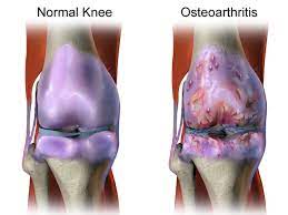
    Your knees work hard every day—absorbing shock, bending, and supporting your weight. But when cartilage wears down, these everyday movements become harder. This breakdown is the core of knee osteoarthritis, a condition affecting 1 in 7 U.S. adults over 25.

    knee joint cartilage breakdown

    Understanding Knee Osteoarthritis

    Healthy cartilage acts like a cushion between bones. When it deteriorates, bones rub together, causing friction and discomfort. Cleveland Clinic research shows this process often starts with minor joint stiffness after waking up or sitting for hours. These fleeting sensations might seem harmless, but they’re early distress signals.

    Nearly 40% of people with joint pain delay seeing a doctor for 6+ months. Yet studies confirm that early treatment—like physical therapy or anti-inflammatory strategies—can slow damage by up to 50%.

    The Importance of Recognizing Mild Symptoms

    Ignoring subtle changes risks irreversible harm. For example:

    • Morning tightness lasting under 30 minutes
    • Mild swelling after gardening or climbing stairs
    • Occasional clicking sounds without pain

    These symptoms often precede severe mobility loss. Annual checkups help catch issues before they escalate. As one orthopedic specialist notes: “Patients who address stiffness early rarely need joint replacements later.”

    Proactive care preserves flexibility. Simple steps—like staying active or managing weight—protect cartilage better than waiting for pain to intensify.

    Recognizing Early Signs of Knee Osteoarthritis: Mild Symptoms You Shouldn’t Ignore

    Many dismiss occasional discomfort as normal aging, but these subtle shifts often reveal cartilage wear. Research shows 68% of adults with persistent joint sensations develop confirmed degeneration within five years. Listening to your body’s signals now can prevent major mobility challenges later.

    Joint Pain and Stiffness

    Mild aches after activity or brief morning tightness often mark the earliest phase of cartilage breakdown. Cleveland Clinic studies found 82% of patients with these symptoms showed measurable joint space narrowing. Unlike typical soreness, osteoarthritis-related discomfort:

    • Worsens after periods of inactivity
    • Eases within 30 minutes of movement
    • Recurs predictably with specific actions

    This pattern suggests deteriorating cushioning between bones. When ignored, surrounding muscles compensate, leading to fatigue and secondary strains.

    Swelling and Unusual Sounds

    Inflammation often follows cartilage erosion as joints produce excess fluid. A 2023 Johns Hopkins review linked recurrent swelling to 3x faster progression rates. Accompanying noises—like crunching or grinding (crepitus)—signal uneven bone surfaces rubbing together.

    Key indicators needing attention:

    • Visible puffiness without injury
    • Persistent warmth around the joint
    • Audible pops during knee bends

    Tracking symptom frequency helps clinicians assess key indicators of joint changes. Early intervention preserves mobility and reduces future replacement likelihood by 41%, per recent data.

    Common Symptoms and What They Mean

    Joint discomfort often whispers before it shouts. Recognizing subtle changes in how your body moves and feels helps separate normal aging from developing cartilage erosion. Let’s decode two frequent complaints and their implications.

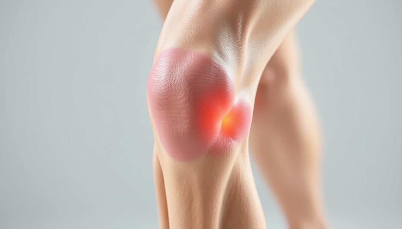
    Unexplained Knee Pain and Tenderness

    Aching that appears without injury often signals wear and tear. Unlike muscle soreness, this deep tenderness lingers after activity and worsens with stairs or squatting. Orthopedic specialists note:

    • Persistent discomfort in one specific area
    • Tenderness when pressing around the kneecap
    • Pain that improves with rest but returns predictably

    These patterns suggest thinning cartilage. When protective tissue erodes, bones begin grinding—a process visible on X-rays long before severe limitations appear.

    Morning Stiffness and Loss of Flexibility

    Struggling to straighten your leg after waking up? Stiffness lasting under 30 minutes often marks early joint changes. Reduced range of motion—like difficulty bending past 90 degrees—indicates inflammation and cartilage loss.

    Symptom Osteoarthritis Rheumatoid Arthritis Gout
    Pain Pattern Worsens with use Symmetrical joints Sudden attacks
    Stiffness Duration <30 minutes >1 hour Variable
    Swelling Mild, localized Warm, tender Intense redness
    Common Triggers Activity Rest Diet

    Cracking sounds during movement (crepitus) occur in 78% of confirmed cases. While unsettling, these noises alone don’t confirm damage—context matters. Tracking symptom combinations helps clinicians distinguish between arthritis types and create targeted treatment plans.

    Causes, Risk Factors, and Underlying Issues

    Our knees bear the brunt of daily life, but some factors accelerate their decline. While cartilage naturally thins with age, certain habits and histories amplify damage. Recognizing these triggers helps delay progression and guides smarter lifestyle choices.

    Wear and Tear and Previous Injuries

    Years of repetitive motion grind down joint cushioning. Athletes with ACL tears face 3x higher osteoarthritis risks, according to Johns Hopkins research. Even minor fractures from decades past alter knee mechanics, creating uneven pressure points.

    Common culprits include:

    • Occupations requiring heavy lifting
    • Untreated meniscus injuries
    • Improperly healed fractures

    Overuse, Obesity, and Inflammation

    Excess weight multiplies joint stress—every 10 pounds adds 40 pounds of pressure per step. Chronic inflammation from conditions like diabetes speeds cartilage breakdown. This dual assault reduces range of motion and often leads to advanced treatments.

    Key connections:

    • BMI over 30 triples knee replacement likelihood
    • High-impact sports accelerate wear patterns
    • Persistent swelling indicates cellular damage

    When conservative measures fail, surgery becomes necessary. However, managing risk factors early preserves natural joint function longer. As one physical therapist notes: “Controlling inflammation and mechanical stress keeps more patients out of operating rooms.”

    Diagnosis and Clinical Evaluations

    Accurate diagnosis forms the foundation for managing joint health effectively. Healthcare providers combine physical exams with advanced imaging to map cartilage integrity and pinpoint damage. Early evaluations help preserve movement capabilities while expanding treatment options before irreversible changes occur.

    How Healthcare Providers Assess Symptoms

    Doctors begin with detailed questions about discomfort patterns and daily limitations. They check for swelling, test range of motion, and apply pressure to identify tender areas. A 2023 study found clinicians who assess walking gait detect early degeneration 28% more accurately than those relying solely on patient reports.

    The Role of X-Rays and Physical Tests

    Imaging reveals what physical exams can’t. X-rays measure joint space narrowing—a key indicator of cartilage loss. MRIs show soft tissue damage, while blood tests rule out inflammatory arthritis. Common diagnostic tools include:

    Method Purpose Key Findings
    X-ray Visualize bone alignment Joint space reduction
    MRI Assess soft tissues Cartilage thinning
    Physical Tests Evaluate mobility Limited flexion/extension
    Blood Work Rule out other conditions Inflammation markers

    Regular assessments guide personalized plans combining exercises, weight management, and anti-inflammatory strategies. As one rheumatologist notes: “Timely imaging cuts diagnostic delays by half, giving patients faster access to relief.” Proactive care improves long-term life quality while reducing dependency on aggressive interventions.

    Treatment Options: Non-Surgical and Home Care Approaches

    Effective management of joint discomfort begins with personalized strategies that address both movement and inflammation. Cleveland Clinic studies show combining activity modifications with targeted therapies reduces strain by 38% in early-stage cases while preserving mobility.

    Exercises, Movement, and Weight Management

    Low-impact activities maintain joint function without worsening injury risks. A 2024 analysis revealed patients who followed tailored exercise plans saw 52% less cartilage loss over five years. Key approaches include:

    • Water aerobics to reduce pressure during movement
    • Cycling with proper seat height alignment
    • Strength training for supporting muscles
    Activity Frequency Benefit
    Tai Chi 3x/week Improves balance
    Elliptical 4x/week Low joint stress
    Leg Raises Daily Builds quad strength

    Medications, Injections, and Pain Relief Strategies

    Anti-inflammatory care complements physical efforts. Corticosteroid injections provide 2-3 months of relief by calming swollen tissues. Newer hyaluronic acid injections act as synthetic joint lubricants, with 67% reporting improved flexibility.

    Daily habits matter too:

    • Alternating heat/ice packs
    • Using assistive devices during flare-ups
    • Tracking symptom patterns in a journal

    As one physiatrist notes: “Patients who combine weight loss with guided care rarely progress to needing surgery.” Regular check-ins help adjust plans as needs change.

    When to Consider Surgical Interventions

    While most joint issues respond to conservative care, advanced cases demand stronger solutions. Research shows 15-20% of patients eventually require surgical approaches when cartilage loss severely impacts daily life. Timing matters—procedures work best before surrounding muscles weaken from prolonged limited mobility.

    Evaluating Surgical Options

    Total knee replacement becomes necessary when:

    • Pain persists despite 6+ months of other treatment options
    • X-rays show bone-on-bone contact
    • Morning stiffness lasts over an hour daily

    Alternatives like osteotomy (bone realignment) or partial replacements suit younger patients with localized damage. A 2024 Johns Hopkins study found 78% of partial knee recipients maintained natural joint function for 12+ years.

    Damage Progression Indicators

    These signs suggest irreversible decline:

    Symptom Non-Surgical Response Surgical Threshold
    Walking Distance Improved with rest Limited to 1 block
    Night Pain Occasional Daily disruption
    Joint Deformity Mild Visible bowing

    Orthopedic surgeon Dr. Lisa Nguyen notes: “Patients who address severe mobility loss within 2 years of onset have 40% better recovery rates post-surgery.” Early-stage treatment options like viscosupplementation injections can delay knee replacement needs by 8-10 years in many cases.

    Lifestyle Modifications and Preventative Measures

    Taking charge of joint health starts with small, consistent actions. Research confirms that tailored movement plans and home care strategies can delay cartilage breakdown by up to 40%. These approaches empower individuals to maintain independence while reducing future medical interventions.

    Daily Exercises and Mobility Routines

    Low-impact activities preserve joint function without straining tissues. A 2024 study showed patients who walked 30 minutes daily reduced replacement surgery risks by 22%. Key routines include:

    • Water-based exercises to support the body’s weight
    • Yoga poses that improve hip and ankle flexibility
    • Resistance band training for muscle balance
    Activity Frequency Joint Benefit
    Swimming 3x/week Reduces pressure
    Cycling 4x/week Enhances circulation
    Leg Slides Daily Maintains range

    Preventative Care and Home Remedies

    Simple habits combat inflammation and protect cartilage. Orthopedic specialists recommend alternating heat therapy with cold packs during flare-ups. Dietary changes—like adding omega-3s—can lower swelling markers by 18%.

    Effective home strategies:

    • Elevating legs after prolonged standing
    • Using ergonomic cushions during seated work
    • Tracking activity levels to avoid overexertion

    Regular communication with your doctor ensures personalized adjustments. As Dr. Ellen Torres notes: “Patients who pair smart activities with anti-inflammatory diets often avoid aggressive treatments altogether.” Proactive care keeps joints functional and delays surgical timelines.

    Conclusion

    Understanding your body’s signals could be the key to preserving mobility. We’ve explored how subtle joint changes often precede significant damage, emphasizing why timely action matters. Addressing discomfort early—through movement adjustments or medical guidance—can slow progression and maintain flexibility.

    Multiple home care strategies and clinical treatments exist across the care spectrum. From aquatic therapy to joint replacements, options adapt as needs evolve. Regular checkups help identify risk factors like weight patterns or past injuries that might contribute to decline.

    Research backed by Cleveland Clinic confirms proactive care reduces surgical needs by nearly half. Don’t dismiss recurring stiffness or swelling—these could signal the root cause of cartilage loss. Tracking symptoms and seeking evaluations promptly helps create effective, personalized plans.

    Your journey toward joint health starts now. Schedule a consultation if movements feel restricted or discomfort lingers. Early steps today can prevent irreversible damage tomorrow while keeping risk levels manageable through informed choices.

    FAQ

    What are the first signs of knee osteoarthritis?

    Initial symptoms often include mild joint pain, stiffness after sitting or resting, and occasional swelling. Some people notice clicking or grinding sounds during movement. These early signs may come and go but tend to worsen over time if ignored.

    Can weight loss help reduce knee osteoarthritis symptoms?

    Yes. Excess weight strains joints, accelerating cartilage wear. Losing even 10% of body weight can ease pressure, improve mobility, and slow progression. Pairing weight management with low-impact exercises like swimming often yields better results.

    How do doctors confirm a knee osteoarthritis diagnosis?

    Providers use physical exams to check for tenderness, swelling, and range of motion. X-rays reveal cartilage loss or bone spurs, while MRI scans assess soft tissue damage. Blood tests may rule out other conditions like rheumatoid arthritis.

    Are corticosteroid injections safe for long-term pain relief?

    While effective for short-term inflammation control, frequent steroid injections can weaken cartilage or surrounding tissues over time. Most doctors limit them to 3-4 doses annually. Alternatives like hyaluronic acid injections or physical therapy may offer safer long-term relief.

    When should someone consider knee replacement surgery?

    Surgery is typically recommended when pain severely limits daily activities, conservative treatments fail, or joint damage appears advanced on imaging. Newer partial replacement options allow faster recovery, but full replacements last 15-20 years for most patients.

    Can exercises worsen knee osteoarthritis symptoms?

    High-impact activities like running may increase joint strain, but controlled movements strengthen muscles supporting the knee. Focus on low-impact exercises like cycling, yoga, or tai chi. Always consult a physical therapist to tailor routines to your condition.

    Does morning stiffness always indicate osteoarthritis?

    Not necessarily. Brief stiffness (

    Are over-the-counter pain medications sufficient for managing symptoms?

    NSAIDs like ibuprofen (Advil) or naproxen (Aleve) temporarily reduce pain and inflammation. However, long-term use risks stomach or kidney issues. Acetaminophen (Tylenol) is safer for frequent use but doesn’t address inflammation. Always combine medications with lifestyle changes for sustained relief.

  • Dealing with Hot Knees: Causes and Solutions

    Dealing with Hot Knees: Causes and Solutions

    Hot knees can be a source of discomfort and inconvenience, impacting daily activities and overall quality of life. Understanding the causes and solutions for hot knees is essential for effective management. In this article, we will explore the anatomy of the knee joint, identify the causes of hot knees, discuss diagnostic techniques, and explore treatment options to alleviate this common issue.

    Key Takeaways

    • Hot knees can be caused by inflammatory conditions, overuse and strain, or environmental factors.
    • Diagnostic techniques for hot knees include physical examination, imaging studies, and laboratory tests.
    • Treatment options for hot knees may involve medication and pain management, physical therapy and rehabilitation, or surgical interventions.
    • Understanding the structure and function of the knee joint is crucial in addressing hot knee problems.
    • Hot knees can significantly impact daily activities, highlighting the importance of effective management strategies.

    Understanding the Anatomy of the Knee Joint

    thumbnail

    Structure and Function of the Knee Joint

    The knee joint is a complex structure that plays a crucial role in supporting our body weight and facilitating movement. It consists of several components, including the femur, tibia, and patella, which work together to provide stability and flexibility. Additionally, the knee joint is surrounded by a network of ligaments and tendons that help maintain its integrity.

    One of the key functions of the knee joint is to absorb shock and distribute the forces generated during activities such as walking, running, and jumping. This is achieved through the presence of specialized cartilage, such as the meniscus, which acts as a cushion and reduces friction between the bones. The intricate interplay of these structures allows for smooth and coordinated motion, essential for our daily activities.

    Component Function
    Femur Weight-bearing bone
    Tibia Supports body weight
    Patella Protects knee joint
    Ligaments Provide stability
    Tendons Facilitate movement

    Common Knee Problems

    Structure and Function of the Knee Joint

    Common Knee Problems

    Hot knees can be caused by a variety of factors, including inflammation, overuse, and environmental factors. These issues can significantly impact our daily activities, making it important to address them promptly and effectively.

    • Inflammatory Conditions
    • Overuse and Strain
    • Environmental Factors

    Impact of Hot Knees on Daily Activities

    We often underestimate the pivotal role our knees play in our daily lives until we experience discomfort like hot knees. This sensation can be a sign of underlying issues that, if left unaddressed, may lead to more serious complications. The impact of hot knees on daily activities can be significant, affecting everything from simple movements to our ability to engage in physical exercise.

    • Mobility and Stability: Our knees are crucial for both mobility and stability. When they’re inflamed or painful, even basic tasks such as walking, climbing stairs, or standing for extended periods become challenging.
    • Sleep Disturbances: Hot knees can also disrupt sleep patterns, as the discomfort may prevent finding a comfortable position, leading to restlessness and fatigue.

    Tip: To minimize the impact on daily activities, it’s essential to address hot knee symptoms promptly with appropriate interventions.

    • Work and Recreation: Professional responsibilities and recreational activities that require knee exertion can be particularly affected, potentially leading to decreased productivity and a diminished quality of life.

    Identifying the Causes of Hot Knees

    thumbnail

    Inflammatory Conditions

    Inflammatory conditions play a significant role in the development of hot knees. Chronic inflammation within the knee joint can lead to discomfort and heat sensation. It is important to consider conditions such as arthritis and synovitis, which are known to contribute to the heat experienced in the knees. Additionally, autoimmune disorders like rheumatoid arthritis can exacerbate the inflammatory response, further intensifying the sensation of heat within the knee joint. Understanding the underlying inflammatory processes is crucial in addressing the root cause of hot knees.

    • Implement a table for presenting structured, quantitative data. Ensure it’s succinct and formatted correctly in Markdown.
    • Use a bulleted or numbered list for less structured content, like steps, qualitative points, or a series of related items.

    Overuse and Strain

    Overuse and strain are common contributors to the development of hot knees. Prolonged and repetitive stress on the knee joint can lead to inflammation and discomfort. It’s important to recognize the signs of overuse and strain to prevent further complications. Here’s a brief overview of the common causes:

    Causes of Hot Knees
    Overexertion
    Misalignment
    Excessive Weight
    Poor Footwear

    Seeking professional guidance and making necessary lifestyle adjustments can help alleviate the symptoms and promote knee health.

    Environmental Factors

    Environmental factors play a significant role in the development of hot knees. Excessive heat exposure and humidity are known to exacerbate knee discomfort, especially in individuals with pre-existing knee conditions. Additionally, prolonged exposure to pollutants and irritants in the environment can contribute to inflammation and discomfort in the knee joint. It’s important to be mindful of our surroundings and take necessary precautions to minimize the impact of environmental factors on knee health.

    • Implement a table for presenting structured, quantitative data. Ensure it’s succinct and formatted correctly in Markdown.
    • Use a bulleted or numbered list for less structured content, like steps, qualitative points, or a series of related items.

    Diagnostic Techniques for Hot Knees

    Physical Examination

    When we approach the issue of hot knees, our initial step is to conduct a thorough physical examination. This process allows us to observe the knee’s range of motion, detect any swelling or redness, and identify areas of tenderness. We systematically assess the knee by palpating specific anatomical landmarks and by asking patients to perform certain movements that may reveal underlying problems.

    Palpation and observation during the physical examination can provide us with valuable clues about the knee’s condition. For instance, warmth and redness over the joint may indicate an inflammatory process, while crepitus— a crackling sensation felt under the skin—could suggest cartilage wear.

    Remember, the physical examination is a non-invasive way to gather important information and should always be performed with care to avoid exacerbating any knee discomfort.

    The following list outlines the key components we examine:

    • Inspection of the knee for signs of swelling and discoloration
    • Assessment of the knee’s range of motion
    • Palpation to pinpoint areas of warmth or tenderness
    • Evaluation of the ligaments and tendons for stability and integrity
    • Observation of gait and posture to identify biomechanical issues that may affect the knee

    Imaging Studies

    After undergoing the Imaging Studies, we can analyze the results to identify any abnormalities or structural issues in the knee joint. This analysis may involve comparing the images to standard measurements and reference values. For example, the size and alignment of the knee joint components can be assessed to determine any deviations from the norm. Additionally, the presence of any inflammation or fluid accumulation can be noted, providing valuable insights into the underlying causes of hot knees.

    Furthermore, it may be beneficial to create a table summarizing the quantitative data obtained from the imaging studies. This table could include measurements of joint space, bone density, and any visible abnormalities. Such a structured presentation of data can aid in the comprehensive evaluation of the knee joint’s condition and guide further diagnostic and treatment decisions.

    Lastly, it’s important to note that the interpretation of imaging studies should be performed by qualified healthcare professionals with expertise in musculoskeletal radiology and diagnostic imaging.

    It is crucial to ensure that the imaging studies are of high quality and accurately capture the details of the knee joint. This can significantly impact the accuracy of the diagnostic findings and subsequent treatment recommendations.

    Laboratory Tests

    After conducting a thorough physical examination and reviewing the results of imaging studies, we proceed to perform laboratory tests to assess specific biomarkers and inflammatory markers. These tests provide valuable quantitative data that aid in confirming the presence of underlying inflammatory conditions or other pathologies. The results of these tests, including levels of C-reactive protein and erythrocyte sedimentation rate, play a crucial role in guiding the subsequent course of treatment and management strategies for hot knees. It is important to interpret these results in conjunction with the clinical presentation and imaging findings to ensure a comprehensive understanding of the patient’s condition. Additionally, the use of a bulleted list can effectively summarize the key laboratory tests:

    Treatment Options for Hot Knees

    thumbnail

    Medication and Pain Management

    In our quest to alleviate the discomfort associated with hot knees, we often turn to medication and pain management strategies. These approaches are designed to reduce inflammation, alleviate pain, and improve joint function. It’s essential to strike a balance between effective pain relief and the minimization of potential side effects.

    Nonsteroidal anti-inflammatory drugs (NSAIDs), such as ibuprofen and naproxen, are commonly prescribed to manage knee pain. However, for more severe cases, stronger painkillers may be necessary. We must be cautious, as researchers have highlighted concerns regarding the prescription of painkillers for inflammatory conditions due to a lack of evidence and the risk of serious side effects.

    It is advisable to consider alternative treatments in conjunction with medication, such as disease-modifying antirheumatic drugs (DMARDs) and tailored exercise programs, to enhance outcomes and reduce dependency on painkillers.

    Here is a list of common medications and their purposes in managing hot knees:

    • NSAIDs: Reduce inflammation and alleviate pain
    • Corticosteroids: Decrease severe inflammation
    • Analgesics: Provide pain relief
    • DMARDs: Slow disease progression and improve joint health
    • Biologic agents: Target specific pathways in the inflammatory process

    Physical Therapy and Rehabilitation

    After undergoing a comprehensive physical examination and imaging studies, physical therapy and rehabilitation play a crucial role in the management of hot knees. Our treatment plan involves a combination of strengthening exercises, flexibility training, and low-impact activities to improve knee function and reduce discomfort. Additionally, we emphasize the importance of proper body mechanics and posture to prevent further strain on the knee joint.

    Furthermore, a structured approach to rehabilitation is essential for achieving optimal outcomes. Our program includes a progressive exercise regimen tailored to individual needs, with a focus on restoring range of motion and building muscle strength. We also provide education on self-care techniques and lifestyle modifications to support long-term knee health.

    For a more detailed overview of the rehabilitation program, please refer to the following table:

    Rehabilitation Program Components Description
    Strengthening Exercises Target major muscle groups to improve stability and support for the knee joint.
    Flexibility Training Focus on enhancing joint mobility and reducing stiffness.
    Low-Impact Activities Incorporate gentle movements to maintain cardiovascular fitness without exacerbating knee discomfort.

    In addition to the structured program, it is important to note that consistency and compliance with the prescribed regimen are key factors in achieving successful rehabilitation outcomes. Our team is dedicated to providing ongoing support and guidance to ensure that each individual receives the best possible care for their hot knees.

    Surgical Interventions

    After considering all treatment options, surgical intervention may be recommended for severe cases of hot knees. This may involve procedures such as arthroscopy or knee replacement surgery. The decision to undergo surgery should be carefully discussed with a qualified orthopedic surgeon. It’s important to weigh the potential benefits against the risks and recovery process. Here is a brief overview of the surgical interventions:

    Procedure Description
    Arthroscopy Minimally invasive procedure to diagnose and treat knee joint problems.
    Knee Replacement Surgery Surgical procedure to replace a damaged knee joint with an artificial implant.

    It’s essential to follow post-operative care instructions diligently to optimize recovery and minimize complications. Maintaining a healthy lifestyle and adhering to the prescribed rehabilitation program are crucial for successful outcomes. Patients should also be aware of potential risks and complications associated with surgical interventions, and seek guidance from their healthcare provider if any concerns arise.

    Conclusion

    In conclusion, hot knees can be caused by a variety of factors, including overuse, injury, and medical conditions. It is important to listen to your body and seek professional medical advice if you experience persistent knee heat. By addressing the underlying causes and implementing appropriate remedies, individuals can effectively manage and alleviate hot knee symptoms, thereby improving their overall quality of life.

    Frequently Asked Questions

    What are the common symptoms of hot knees?

    Common symptoms of hot knees include warmth, redness, swelling, and pain in the knee joint.

    Can hot knees be a sign of a serious medical condition?

    In some cases, hot knees can indicate underlying inflammatory conditions or joint diseases that require medical attention.

    How can I relieve hot knees at home?

    Applying ice packs, elevating the knee, and resting can help relieve hot knees at home.

    Are there specific exercises that can help with hot knees?

    Low-impact exercises such as swimming, cycling, and gentle stretching can help improve knee mobility and reduce discomfort.

    What are the potential complications of untreated hot knees?

    Untreated hot knees can lead to chronic pain, joint damage, and limited mobility, affecting daily activities and quality of life.

    When should I seek medical advice for hot knees?

    It is advisable to seek medical advice if hot knees persist, worsen over time, or are accompanied by fever, severe swelling, or difficulty bearing weight on the affected knee.

  • Iliotibial Syndrome: Causes and Treatment

    Iliotibial syndrome, also known as IT band syndrome, is a common overuse injury that affects the iliotibial band, a thick band of fascia running along the outside of the thigh. This article explores the anatomy and function of the iliotibial band, the causes of iliotibial syndrome, diagnosis methods, and treatment options.

    Key Takeaways

    • Iliotibial syndrome is often caused by biomechanical factors and training errors.
    • Clinical evaluation is an important diagnostic tool for identifying iliotibial syndrome.
    • Conservative management, including rest, stretching, and physical therapy, is the primary treatment approach for iliotibial syndrome.
    • Surgical interventions may be considered for severe or persistent cases of iliotibial syndrome.
    • Prevention strategies, such as proper warm-up, cross-training, and gradual progression of activity, can help reduce the risk of developing iliotibial syndrome.

    Anatomy and Function of the Iliotibial Band

    thumbnail

    Structure of the Iliotibial Band

    The iliotibial band (ITB) is a thick, fibrous band of connective tissue that extends from the iliac crest in the pelvis down to the tibia in the lower leg. Its primary composition is fascial tissue, which is dense and fibrous, providing both flexibility and tensile strength. The ITB plays a crucial role in the stabilization of the knee, especially during activities that involve running or cycling.

    Anatomically, the ITB is not a discrete structure but rather a thickening of the tensor fasciae latae muscle’s fascia. This integration with muscle tissue is what allows the ITB to assist in hip abduction, flexion, and rotation. The distal portion of the ITB also contributes to lateral knee stabilization by attaching to the Gerdy’s tubercle on the tibia.

    • The ITB originates from the iliac crest.
    • It is comprised of fascial tissue.
    • It attaches to the Gerdy’s tubercle on the tibia.

    Tip: A well-balanced strength and flexibility training program can help maintain the proper function of the ITB and prevent overuse injuries.

    Function of the Iliotibial Band

    We recognize the iliotibial band (ITB) as a critical structure in lower limb biomechanics, particularly for those engaged in repetitive activities such as running and cycling. Its primary function is to stabilize the knee during gait, working in concert with other muscles around the hip and thigh.

    Stabilization is not the ITB’s only role; it also assists in the abduction of the hip, which is essential for maintaining pelvic balance during single-leg support phases of walking or running. This secondary function underscores the ITB’s importance in coordinated movement and injury prevention.

    • The ITB contributes to lateral knee stabilization.
    • It aids in hip abduction.
    • The band plays a role in shock absorption, reducing the impact on the knee joint.

    Tip: A well-conditioned ITB is less susceptible to overuse injuries, emphasizing the need for targeted strength and flexibility exercises in preventative care.

    Causes of Iliotibial Syndrome

    thumbnail

    Biomechanical Factors

    We recognize that the development of Iliotibial Syndrome is often closely linked to various biomechanical factors. These factors can include abnormalities in leg length, muscle imbalances, and foot structure, which may contribute to the stress on the iliotibial band during activity. For instance, a discrepancy in leg length can lead to an uneven gait, placing more strain on one side of the body.

    • Overpronation of the foot, where the foot rolls inward excessively when walking or running, can also increase tension in the iliotibial band. Similarly, weak hip abductor muscles may fail to stabilize the pelvis, resulting in increased movement and stress on the IT band. It’s essential to identify and address these biomechanical issues to prevent or alleviate symptoms.

    Here is a list of common biomechanical factors that may lead to Iliotibial Syndrome:

    • Leg length discrepancy
    • Muscle imbalances
    • Overpronation of the foot
    • Weak hip abductor muscles

    Tip: Regular strength training and stretching exercises targeting the hip abductors can help in correcting muscle imbalances and reducing the risk of Iliotibial Syndrome.

    Training Errors

    Training errors, such as overtraining or sudden increases in training intensity, can contribute to the development of iliotibial syndrome. It is important to gradually increase training volume and intensity to prevent overuse injuries. Additionally, improper footwear and running on uneven surfaces can also exacerbate the condition. Here’s a list of common training errors that may lead to iliotibial syndrome:

    1. Overtraining
    2. Sudden increases in training intensity
    3. Improper footwear
    4. Running on uneven surfaces

    It is crucial to address these training errors to reduce the risk of developing iliotibial syndrome. As a reminder, proper training techniques and gradual progression are key to preventing this condition.

    Diagnosis of Iliotibial Syndrome

    thumbnail

    Clinical Evaluation

    Following the initial consultation, we conduct a thorough clinical evaluation to diagnose Iliotibial Syndrome. This involves a detailed history taking to understand the onset and progression of symptoms. We pay close attention to the patient’s activity level and any recent changes in training routines or equipment.

    Physical examination is pivotal. We assess for tenderness along the iliotibial band, especially at its insertion on the lateral epicondyle of the knee. The Ober test, which evaluates the tightness of the iliotibial band, is a key component of our assessment. Additionally, we may perform the Noble compression test to provoke symptoms and confirm the diagnosis.

    It’s essential to rule out other conditions that can mimic Iliotibial Syndrome. Therefore, we consider the differential diagnosis carefully. Here’s a simplified list of conditions we consider:

    • Lateral meniscus tear
    • Lateral collateral ligament injury
    • Popliteal tendonitis
    • Bursitis

    Tip: A comprehensive clinical evaluation is crucial for an accurate diagnosis. It’s important not to rush this process and to consider all potential contributing factors.

    Imaging Studies

    Following a thorough clinical evaluation, we often turn to imaging studies to confirm the diagnosis of iliotibial syndrome. While MRI scans are not routinely used, they can be particularly helpful in visualizing the soft tissue structures and any associated inflammation of the iliotibial band. Ultrasound imaging is another tool at our disposal, offering the advantage of dynamic assessment while the joint is in motion.

    Typically, imaging is reserved for cases where the diagnosis is uncertain or if there is a lack of response to conservative treatment. In such scenarios, imaging can provide valuable insights into the extent of the injury and rule out other potential causes of lateral knee pain.

    Remember, imaging should complement, not replace, a comprehensive clinical evaluation.

    When interpreting imaging results, it’s crucial to correlate findings with the patient’s symptoms and physical exam results. This ensures a holistic approach to diagnosis and subsequent treatment planning.

    Treatment Options for Iliotibial Syndrome

    thumbnail

    Conservative Management

    After exploring various conservative management techniques, we have found that rest and stretching are crucial components of recovery. Additionally, we recommend incorporating strength training exercises to address muscle imbalances. Below is a table summarizing the recommended exercises and their frequency:

    Exercise Frequency
    Stretching Daily
    Strength Training 3 times a week

    We advise patients to gradually increase activity levels and to avoid overtraining, as this can exacerbate symptoms. It is important to monitor progress and adjust the treatment plan accordingly. We emphasize the significance of a comprehensive approach that addresses both the symptoms and underlying causes of the syndrome.

    Surgical Interventions

    After considering the various conservative management approaches, surgical interventions may be recommended for individuals with severe and persistent symptoms. These interventions typically involve the release or lengthening of the iliotibial band to alleviate tension and reduce friction. In some cases, a table summarizing the success rates and potential risks of surgical procedures may aid in decision-making. It’s important to note that surgical interventions should be considered as a last resort and only after thorough evaluation and discussion with a healthcare professional. Post-surgery, a comprehensive rehabilitation program is essential for optimal recovery and long-term success.

    Conclusion

    In conclusion, Iliotibial Syndrome is a complex condition that can be caused by a variety of factors, including overuse and biomechanical issues. Treatment typically involves a combination of physical therapy, rest, and targeted exercises to address muscle imbalances. It is important for individuals suffering from this syndrome to seek professional medical advice and adhere to a comprehensive treatment plan in order to achieve long-term relief and prevent recurrence.

    Frequently Asked Questions

    What are the common symptoms of Iliotibial Syndrome?

    Common symptoms include pain on the outside of the knee, swelling, and a clicking sensation during movement.

    Can Iliotibial Syndrome be prevented?

    Yes, it can be prevented by avoiding sudden increases in physical activity, maintaining proper running form, and using proper footwear.

    Is stretching helpful for Iliotibial Syndrome?

    Yes, stretching the iliotibial band and surrounding muscles can help reduce tension and alleviate symptoms.

    What are the risk factors for developing Iliotibial Syndrome?

    Risk factors include overuse, excessive running or cycling, improper footwear, and muscle imbalances.

    Can I continue physical activity if I have Iliotibial Syndrome?

    It is recommended to modify physical activity to avoid exacerbating symptoms, and to consult with a healthcare professional for personalized recommendations.

    How long does it take to recover from Iliotibial Syndrome?

    Recovery time varies depending on the severity of the condition and individual factors, but with proper treatment, most individuals can recover within a few weeks to a few months.

  • Knee Cartilage Degeneration

    Knee Cartilage Degeneration

    Knee Cartilage Degeneration: Causes, Symptoms, and Treatment Options

    Knee cartilage degeneration is a common condition that affects millions of people worldwide. It occurs when the cartilage that cushions the knee joint begins to wear away, leading to pain, stiffness, and limited mobility. While this condition can occur at any age, it is more common in older adults and athletes who put a lot of stress on their knees.

    v2 2idq2 hgbrt

    Understanding Knee Cartilage Degeneration is important for anyone who wants to maintain healthy knees. The knee joint is made up of three bones: the femur, tibia, and patella. Between these bones is a layer of cartilage that acts as a shock absorber, preventing the bones from rubbing against each other. When this cartilage begins to break down, it can cause pain and inflammation in the knee joint. There are several factors that can contribute to knee cartilage degeneration, including age, genetics, obesity, and injury.

    If left untreated, knee cartilage degeneration can lead to more serious complications, such as osteoarthritis. Fortunately, there are several treatment options available to manage this condition, including physical therapy, medication, and surgery. By taking proactive steps to prevent knee cartilage degeneration and seeking prompt treatment when symptoms arise, individuals can maintain healthy knees and enjoy an active lifestyle for years to come.

    Key Takeaways

    • Knee cartilage degeneration is a common condition that affects the knee joint and can cause pain, stiffness, and limited mobility.
    • Factors that contribute to knee cartilage degeneration include age, genetics, obesity, and injury.
    • Treatment options for knee cartilage degeneration include physical therapy, medication, and surgery, and early intervention can help prevent more serious complications.

    Understanding Knee Cartilage Degeneration

    Knee cartilage degeneration is a common condition that occurs when the cartilage in the knee joint wears away. This can lead to pain, swelling, and stiffness in the knee, making it difficult to perform daily activities.

    Causes of Degeneration

    There are several factors that can contribute to knee cartilage degeneration. Osteoarthritis is a common cause, which occurs when the articular cartilage that covers the ends of the tibia and femur bones in the knee joint breaks down over time. This can be due to aging, injury, trauma, wear and tear, or genetics.

    Other factors that can contribute to knee cartilage degeneration include joint damage, physical activity, and obesity. In some cases, there may be no obvious cause for the degeneration.

    Symptoms and Diagnosis

    The symptoms of knee cartilage degeneration can vary depending on the severity of the condition. Common symptoms include knee pain, swelling, stiffness, and limited range of motion. In some cases, there may be a clicking or popping sound when the knee is moved.

    Diagnosis of knee cartilage degeneration typically involves a physical exam, medical history, and diagnostic tests such as x-rays and magnetic resonance imaging (MRI). These tests can help to identify the extent of the damage and determine the best course of treatment.

    Overall, understanding the causes and symptoms of knee cartilage degeneration is important for managing the condition and preventing further damage to the knee joint. If you are experiencing knee pain or other symptoms, it is important to seek medical attention and discuss your treatment options with your healthcare provider.

    Risk Factors and Complications

    v2 2idqa

    Knee cartilage degeneration can be caused by several risk factors that increase the likelihood of developing the condition. In this section, we will discuss identifying risk factors and potential complications associated with knee cartilage degeneration.

    Identifying Risk Factors

    The following factors may increase the risk of knee cartilage degeneration:

    • Age: Cartilage naturally wears down over time, making older adults more susceptible to the condition.
    • Gender: Women are more likely than men to develop knee cartilage degeneration.
    • Obesity: Excess weight puts additional pressure on the knees, which can lead to cartilage damage.
    • Previous injury: A history of knee injuries can increase the risk of developing knee cartilage degeneration.
    • Occupation: Jobs that require repetitive knee movements, such as construction work, can increase the risk of developing the condition.

    Potential Complications

    Knee cartilage degeneration can lead to several complications, including:

    • Joint damage: As cartilage wears down, bones in the knee may rub against each other, causing joint damage.
    • Disability: Severe cases of knee cartilage degeneration can limit mobility and lead to disability.
    • Instability: As cartilage wears down, the knee joint may become unstable, making it more difficult to walk or stand.
    • Medical conditions: Knee cartilage degeneration can increase the risk of developing other medical conditions, such as diabetes and fever.
    • Deformities: In some cases, knee cartilage degeneration can cause the knee joint to become misaligned or deformed.

    Overall, understanding the risk factors and potential complications associated with knee cartilage degeneration is important for early detection and treatment of the condition.

    Treatment and Management

    When it comes to knee cartilage degeneration, there are a variety of treatment options available. The best course of action will depend on the severity of the condition, as well as the individual needs and goals of the patient.

    Non-Surgical Treatments

    For those with mild to moderate knee cartilage degeneration, non-surgical treatments may be effective. These can include:

    • Physical therapy: This can help improve mobility and flexibility, as well as strengthen the muscles around the knee joint.
    • Medications: Anti-inflammatory drugs such as NSAIDs or corticosteroids can help reduce inflammation and pain.
    • Injections: Platelet-rich plasma (PRP) or hyaluronic acid injections may be used to promote healing and reduce pain.
    • Braces: Knee braces can help stabilize the joint and reduce pain during physical activity.

    Surgical Treatments

    In more severe cases of knee cartilage degeneration, surgical treatments may be necessary. These can include:

    • Arthroscopy: This minimally invasive procedure involves inserting a small camera into the knee joint to remove damaged tissue or bone spurs.
    • Autologous chondrocyte implantation: This procedure involves removing healthy cartilage cells from the patient’s knee, growing them in a lab, and then implanting them back into the knee joint to promote healing.
    • Joint replacement surgery: This involves removing the damaged knee joint and replacing it with an artificial joint.

    It’s important to note that surgery is generally considered a last resort, and conservative treatment options should be exhausted before considering surgical intervention.

    If you are experiencing knee pain, redness, or swelling, or if you are having difficulty with mobility or flexibility in your knee joint, it’s important to see a doctor. They can help diagnose the underlying cause of your symptoms and recommend appropriate treatment options.

    Prevention is also key when it comes to knee cartilage degeneration. Maintaining a healthy weight, avoiding repetitive stress on the knee joint, and engaging in regular exercise can all help prevent degenerative joint disease from developing.

    Frequently Asked Questions

    v2 2idqi 91yw6

    What are the early symptoms of knee arthritis?

    The early symptoms of knee arthritis include pain, stiffness, and swelling in the knee joint. You may also experience a decrease in range of motion and difficulty walking. If you are experiencing any of these symptoms, it is recommended that you see a doctor for an evaluation.

    What are some new treatments for osteoarthritis of the knee?

    There are several new treatments for osteoarthritis of the knee, including stem cell therapy, platelet-rich plasma (PRP) therapy, and hyaluronic acid injections. These treatments can help to reduce pain, inflammation, and promote healing of damaged cartilage.

    What causes degeneration of cartilage?

    Degeneration of cartilage can be caused by a variety of factors, including aging, injury, and genetics. Other factors that can contribute to cartilage degeneration include obesity, repetitive stress on the joints, and certain medical conditions such as rheumatoid arthritis.

    Can knee arthritis pain radiate down the leg?

    Yes, knee arthritis pain can radiate down the leg. This is because the nerves that supply sensation to the knee joint also supply sensation to other parts of the leg, such as the thigh and calf.

    What are the symptoms of having no cartilage in the knee?

    The symptoms of having no cartilage in the knee include pain, swelling, stiffness, and a decrease in range of motion. You may also experience a grinding or popping sensation when you move your knee.

    How can knee cartilage damage be treated effectively?

    Knee cartilage damage can be treated effectively through a variety of treatments, including physical therapy, medications, and surgery. The type of treatment that is recommended will depend on the severity of the damage and the underlying cause of the damage. It is important to work closely with your doctor to determine the best course of treatment for you.