Tag: knee fonctions

  • Effective Treatment for Jumper’s Knee: Management and Prevention

    Effective Treatment for Jumper’s Knee: Management and Prevention

    Jumper’s knee, also known as patellar tendinitis, is a common overuse injury that affects the patellar tendon. Athletes, particularly those involved in jumping sports, are at a higher risk of developing this condition. Understanding the causes, symptoms, and effective treatment options is crucial for managing and preventing jumper’s knee.

    Key Takeaways

    • Proper diagnosis is essential for effective treatment of jumper’s knee.
    • Conservative treatments such as rest and ice can help alleviate symptoms of jumper’s knee.
    • Medical interventions like corticosteroid injections may be recommended for severe cases of jumper’s knee.
    • Rehabilitation and physical therapy play a vital role in the recovery and prevention of jumper’s knee.
    • Preventive measures such as proper exercise, conditioning, and technique are key to minimizing the risk of developing jumper’s knee.

    Understanding Jumper’s Knee
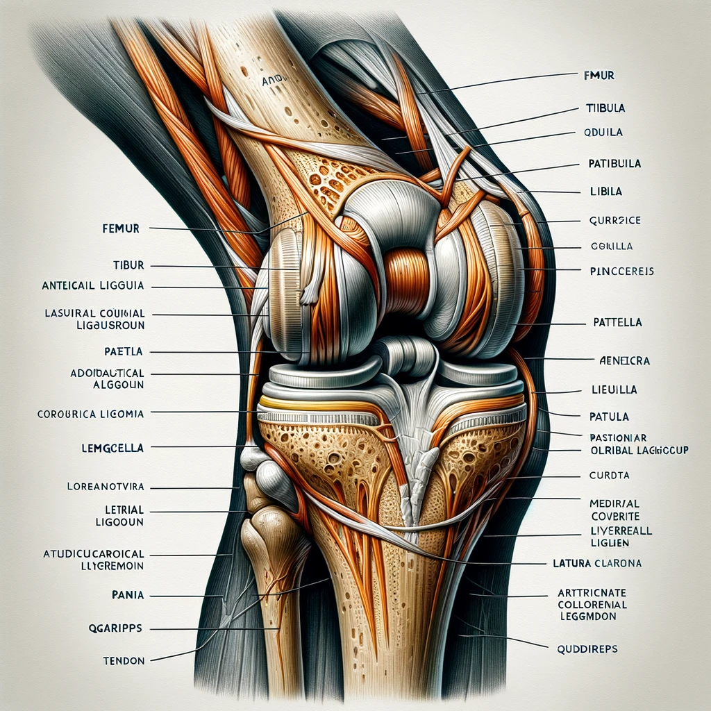
    thumbnail

    Anatomy of the Knee

    Jumper’s knee, also known as patellar tendinitis, is a common overuse injury affecting the knee. It primarily impacts athletes involved in jumping sports, such as basketball and volleyball. The condition is characterized by pain and inflammation in the patellar tendon, which connects the kneecap to the shinbone. This can lead to difficulty in performing physical activities and may require a period of rest to heal properly. Understanding the anatomical structures involved in this condition is crucial for effective management and prevention. Below is a table summarizing the key anatomical components of the knee:| Anatomical Component | Description | |———————|————-| | Patellar Tendon | Connects the kneecap to the shinbone | | Quadriceps Tendon | Attaches the quadriceps muscle to the kneecap | | Patella | Kneecap that protects the knee joint |

    Causes of Jumper’s Knee

    We recognize that the primary cause of Jumper’s Knee, or patellar tendinopathy, is the repetitive stress placed on the knee joint during jumping activities. This overuse leads to microtears in the patellar tendon, which, over time, can result in inflammation and pain.

    Overloading of the tendon is often due to a sudden increase in activity or intensity without adequate conditioning. However, we must also consider intrinsic factors such as muscle imbalance or weakness, particularly in the quadriceps and hamstrings, which can contribute to the development of this condition.

    Biomechanical issues, such as poor alignment or flat feet, can also predispose individuals to Jumper’s Knee. Here’s a brief list of common causes:

    • Repetitive jumping or impact activities
    • Sudden increase in physical activity
    • Muscle imbalance or weakness
    • Biomechanical irregularities

    Tip: Gradual progression in training intensity and volume can help mitigate the risk of overloading the patellar tendon.

    Symptoms and Diagnosis

    Anatomy of the knee is crucial in understanding the complexities of Jumper’s Knee. Patellar tendon and quadriceps muscles play a significant role in this condition. Understanding the causes of Jumper’s Knee is essential for effective management. Overuse, sudden increase in physical activity, and inadequate warm-up are common culprits. Diagnosis involves a thorough physical examination and may include imaging tests such as MRI or ultrasound. It’s important to consult a healthcare professional for accurate diagnosis and treatment planning.

    Treatment Options

    thumbnail

    Conservative Treatments

    In our approach to managing Jumper’s Knee, we prioritize conservative treatments before considering more invasive options. These treatments are designed to alleviate pain and promote healing without the need for surgery.

    The cornerstone of conservative treatment is rest, allowing the inflamed tendon to recover. However, rest does not mean complete inactivity. We encourage patients to engage in low-impact activities that do not exacerbate the knee pain. Alongside rest, we recommend the following:

    • Ice therapy: Applying ice to the affected area to reduce swelling and pain.
    • Compression: Using an elastic bandage or brace to provide support.
    • Elevation: Keeping the knee raised above heart level whenever possible.

    Tip: Consistency in applying these methods is key to reducing symptoms effectively.

    Additionally, over-the-counter anti-inflammatory medications can be beneficial in managing pain and inflammation. It’s important to note that while these treatments can provide relief, they do not address the underlying causes of Jumper’s Knee. Therefore, they should be complemented with physical therapy and exercises tailored to strengthen the knee and improve flexibility.

    Medical Interventions

    When conservative treatments for jumper’s knee do not yield the desired results, we may consider medical interventions. These are typically more invasive and are reserved for cases where pain and dysfunction persist despite exhaustive non-surgical approaches. One common procedure is the injection of platelet-rich plasma (PRP), which utilizes the patient’s own blood components to promote healing in the injured tendon. Corticosteroid injections are also used, though with caution, as they can provide short-term pain relief but may potentially weaken the tendon over time.

    Surgery is an option we consider as a last resort, often for those who have not responded to other treatments over a six-month period. The goal of surgical intervention is to remove damaged tissue and stimulate the growth of healthy tissue. Post-surgery, a structured rehabilitation program is crucial for recovery.

    Tip: Always discuss the potential risks and benefits of medical interventions with your healthcare provider to make an informed decision.

    Here is a brief overview of the medical interventions we might explore:

    • PRP Injections: Encourages natural healing by concentrating growth factors.
    • Corticosteroid Injections: Offers temporary relief but should be used sparingly.
    • Surgery: Considered when other treatments fail, focusing on tissue repair and regeneration.

    Rehabilitation and Physical Therapy

    After undergoing rehabilitation and physical therapy, consistent exercise and proper conditioning are crucial for long-term recovery. Our treatment plan emphasizes a combination of strength training, flexibility exercises, and targeted stretching to improve knee stability and function. Additionally, we recommend incorporating low-impact activities such as swimming or cycling to maintain cardiovascular fitness without exacerbating the injury.

    Furthermore, it’s important to monitor progress and adjust the exercise regimen accordingly. Below is a table summarizing the recommended exercise program:

    Exercise Type Frequency Duration
    Strength Training 3-4 times per week 30-45 mins
    Flexibility Exercises Daily 10-15 mins
    Targeted Stretching 5 times per week 15-20 mins

    In addition to the structured exercise program, here are some key points to keep in mind:

    • Gradually increase the intensity and duration of exercises to avoid overexertion.
    • Utilize proper technique and form during all exercises to prevent further strain on the knee.
    • Ensure that footwear and equipment are appropriate for the chosen activities.

    Our experience has shown that adherence to these guidelines significantly contributes to successful recovery and reduces the risk of re-injury.

    Preventive Measures

    thumbnail

    Exercise and Conditioning

    When it comes to exercise and conditioning, consistent training is key. We should focus on a well-rounded program that includes strength training, flexibility exercises, and cardiovascular activities. A balanced approach to exercise can help improve muscle strength and joint stability, reducing the risk of injury. Additionally, proper warm-up and cool-down routines are essential for injury prevention and overall performance enhancement. Remember, gradual progression is crucial to avoid overuse injuries and achieve long-term success. Here’s a simple table to illustrate the components of an effective conditioning program:

    Component Description
    Strength Training Builds muscle strength and endurance
    Flexibility Exercises Improves range of motion and joint flexibility
    Cardiovascular Activities Enhances heart health and overall fitness

    Lastly, it’s important to listen to our bodies and adjust the intensity and duration of our workouts accordingly. As we continue to prioritize exercise and conditioning, we can make significant strides in preventing and managing jumper’s knee effectively.

    Proper Technique and Form

    When it comes to preventing jumper’s knee, proper technique and form are crucial. We must ensure that our movements are aligned and controlled, reducing the strain on the knee. Additionally, maintaining a balanced stance during physical activities is essential for minimizing the risk of injury. Here are some key points to keep in mind:

    • Maintain proper alignment of the body during movements
    • Distribute weight evenly between both legs
    • Engage core muscles to stabilize the body

    It’s important to remember that even small adjustments in technique can have a significant impact on knee health and injury prevention.

    Equipment and Gear

    In our pursuit to prevent Jumper’s Knee, we must not overlook the significance of equipment and gear. Proper footwear, for instance, can dramatically reduce the stress on the knees by providing adequate cushioning and support. Here’s a brief rundown of essential gear:

    • Footwear: Shoes with good shock absorption and stability.
    • Knee braces: To offer support and limit harmful movements.
    • Compression sleeves: Enhance circulation and provide mild support.
    • Insoles: Custom orthotics can correct biomechanical imbalances.

    Tip: Always consult a professional to find the gear that best suits your individual needs and biomechanics.

    It’s crucial to replace equipment before it wears out to maintain its protective benefits. A worn-out shoe or brace may not only fail to prevent injury but could also actively contribute to its occurrence. We encourage athletes to keep a close eye on the condition of their gear and make timely replacements a priority.

    Conclusion

    In conclusion, the management and prevention of Jumper’s Knee are crucial for athletes and active individuals. With a focus on early intervention and comprehensive treatment strategies, the impact of this condition can be minimized. It is imperative for healthcare professionals and sports practitioners to prioritize patient education and implement evidence-based practices to ensure the long-term well-being of individuals affected by Jumper’s Knee.

    Frequently Asked Questions

    What are the common causes of Jumper’s Knee?

    Jumper’s Knee is commonly caused by repetitive stress or overuse of the knee, such as frequent jumping or running on hard surfaces.

    What are the main symptoms of Jumper’s Knee?

    The main symptoms include pain and tenderness around the kneecap, especially during activities that involve jumping or bending the knee.

    How is Jumper’s Knee diagnosed?

    Jumper’s Knee is typically diagnosed through a physical examination, medical history review, and imaging tests such as X-rays or MRI scans.

    What are some effective conservative treatments for Jumper’s Knee?

    Conservative treatments may include rest, ice therapy, compression, elevation, and the use of supportive braces or straps.

    Are there surgical options for treating Jumper’s Knee?

    In some cases, surgical interventions such as arthroscopic surgery or patellar tendon repair may be considered for severe or persistent cases of Jumper’s Knee.

    What preventive measures can help reduce the risk of Jumper’s Knee?

    Preventive measures may include proper warm-up and stretching, strengthening exercises for the quadriceps and hamstrings, and using appropriate footwear and protective gear during physical activities.

  • Knee Anatomy and Functions And the Key Components of Your Knee

    Knee Anatomy and Functions And the Key Components of Your Knee

    As a physical therapist, I have seen many patients with knee injuries and conditions. Understanding the anatomy and function of the knee joint is essential to prevent injuries, manage pain, and recover from surgery. In this article, I will explain the basic structure of the knee, the role of ligaments and tendons, and the muscles responsible for movement.

    v2 2i6y0

    The knee joint is the largest joint in the body and connects the thigh bone (femur) to the shin bone (tibia). The kneecap (patella) is a small bone that sits in front of the knee joint and protects the joint. The knee joint is a hinge joint that allows for flexion and extension of the leg. It also has some rotational movement, which is important for activities such as walking and running.

    The knee joint is supported by several ligaments and tendons. Ligaments are tough bands of tissue that connect bones to bones, while tendons connect muscles to bones. The ligaments and tendons around the knee joint work together to provide stability and support to the joint. In the next section, I will discuss the role of these structures in more detail.

    Key Takeaways

    • The knee joint is the largest joint in the body and allows for flexion, extension, and some rotational movement.
    • The knee joint is supported by ligaments and tendons that provide stability and support to the joint.
    • Understanding the basic structure and function of the knee joint is essential for preventing injuries, managing pain, and recovering from surgery.

    Basic Structure of the Knee

    As the largest joint in the body, the knee is a complex structure that allows us to walk, run, jump, and perform other physical activities. It is a synovial joint, meaning it contains a fluid-filled capsule that lubricates the joint and reduces friction during movement.

    Bones and Joints

    The knee joint is formed by the articulation of three bones: the femur, tibia, and patella. The femur, or thigh bone, is the longest bone in the body and forms the upper part of the knee joint. The tibia, or shin bone, is the larger of the two bones in the lower leg and forms the lower part of the knee joint. The fibula is the smaller bone in the lower leg and is not directly involved in the knee joint.

    The knee joint is actually two joints in one: the tibiofemoral joint and the patellofemoral joint. The tibiofemoral joint is the main joint between the femur and tibia, while the patellofemoral joint is the joint between the patella and the femur.

    Patella

    The patella, or kneecap, is a sesamoid bone that sits in front of the knee joint and helps to protect the knee and improve the leverage of the quadriceps muscle. The patella is unique in that it is not directly attached to any other bone in the body. Instead, it is connected to the quadriceps tendon and the patellar ligament.

    In summary, the knee joint is a complex structure that is formed by the articulation of three bones: the femur, tibia, and patella. The knee joint is actually two joints in one: the tibiofemoral joint and the patellofemoral joint. The patella, or kneecap, is a sesamoid bone that sits in front of the knee joint and helps to protect the knee and improve the leverage of the quadriceps muscle.

    Knee Ligaments and Tendons

    The knee joint is stabilized and supported by a network of ligaments and tendons. These structures work together to provide strength and stability to the knee joint, allowing us to perform various activities such as walking, running, and jumping.

    Collateral Ligaments

    The collateral ligaments are located on the sides of the knee joint. The medial collateral ligament (MCL) is located on the inner side of the knee, while the lateral collateral ligament (LCL) is found on the outer side. These ligaments help to prevent excessive side-to-side movement of the knee joint.

    Cruciate Ligaments

    The cruciate ligaments are located inside the knee joint and cross each other to form an “X” shape. The anterior cruciate ligament (ACL) is located in the front of the knee, while the posterior cruciate ligament (PCL) is located at the back. These ligaments help to prevent excessive forward and backward movement of the knee joint.

    Tendons

    The patellar tendon and quadriceps tendon are two important tendons that are located in the knee joint. The patellar tendon connects the patella (kneecap) to the tibia (shinbone), while the quadriceps tendon connects the quadriceps muscle to the patella. These tendons help to provide stability to the knee joint and allow us to perform various movements such as jumping and climbing stairs.

    In summary, the knee joint is stabilized and supported by a network of ligaments and tendons. The collateral ligaments help to prevent excessive side-to-side movement of the knee joint, while the cruciate ligaments help to prevent excessive forward and backward movement. The patellar and quadriceps tendons provide stability to the knee joint and allow us to perform various movements.

    Muscles and Movement of the Knee

    v2 2i6yj oazrc

    The knee joint is a hinge joint that allows for flexion and extension of the lower leg. The movement of the knee is controlled by a complex system of muscles, tendons, and ligaments that work together to stabilize and move the joint.

    9d23c210 985b 4333 a7fa ab54f1b02ac7

    Anatomical illustration of the human knee joint, showing all the components along with their names. This includes the bones, ligaments, cartilage, and tendons, with clear labels for each part such as the femur, tibia, fibula, patella, meniscus, and various ligaments and tendons.

    Quadriceps Muscles

    The quadriceps muscles are a group of four muscles located on the front of the thigh that work together to extend the knee joint. These muscles include the rectus femoris, vastus lateralis, vastus intermedius, and vastus medialis. The rectus femoris also works to flex the hip joint.

    Hamstring Muscles

    The hamstring muscles are a group of three muscles located on the back of the thigh that work together to flex the knee joint. These muscles include the biceps femoris, semitendinosus, and semimembranosus. The gracilis muscle also works to flex the knee joint.

    Movements of the Knee

    The knee joint allows for a variety of movements, including flexion, extension, lateral rotation, and medial rotation. Flexion is the movement that brings the heel towards the buttocks, while extension is the movement that straightens the leg. Lateral rotation is the movement that turns the lower leg outward, while medial rotation is the movement that turns the lower leg inward.

    The popliteus muscle is a small muscle located at the back of the knee joint that works to unlock the knee joint during flexion. The flexors and extensors of the knee joint work together to stabilize the joint during movement.

    The tibiofemoral joint is the main joint of the knee, while the patellofemoral joint is the joint between the kneecap and the femur. The articularis genus muscle is a small muscle located at the front of the knee joint that works to pull the synovial membrane of the joint upward during extension.

    The gastrocnemius and plantaris muscles are located at the back of the knee joint and work to plantarflex the ankle joint. Instability of the knee joint can lead to pain and difficulty with activities such as running and walking.

    Knee Injuries and Treatment

    v2 2i6yr 6vmiv

    Common Knee Injuries

    The knee is a complex joint that is susceptible to a variety of injuries. Some common knee injuries include:

    • Anterior Cruciate Ligament (ACL) Tear: This is a common injury that occurs when the ACL is stretched or torn. It can happen during sports that involve sudden stops or changes in direction, such as basketball or soccer. Symptoms include pain, swelling, and instability in the knee.
    • Meniscus Tear: The meniscus is a piece of cartilage that helps cushion the knee joint. A tear can occur when the knee is twisted or bent forcefully. Symptoms include pain, swelling, and difficulty moving the knee.
    • Patellar Tendinitis: This is an injury to the tendon that connects the kneecap to the shinbone. It is often caused by overuse and can result in pain and swelling in the front of the knee.

    how to prevent knee injuries and arthritis

    SectionDescription
    1.Maintain a healthy weight to reduce stress on the knee joint.
    2.Wear appropriate shoes for the activity you are doing.
    3.Warm up before exercising to increase blood flow to the muscles.
    4.Use proper technique when exercising to avoid unnecessary stress on the knee joint.
    5.Incorporate exercises that strengthen the muscles around the knee joint.
    6.Avoid activities that put excessive stress on the knee joint, such as jumping or running on hard surfaces.
    7.Take breaks during activities to rest and stretch the knee joint.
    8.Use knee pads or braces for added support during high-impact activities.
    9.Stay hydrated to help keep the joints lubricated.
    10.Consult with a healthcare professional before starting a new exercise program.

    Treatment and Rehabilitation

    Treatment for knee injuries varies depending on the severity and type of injury. Some common treatments include:

    • Rest: Resting the knee and avoiding activities that aggravate the injury can help reduce pain and swelling.
    • Ice: Applying ice to the knee can help reduce pain and swelling. Ice should be applied for 15-20 minutes at a time, several times a day.
    • Physical therapy: Physical therapy can help strengthen the muscles around the knee joint and improve flexibility and range of motion.
    • Medication: Over-the-counter pain relievers, such as ibuprofen or acetaminophen, can help reduce pain and swelling.

    In some cases, surgery may be necessary to repair a knee injury. Rehabilitation after surgery may include physical therapy and exercises to help regain strength and range of motion.

    Overall, it is important to take steps to prevent knee injuries and to seek treatment promptly if an injury does occur. With proper care and treatment, many knee injuries can be successfully treated, allowing individuals to return to their normal activities.

    Frequently Asked Questions

    v2 2i6yz bljme

    What is the knee joint called?

    The knee joint is a synovial joint that connects three bones: the femur, tibia, and patella. It is a complex hinge joint composed of two articulations: the tibiofemoral joint and patellofemoral joint.

    Is the knee a hinge joint?

    Yes, the knee is a hinge joint. It allows for flexion and extension, as well as a small degree of medial and lateral rotation.

    What are the 3 most commonly injured knee structures?

    The three most commonly injured knee structures are the anterior cruciate ligament (ACL), medial collateral ligament (MCL), and meniscus.

    What are the major anatomical features of the knee?

    The major anatomical features of the knee include bones (femur, tibia, and patella), cartilage, ligaments, tendons, and muscles.

    What is the functional anatomy of the knee?

    The knee joint is responsible for weight-bearing and movement, allowing for flexion and extension, as well as a small degree of medial and lateral rotation. The major muscles involved in knee movement include the quadriceps, hamstrings, and calf muscles.

    What are the symptoms of a torn ligament in your knee?

    Symptoms of a torn ligament in your knee include pain, swelling, instability, and difficulty bearing weight. Depending on the severity of the tear, surgery may be necessary to repair the ligament.