Common Causes of Knee Pain
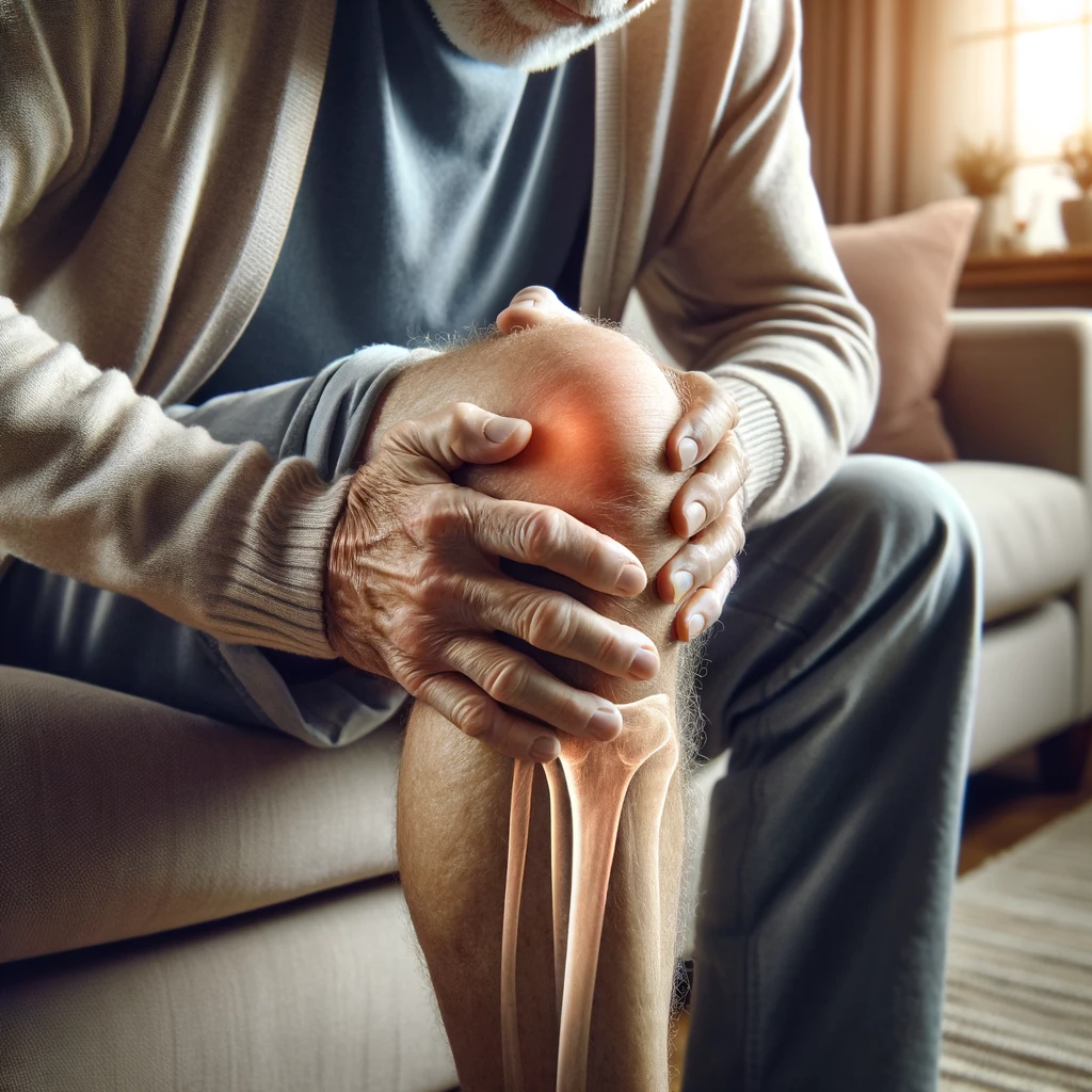
Osteoarthritis
Osteoarthritis involves the breakdown of cartilage that normally provides a cushion between the bones in the knee joint. As this cartilage cushion wears down, the bones begin to rub together, causing pain, swelling, and stiffness. Bony spurs may also form around the joint. Osteoarthritis progresses gradually over years and often affects both knees. Factors that increase risk for knee osteoarthritis include aging, obesity, prior knee injury, overuse, and genetic predisposition. Osteoarthritis cannot be reversed, but symptoms can be effectively managed with a combination of lifestyle changes, medication, injections, physical therapy, assistive devices, and possibly surgery in advanced cases.
ACL Tears
The anterior cruciate ligament (ACL) is one of the key ligaments providing internal stability to the knee joint. ACL tears are a very common athletic knee injury, especially in sports that involve sudden stops, changes in direction, landing from jumps, and pivoting motions. Symptoms of an ACL tear may include hearing a “pop” at the time of injury, knee instability, buckling of the knee, and swelling over the first 24 hours. ACL tears are often caused by an abrupt change in speed or direction combined with deceleration, pivoting with a fixed foot, or landing awkwardly from a jump. Outward forces on the knee can also cause ACL tears. Treatment depends on the patient’s activity level, with options ranging from bracing and physical therapy to surgical ACL reconstruction.
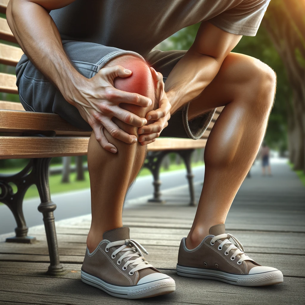
Patellofemoral Pain Syndrome
Patellofemoral pain syndrome (PFPS) is characterized by pain around or behind the kneecap (patella). It results from irritation of the soft tissues of the knee between the patella and the femur. Symptoms include pain and tenderness when bending the knee, using stairs, squatting down, or sitting with knees bent for prolonged periods. PFPS is often caused by repetitive overuse activities that stress the knee joint, such as running. Muscle imbalances of the thigh can also contribute by pulling the kneecap out of alignment. Treatment involves rest and activity modification, physical therapy to strengthen muscles and improve tracking of the kneecap, knee bracing, anti-inflammatory medications, and sometimes surgery.
Treatment Options
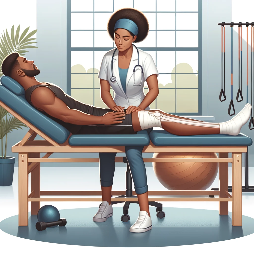
Physical Therapy
Physical therapy is often a key part of knee pain treatment, especially during recovery from injuries. A physical therapist will evaluate areas of muscle weakness or imbalance and design a customized program of flexibility, strengthening, and neuromuscular control exercises. Other physical therapy treatments that may provide relief include manual therapy techniques, ultrasound, ice, heat, electrical stimulation, and compression. For knee osteoarthritis, low-impact exercises to improve mobility and strengthen muscles around the joint are particularly helpful.
Medications
Medications used for knee pain include oral non-steroidal anti-inflammatories (NSAIDs) such as ibuprofen, topical NSAIDs, and analgesics like acetaminophen. These help control pain and swelling. For additional relief, corticosteroid injections can reduce inflammation, while hyaluronic acid injections act as a lubricant and shock absorber. Some supplements like glucosamine may also benefit knee arthritis symptoms. Medications carry potential side effects, so discuss options with your doctor.
Surgery
Surgery may be considered for severe knee ligament and meniscus tears, joint damage from arthritis, or painful misalignment. Common surgeries include arthroscopic debridement/repair, osteotomy realignment, and total knee replacement. Partial knee replacement is also an option for arthritis limited to just one area of the joint. Surgery can relieve pain and improve function, but recovery time and rehabilitation is extensive. Nonsurgical options are usually tried first. Discuss the pros and cons of surgery with your orthopedic specialist.