Tag: leash

  • Understanding the Anatomy of Knee Joint Bones

    Understanding the Anatomy of Knee Joint Bones

    The knee joint is a complex structure that plays a crucial role in supporting the body and facilitating movement. Understanding the anatomy of the knee joint, including its bones and surrounding structures, is essential for both medical professionals and individuals seeking to maintain optimal joint health. In this article, we will explore the structure of the knee joint, focusing on the femur and tibia bones, the patella bone, the ligaments and tendons, and the articular cartilage.

    Key Takeaways

    • The femur and tibia bones form the major weight-bearing components of the knee joint.
    • The patella bone acts as a protective shield for the knee joint and assists in the extension of the leg.
    • Ligaments and tendons provide stability and support to the knee joint, enabling various movements and preventing excessive strain.
    • Articular cartilage helps in reducing friction and providing smooth movement within the knee joint.
    • Understanding the anatomy of the knee joint is crucial for diagnosing and treating knee-related injuries and conditions.

    The Structure of the Knee Joint

    thumbnail

    The Femur and Tibia Bones

    The femur and tibia bones are the primary bones that form the knee joint. These bones play a crucial role in supporting the body’s weight and facilitating movement. The femur is the longest and strongest bone in the body, while the tibia is the second longest and provides stability to the knee joint. The interaction between these two bones is essential for the proper functioning of the knee joint.

    • The femur and tibia bones form the major weight-bearing structure of the knee joint.
    • The alignment and articulation of these bones are critical for stability and mobility.
    • Proper care and attention to these bones are essential for maintaining overall knee health.

    The Patella Bone

    Moving beyond the femur and tibia, we encounter the patella, or kneecap, which plays a crucial role in the knee joint’s function. The patella is a small, triangular bone that protects the knee joint and improves the leverage of the thigh muscles, which are essential for walking, running, and jumping.

    The patella’s posterior surface is lined with articular cartilage, which aids in smooth movement against the femur. This cartilage is vital for absorbing stress and reducing friction during knee motion.

    • The patella increases the leverage of the thigh muscles.
    • It serves as a protective shield for the knee joint.
    • Articular cartilage on the patella’s surface helps in smooth knee movements.

    Remember, the health of the patella’s articular cartilage is key to maintaining knee mobility and reducing the risk of injury.

    The Ligaments and Tendons

    After discussing the ligaments and tendons, we must emphasize the importance of proper treatment for common knee injuries. Common knee injuries requiring surgery include ACL tears, fractures in the kneecap, torn meniscus, and patellar tendonitis. Proper treatment is crucial to avoid chronic pain and complications. It is essential to consult a healthcare professional for accurate diagnosis and personalized treatment plans. Additionally, rehabilitation and physical therapy play a vital role in the recovery process. We cannot stress enough the significance of early intervention and adherence to the prescribed treatment regimen. We must prioritize the long-term health and functionality of the knee joint.

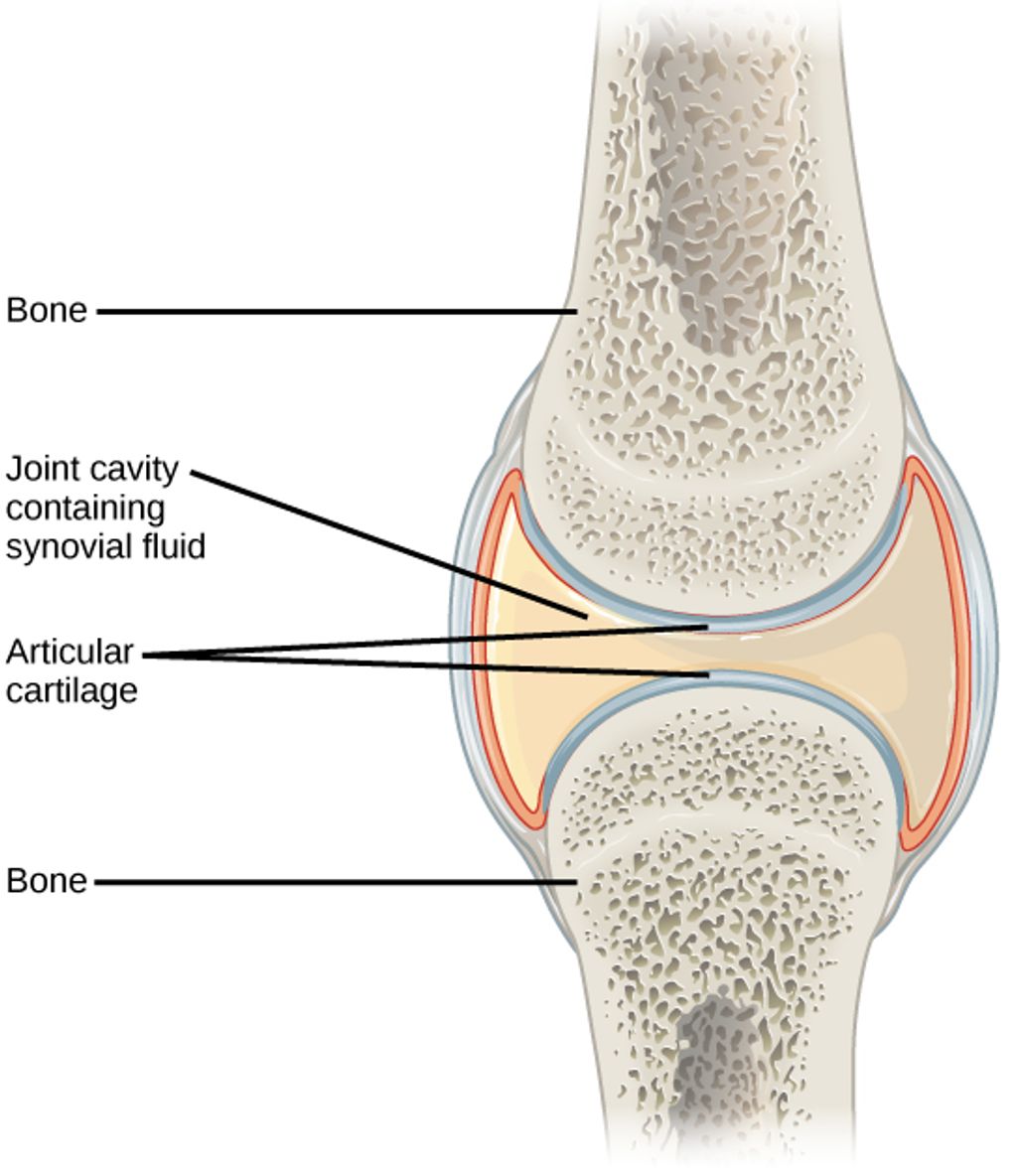
    The Articular Cartilage

    As we delve deeper into the knee joint’s anatomy, we encounter the articular cartilage, a pivotal element in facilitating smooth joint movement. This specialized structure coats the surfaces of the bones within the joint, notably the femur, tibia, and patella, providing a slick, frictionless interface that aids in the effortless bending and extending of the knee.

    The health of the articular cartilage is crucial for maintaining knee function. Over time, it can wear down or become damaged, leading to conditions such as osteoarthritis. To preserve the integrity of this cartilage, it is essential to understand its composition and the factors that affect its well-being.

    • Hyaline cartilage, the most common type found in the knee
    • Collagen fibers, providing tensile strength
    • Proteoglycans, contributing to elasticity
    • Water content, which is high in healthy cartilage

    Tip: Regular exercise and maintaining a healthy weight can help reduce the stress on knee cartilage, potentially slowing the progression of degenerative conditions.

    In our comprehensive guide to knee anatomy, we also explore the morphology and function of the knee joint, as well as common issues that can arise. Effective management of knee health includes understanding over-the-counter solutions for knee pain and adopting strategies for managing discomfort during activities such as running and walking.

    Conclusion

    In conclusion, the anatomy of the knee joint bones is a complex and intricate system that plays a crucial role in human mobility and stability. Understanding the structure and function of these bones is essential for medical professionals, researchers, and individuals seeking to maintain optimal joint health. Further exploration of the interplay between the various components of the knee joint bones holds promise for advancements in orthopedic medicine and the treatment of musculoskeletal conditions.

    Frequently Asked Questions

    What is the function of the femur and tibia bones in the knee joint?

    The femur and tibia bones form the major weight-bearing structure of the knee joint and are responsible for stability and movement.

    What is the purpose of the patella bone in the knee joint?

    The patella bone acts as a protective covering for the knee joint and provides leverage for the quadriceps muscles.

    What are ligaments and tendons in the context of the knee joint?

    Ligaments are tough bands of tissue that connect bones to each other, providing stability to the knee joint. Tendons are fibrous cords that attach muscles to bones, allowing movement of the joint.

    What is the role of articular cartilage in the knee joint?

    Articular cartilage covers the ends of the bones in the knee joint, providing a smooth and low-friction surface for movement and absorbing shock during weight-bearing activities.

    How does the knee joint support the body during various activities?

    The knee joint supports the body by distributing the weight and forces from activities such as walking, running, jumping, and standing.

    What are common injuries or conditions associated with the knee joint?

    Common knee joint injuries and conditions include ligament tears (such as ACL or MCL tears), meniscus tears, arthritis, and patellar dislocation.

  • Effective Treatment for Jumper’s Knee: Management and Prevention

    Effective Treatment for Jumper’s Knee: Management and Prevention

    Jumper’s knee, also known as patellar tendinitis, is a common overuse injury that affects the patellar tendon. Athletes, particularly those involved in jumping sports, are at a higher risk of developing this condition. Understanding the causes, symptoms, and effective treatment options is crucial for managing and preventing jumper’s knee.

    Key Takeaways

    • Proper diagnosis is essential for effective treatment of jumper’s knee.
    • Conservative treatments such as rest and ice can help alleviate symptoms of jumper’s knee.
    • Medical interventions like corticosteroid injections may be recommended for severe cases of jumper’s knee.
    • Rehabilitation and physical therapy play a vital role in the recovery and prevention of jumper’s knee.
    • Preventive measures such as proper exercise, conditioning, and technique are key to minimizing the risk of developing jumper’s knee.

    Understanding Jumper’s Knee

    thumbnail

    Anatomy of the Knee

    Jumper’s knee, also known as patellar tendinitis, is a common overuse injury affecting the knee. It primarily impacts athletes involved in jumping sports, such as basketball and volleyball. The condition is characterized by pain and inflammation in the patellar tendon, which connects the kneecap to the shinbone. This can lead to difficulty in performing physical activities and may require a period of rest to heal properly. Understanding the anatomical structures involved in this condition is crucial for effective management and prevention. Below is a table summarizing the key anatomical components of the knee:| Anatomical Component | Description | |———————|————-| | Patellar Tendon | Connects the kneecap to the shinbone | | Quadriceps Tendon | Attaches the quadriceps muscle to the kneecap | | Patella | Kneecap that protects the knee joint |

    Causes of Jumper’s Knee

    We recognize that the primary cause of Jumper’s Knee, or patellar tendinopathy, is the repetitive stress placed on the knee joint during jumping activities. This overuse leads to microtears in the patellar tendon, which, over time, can result in inflammation and pain.

    Overloading of the tendon is often due to a sudden increase in activity or intensity without adequate conditioning. However, we must also consider intrinsic factors such as muscle imbalance or weakness, particularly in the quadriceps and hamstrings, which can contribute to the development of this condition.

    Biomechanical issues, such as poor alignment or flat feet, can also predispose individuals to Jumper’s Knee. Here’s a brief list of common causes:

    • Repetitive jumping or impact activities
    • Sudden increase in physical activity
    • Muscle imbalance or weakness
    • Biomechanical irregularities

    Tip: Gradual progression in training intensity and volume can help mitigate the risk of overloading the patellar tendon.

    Symptoms and Diagnosis

    Anatomy of the knee is crucial in understanding the complexities of Jumper’s Knee. Patellar tendon and quadriceps muscles play a significant role in this condition. Understanding the causes of Jumper’s Knee is essential for effective management. Overuse, sudden increase in physical activity, and inadequate warm-up are common culprits. Diagnosis involves a thorough physical examination and may include imaging tests such as MRI or ultrasound. It’s important to consult a healthcare professional for accurate diagnosis and treatment planning.

    Treatment Options

    thumbnail

    Conservative Treatments

    In our approach to managing Jumper’s Knee, we prioritize conservative treatments before considering more invasive options. These treatments are designed to alleviate pain and promote healing without the need for surgery.

    The cornerstone of conservative treatment is rest, allowing the inflamed tendon to recover. However, rest does not mean complete inactivity. We encourage patients to engage in low-impact activities that do not exacerbate the knee pain. Alongside rest, we recommend the following:

    • Ice therapy: Applying ice to the affected area to reduce swelling and pain.
    • Compression: Using an elastic bandage or brace to provide support.
    • Elevation: Keeping the knee raised above heart level whenever possible.

    Tip: Consistency in applying these methods is key to reducing symptoms effectively.

    Additionally, over-the-counter anti-inflammatory medications can be beneficial in managing pain and inflammation. It’s important to note that while these treatments can provide relief, they do not address the underlying causes of Jumper’s Knee. Therefore, they should be complemented with physical therapy and exercises tailored to strengthen the knee and improve flexibility.

    Medical Interventions

    When conservative treatments for jumper’s knee do not yield the desired results, we may consider medical interventions. These are typically more invasive and are reserved for cases where pain and dysfunction persist despite exhaustive non-surgical approaches. One common procedure is the injection of platelet-rich plasma (PRP), which utilizes the patient’s own blood components to promote healing in the injured tendon. Corticosteroid injections are also used, though with caution, as they can provide short-term pain relief but may potentially weaken the tendon over time.

    Surgery is an option we consider as a last resort, often for those who have not responded to other treatments over a six-month period. The goal of surgical intervention is to remove damaged tissue and stimulate the growth of healthy tissue. Post-surgery, a structured rehabilitation program is crucial for recovery.

    Tip: Always discuss the potential risks and benefits of medical interventions with your healthcare provider to make an informed decision.

    Here is a brief overview of the medical interventions we might explore:

    • PRP Injections: Encourages natural healing by concentrating growth factors.
    • Corticosteroid Injections: Offers temporary relief but should be used sparingly.
    • Surgery: Considered when other treatments fail, focusing on tissue repair and regeneration.

    Rehabilitation and Physical Therapy

    After undergoing rehabilitation and physical therapy, consistent exercise and proper conditioning are crucial for long-term recovery. Our treatment plan emphasizes a combination of strength training, flexibility exercises, and targeted stretching to improve knee stability and function. Additionally, we recommend incorporating low-impact activities such as swimming or cycling to maintain cardiovascular fitness without exacerbating the injury.

    Furthermore, it’s important to monitor progress and adjust the exercise regimen accordingly. Below is a table summarizing the recommended exercise program:

    Exercise Type Frequency Duration
    Strength Training 3-4 times per week 30-45 mins
    Flexibility Exercises Daily 10-15 mins
    Targeted Stretching 5 times per week 15-20 mins

    In addition to the structured exercise program, here are some key points to keep in mind:

    • Gradually increase the intensity and duration of exercises to avoid overexertion.
    • Utilize proper technique and form during all exercises to prevent further strain on the knee.
    • Ensure that footwear and equipment are appropriate for the chosen activities.

    Our experience has shown that adherence to these guidelines significantly contributes to successful recovery and reduces the risk of re-injury.

    Preventive Measures

    thumbnail

    Exercise and Conditioning

    When it comes to exercise and conditioning, consistent training is key. We should focus on a well-rounded program that includes strength training, flexibility exercises, and cardiovascular activities. A balanced approach to exercise can help improve muscle strength and joint stability, reducing the risk of injury. Additionally, proper warm-up and cool-down routines are essential for injury prevention and overall performance enhancement. Remember, gradual progression is crucial to avoid overuse injuries and achieve long-term success. Here’s a simple table to illustrate the components of an effective conditioning program:

    Component Description
    Strength Training Builds muscle strength and endurance
    Flexibility Exercises Improves range of motion and joint flexibility
    Cardiovascular Activities Enhances heart health and overall fitness

    Lastly, it’s important to listen to our bodies and adjust the intensity and duration of our workouts accordingly. As we continue to prioritize exercise and conditioning, we can make significant strides in preventing and managing jumper’s knee effectively.

    Proper Technique and Form

    When it comes to preventing jumper’s knee, proper technique and form are crucial. We must ensure that our movements are aligned and controlled, reducing the strain on the knee. Additionally, maintaining a balanced stance during physical activities is essential for minimizing the risk of injury. Here are some key points to keep in mind:

    • Maintain proper alignment of the body during movements
    • Distribute weight evenly between both legs
    • Engage core muscles to stabilize the body

    It’s important to remember that even small adjustments in technique can have a significant impact on knee health and injury prevention.

    Equipment and Gear

    In our pursuit to prevent Jumper’s Knee, we must not overlook the significance of equipment and gear. Proper footwear, for instance, can dramatically reduce the stress on the knees by providing adequate cushioning and support. Here’s a brief rundown of essential gear:

    • Footwear: Shoes with good shock absorption and stability.
    • Knee braces: To offer support and limit harmful movements.
    • Compression sleeves: Enhance circulation and provide mild support.
    • Insoles: Custom orthotics can correct biomechanical imbalances.

    Tip: Always consult a professional to find the gear that best suits your individual needs and biomechanics.

    It’s crucial to replace equipment before it wears out to maintain its protective benefits. A worn-out shoe or brace may not only fail to prevent injury but could also actively contribute to its occurrence. We encourage athletes to keep a close eye on the condition of their gear and make timely replacements a priority.

    Conclusion

    In conclusion, the management and prevention of Jumper’s Knee are crucial for athletes and active individuals. With a focus on early intervention and comprehensive treatment strategies, the impact of this condition can be minimized. It is imperative for healthcare professionals and sports practitioners to prioritize patient education and implement evidence-based practices to ensure the long-term well-being of individuals affected by Jumper’s Knee.

    Frequently Asked Questions

    What are the common causes of Jumper’s Knee?

    Jumper’s Knee is commonly caused by repetitive stress or overuse of the knee, such as frequent jumping or running on hard surfaces.

    What are the main symptoms of Jumper’s Knee?

    The main symptoms include pain and tenderness around the kneecap, especially during activities that involve jumping or bending the knee.

    How is Jumper’s Knee diagnosed?

    Jumper’s Knee is typically diagnosed through a physical examination, medical history review, and imaging tests such as X-rays or MRI scans.

    What are some effective conservative treatments for Jumper’s Knee?

    Conservative treatments may include rest, ice therapy, compression, elevation, and the use of supportive braces or straps.

    Are there surgical options for treating Jumper’s Knee?

    In some cases, surgical interventions such as arthroscopic surgery or patellar tendon repair may be considered for severe or persistent cases of Jumper’s Knee.

    What preventive measures can help reduce the risk of Jumper’s Knee?

    Preventive measures may include proper warm-up and stretching, strengthening exercises for the quadriceps and hamstrings, and using appropriate footwear and protective gear during physical activities.