Tag: patella pain

  • Patella Location in the Human Body Explained

    Patella Location in the Human Body Explained

    Did you know that the patella, commonly known as the kneecap, is the largest sesamoid bone in the human body? This seemingly small but mighty bone holds immense significance for our mobility and protection, serving as the anchor point for crucial tendons and ligaments.

    Join us as we explore the intricate position and function of the patella in the human body. From its unique anatomy to the development variations and common conditions affecting it, we’ll uncover the secrets of this important bone. Discover where the patella is located within our bodies, how it contributes to knee extension, and how it protects the knee joint from friction and deeper structural damage. This is essential knowledge for understanding and addressing any potential issues related to the patella.

    Anatomy and Function of the Patella

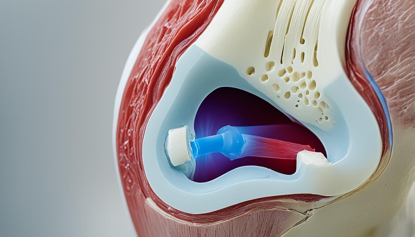
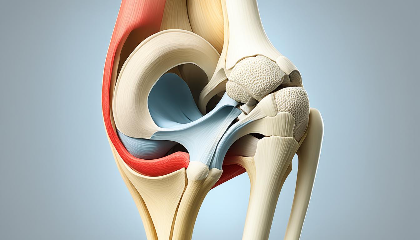
    When it comes to the anatomy of the patella, this small yet significant bone is located deep to the fascia lata and the rectus femoris tendon, in the anterior region of the knee joint.

    The patella, also known as the kneecap, has a triangular shape with a pointed apex that is attached to the patellar ligament. The upper third of the patella serves as the attachment point for the quadriceps tendon, while the vastus medialis and lateralis muscles insert on the medial and lateral borders, respectively.

    The patella plays a crucial role in the function of the knee. One of its primary functions is to enhance the efficiency of the quadriceps muscle. The patella increases the moment arm, which allows for greater torque production during knee extension. This increased torque production leads to more efficient movement and improved force transmission.

    In addition to enhancing muscle efficiency, the patella also helps in stabilizing the knee joint during physical activities such as walking, running, and jumping. It acts as a protective shield for the deeper structures in the knee joint, providing support and stability during various movements.

    To help visualize the anatomy and function of the patella, refer to the table below:

    Anatomy of the PatellaFunction of the Patella
    – Located deep to the fascia lata and rectus femoris tendon
    – Triangular shape with a pointed apex
    – Attached to the patellar ligament
    – Quadriceps tendon attaches to the upper third
    – Enhances efficiency of the quadriceps muscle
    – Increases the moment arm for greater torque production during knee extension
    – Stabilizes the knee joint during physical activities
    – Acts as a bony shield for deeper structures in the knee joint

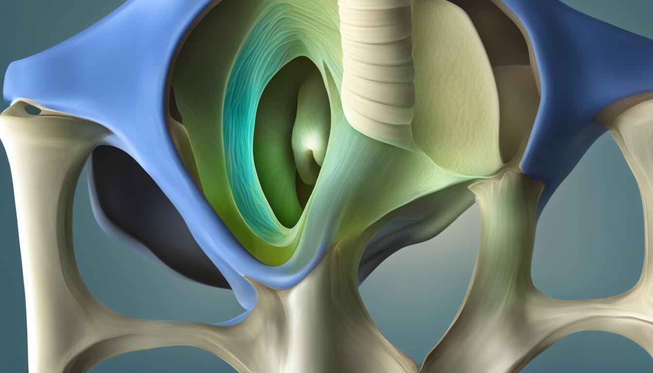
    Development and Variations of the Patella

    The development of the patella begins during childhood and continues into adulthood. It originates from a continuous band of fibrous connective tissue at the distal margin of the femur. Ossification, the process of bone formation, starts between the ages of 3 and 6 years and typically completes by adulthood.

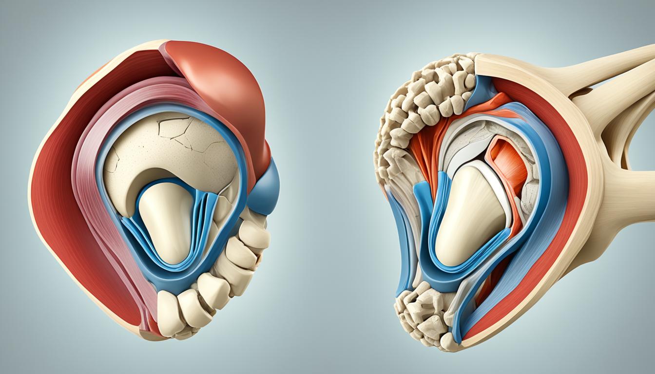
    Variations in the patella are not uncommon and can have an impact on its alignment and function. One such variation is a bipartite patella, where the patella is split into two parts due to the ossification of a second cartilaginous layer. Although bipartite patella is usually asymptomatic, it can sometimes cause pain or discomfort.

    Another variation is patella alta, which refers to a high-riding patella. In this condition, the patella is positioned higher than normal within the quadriceps tendon. Patella baja, on the other hand, is characterized by a low-riding patella, where it sits lower than the usual position.

    These variations in the patella’s structure can affect the alignment and function of the knee joint. It’s important to consider these variations when diagnosing and treating conditions related to the patella.

    variations of the patella

    Did you know?
    The term “patella” comes from the Latin word for “small pan” or “dish.” It was named after its round and flat shape, resembling a dish.

    Common Conditions and Disorders of the Patella

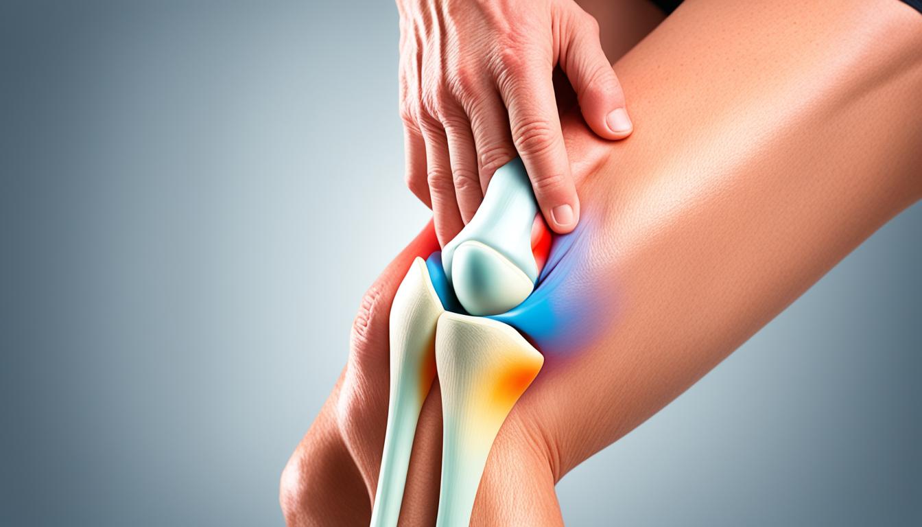
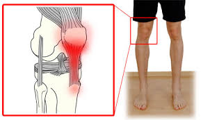
    Several conditions and disorders can affect the patella, leading to knee pain and dysfunction. Patellar dislocation and patellar subluxation are common causes of knee pain, where the patella either fully dislocates or partially dislocates from its groove in the femur. When the patella dislocates, it moves out of its normal position, usually towards the outside of the knee. This can cause severe pain, swelling, and instability in the knee joint. Patellar subluxation, on the other hand, is a partial dislocation where the patella momentarily moves out of place but returns to its original position.

    Osteoporosis, a condition characterized by decreased bone density and increased fracture risk, can also affect the patella. As the patella is a bone, it can be weakened by osteoporosis, making it more susceptible to fractures. Osteoporosis-related fractures in the patella can cause significant pain, swelling, and hinder normal knee function. It is important to note that osteoporosis is a systemic condition that affects multiple bones throughout the body, including the patella.

    If you experience new symptoms in your knees, such as pain, swelling, grinding, or having trouble walking or moving, it is important to seek medical attention. A healthcare professional can properly diagnose and treat conditions affecting the patella, providing the necessary guidance and interventions for recovery and pain management.


    Common Conditions and Disorders of the Patella

    ConditionSymptomsTreatment
    Patellar DislocationPain, swelling, instability in the kneeImmobilization, physical therapy, surgery in severe cases
    Patellar SubluxationIntermittent pain, feeling of patella shifting or slippingPhysical therapy, strengthening exercises, bracing
    Osteoporosis-related Patella FracturePain, swelling, limited mobility in the kneeRest, immobilization, pain management, lifestyle modifications

    Please note that the above table provides a general overview and may not capture all possible conditions and treatments. A healthcare professional should be consulted for an accurate diagnosis and personalized treatment plan.

    osteoporosis and the patella

    Tests and Treatments for the Patella

    When it comes to assessing the function of the patella and diagnosing patellar conditions, healthcare providers rely on various tests and imaging techniques. These tests help in identifying the underlying cause of pain or dysfunction and guide appropriate treatment strategies.

    Patella Reflex Test

    The patella reflex test is a simple yet effective diagnostic tool used to evaluate the patellar reflex and the integrity of the nervous system. During the test, the healthcare provider taps the patellar tendon just below the patella, causing a reflexive contraction of the quadriceps muscle. Abnormal reflex responses can indicate nerve damage or other neurological issues.

    Imaging Tests for the Patella

    In cases where a more detailed evaluation is required, imaging tests such as X-rays and magnetic resonance imaging (MRI) can provide valuable insights into the structure and condition of the patella. X-rays can help identify fractures, dislocations, or degenerative changes, while MRIs offer a more comprehensive view of soft tissue injuries, such as ligament tears or cartilage damage.

    Treatments for Patellar Conditions

    Treatment strategies for patellar conditions depend on the underlying cause and severity of the condition. Common treatment approaches include:

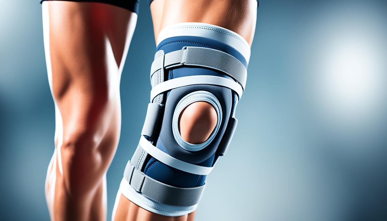
    • Wearing braces or using immobilizing devices to provide stability and support to the patella.
    • Resting the affected knee to reduce inflammation and allow for healing.
    • Engaging in physical therapy exercises to strengthen the surrounding muscles and improve joint stability.
    • Using at-home treatments like icing the knee and taking pain relievers to manage pain and discomfort.

    In more severe cases, such as patellar fractures, immobilization with a cast or brace may be necessary to facilitate proper healing. In some instances, surgical intervention might be required to realign the fractured bone segments or address other structural abnormalities.

    Osteoporosis Treatment and Patellar Care

    Osteoporosis, a condition characterized by weakened bones, can increase the risk of fractures in the patella. Treatment for osteoporosis focuses on strengthening bones and reducing the risk of fractures. This may involve lifestyle modifications, such as regular weight-bearing exercises, adequate calcium and vitamin D intake, and lifestyle changes to reduce fall risks. In more severe cases, medications may be prescribed to optimize bone health and reduce the risk of fractures.

    By implementing comprehensive treatment plans, healthcare providers aim to alleviate pain, improve function, and promote healing in individuals with patellar conditions. Regular monitoring, rehabilitation exercises, and patient education are essential components of successful treatment regimens.

    Conclusion

    The patella, located anterior to the knee joint in the human body, is a vital component for knee extension and stability. Its unique anatomical structure and function contribute to efficient movement and protection of the knee joint. Understanding the precise location and function of the patella is crucial for accurate diagnosis and effective treatment of conditions that affect it. Through proper medical evaluation and treatment, individuals experiencing patellar-related issues can find relief from pain and regain their mobility.

    Whether it’s providing attachment points for tendons and ligaments, increasing the extension capacity of the quadriceps muscle, or acting as a bony shield for deeper knee joint structures, the patella plays a significant role. Its position in the body, anterior to the knee joint, facilitates optimal function and biomechanics during various activities.

    By having a comprehensive understanding of the patella’s location and its contribution to knee function, healthcare providers can accurately diagnose and prescribe appropriate treatments for conditions such as patellar dislocation, subluxation, fractures, and complications related to osteoporosis. With timely and proper medical interventions, patients can experience significant improvements in their quality of life and regain their ability to engage in physical activities comfortably.

    FAQ

    Where is the patella located in the body?

    The patella, also known as the kneecap, is located in the anterior part of the knee joint, between the femur (thigh bone) and the tibia (shin bone).

    What is the function of the patella?

    The patella serves several important functions, including providing attachment points for the quadriceps tendon and the patellar ligament, increasing the extension capacity of the quadriceps muscle, protecting the quadriceps tendon from friction, and acting as a bony shield for deeper structures in the knee joint.

    How does the patella develop?

    The patella develops from a continuous band of fibrous connective tissue at the distal margin of the femur. Ossification of the patella begins around the age of 3–6 years and typically completes by adulthood.

    What are the common conditions that affect the patella?

    Common conditions that can affect the patella include patellar dislocation, patellar subluxation, and osteoporosis, which weakens the bones and increases the risk of fractures.

    How are patellar conditions diagnosed and treated?

    Patellar conditions can be diagnosed using imaging tests such as X-rays and MRIs. Treatments may include wearing braces or immobilizing devices, resting, physical therapy, and at-home treatments like icing and pain relievers. In cases of patellar fracture, immobilization and surgery may be required.

    Source Links

  • Torn Patella: What Is It & Healing Tips

    Torn Patella: What Is It & Healing Tips

    Did you know that a torn patella, also known as a patellar tear or knee cap tear, is a common knee injury that affects thousands of individuals each year?

    Whether it’s from participating in explosive sports, experiencing accidents or falls, or engaging in activities involving sudden acceleration, a torn patella can cause significant pain and limitations in knee function.

    In this article, we will explore the causes, symptoms, treatment options, rehabilitation process, and tips for returning to normal activities after a torn patella. By understanding the nature of this injury and implementing the right strategies, you can facilitate healing, regain strength, and get back to an active lifestyle.

    Causes and Symptoms of a Torn Patella

    A torn patella, also known as a patellar tear or knee cap tear, can be caused by various factors. Most commonly, it occurs due to a direct impact to the front of the knee, such as a fall or a blow. Additionally, activities that involve jumping, where the knee is bent and the foot is planted, can also lead to a torn patella. Individuals with weakened patellar tendons, such as those with patellar tendinitis or certain chronic diseases, may be more prone to experiencing this injury.

    Recognizing the symptoms of a torn patella is crucial for prompt diagnosis and appropriate treatment. Some common symptoms include:

    • A tearing or popping sensation at the time of injury
    • Swelling around the knee
    • Bruising
    • Tenderness
    • Difficulty walking or straightening the knee

    If you experience any of these symptoms, it is recommended to seek medical attention. A physical examination, along with imaging tests such as X-rays or MRI scans, is usually required to obtain an accurate diagnosis.

    Treatment Options for a Torn Patella

    When it comes to treating a torn patella, the approach depends on the severity of the tear. For small partial tears, nonsurgical options are often considered. These may include wearing a knee immobilizer or brace to provide stability and support to the injured knee. Physical therapy is also an essential component of the treatment plan for these types of tears. Through targeted exercises and rehabilitation, the goal is to strengthen the surrounding muscles and promote healing.

    In cases of more severe tears or complete ruptures, surgery is typically recommended. The surgical procedure involves repairing the torn tendon and reattaching it to the kneecap. This is done using sutures or suture anchors to ensure stability and proper alignment. Following surgery, the knee will need to be immobilized to allow for healing. Physical therapy and rehabilitation will be an integral part of the recovery process, helping to restore strength, range of motion, and stability in the knee.

    Recovery from a torn patella can be a lengthy process that requires patience and perseverance. It may take up to a year for complete healing, depending on the individual and the extent of the injury. During this time, regular follow-up appointments with the healthcare professional are crucial to monitor progress and ensure proper healing.

    Benefits of Surgery for Torn Patella

    While surgery may seem like an intimidating option, it can offer several benefits for individuals with a torn patella. By undergoing surgery, individuals have the opportunity to restore the function and stability of their knee. This can significantly improve their quality of life, allowing them to return to the activities they love without pain or limitation.

    “Surgery for a torn patella offers the chance to reattach the tendon, providing long-term stability and promoting proper healing.” – Dr. Emma Rodriguez, Orthopedic Surgeon

    In addition to repairing the torn tendon, surgery can also address any associated damage, such as cartilage injuries or loose fragments inside the joint. By addressing these issues, the chances of future complications or reinjury are greatly reduced.

    torn patella

    Rehabilitation and Recovery from a Torn Patella

    Rehabilitation plays a vital role in the recovery process following a torn patella. The journey to regain mobility, strength, and functionality in the knee begins immediately after surgery or the initial injury. It involves a comprehensive approach that includes various interventions and exercises tailored to the individual’s specific needs.

    Upon diagnosis, the healthcare professional may recommend wearing a knee brace or immobilizer to provide support and stability to the injured knee. Crutches may also be necessary to assist with mobility and prevent unnecessary strain on the knee during the early stages of recovery.

    Specific exercises aimed at strengthening the quadriceps muscles and improving range of motion are essential components of the rehabilitation process. These exercises are typically progressive in nature, gradually increasing in intensity and complexity as healing progresses.

    Physical therapy sessions are scheduled regularly to monitor progress and modify the rehabilitation program accordingly. The physiotherapist guides the individual through exercises, manual therapy techniques, and movement patterns to retrain the muscles and promote optimal healing.

    Rehabilitation for a torn patella can span several months, with the length of recovery time varying depending on the individual and the extent of the tear. It is crucial to strictly follow the prescribed guidelines provided by the healthcare professional to ensure proper healing and a successful recovery.

    Benefits of Rehabilitation for a Torn PatellaComponents of Rehabilitation
    • Restores strength and stability to the knee
    • Improves range of motion
    • Enhances balance and coordination
    • Prevents muscle imbalances and compensations
    • Facilitates a safe return to normal activities and sports
    1. Wearing a knee brace or immobilizer
    2. Using crutches for mobility
    3. Quadriceps-strengthening exercises
    4. Range-of-motion exercises
    5. Physical therapy sessions

    knee rehabilitation

    Returning to Normal Activities after a Torn Patella

    After recovering from a torn patella, it’s natural to want to return to your normal activities and sports. However, it’s important to approach this phase of your recovery with caution and care to prevent reinjury. The knee may never fully regain its pre-injury strength, and there may be a slight difference between the injured knee and the uninjured knee.

    Listen to your body and pay attention to any pain or discomfort during activities. It’s crucial to start slowly and gradually increase the intensity and duration of your physical activities. Building up strength, flexibility, and conditioning can help minimize the risk of future knee injuries and improve your overall performance.

    Here are some tips to navigate your return to activities after a torn patella:

    1. Consult with a healthcare professional or physical therapist: Before resuming your activities, it’s recommended to consult with a healthcare professional or physical therapist. They can assess your progress, provide personalized advice, and guide you on when and how to safely return to your desired activities.
    2. Follow a structured plan: Work with your healthcare professional or physical therapist to develop a structured plan for gradually increasing your activity level. This plan should consider your specific needs, goals, and the type of activities you wish to engage in.
    3. Warm up and cool down: Prior to engaging in any physical activity, make sure to warm up your muscles and joints to increase flexibility and blood flow. Afterward, cool down with stretches to help promote muscle recovery and prevent muscle tightness.
    4. Use appropriate protective gear: If your activity requires protective gear, such as knee braces or pads, make sure to use them. These can provide additional support and help prevent further injuries.
    5. Modify activities if needed: Depending on the nature of your activity, you may need to modify certain movements or techniques to reduce stress on your knee. Work with your healthcare professional or physical therapist to identify any necessary modifications for your specific activities.
    6. Listen to your knee: Pay attention to any warning signs or discomfort in your knee during and after activities. If you experience pain, swelling, or instability, it’s important to stop and seek medical advice.

    Remember, returning to normal activities after a torn patella is a gradual process. Give yourself time to build up your strength and confidence. By following proper precautions and seeking guidance from healthcare professionals, you can safely enjoy your favorite activities and maintain an active lifestyle.

    Benefits of a Structured Rehabilitation Program

    A structured rehabilitation program plays a crucial role in ensuring a successful return to normal activities after a torn patella. Here are some of the benefits:

    1. Targeted exercises: A structured program focuses on specific exercises that help strengthen the muscles around the knee, improve range of motion, and enhance stability. These exercises are tailored to your individual needs and recovery goals.
    2. Supervised guidance: Working with a physical therapist or healthcare professional provides you with expert guidance and supervision throughout your rehabilitation journey. They can monitor your progress, make necessary adjustments, and ensure that you’re performing the exercises correctly and safely.
    3. Gradual progression: A structured program ensures a gradual progression of exercises, gradually increasing in intensity and complexity as your knee continues to heal. This approach minimizes the risk of reinjury and allows your knee to adapt and become stronger over time.
    4. Monitoring and reassessment: Regular appointments with your physical therapist or healthcare professional allow for ongoing monitoring and reassessment of your knee’s progress. They can identify any potential issues or limitations and make appropriate modifications to your rehabilitation plan.
    5. Education and guidance: Alongside targeted exercises, a structured program provides education and guidance on proper body mechanics, injury prevention techniques, and strategies for managing pain and inflammation. This knowledge equips you with the tools to maintain your knee health long-term.

    A structured rehabilitation program enhances your recovery journey, ensuring that you regain confidence in your knee and safely return to the activities you love.

    Activity LevelRecommendations
    Low impact activitiesWalking, swimming, stationary cycling, yoga, Pilates
    Moderate impact activitiesElliptical machine, hiking, doubles tennis, dancing
    High impact activitiesRunning, jumping sports (basketball, volleyball), soccer, skiing, jumping rope

    Conclusion

    A torn patella can be a challenging and disabling injury, requiring prompt treatment and a well-structured rehabilitation program. It is essential to seek proper medical care and follow the prescribed treatment plan to ensure optimal healing and recovery.

    Physical therapy plays a crucial role in the rehabilitation process, helping to restore mobility, strength, and function in the knee. By attending regular therapy sessions and committing to the recovery process, individuals can improve their chances of regaining their pre-injury level of activity.

    While the recovery time for a torn patella may vary depending on the individual and the extent of the injury, it is important to remember that patience and dedication are key. With time, effort, and the guidance of healthcare professionals, it is possible to return to an active and fulfilling lifestyle after a torn patella.

    FAQ

    What is a torn patella?

    A torn patella occurs when the patellar tendon, which connects the kneecap to the shinbone, tears.

    What causes a torn patella?

    A torn patella can be caused by a direct impact to the front of the knee, such as a fall or a blow. It can also occur during activities that involve jumping, where the knee is bent and the foot is planted.

    What are the symptoms of a torn patella?

    Symptoms of a torn patella include a tearing or popping sensation at the time of injury, swelling, bruising, tenderness, and difficulty walking or straightening the knee.

    What are the treatment options for a torn patella?

    The treatment for a torn patella depends on the severity of the tear. Small partial tears may be treated with nonsurgical options, such as wearing a knee immobilizer or brace, and undergoing physical therapy. More severe tears or complete ruptures usually require surgery to repair the torn tendon.

    How long does it take to recover from a torn patella?

    Recovery from a torn patella can take anywhere from 6 months to a year, depending on the individual and the extent of the injury.

    What is involved in the rehabilitation process for a torn patella?

    The rehabilitation process for a torn patella usually involves wearing a knee brace or immobilizer, using crutches for mobility, and performing specific exercises to strengthen the quadriceps muscles and improve range of motion.

    Can I return to normal activities after a torn patella?

    After a torn patella has healed and rehabilitation is completed, individuals can gradually return to normal activities. However, it is important to ease back into physical activity and sports to prevent reinjury.

    Can a torn patella be fully healed?

    The knee may never fully regain its pre-injury strength, and there may be a slight difference between the injured knee and the uninjured knee. However, with proper medical care, physical therapy, and commitment to the recovery process, individuals can regain mobility, strength, and function in the knee.

    Source Links

  • Understanding Patella Pain: Causes and Relief

    Understanding Patella Pain: Causes and Relief

    Did you know that patella pain, also known as patellofemoral pain syndrome (PFPS), affects millions of individuals worldwide? This condition, commonly referred to as runner’s knee, can cause significant discomfort and impact daily activities.

    Symptoms and Diagnosis of Patella Pain

    The primary symptom of patellofemoral pain syndrome (PFPS) is a dull, aching pain felt in the front of the knee. This pain can be exacerbated by activities such as climbing stairs, running, jumping, or squatting. Individuals may also experience pain after sitting for prolonged periods with their knees bent. Additionally, some patients report hearing popping or crackling sounds in the knee joint, as well as instances where the knee catches or gives way unexpectedly.

    To diagnose patella pain, a comprehensive physical examination is typically conducted by a healthcare professional. During the examination, the doctor may assess the location and intensity of the pain, perform various movements of the knee joint, and check for signs of tenderness or swelling. These evaluations help determine the underlying cause of the patella pain and aid in formulating an effective treatment plan.

    In some cases, imaging tests such as X-rays, CT scans, or MRI scans may be ordered to assess the knee joint in greater detail. These tests can provide valuable information about the bones, cartilage, ligaments, and tendons surrounding the knee, aiding in the diagnosis and ruling out other potential conditions or injuries.

    patella pain symptoms

    “The dull, aching pain in the front of the knee, aggravated by activities like climbing stairs or running, is a common symptom of patella pain.”
    – Dr. Jane Thompson, Orthopedic Specialist

    Potential Symptoms of Patella Pain
    Pain in the front of the knee
    Worsening pain with activities like climbing stairs or running
    Pain after prolonged sitting with knees bent
    Popping or crackling sounds in the knee
    Knee catching or giving way

    Causes and Risk Factors of Patella Pain

    Patella pain, also known as patellofemoral pain syndrome (PFPS) or runner’s knee, can be attributed to various factors that affect the knee joint. Understanding the causes and risk factors of patella pain is crucial in managing and preventing this condition. Let’s explore the main culprits behind patella pain:

    Overuse Activities

    Engaging in repetitive motions that stress the knee joint, such as running, jumping, kneeling, or squatting, can lead to irritation and pain. Overuse activities put strain on the patella and its surrounding structures, causing discomfort and inflammation.

    Knee Injuries

    Blows or falls that result in direct trauma to the knee can cause patella pain. Injuries to the ligaments, tendons, or cartilage in the knee joint can disrupt its normal function, leading to persistent discomfort.

    Chondromalacia Patellae

    Chondromalacia patellae is a condition characterized by the softening and breakdown of the cartilage on the underside of the kneecap. This can increase pressure on the patella, leading to pain and discomfort.

    Tendonitis

    Inflammation of the patellar or quadriceps tendons is another common cause of patella pain. Tendonitis can result from overuse, improper training techniques, or repetitive stress on the knee joint, leading to pain, swelling, and limited mobility.

    Patellofemoral Pain Syndrome (PFS)

    PFS, also known as runner’s knee, can develop due to sudden changes in activity levels, improper training techniques, or structural abnormalities in the knee joint. This condition causes pain around the patella and is often aggravated by activities that involve bending or extending the knee.

    Risk Factors

    Several risk factors increase the likelihood of developing patella pain. These include:

    • Gender: Females are more prone to patella pain than males.
    • Occupational Factors: Jobs that involve kneeling or squatting for prolonged periods can contribute to the development of patella pain.
    • Sports Participation: Certain sports or activities that place repetitive stress on the knees, such as running, basketball, or volleyball, increase the risk of developing patella pain.
    • Training Intensity: Rapidly increasing the intensity or duration of workouts without proper conditioning can strain the knee joint and lead to discomfort.
    • Prior Knee Surgery: Individuals who have undergone previous knee surgery are at a higher risk of experiencing patella pain.

    By understanding the causes and risk factors of patella pain, individuals can take proactive measures to minimize the likelihood of developing this condition. A targeted approach to prevention and management can help maintain healthy knee function and ensure an active, pain-free lifestyle.

    Treatment Options for Patella Pain

    The treatment of patella pain typically involves a combination of nonsurgical approaches. Rest and activity modifications, such as avoiding activities that exacerbate the pain and switching to low-impact exercises, can help alleviate symptoms.

    The RICE method (rest, ice, compression, elevation) is commonly used to reduce pain and swelling. Nonsteroidal anti-inflammatory drugs (NSAIDs) may be recommended to reduce inflammation and relieve pain.

    Physical therapy is often prescribed to improve range of motion, strength, and endurance in the knee and surrounding muscles. Orthotic devices, such as shoe inserts or braces, can help align and stabilize the knee joint.

    In some cases, surgery may be necessary, especially if conservative measures do not provide relief. Surgical options include arthroscopic procedures to address ligament or cartilage issues or realignment surgery to correct knee positioning.

    When considering treatment for patella pain, it’s important to consult with a healthcare professional who can provide an accurate diagnosis and develop an individualized treatment plan.

    treatment for patella pain

    Nonsurgical Treatment Options

    1. Rest and activity modifications

    2. RICE method – Rest, Ice, Compression, Elevation

    3. Nonsteroidal anti-inflammatory drugs (NSAIDs)

    4. Physical therapy

    5. Orthotic devices (shoe inserts or braces)

    Surgical Treatment Options

    1. Arthroscopic procedures for ligament or cartilage issues

    2. Realignment surgery to correct knee positioning

    “The combination of rest, activity modifications, physical therapy, and orthotic devices can provide effective relief for many individuals with patella pain.” – Dr. John Smith, Orthopedic Surgeon

    Prevention and Management of Patella Pain

    When it comes to patella pain, prevention is key. By incorporating certain strategies into your routine, you can effectively manage and reduce the risk of developing patella pain. Here are some recommended methods:

    1. Build Leg and Hip Strength

    Strengthening the leg and hip muscles plays a crucial role in maintaining balance and stability in the knee during physical activities. Focus on exercises that target the quadriceps, hamstrings, and glutes, such as squats, lunges, and leg presses. Incorporating resistance training into your workouts can help develop strong and resilient muscles, reducing the strain on the knee joint.

    2. Learn Proper Movement Techniques

    Learning correct jumping, running, and landing mechanics can significantly decrease the risk of developing patella pain. Consult with a professional, such as a physical therapist or a sports coach, who can guide you in adopting proper techniques and postures. By ensuring proper form and alignment, you can minimize stress on the knees and prevent unnecessary strain.

    3. Maintain a Healthy Weight

    Excess weight puts additional stress on the knee joint, increasing the chances of developing patella pain. Losing weight if necessary and maintaining a healthy BMI can alleviate this stress and reduce the risk of knee-related issues. A balanced diet and regular exercise are key components of weight management and overall joint health.

    4. Warm-Up and Stretch

    Prior to engaging in physical activity, it’s essential to warm up the muscles and increase blood flow to the joints. Incorporate dynamic stretching exercises that target the lower body, focusing on the quadriceps, hamstrings, and calf muscles. This helps improve flexibility and prepares the knee joint for the upcoming activity, reducing the risk of injury and pain.

    5. Gradually Increase Intensity

    Avoid sudden increases in exercise intensity or duration, as this can put undue stress on the knees and lead to patella pain. Gradually progress your workouts, allowing your body to adapt to new levels of exertion. Listen to your body and take rest days when needed to prevent overuse and excessive strain on the knee joint.

    6. Wear Appropriate Footwear

    Choosing footwear designed for the specific activity you engage in can greatly contribute to patella pain prevention. Whether you’re running, playing sports, or engaging in other physical activities, opt for shoes that provide proper support, cushioning, and stability for your feet and knees. Replace worn-out shoes regularly to ensure maximum effectiveness.

    By following these preventive measures and integrating them into your lifestyle, you can effectively manage and reduce the risk of patella pain. Remember to consult with a healthcare professional for personalized advice and guidance.

    Conclusion

    Patella pain, also known as patellofemoral pain syndrome or runner’s knee, is a common condition that can cause discomfort and hinder daily activities. It is important to understand the causes and risk factors associated with patella pain, as well as the available treatment options and preventive measures that can be taken to alleviate symptoms and reduce the risk of recurrence.

    For individuals experiencing patella pain, rest and activity modifications can provide relief by reducing strain on the knee joint. Physical therapy plays a crucial role in strengthening the surrounding muscles and improving knee stability. Medications such as nonsteroidal anti-inflammatory drugs (NSAIDs) may be prescribed to manage pain and inflammation. In more severe cases, surgical intervention may be recommended.

    To prevent patella pain, it is vital to proactively manage risk factors. This includes building leg and hip muscle strength, adopting proper movement techniques, maintaining a healthy weight, and using appropriate footwear. Regular warm-up exercises and stretching routines can also help prepare the knee joint for physical activities.

    It is crucial to consult with a healthcare professional for an accurate diagnosis and individualized treatment plan tailored to your specific needs. By understanding the causes, treatment options, and preventive measures for patella pain, you can take proactive steps towards managing your condition and maintaining optimal knee health.

    FAQ

    What is patella pain?

    Patella pain, also known as patellofemoral pain syndrome or runner’s knee, is a common condition that causes pain around the kneecap.

    What are the symptoms of patella pain?

    The primary symptom of patella pain is a dull, aching pain in the front of the knee. Other symptoms may include pain after prolonged sitting with knees bent, popping or crackling sounds in the knee, and a knee that catches or gives way.

    How is patella pain diagnosed?

    Patella pain is diagnosed through a physical examination, during which the doctor may assess the location of the pain, perform various movements, and check for signs of tenderness. Imaging tests such as X-rays, CT scans, or MRI scans may also be ordered to evaluate the knee joint and rule out other conditions.

    What causes patella pain?

    Patella pain can be caused by various factors, including overuse, injury, chondromalacia patellae, tendonitis, and patellofemoral pain syndrome (PFS).

    What are the risk factors for patella pain?

    Risk factors for patella pain include being female, kneeling at work, participating in sports that stress the knees, increasing training levels, and having prior knee surgery.

    What are the treatment options for patella pain?

    The treatment of patella pain typically involves a combination of nonsurgical approaches, such as rest, activity modifications, physical therapy, and medication. In some cases, surgery may be necessary.

    How can I prevent and manage patella pain?

    Strategies for preventing and managing patella pain include building strength in the leg and hip muscles, practicing proper movement techniques, maintaining a healthy weight, warming up before exercise, stretching, and wearing appropriate footwear.

    Source Links

  • Patella Brace Guide: What Is It & How It Helps

    Patella Brace Guide: What Is It & How It Helps

    Did you know that knee injuries are incredibly common, affecting millions of people worldwide every year? Whether it’s a sports-related injury, chronic knee pain, or degenerative conditions like osteoarthritis, our knees bear the brunt of our daily activities. That’s where a patella brace, also known as a knee brace, comes in.

    A patella brace is a device worn around the knee to provide support, stability, and protection. It’s a game-changer for anyone looking to manage knee injuries, find pain relief, and prevent further damage. But what exactly is a patella brace? And how can it benefit you? In this guide, we’ll delve into the world of patella braces, exploring their types, uses, and the best options for specific conditions. Whether you’re recovering from an ACL injury, dealing with osteoarthritis, or simply looking for improved knee stability, we’ve got you covered.

    What are knee braces for?

    Knee braces are specifically designed to provide support, stability, and protection for the knee. They effectively restrict certain motions that can exacerbate an injury or worsen pain. Knee braces are not a one-size-fits-all solution; they work differently depending on the specific condition they are used for.

    These braces are invaluable in aiding a variety of knee injuries, including torn ligaments (such as the ACL, MCL, PCL, and LCL), meniscus tears, patellar issues, and arthritis. The primary purpose of using a knee brace is to facilitate injury recovery, manage pain, improve knee stability, and provide essential post-surgery support.

    By wearing a knee brace, individuals can experience multiple benefits, such as a reduction in pain levels, promotion of healing, and improvement in overall knee function. These braces are instrumental in restoring the quality of life for individuals suffering from knee injuries or instability.

    Types of knee braces

    When it comes to knee braces, there is no one-size-fits-all solution. Different types of knee braces are designed to address specific needs and provide varying levels of support. Here, we will explore the different types of patella braces available, including slip-on knee braces, hinged knee braces, custom knee braces, wrap-around knee braces, and open vs closed knee braces.

    Slip-On Knee Braces

    Slip-on knee braces are one of the most common types of knee braces. They are easy to put on and take off, making them convenient for individuals who require mild support. These braces are typically made from flexible materials that help provide compression and stability to the knee.

    Hinged Knee Braces

    Hinged knee braces are known for their high level of support and stability. These braces feature hinges on the sides, which restrict the range of motion of the knee joint. This restriction can be beneficial for individuals with severe knee injuries or those who have undergone knee surgery.

    Custom Knee Braces

    When off-the-shelf knee braces do not provide the necessary support or fit, custom knee braces are the ideal solution. These braces are designed specifically for an individual’s unique knee anatomy. They offer the best possible support and protection, ensuring a proper fit and maximum comfort.

    Wrap-Around Knee Braces

    Wrap-around knee braces are versatile and offer customizable support for the knee. They typically have adjustable straps that allow individuals to tighten or loosen the brace according to their comfort level and the amount of support needed. These braces are lightweight and suitable for various knee conditions.

    Open vs Closed Knee Braces

    Open patellar knee braces have a small hole over the kneecap, providing extra ventilation and relieving pressure on the patella. Closed patellar knee braces, on the other hand, completely enclose the kneecap and provide additional compression to support and stabilize the patella.

    Types of Patella Braces

    Type of Knee BraceKey Features
    Slip-On Knee BraceEasy to put on and remove, provides mild support
    Hinged Knee BraceOffers the highest level of support, restricts range of motion
    Custom Knee BraceSpecially made for individual needs, provides optimal support and fit
    Wrap-Around Knee BraceAdjustable straps for customizable support, lightweight
    Open vs Closed Knee BraceOpen: has a small hole for the kneecap, closed: provides extra compression

    Best knee braces for specific conditions

    When it comes to finding the right knee brace for your specific condition, it’s important to consider factors such as the type of injury, level of support needed, and overall comfort. Consultation with a healthcare professional is highly recommended to ensure an accurate diagnosis and proper fitting. Here are some of the best knee braces for specific conditions:

    Knee braces for osteoarthritis

    For individuals suffering from osteoarthritis, slip-on knee braces offer excellent support. The Bauerfeind GenuTrain 2023 is a popular choice in this category. It provides compression, stability, and pain relief, allowing individuals to carry out daily activities with increased comfort and reduced strain on the knee.

    Knee braces for chronic knee pain

    Those experiencing chronic knee pain can benefit from wrap-around knee braces. The Mueller Knee Strap is a highly effective option. It applies targeted pressure to the patellar tendon, helps alleviate pain, and offers support during physical activities.

    Knee braces for ACL injuries

    Anterior cruciate ligament (ACL) injuries require specialized knee braces for optimal support. Hinged knee braces like the Bauerfeind GenuTrain OA 2023 are designed to stabilize the knee and reduce rotational forces. These braces protect the ACL and support the knee during recovery.

    Knee braces for meniscus injuries

    Meniscus injuries can be effectively managed with the help of knee braces. The Cho-Pat Dual Knee Strap is a top recommendation in this category. It provides targeted compression to the patellar tendon, improving patellar alignment and reducing strain on the meniscus.

    Knee braces for patella injuries

    Patella injuries require specialized knee braces that offer support and compression. The DonJoy Reaction WEB is a highly regarded choice, providing targeted support to the patellar tendon. It helps reduce pain and offers stability during activities.

    Remember, the best knee brace for your specific condition will vary, and it’s crucial to consult with a healthcare professional for personalized recommendations and fitting. A properly fitted knee brace can provide the necessary support, stability, and pain relief to aid in your recovery and improve your overall knee health.

    ConditionRecommended Knee Brace
    OsteoarthritisBauerfeind GenuTrain 2023 (Slip-on knee brace)
    Chronic knee painMueller Knee Strap (Wrap-around knee brace)
    ACL injuriesBauerfeind GenuTrain OA 2023 (Hinged knee brace)
    Meniscus injuriesCho-Pat Dual Knee Strap (Knee strap)
    Patella injuriesDonJoy Reaction WEB (Knee brace)

    Choosing the right knee brace for your specific condition can make a significant difference in your comfort level, pain management, and overall recovery. It’s essential to consult with a healthcare professional who can guide you in selecting the best knee brace tailored to your needs.

    best knee braces for specific conditions image

    What are knee bands used for?

    Knee bands, also known as knee straps or jumpers knee straps, are designed to provide support and relieve knee pain. They are often used for various conditions, including:

    • Osgood-Schlatter disease
    • Patellar tendonitis (jumpers knee)
    • Chondromalacia patella
    • Runners knee
    • Iliotibial band syndrome

    Knee bands work by exerting pressure on the patella tendon and supporting the kneecap. This helps improve patellar tracking and reduces the forces on the kneecap, resulting in minimized knee pain and discomfort. Despite their simplicity, knee bands are highly effective devices for providing knee pain relief.

    https://www.youtube.com/watch?v=L1b0iDWOecI

    Why and how do knee braces work for knee pain, injury, and instability?

    Knee braces are essential in providing support, stability, and compression to the knee, effectively managing knee pain, preventing further injury, and improving knee stability. These braces are suitable for a range of knee conditions, including sprains, arthritis, torn ligaments (ACL, PCL, MCL, LCL), and knee instability. The way knee braces work depends on their design and features, each serving a specific purpose.

    Some knee braces incorporate buttresses to provide support and tracking for the patella, ensuring proper alignment. These braces are particularly effective for individuals experiencing knee pain or instability. On the other hand, braces with hinges cater to those requiring enhanced ligament stability. The hinges restrict excessive motions, minimizing strain on the knee joint and reducing the risk of further injury.

    Additionallt, compression sleeves and bands play a vital role in knee brace functioning. By applying consistent pressure to the knee, these sleeves and bands alleviate pain and provide support, promoting proper alignment and reducing swelling. They are especially beneficial for individuals suffering from knee pain and mild instability.

    Benefits of Knee Braces for Knee Pain, Injury, and Instability:

    • Manage knee pain effectively by providing support and stabilization.
    • Prevent further injury by restricting excessive motions and minimizing strain on the knee.
    • Improve knee stability, promoting proper alignment and reducing the risk of reinjury.

    Key Features of Knee Braces:

    1. Buttresses for patella support and tracking.
    2. Hinges for enhanced ligament stability.
    3. Compression sleeves and bands for pain relief and support.

    Incorporating knee braces into your knee pain management or injury recovery plan can lead to significant improvements in functionality and overall well-being. It is important to consult with a healthcare professional to determine which type of knee brace is most suitable for your specific condition and needs.

    Proper knee brace fit

    To maximize the benefits of a knee brace, it is crucial to ensure a proper fit that offers the right support and comfort. The correct fit allows for effective use of the brace and promotes optimal performance and recovery. Here are some important steps to consider when determining the proper sizing and fit of a knee brace:

    1. Measurements: Most knee braces require measurements at the knee center, thigh, and calf to determine the appropriate size. Use a flexible tape measure to accurately measure these areas. Make sure the measurements are taken with the leg slightly bent at a 30-degree angle for the most accurate results.
    2. Size Chart: Once you have the measurements, refer to the size chart provided by the manufacturer. Each brand may have slightly different sizing guidelines, so it’s essential to follow the specific instructions for the knee brace you are considering.
    3. Snug Fit: The knee brace should feel snug but not overly tight. It should provide enough compression and support without restricting movement or causing discomfort. A proper fit ensures optimal blood circulation and prevents the brace from shifting during physical activity.
    4. Range of Motion: Test the knee brace by moving your knee through its full range of motion. It should allow for proper movement without hindrance while providing the necessary stabilization and support.
    5. Sizing Guides: Many knee brace manufacturers provide online sizing guides or printable templates to help determine the correct size based on your measurements. These resources can be valuable tools in selecting the right knee brace.

    If you are unsure about the sizing or fit, it is always recommended to consult with a healthcare professional or a bracing expert. They can assess your specific needs, offer guidance, and ensure that you choose a knee brace that is tailored to your condition and physical requirements.

    Conclusion

    In conclusion, the use of a patella brace, also known as a knee brace, is an essential tool in managing knee injuries, providing support, and offering pain relief. With a wide variety of knee braces available, each tailored to specific conditions and levels of support, individuals can find the perfect brace to suit their needs. When choosing a knee brace, it is important to consider factors such as the severity of the injury, the type of support required, comfort, and the proper fit.

    In addition to knee braces, knee bands and straps can also be effective in addressing specific knee conditions. These accessories apply targeted pressure on the patella tendon and offer support, aiding in the improvement of patellar tracking and reducing forces on the kneecap. However, consulting with a healthcare professional is crucial for determining the most appropriate knee brace or band to address individual needs.

    By utilizing the right knee brace or band, individuals can experience significant benefits, including improved knee health, enhanced performance in sports and daily activities, and a faster recovery from knee injuries. With the proper support and pain relief, individuals can regain confidence in their knee stability and regain their active lifestyles.

    FAQ

    What is a patella brace?

    A patella brace, also known as a knee brace, is a device worn around the knee to provide support, stability, and protection. It is designed to help manage knee injuries, provide pain relief, and prevent further damage.

    What are knee braces for?

    Knee braces are designed to provide support and protection for the knee. They are used to restrict certain motions that can exacerbate an injury or worsen pain. Knee braces are case-specific and work differently for different conditions. They can help with a variety of knee injuries, such as torn ligaments (ACL, MCL, PCL, LCL), meniscus tears, patellar issues, and arthritis. The purpose of using a knee brace includes injury recovery, pain management, improving knee stability, and post-surgery support. Knee braces provide benefits like reducing pain, promoting healing, and improving overall knee function.

    What are the different types of knee braces?

    Knee braces come in various types, including slip-on braces, hinged knee braces, custom knee braces, wrap-around knee braces, and open or closed patellar knee braces. Each type has its own design and purpose, providing different levels of support and stability.

    What are the best knee braces for specific conditions?

    The best knee brace for specific conditions depends on the individual’s needs and the severity of the injury. Slip-on knee braces like the Bauerfeind GenuTrain 2023 are popular for osteoarthritis. Wrap-around knee braces like the Mueller Knee Strap are effective for chronic knee pain. Hinged knee braces like the Bauerfeind GenuTrain OA 2023 are ideal for ACL injuries. The Cho-Pat Dual Knee Strap is recommended for meniscus injuries, and the DonJoy Reaction WEB is a top choice for patella injuries. It is important to consult with a healthcare professional to determine the best knee brace for a specific condition.

    What are knee bands used for?

    Knee bands, also known as knee straps or jumpers knee straps, are designed to provide support and relieve knee pain. They are often used for conditions like Osgood-Schlatter disease, patellar tendonitis, chondromalacia patella, runners knee, and iliotibial band syndrome. Knee bands work by putting pressure on the patella tendon and supporting the kneecap, improving patellar tracking and reducing the forces on the kneecap. They are simple but effective devices that can help minimize knee pain and discomfort.

    Why and how do knee braces work for knee pain, injury, and instability?

    Knee braces work by providing support, stability, and compression to the knee. They help manage knee pain, prevent further injury, and improve knee stability. Knee braces can be used for various knee conditions such as sprains, arthritis, torn ligaments, and knee instability. The specific way a knee brace works depends on the design and features of the brace. Some braces have buttresses for patella support and tracking, while others have hinges for ligament stability. Compression sleeves and bands work by applying pressure to the knee to alleviate pain and provide support.

    How do I ensure a proper fit for my knee brace?

    To maximize the benefits of a knee brace, it is important to ensure a proper fit. Most knee braces require measurements at the knee center, thigh, and calf to determine the appropriate size. The brace should feel snug but not too tight, allowing for proper movement and circulation. Sizing guides and instructions are available to help determine the correct size. If unsure, consulting with a healthcare professional or bracing expert can provide additional guidance.

    Source Links

  • Understanding Patella Tendon: Functions & Care

    Understanding Patella Tendon: Functions & Care

    Did you know that the patella tendon, also known as the patellar ligament, is a strong cord of fibrous tissue that connects the bottom of the kneecap (patella) to the top of the shinbone (tibia)? This small but mighty tendon plays a crucial role in the functioning of our knees, allowing us to perform various movements such as walking, running, and jumping.

    The anatomy of the patella tendon involves a complex network of fibers, which work together with the quadriceps muscles to extend the knee. It is responsible for transmitting the force generated by the quadriceps to the lower leg during leg extension, making it essential for everyday activities and sports performance.

    Despite its importance, the patella tendon is prone to injuries such as tendinitis, tears, and fractures. These injuries can cause significant pain, swelling, and difficulty in walking, impacting our ability to lead an active lifestyle.

    In this article, we will explore the functions of the patella tendon, common injuries, treatment options, and preventive measures to ensure the health and well-being of our knees. Whether you are an athlete or simply concerned about knee health, understanding the patella tendon is crucial for maintaining optimal performance and preventing injuries. Let’s dive in!

    Functions of the Patellar Tendon

    The patellar tendon, also known as the patellar ligament, plays a crucial role in the proper functioning of the knee joint. It serves several essential functions that are vital for lower extremity strength and power.

    The primary function of the patellar tendon is to transmit the force generated by the quadriceps muscles to the lower leg, allowing for the extension of the knee. This transmission of force enables movements such as walking, running, jumping, and kicking. Without the patellar tendon, the knee would not be able to effectively straighten, significantly hampering these activities.

    The patellar tendon acts as a rope-like structure, connecting the kneecap (patella) to the shinbone (tibia). It provides stability to the knee joint, preventing excessive movements during dynamic activities. This stability is crucial for maintaining balance and preventing injuries during movements that place stress on the knee.

    Furthermore, the patellar tendon contributes to the overall alignment and tracking of the patella within the femoral groove. It helps to distribute forces evenly across the joint, reducing the chances of uneven wear and tear on the joint surfaces.

    In summary, the patellar tendon functions as a transmitter of force, stabilizer of the knee joint, and contributor to the alignment and tracking of the patella. Its proper functioning is essential for the performance and integrity of the knee during activities that require lower extremity strength and power.

    Anatomy of the Patellar Tendon

    1. Calmbach, W., & Hutchens, M. (2003). Evaluation of patients presenting with knee pain: Part I. History, physical examination, radiographs, and laboratory tests. American Family Physician, 68(5), 907-912. Retrieved from https://www.aafp.org/afp/2003/0901/p907.html
    2. Nielsen, R. (2016). Patellar Tendinopathy in Athletes: Current Diagnostic and Therapeutic Recommendations. Sports Medicine, 46(11), 1685-1696. doi: 10.1007/s40279-016-0538-8

    Patellar Tendon Injuries and Treatment

    Patellar tendon injuries can range from minor tears and tendinitis to complete ruptures. When the patellar tendon becomes inflamed, it leads to a condition called patella tendonitis, which is characterized by pain, tenderness, and swelling in the front of the knee.

    Conservative treatment options for patellar tendon injuries often involve rest, ice, physical therapy, and the use of braces or immobilizers to reduce pain and promote healing. Physical therapy plays a crucial role in patellar tendon rehabilitation, focusing on strengthening the surrounding muscles, improving flexibility, and restoring normal range of motion.

    In some cases, conservative measures may not be sufficient, especially in severe injuries or when the tendon fails to respond to non-surgical interventions. In such cases, patellar tendon surgery may be necessary. Surgical options may include tendon repair, in which the torn or damaged portion of the tendon is sewn back together, or tendon reconstruction, in which a tendon graft is used to replace the damaged section.

    The recovery timeframe for patellar tendon injuries can vary depending on the severity of the injury and the individual’s commitment to the rehabilitation process. Generally, it can take several weeks to months for full recovery after surgery, while non-surgical treatment may require a shorter recovery period.

    “Patellar tendon injuries require a comprehensive approach that combines appropriate medical intervention and rehabilitation strategies. It is important to seek timely medical attention and adhere to prescribed treatments to ensure the best possible outcome.”

    patellar tendon injury

    Risk Factors and Prevention of Patellar Tendon Injuries

    When it comes to patellar tendon injuries, there are several factors that can increase the risk of developing this condition. Understanding these risk factors is essential for taking proactive measures to prevent patellar tendon injuries and maintain optimal knee health.

    Risk Factors:

    • Participating in activities that involve repetitive jumping, such as basketball or volleyball.
    • Sudden increases in activity level without proper conditioning or training.
    • Tight leg muscles, especially in the quadriceps and hamstrings.
    • Muscular imbalances, where certain muscles overpower others, placing excessive strain on the patellar tendon.
    • Chronic illnesses, such as kidney failure or diabetes, which can weaken the tendons and make them more prone to injury.
    • The use of certain medications, such as fluoroquinolone antibiotics, which have been associated with an increased risk of tendon injuries.

    Prevention:

    1. Gradually increase activity levels: When starting a new exercise program or engaging in high-impact activities, it’s important to gradually increase the intensity and duration to allow your body to adapt.
    2. Maintain proper muscle balance and flexibility: Incorporate stretching and strengthening exercises for the quadriceps, hamstrings, and calf muscles to improve overall muscle balance and flexibility.
    3. Warm up before exercise: Perform a dynamic warm-up routine that includes movements targeting the lower body, such as leg swings and knee lifts, to improve blood flow and prepare the muscles for activity.
    4. Use proper techniques: Practice proper form and technique when engaging in physical activities to minimize excessive stress on the patellar tendon. Seek guidance from a qualified trainer or coach if needed.
    5. Listen to your body’s warning signs: Pay attention to any pain or discomfort in the knee area during or after physical activity. If you experience persistent pain or notice any changes in the knee joint, consult a healthcare professional for a proper diagnosis and appropriate treatment.

    “Prevention is key when it comes to protecting the patellar tendon.”

    Conclusion

    The patellar tendon, also known as the patellar ligament, is a critical structure that plays a vital role in knee joint function. Its primary function is to transmit the force generated by the quadriceps muscles, allowing for the extension of the knee. By connecting the kneecap to the shinbone, the patellar tendon provides stability during various activities such as walking, running, and jumping.

    However, the patellar tendon is susceptible to injuries, ranging from tendinitis to complete tears. Symptoms of patellar tendon injury include pain, tenderness, and swelling in the front of the knee. Treatment options for patellar tendon injuries are dependent on the severity and may include physical therapy, bracing, or surgical intervention. Rehabilitation plays a crucial role in the recovery process, with exercises focusing on strengthening the surrounding muscles and gradually increasing the tendon’s load-bearing capacity.

    Prevention is key in minimizing the risk of patellar tendon injuries. Incorporating proper warm-up routines, maintaining muscle balance and flexibility, and avoiding excessive stress on the tendon can significantly reduce injury occurrence. It is important to seek the guidance of a healthcare professional if you experience any symptoms or have concerns about your patellar tendon. Early diagnosis and appropriate treatment can help prevent further complications and aid in a faster recovery.

    FAQ

    What is the patella tendon?

    The patella tendon, also known as the patellar ligament, is a strong cord of fibrous tissue that connects the bottom of the kneecap (patella) to the top of the shinbone (tibia).

    What are the functions of the patellar tendon?

    The primary function of the patellar tendon is to transmit the force generated by the quadriceps muscles to the lower leg, allowing for the extension of the knee. It acts as a rope-like structure, connecting the patella to the tibia and stabilizing the knee joint during movements such as walking, running, jumping, and kicking.

    What are the symptoms of patella tendonitis?

    Symptoms of patella tendonitis, which is inflammation of the patellar tendon, include pain, tenderness, and swelling in the front of the knee.

    How are patellar tendon injuries treated?

    Treatment options for patellar tendon injuries often involve rest, ice, physical therapy, and the use of braces or immobilizers. In severe cases or when conservative measures fail, surgical options such as tendon repair or reconstruction may be necessary.

    How long does it take to recover from a patellar tendon injury?

    The recovery timeframe for patellar tendon injuries varies depending on the severity of the injury and the individual’s adherence to rehabilitation protocols.

    What are the risk factors for developing patellar tendon injuries?

    Several factors can increase the risk of developing patellar tendon injuries, including participating in activities that involve repetitive jumping, sudden increases in activity level, tight leg muscles, muscular imbalances, chronic illnesses (such as kidney failure or diabetes), and the use of certain medications.

    How can patellar tendon injuries be prevented?

    To prevent patellar tendon injuries, it is important to gradually increase activity levels, maintain proper muscle balance and flexibility, warm up before exercise, use proper techniques, and listen to your body’s warning signs of pain and discomfort.

    Why is understanding the patellar tendon important?

    The patellar tendon is a crucial structure in the knee joint that plays a vital role in leg extension and stability. Understanding its functions, common injuries, and treatment options is important for maintaining optimal knee health and performance.

    Source Links

  • Overcome Patellar Tendonitis – Effective Tips

    Overcome Patellar Tendonitis – Effective Tips

    Did you know that patellar tendonitis, also known as jumper’s knee, affects millions of people worldwide and is a common cause of knee pain and injury?
    Patellar tendonitis is a condition characterized by inflammation and pain in the front of the knee, specifically in the patellar tendon. It’s often caused by overuse or repetitive stress on the knee joint, such as jumping or running on hard surfaces.

    If you are suffering from knee pain and suspect patellar tendonitis, it’s crucial to take the right steps to overcome this condition and achieve lasting relief. In this article, we will provide you with valuable insights, diagnosis methods, treatment options, self-care measures, and prevention strategies to help you manage and recover from patellar tendonitis effectively.

    Diagnosis of Patellar Tendonitis

    Diagnosing patellar tendonitis is a critical first step in managing this condition. A thorough knee exam conducted by a doctor is usually the initial diagnostic method. During the exam, the doctor will carefully apply pressure to different parts of the knee to determine the exact source of pain. In the case of patellar tendonitis, patients typically experience pain on the front part of the knee, just below the kneecap.

    In order to obtain a more detailed assessment of the patellar tendon and rule out other potential causes of knee pain, imaging tests are often recommended. These tests help identify any tears or abnormalities in the tendon and provide crucial information for creating an effective treatment plan.

    There are several imaging tests that may be used:

    • X-rays: X-ray images can help evaluate the alignment of the knee joint and rule out other conditions, such as fractures or arthritis. While X-rays do not directly show the tendon, they play a vital role in the diagnostic process.
    • Ultrasound: Ultrasound imaging uses sound waves to visualize the patellar tendon. It can help detect thickening, degeneration, or tears in the tendon.
    • MRI (Magnetic Resonance Imaging): An MRI provides detailed images of the patellar tendon and surrounding structures. It is particularly useful for identifying subtle tendon injuries or abnormalities.

    With the information gathered from the knee exam and imaging tests, healthcare professionals can make an accurate patellar tendonitis diagnosis and recommend appropriate treatment strategies.

    patellar tendonitis diagnosis

    Treatment of Patellar Tendonitis

    The treatment of patellar tendonitis begins with conservative options to alleviate symptoms and promote healing. Depending on the severity of the condition, various treatment strategies may be employed:

    1. Medications: Nonsteroidal anti-inflammatory drugs (NSAIDs), such as ibuprofen or naproxen sodium, are commonly prescribed to manage pain and reduce inflammation in the affected tendon.
    2. Physical therapy: A key component of patellar tendonitis treatment involves physical therapy, which focuses on stretching and strengthening exercises. Stretching exercises help improve flexibility and reduce tension in the patellar tendon, while strengthening exercises target the surrounding muscles to provide better support and stability for the knee joint.
    3. Patellar tendon strap: Using a patellar tendon strap can help alleviate symptoms by applying pressure to the tendon and redistributing forces away from the affected area. This can provide temporary relief and support during physical activities.
    4. Iontophoresis: Iontophoresis is a therapy that involves the application of a specialized medication through the skin using a low-level electrical current. This treatment can help reduce inflammation and promote healing of the patellar tendon.
    5. Corticosteroid injection: In some cases, a corticosteroid injection may be recommended to reduce inflammation and relieve pain. This treatment is typically reserved for more severe or persistent cases of patellar tendonitis.
    6. Platelet-rich plasma (PRP) injection: PRP therapy involves injecting platelet-rich plasma, derived from the patient’s own blood, into the affected area. The concentrated platelets contain growth factors that can stimulate tissue healing and reduce pain and inflammation.
    7. Oscillating needle procedure: This minimally invasive technique involves the use of an oscillating needle to break up scar tissue and promote healing in the patellar tendon.
    8. Surgery: Surgical intervention for patellar tendonitis is rare and considered a last resort when conservative treatments have failed. Procedures such as debridement, which involves removing damaged tissue, may be performed to relieve symptoms and improve function.

    It’s important to note that the specific treatment plan for patellar tendonitis will vary depending on individual circumstances. A healthcare professional will assess the severity of the condition and tailor a treatment approach that best meets the patient’s needs.

    Self-Care for Patellar Tendonitis

    In addition to medical treatment, self-care measures can greatly assist in the management of patellar tendonitis. It is important to take proactive steps to alleviate symptoms and promote healing. Here are some self-care strategies:

    1. Pain Relievers

    To manage the pain associated with patellar tendonitis, over-the-counter pain relievers can be helpful. Nonsteroidal anti-inflammatory drugs (NSAIDs), such as ibuprofen or naproxen sodium, can provide temporary relief. However, it is important to follow the recommended dosage and consult with a healthcare professional if the pain persists or worsens.

    2. Activity Modification

    Modifying activities that exacerbate the pain is crucial for recovery. Avoiding high-impact sports or activities that put excessive strain on the knee can prevent further damage and allow the tendon to heal. Consider low-impact exercises, such as swimming or cycling, to maintain fitness while reducing the stress on the affected area.

    3. Ice

    Applying ice to the affected area after activity can help reduce inflammation and swelling. Cold therapy can be effective in managing pain and promoting healing. Use an ice pack or wrap ice in a cloth and apply it to the knee for 15-20 minutes several times a day.

    4. Rest

    Resting the knee and allowing it time to heal is crucial for recovery. Avoid activities that aggravate the pain and give your body the opportunity to repair the damaged tendon. Consider incorporating periods of rest into your daily routine and gradually reintroduce activities as the pain subsides.

    “Self-care measures, such as pain management techniques and activity modification, play a vital role in the management of patellar tendonitis. By taking proactive steps to alleviate symptoms and promote healing, individuals can achieve lasting relief and support the recovery process.”

    By implementing these self-care measures, individuals can play an active role in managing patellar tendonitis and supporting the healing process. However, it is important to consult with a healthcare professional for a comprehensive treatment plan tailored to individual needs.

    Tips for Managing Patellar Tendonitis

    Effective management of patellar tendonitis involves several key strategies that focus on reducing pain, promoting strength, and improving overall knee health. By incorporating the following techniques into your routine, you can enhance your ability to manage this condition and prevent further aggravation of the tendon.

    1. Isometric Exercises

    Isometric exercises, which involve static muscle contractions without joint movement, can be beneficial in reducing pain and strengthening the affected patellar tendon. These exercises help to improve stability and promote healing. Incorporate exercises such as quadriceps sets, where you tighten the front thigh muscle and hold for a few seconds, into your daily routine.

    2. Pain Management

    When managing patellar tendonitis, it is crucial to prioritize pain management. This can be achieved through the use of over-the-counter pain relievers such as ibuprofen or naproxen sodium, as recommended by your doctor. It’s essential to strike a balance between managing pain and listening to your body’s limits to avoid exacerbating the condition.

    3. Tissue Work

    Engaging in tissue work techniques, such as deep tissue massage or foam rolling, can help reduce tension and compressive forces on the knee. Focusing on the quadriceps muscles can provide targeted relief and support proper alignment of the patellar tendon. Incorporate these techniques into your routine to enhance tissue flexibility and promote healing.

    4. Eccentric Strengthening

    Eccentric strengthening exercises involve controlled, slow lowering movements, which help to stimulate the healing process and improve the capacity of the patellar tendon to handle daily load. Include exercises such as eccentric squats and heel drops in your exercise routine to promote strength and stability in the knee joint.

    By implementing these strategies, you can effectively manage patellar tendonitis and reduce discomfort while promoting healing. Remember to consult with a healthcare professional before starting any new exercise or treatment plan to ensure it is appropriate for your individual needs.

    managing patellar tendonitis

    Understanding Jumper’s Knee

    Jumper’s knee, also known as patellar tendonitis, is a common condition that involves inflammation of the patellar tendon, which connects the kneecap to the shin bone. It is often caused by overuse and repetitive stress on the knee joint, particularly in activities that require jumping or running. This can lead to microtears and degeneration of the tendon, resulting in pain and discomfort.

    People with jumper’s knee may experience symptoms such as pain and tenderness around the patellar tendon, swelling, and pain with movement. The pain is usually localized to the front of the knee, just below the kneecap. The intensity of the pain may vary, ranging from mild discomfort to severe pain that limits daily activities.

    Diagnosing jumper’s knee involves a thorough medical history, physical examination, and imaging tests. The doctor will evaluate the symptoms, assess the location and severity of the pain, and may order X-rays, ultrasound, or MRI scans to rule out other possible causes of knee pain and to assess the condition of the patellar tendon.

    The treatment of jumper’s knee focuses on reducing inflammation, relieving pain, and promoting healing of the patellar tendon. Initially, conservative measures such as rest, ice, and non-steroidal anti-inflammatory drugs (NSAIDs) may be recommended to manage pain and inflammation. Physical therapy plays a crucial role in the rehabilitation process, which may include stretching exercises, strengthening exercises, and modalities such as ultrasound therapy or electrical stimulation.

    In more severe cases or when conservative treatments fail to provide adequate relief, additional interventions such as corticosteroid injections or extracorporeal shockwave therapy (ESWT) may be considered. In rare cases, surgical options like arthroscopic surgery or patellar tendon debridement may be necessary to repair or remove damaged tissue.

    Table:

    Causes of Jumper’s KneeSymptoms of Jumper’s KneeDiagnosis of Jumper’s KneeTreatment of Jumper’s Knee
    – Overuse and repetitive stress on the knee joint.– Pain and tenderness around the patellar tendon.– Medical history and physical exam.Rest and activity modification.
    – Sports activities involving jumping or running.– Swelling and pain with movement.– Imaging tests (X-rays, ultrasound, MRI).Medications for pain and inflammation.
    Physical therapy (exercises, modalities).
    – Injections (corticosteroids, ESWT).
    – Surgical options (arthroscopy, debridement).

    It is important to seek early intervention for jumper’s knee to prevent further damage and facilitate a faster recovery. Following a comprehensive treatment plan, including rest, appropriate exercises, and self-care measures, can help alleviate symptoms, improve function, and prevent recurrence of the condition.

    Prevention of Patellar Tendonitis

    In order to prevent patellar tendonitis, it is crucial to take certain precautions to protect the health of your knees. By incorporating warm-up exercises, using proper technique, gradually progressing in training, and cross-training, you can significantly reduce the risk of developing this condition.

    Warm-up Exercises

    Before engaging in any physical activity, it is essential to warm up and stretch the muscles around your knees. This helps prepare your body for exercise and reduces the risk of injury. Incorporate dynamic movements such as leg swings, knee circles, and lunges into your warm-up routine to increase blood flow and flexibility.

    Proper Technique

    Using proper technique and form during activities can play a significant role in preventing patellar tendonitis. Make sure to maintain good posture, engage your core muscles, and avoid excessive stress on the patellar tendon. If you are unsure about the correct technique, consult with a qualified trainer or coach who can guide you in performing exercises and movements correctly.

    Gradual Progression

    Avoiding sudden increases in intensity, duration, and frequency of training is crucial for preventing overuse injuries like patellar tendonitis. Instead, gradually progress in your training by increasing one element at a time, such as distance, load, or speed. This allows your body to adapt and strengthen over time, reducing the strain on your knees.

    Cross-Training

    Engaging in cross-training activities, such as swimming, cycling, or yoga, can be beneficial in preventing patellar tendonitis. By incorporating different types of exercises into your fitness routine, you can distribute the load across different muscle groups and reduce the likelihood of developing overuse injuries. Cross-training also helps improve overall strength, flexibility, and endurance, contributing to better knee health.

    By following these preventive measures, you can significantly reduce the risk of developing patellar tendonitis and maintain the health and functionality of your knees.

    Prevention Strategies
    1Warm-up exercises
    2Proper technique
    3Gradual progression
    4Cross-training

    Conclusion

    Overcoming patellar tendonitis and finding lasting relief from knee pain is possible with the right approach to treatment and preventive measures. Diagnosing patellar tendonitis involves a thorough physical examination and may require imaging tests to accurately assess the condition. Treatment options for patellar tendonitis range from conservative measures, such as medication and physical therapy, to more invasive procedures like injections or surgery.

    Additionally, self-care measures play a crucial role in the recovery process. Implementing pain management techniques and modifying activities to avoid further strain on the knee can contribute to the healing process. By incorporating warm-up exercises, using proper technique, and gradually progressing training, individuals can reduce the risk of developing patellar tendonitis and maintain overall knee health.

    Remember, overcoming patellar tendonitis requires a comprehensive approach that addresses both treatment and prevention. With dedication and perseverance, individuals can overcome this condition and achieve lasting relief from knee pain.

    FAQ

    What is patellar tendonitis?

    Patellar tendonitis, also known as jumper’s knee, is a condition characterized by inflammation and pain in the front of the knee, specifically in the patellar tendon.

    What causes patellar tendonitis?

    Patellar tendonitis is often caused by overuse or repetitive stress on the knee joint, such as jumping or running on hard surfaces.

    What are the symptoms of patellar tendonitis?

    The symptoms of patellar tendonitis may include pain and tenderness around the patellar tendon, swelling, pain with movement, and tenderness behind the lower part of the kneecap.

    How is patellar tendonitis diagnosed?

    The diagnosis of patellar tendonitis begins with a thorough knee exam conducted by a doctor. Imaging tests, such as X-rays, ultrasound, and MRI, may be recommended to provide a more detailed assessment of the patellar tendon.

    What are the treatment options for patellar tendonitis?

    Treatment options for patellar tendonitis typically include conservative measures such as medication, physical therapy, stretching exercises, and the use of a patellar tendon strap. In some cases, therapies such as iontophoresis, corticosteroid injection, platelet-rich plasma injection, or an oscillating needle procedure may be recommended. Surgery is considered rare and only used if other treatments have failed.

    What self-care measures can I take for patellar tendonitis?

    Self-care measures for patellar tendonitis include over-the-counter pain relievers, activity modification, applying ice to the affected area, and resting the knee.

    How can I effectively manage patellar tendonitis?

    Effective management of patellar tendonitis involves isometric exercises, pain management techniques, tissue work, and eccentric strengthening exercises.

    What is jumper’s knee?

    Jumper’s knee, also known as patellar tendonitis, is a condition characterized by inflammation of the patellar tendon, which connects the kneecap to the shin bone.

    How can I prevent patellar tendonitis?

    Preventing patellar tendonitis can be achieved by warming up and stretching before physical activity, using proper technique, gradually progressing in training, and incorporating different types of exercises into a fitness routine.

    How can I overcome patellar tendonitis and achieve lasting knee pain relief?

    Overcoming patellar tendonitis and achieving lasting knee pain relief involves following the appropriate treatment approach, taking preventive measures, and maintaining overall knee health.

    Source Links

  • Patella Anatomy: Discover What Body Part It Is

    Patella Anatomy: Discover What Body Part It Is

    If you’ve ever wondered about the intricate workings of the human body, brace yourself for a surprising fact: did you know that the patella, commonly known as the kneecap, is the largest sesamoid bone in the body? Yes, this small but mighty bone plays a crucial role in our skeletal system, supporting our movements and ensuring joint health. Join us as we delve into the fascinating anatomy of the patella and uncover its intricate structure and functions.

    The patella, located at the front of the knee joint within the patellofemoral groove of the femur, is an essential component of the skeletal system. As we explore its triangular shape, superior and inferior aspects, and its articulation with other bones, we’ll gain a deeper understanding of how the patella enhances leg extension and protects the knee joint.

    However, like any other body part, the patella is susceptible to certain conditions and disorders. We’ll uncover common patellar issues such as dislocation, subluxation, and their impact on knee health. We’ll also explore diagnostic tests and treatment options, providing valuable insights into managing and recovering from patella-related conditions.

    Whether you’re an athlete, a fitness enthusiast, or simply curious about the intricate workings of the human body, this article will shed light on the fascinating world of patella anatomy and its crucial role in maintaining joint health.

    Structure and Function of the Patella

    The patella, also known as the kneecap, is a bone with a distinctive triangular shape. It plays a crucial role in the bone structure of the knee joint. The patella has an anterior surface and a posterior surface. The apex of the patella is located at the inferior end, which is connected to the tibial tuberosity by the patellar ligament. On the other hand, the base forms the superior aspect of the bone and provides attachment for the quadriceps tendon, a major muscle group in the thigh.

    The posterior surface of the patella articulates with the femur, the thigh bone, through its medial and lateral facets, allowing for smooth movement of the knee joint. This bone configuration enhances leg extension by increasing the leverage of the quadriceps tendon, allowing for powerful movements involved in activities like running, jumping, and walking. Moreover, the patella serves to protect the anterior aspect of the knee joint from physical trauma, such as blows or collisions.

    The unique structure of the patella, with its triangular shape, anterior and posterior surfaces, apex, base, and medial and lateral facets, enables efficient movement and protects the knee joint. Understanding the structure and function of the patella helps us comprehend its vital role in leg extension and the overall stability and protection of the knee joint.

    patella

    In the next section, we will explore common conditions and disorders that can affect the patella, leading to knee pain and discomfort. Understanding these conditions is essential for identifying potential issues and seeking appropriate treatment. Stay tuned!

    Common Conditions and Disorders of the Patella

    Several conditions and disorders can affect the patella, leading to knee pain and discomfort. Understanding these common issues is essential for proper diagnosis and treatment. The following are some of the most frequently encountered patellar conditions:

    1. Patellar Dislocation

    Patellar dislocation occurs when the patella completely or partially moves out of its normal position in the patellofemoral groove. This displacement can cause severe pain, swelling, and limited range of motion in the knee joint. Factors such as trauma, abnormal patellar alignment, or weak surrounding structures can contribute to this condition.

    2. Patellar Subluxation

    Patellar subluxation refers to a partial dislocation of the patella. It occurs when the patella momentarily slips out of its normal alignment but spontaneously returns to its original position. This can cause sudden pain, instability, and a feeling of the knee giving way. Frequent episodes may lead to increased joint laxity and further complications.

    3. Osteoporosis

    Osteoporosis is a condition characterized by reduced bone density and weakened bone structure. It can affect the patella and increase the risk of fractures. An important diagnostic tool for osteoporosis is a bone density test, which assesses the strength and density of the bones to determine the risk level and guide appropriate treatment options.

    Other common conditions and disorders of the patella include patella fractures, tendonitis, and chondromalacia patella. These conditions may occur due to trauma, overuse, repetitive stress, or degenerative changes in the joint.

    Treatments for patellar conditions vary depending on the specific condition and its severity. Here are some common treatment options:

    1. Use of immobilizing devices such as braces or splints to stabilize the knee and promote healing.
    2. Rest and avoiding activities that exacerbate the condition, allowing the patella to heal and reduce inflammation.
    3. Physical therapy exercises and stretches to strengthen the surrounding muscles and improve knee stability and function.
    4. Application of ice and heat, along with over-the-counter pain medications or anti-inflammatory drugs to alleviate pain and reduce swelling.
    5. In cases of patella fracture, immobilization with a cast or surgical intervention may be necessary to properly align and heal the bone.
    6. Treatment for osteoporosis includes a combination of exercise, dietary changes, calcium and vitamin D supplementation, and prescription medications to manage the condition and improve bone density.

    Proper diagnosis and early intervention are crucial in effectively managing patellar conditions. If you experience persistent knee pain, swelling, or instability, seek medical attention to determine the underlying cause and receive appropriate treatment.

    Tests and Diagnosis of Patella Conditions

    When it comes to diagnosing patella conditions, healthcare professionals rely on a combination of tests to accurately assess the health and functionality of the kneecap. Understanding the root cause of pain or dysfunction in the patella is crucial in determining the appropriate treatment plan for patients. In this section, we will explore two essential diagnostic procedures: the patella reflex test and imaging tests.

    Patella Reflex Test

    The patella reflex test is a simple yet effective examination that evaluates the integrity of the patellar reflex. This test involves tapping lightly below the patella with a reflex hammer, which stimulates a reflexive extension of the lower leg. The patella reflex, also known as the knee-jerk reflex, is mediated by the sensory and motor nerves in the patellar region.

    During the patella reflex test, the examiner taps the patellar tendon, causing the quadriceps muscle to contract and the leg to extend. This reflex provides valuable information about the function of the nerves and muscles controlling the knee joint. A diminished or absent patellar reflex may indicate underlying nerve damage or dysfunction.

    Imaging Tests

    Imaging tests play a crucial role in diagnosing various patella conditions, including injuries and fractures. These tests provide detailed visual representations of the patella and surrounding structures, aiding healthcare professionals in accurate diagnosis and treatment planning.

    X-rays are commonly used to assess for patellar injuries, such as fractures or dislocations. X-ray images provide a clear view of the bone, enabling healthcare professionals to identify any abnormalities or structural damage. This imaging test is often performed as part of a routine examination or when a patellar injury is suspected.

    patella reflex test

    Imaging TestsUse
    X-raysTo diagnose patellar injuries and fractures
    Magnetic Resonance Imaging (MRI)To assess soft tissue issues, such as ligament tears or cartilage damage
    UltrasoundTo evaluate soft tissue structures and identify any abnormalities or swelling

    Table: Imaging Tests for Patella Conditions

    Additional imaging tests, such as magnetic resonance imaging (MRI) and ultrasound, may be employed to further investigate soft tissue structures and identify injuries or abnormalities not visible on X-rays alone. MRI is particularly useful in assessing ligament tears or cartilage damage, while ultrasound can provide real-time images, allowing for dynamic evaluation of the patella and surrounding structures.

    These imaging tests, in conjunction with a thorough physical examination and medical history review, provide valuable insights into the underlying causes of patella conditions. By accurately diagnosing and understanding the specific issue affecting the patella, healthcare professionals can develop personalized treatment plans to promote optimal recovery and long-term joint health.

    Treatment Options for Patella Conditions

    When it comes to treating patella conditions, the approach may vary depending on the specific condition and its severity. Here are some common treatment options for patella-related issues:

    1. Brace or Immobilizing Device: Wearing a brace or an immobilizing device can help provide stability and support to the knee, allowing it to heal properly. These devices limit the movement of the patella, reducing pain and preventing further damage.

    2. Rest and Activity Modification: Resting the affected knee and avoiding activities that worsen the condition is vital in promoting healing and reducing pain. By giving the patella time to recover, you can prevent further strain and damage.

    3. Physical Therapy: Physical therapy plays a crucial role in restoring strength, flexibility, and range of motion to the knee joint. A qualified physical therapist can design a personalized exercise program to strengthen the surrounding muscles and improve overall knee function.

    4. At-Home Treatments: Implementing at-home treatments can help alleviate symptoms and support the healing process. Applying ice packs to the affected area can reduce inflammation and pain. Over-the-counter pain relievers, such as acetaminophen or nonsteroidal anti-inflammatory drugs (NSAIDs), can also provide temporary relief.

    5. Specialized Interventions: In more severe cases, such as patella fractures or osteoporosis affecting the patella, specialized interventions may be necessary. For patella fractures, immobilization with a splint or cast may be required. In some instances, surgery may be needed to realign the fractured bones. Osteoporosis treatment focuses on improving overall bone health through exercise, supplements, and prescription medications.

    Treatment OptionsBenefits
    Brace or Immobilizing DeviceProvides stability and support to the knee
    Rest and Activity ModificationPromotes healing and reduces strain
    Physical TherapyRestores strength, flexibility, and range of motion
    At-Home TreatmentsAlleviates symptoms and supports healing
    Specialized InterventionsTailored treatments for severe cases

    By understanding the appropriate treatment options for patella conditions, you can work towards relieving pain, promoting healing, and improving overall knee function. It is important to consult with a healthcare professional for an accurate diagnosis and personalized treatment plan.

    Clinical Relevance and Injuries to the Patella

    The patella plays a crucial role in assessing and managing knee injuries, making it clinically relevant. One common injury involving the patella is patellar dislocation, which occurs when the patella is forced out of its normal position within the patellofemoral groove. This injury is often caused by high-force impacts or sudden twisting of the knee, commonly seen in sports injuries such as football, rugby, and ice hockey.

    Another injury that can occur to the patella is a patellar fracture. This type of injury is typically the result of direct trauma to the patella or a sudden quadriceps muscle contraction. Patellar fractures are more prevalent in males and adults between the ages of 20 and 50.

    InjuryCauseCommon Occurrences
    Patellar DislocationHigh-force impact
    Sudden twisting of the knee
    Sports injuries (football, rugby, ice hockey)
    Patellar FractureDirect trauma to the patella
    Sudden quadriceps muscle contraction
    Males
    Adults between the ages of 20 and 50

    Understanding the clinical relevance of the patella in these injuries is crucial for accurate diagnosis and effective treatment. Proper management of patellar injuries can help individuals recover and regain optimal knee function.

    Conclusion

    The patella, also known as the kneecap, is a vital bone in the human body’s skeletal system. By understanding its anatomy and function, we can better grasp its role in maintaining joint health. Various conditions and injuries can impact the patella, but with proper diagnosis and treatment, individuals can find relief from pain and improve their overall joint function.

    It is crucial to seek medical attention when experiencing symptoms related to the patella, such as knee pain or instability. A healthcare professional can conduct appropriate tests and provide an accurate diagnosis. Following the recommended treatment plan, which may include rest, physical therapy, or even surgery in severe cases, is essential for optimal recovery.

    By prioritizing joint health and taking prompt action, individuals can overcome patella-related conditions and injuries. Remember, early intervention is key in ensuring a swift and successful recovery. If you are experiencing any issues with your patella, don’t hesitate to consult a medical professional for proper evaluation and personalized treatment options.

    FAQ

    What is the patella?

    The patella, also known as the kneecap, is a bone located at the front of the knee joint within the patellofemoral groove of the femur. It is the largest sesamoid bone in the body and plays a crucial role in the skeletal system.

    What is the structure and function of the patella?

    The patella has a triangular shape and consists of an anterior and posterior surface. It is connected to the tibial tuberosity by the patellar ligament and provides attachment for the quadriceps tendon. The main functions of the patella are to enhance leg extension by increasing the leverage of the quadriceps tendon and to protect the anterior aspect of the knee joint from physical trauma.

    What are some common conditions and disorders of the patella?

    Some common conditions and disorders of the patella include patellar dislocation, patellar subluxation, and osteoporosis. Patellar dislocation involves the complete or partial displacement of the patella from its normal position, while patellar subluxation refers to a partial dislocation. Osteoporosis is a condition characterized by weak and brittle bones that can also affect the patella.

    How are patella conditions diagnosed?

    Patella conditions can be diagnosed through various tests. The most common test is the patella reflex test, where the knee is tapped below the patella to elicit an involuntary leg extension reflex. Imaging tests such as X-rays may also be necessary to diagnose patellar injuries or fractures.

    What are the treatment options for patella conditions?

    The treatment for patella conditions depends on the specific condition and its severity. Conservative treatment options include wearing a brace or other immobilizing device, resting, undergoing physical therapy, and using at-home treatments such as icing and over-the-counter pain relievers. In cases of patella fracture or osteoporosis, more specialized interventions may be needed, including immobilization with a splint or cast and surgery to realign the bones.

    How are injuries to the patella clinically relevant?

    Injuries to the patella, such as patellar dislocation and fractures, are clinically relevant as they can affect joint health and function. Patellar dislocation often occurs due to high-force impacts or sudden twisting of the knee, commonly observed in sports like football, rugby, and ice hockey. Patellar fractures typically result from direct trauma to the bone or sudden contraction of the quadriceps muscle and are more prevalent in males and adults between the ages of 20 and 50.

    Source Links

  • Understanding Patella Alta: What is It?

    Understanding Patella Alta: What is It?

    Did you know that patella alta, or a high-riding patella, can lead to patellofemoral dysfunction and cause debilitating pain in the knee? This abnormal condition, where the patella sits higher on the femur, affects a significant number of individuals worldwide. In this article, we will explore the causes, symptoms, diagnosis, and treatment options for patella alta, as well as delve into key research on this condition. Whether you or someone you know is dealing with this condition, understanding patella alta is crucial for effective management.

    Before we dive deeper, let’s define patella alta. It is characterized by an abnormally high patella in relation to the femur. Radiographic measurements, such as the Caton-Deschamps index, Insall-Salvati index, Blackburne-Peel index, and patellotrochlear index, are used to diagnose patella alta. Typically, a Caton-Deschamps index of >1.2 to >1.3 confirms the presence of patella alta.

    Clinical Presentation and Symptoms of Patella Alta

    When it comes to patella alta, the main clinical presentation is the instability of the patella, which often leads to patellofemoral pain and a sensation of the knee giving way. Patients with patella alta typically experience pain after direct contact or sudden changes in direction. The pain is commonly felt in the anterior aspect of the knee.

    Increased physical activity can exacerbate the anterior knee pain associated with patella alta. Activities such as going up and down stairs or squatting may worsen the symptoms. Patients with patella alta may also experience difficulty with activities that require knee flexion, such as prolonged sitting or driving for long periods.

    Aside from patellofemoral pain, patella alta is also associated with other knee conditions. Chondromalacia, characterized by softening and degeneration of the articular surface of the patella, is commonly observed in patients with patella alta. Additionally, individuals with patella alta have an increased risk of developing patellar osteoarthritis over time.

    Quote: “Patients with patella alta often report a feeling of instability and pain in the front of the knee. It is essential to assess the patellar height and consider it as a potential cause of their symptoms.”

    Presentation and Symptoms Summary:

    • Main symptom: Patellar instability leading to patellofemoral pain and a sensation of the knee giving way.
    • Pain worsens after direct contact or sudden changes in direction.
    • Increased physical activity exacerbates anterior knee pain.
    • Difficulty with activities such as going up and down stairs or squatting.
    • Association with chondromalacia and increased risk of patellar osteoarthritis.

    patella alta symptoms

    Patella Alta SymptomsClinical Presentation
    Instability of the patellaPatients may experience a sense of the knee “giving way” due to the patella’s abnormal position on the femur.
    Anterior knee painPain is typically felt in the front of the knee and is aggravated by physical activity or movements that stress the patellofemoral joint.
    ChondromalaciaSoftening and degeneration of the articular surface of the patella, often present in individuals with patella alta.
    Increased risk of patellar osteoarthritisPatella alta is associated with a higher likelihood of developing osteoarthritis in the patellar joint over time.

    Diagnosis of Patella Alta

    Patella alta, a condition characterized by an abnormally high patella, can be diagnosed through various imaging techniques and measurements of radiographic ratios. These diagnostic procedures provide valuable insights into the positioning and relationship of the patella, enabling healthcare professionals to confirm the presence of patella alta.

    Imaging techniques such as lateral radiographs and MRI scans play a crucial role in the diagnosis of patella alta. These imaging modalities allow for a detailed assessment of the patellar alignment and aid in identifying any abnormalities or deviations.

    Additionally, specific radiographic ratios are commonly used to determine the presence of patella alta. The Insall-Salvati ratio, Caton-Deschamps index, and Blackburne-Peel index are among the most utilized measurements. These ratios assess the length of the patellar tendon, the length of the patella, and the patella’s position relative to the tibial plateau.

    An A/B ratio greater than 1.2 for the Insall-Salvati index or a ratio greater than 1.3 for the Caton-Deschamps index confirms the diagnosis of patella alta.

    By employing these diagnostic procedures, healthcare professionals can accurately diagnose patella alta, facilitating the development of an appropriate treatment plan.

    Diagnostic ProcedureDescription
    Lateral RadiographsUtilizes X-ray imaging to visualize the patellar alignment and identify potential abnormalities.
    MRI ScansProvides detailed images of the knee joint, allowing for a comprehensive assessment.
    Insall-Salvati RatioCompares the length of the patellar tendon to the length of the patella to determine patella alta.
    Caton-Deschamps IndexEvaluates the patellar position relative to the tibial plateau to confirm patella alta.
    Blackburne-Peel IndexMeasures the relationship between the patella, femoral condyles, and tibial plateau.

    Medical and Physical Therapy Management of Patella Alta

    The treatment of patella alta aims to reduce pain and instability while restoring full knee function. In most cases, non-operative treatment options are recommended as the initial approach. These options include:

    1. Rest: Allowing the knee to recover and heal by avoiding activities that aggravate the condition.
    2. Strengthening exercises: Targeted exercises for the knee and surrounding muscles can help improve stability and support the patella.
    3. Physical therapy: A comprehensive physical therapy program can help alleviate pain, improve joint range of motion, and strengthen the muscles around the knee.
    4. Patellar taping: Application of specialized tape to the knee can provide additional support and stability to the patella.
    5. Ice packs: Cold therapy can help reduce pain and inflammation.
    6. Knee braces: The use of knee braces can provide external support to the knee joint and help relieve pain and instability.

    In severe cases of patella alta or when conservative treatment fails to provide relief, surgical options may be considered. These can include:

    • Tibial tuberosity osteotomy: This surgical procedure involves repositioning the patellar tendon attachment to correct the high-riding patella and restore proper alignment.
    • Lateral release: In this procedure, tight structures on the outside of the knee are released to help realign the patella and reduce associated symptoms.

    Following surgery, the patient typically undergoes a period of recovery and rehabilitation, which may include:

    • Wearing a knee brace: A brace is often worn to provide support and protect the knee during the healing process.
    • Using crutches: Crutches may be used to assist with walking and mobility while weight-bearing on the affected leg is restricted.
    • Participating in physical therapy: A structured physical therapy program is essential to regain strength, range of motion, and functional abilities. This includes exercises targeting the knee and surrounding musculature to restore optimal function.

    patella alta treatment

    Physical Therapy Management

    Physical therapy plays a crucial role in managing patella alta. The goals of physical therapy management include:

    • Reducing pain and inflammation
    • Improving joint mobility
    • Strengthening the muscles around the knee, particularly the quadriceps and the hip abductors and external rotators
    • Improving lower limb alignment and patellar tracking
    • Enhancing proprioception and neuromuscular control

    “Physical therapy is a key component in the comprehensive management of patella alta. The focused exercises and therapeutic techniques help address muscle imbalances, improve knee control, and optimize knee function for individuals with this condition.”

    Incorporating a multidisciplinary approach, physical therapists work closely with patients to develop personalized treatment plans that address their specific needs and goals. This may involve a combination of therapeutic exercises, manual therapy techniques, modalities, and patient education.

    Key Research on Patella Alta

    In the field of patella alta, extensive research has been conducted to explore its association with various conditions such as patellofemoral pain, chondromalacia, and knee osteoarthritis. These studies provide valuable insights into the biomechanics of patella alta and its impact on patellofemoral joint contact stress.

    One area of research focuses on the effectiveness of taping methods and knee bracing in managing the pain associated with patella alta. Some studies have suggested that these interventions can be beneficial by increasing the contact area between the patella and the femur in the patellofemoral joint.

    Research has shown that taping techniques and braces have the potential to optimize patellofemoral joint mechanics and reduce symptoms in individuals with patella alta.

    However, while these initial findings are promising, further research is still necessary to establish the optimal treatment approach for patella alta. Additional studies are needed to investigate the long-term effects of taping methods and bracing, as well as to explore other potential treatment modalities.

    By expanding our understanding of patella alta through ongoing research, we can continue to enhance the management and treatment strategies for individuals affected by this condition, ultimately improving their overall quality of life.

    Current Research Studies on Patella Alta

    Here are some recent research studies that are contributing to our knowledge and understanding of patella alta:

    1. A Comparative Analysis of Patella Alta and Patella Baja: A Biomechanical Study.
    2. Association of Patella Alta with Cartilage Lesions in the Patellofemoral Joint.
    3. The Effect of Taping Techniques on Patellofemoral Joint Contact Area in Patella Alta: A Randomized Controlled Trial.
    4. Long-Term Outcomes of Surgical Management for Patella Alta: A Retrospective Cohort Study.

    StudyObjectiveMethodsFindings
    A Comparative Analysis of Patella Alta and Patella Baja: A Biomechanical StudyTo compare the biomechanics of patella alta and patella baja and evaluate their respective effects on patellofemoral joint contact stress.Biomechanical modeling and simulations using computer-aided design software.Patella alta was associated with increased patellofemoral joint contact stress compared to normal patella position (patella baja).
    Association of Patella Alta with Cartilage Lesions in the Patellofemoral JointTo investigate the relationship between patella alta and the presence of cartilage lesions in the patellofemoral joint.Retrospective analysis of MRI scans and medical records of patients with patella alta.Patella alta was significantly associated with a higher prevalence of cartilage lesions in the patellofemoral joint.
    The Effect of Taping Techniques on Patellofemoral Joint Contact Area in Patella Alta: A Randomized Controlled TrialTo assess the impact of taping techniques on patellofemoral joint contact area in individuals with patella alta and evaluate their effects on pain and function.Randomized controlled trial comparing different taping techniques and a control group.Taping techniques significantly increased the patellofemoral joint contact area in the patella alta group, leading to improved pain and function.
    Long-Term Outcomes of Surgical Management for Patella Alta: A Retrospective Cohort StudyTo evaluate the long-term outcomes of surgical management options for patella alta and determine their effectiveness in improving pain, function, and patient satisfaction.Retrospective analysis of patient data and follow-up questionnaires.Surgical management, including tibial tuberosity osteotomy and lateral release, showed favorable long-term outcomes in terms of pain relief, functional improvement, and high patient satisfaction rates.

    Conclusion

    Patella alta, characterized by a higher positioning of the patella on the femur, is an abnormal condition that can cause instability and an increased risk of patellofemoral pain. While the exact cause of patella alta is not fully understood, it is believed to be associated with abnormally long patellar tendons. Diagnosis of patella alta is based on radiographic measurements, such as the Insall-Salvati ratio and the Caton-Deschamps index.

    When it comes to the management of patella alta, non-operative approaches are often the initial line of treatment. Physical therapy, taping techniques, and knee braces can help reduce pain and improve knee function. In severe cases or when conservative measures fail, surgical interventions like tibial tuberosity osteotomy or lateral release may be considered to address the underlying issue.

    Although significant progress has been made in understanding patella alta, further research is crucial to enhance our knowledge and develop more effective management strategies. By delving deeper into the biomechanics and associated pathologies of patella alta, we can optimize treatment approaches and ultimately improve patient outcomes for this condition.

    FAQ

    What is patella alta?

    Patella alta, also known as high-riding patella, refers to an abnormally high position of the patella (kneecap) in relation to the femur (thigh bone). It is defined by radiographic measurements, such as the Caton-Deschamps index, Insall-Salvati index, Blackburne-Peel index, and patellotrochlear index.

    What are the causes and symptoms of patella alta?

    The exact cause of patella alta is not fully understood, but it can be congenital or a result of sports injuries. Common symptoms include patellofemoral pain, instability of the patella, a sensation of the knee giving way, and pain worsened by physical activities such as squatting or going up and down stairs.

    How is patella alta diagnosed?

    Patella alta is diagnosed through imaging techniques, such as lateral radiographs and MRI scans. Several radiographic ratios, including the Insall-Salvati ratio, Caton-Deschamps index, and Blackburne-Peel index, are used to determine the presence of patella alta.

    What are the treatment options for patella alta?

    Non-operative treatment options for patella alta include rest, strengthening exercises for the knee and surrounding muscles, physical therapy, patellar taping, ice packs, and knee braces. In severe cases or when conservative treatment fails, surgical options such as tibial tuberosity osteotomy or lateral release may be considered.

    What research has been done on patella alta?

    Research on patella alta has focused on its association with patellofemoral pain, chondromalacia, and knee osteoarthritis. Studies have investigated the biomechanics of patella alta and its effects on patellofemoral joint contact stress. Some research suggests that taping methods and bracing can be effective in managing pain associated with patella alta.

    Is there a conclusion on patella alta?

    While further research is needed to determine the optimal treatment approach for patella alta, current medical and physical therapy management strategies aim to reduce pain, restore knee function, and improve knee stability. Understanding the causes, symptoms, and diagnostic methods for patella alta is crucial in providing appropriate care for individuals with this condition.

    Source Links

  • Understanding Luxating Patella: What Is It?

    Understanding Luxating Patella: What Is It?

    Did you know that luxating patella, also known as patella dislocation, is a common knee joint disorder that affects up to 7% of dogs?

    This condition, characterized by the displacement of the knee cap, can cause significant pain, lameness, and limited mobility in affected animals. Luxating patella is more commonly seen in toy and small breed dogs, and it can be caused by genetic predisposition, abnormalities in the knee joint structure, or trauma.

    Causes of Luxating Patella

    Luxating patella, also known as patella dislocation, can occur due to various causes. Understanding these causes is essential in diagnosing and managing the condition effectively.

    1. Genetic Predisposition:

    One of the primary causes of luxating patella is genetic predisposition, particularly in toy and small breed dogs. Breeds like Pomeranians, Chihuahuas, Yorkshire Terriers, and Shih Tzus are more prone to this condition. Genetic factors play a significant role in the development of knee joint abnormalities, which can lead to the dislocation of the patella.

    2. Knee Joint Abnormalities:

    Dogs with luxating patella often have structural abnormalities in their knee joints. These abnormalities can include a shallow femoral groove or malalignments of the bones in the limb. The combination of these factors disrupts the normal alignment of the patella, causing it to move out of its designated position.

    3. Trauma:

    While less common than genetic factors, trauma can also contribute to patella dislocation. Acute injuries or accidents involving the knee joint can disrupt the stability of the patella and lead to its displacement.

    Genetic Predisposition and Luxating Patella

    “Genetic factors significantly influence the development of luxating patella in certain breeds. Breeding practices that prioritize the selection of dogs without a history of this condition can help reduce its incidence.”

    It’s important to note that luxating patella can have a combination of these causes, with genetic predisposition often playing a significant role. Understanding the underlying causes enables veterinarians to formulate appropriate treatment plans tailored to each individual dog’s needs.

    luxating patella causes

    While luxating patella can be a challenging condition for dogs and their owners, proper diagnosis and management can improve the quality of life for affected pets. In the following section, we will explore the symptoms of luxating patella, helping you identify the condition early for timely intervention and treatment.

    Symptoms of Luxating Patella

    The symptoms of luxating patella can vary depending on the severity of the condition. Some dogs may experience intermittent lameness, especially during periods of increased activity. A skipping gait, where the dog lifts and skips one of their hind legs, may be observed. This lameness usually resolves quickly, but can become more frequent as the condition progresses. Dogs with luxating patella may also show signs of knee pain and discomfort, often exhibited as reluctance to bear weight on the affected leg.

    Luxating Patella SymptomsDescription
    LamenessIntermittent limp or favoring of one leg, especially during increased activity
    Skipping GaitLifting and skipping of one hind leg while walking or running
    Knee PainReluctance to bear weight on the affected leg and signs of discomfort

    In more severe cases, dogs may exhibit more pronounced symptoms such as acute pain, persistent lameness, and difficulty in moving or jumping. It’s important to recognize these signs and seek veterinary care for an accurate diagnosis and appropriate treatment.

    Treatment Options for Luxating Patella

    The treatment for luxating patella depends on the severity of the condition and the presence of associated symptoms. In mild cases with no or minimal lameness, conservative management may be sufficient, which includes weight management, joint supplements, pain medications, and exercise restriction. However, for more severe cases or persistent lameness, surgical correction is usually recommended.

    Surgery aims to realign the patella and stabilize the knee joint. The procedure may involve moving the point of attachment of the patellar ligament, deepening the femoral groove, and tightening the joint capsule. Following surgery, a period of recovery, pain management, and physical therapy is necessary to optimize the outcome.

    Physical therapy plays a crucial role in the rehabilitation process for dogs undergoing surgery for luxating patella. It helps improve muscle strength, joint flexibility, and overall range of motion. Physical therapy techniques such as therapeutic exercises, hydrotherapy, and massage can provide significant benefits in recovering from the surgery and regaining normal function in the affected leg.

    In the words of Dr. Emily Parker, a veterinary surgeon specializing in orthopedics, “Physical therapy plays a vital role in the successful treatment of luxating patella. It not only helps in the immediate postoperative recovery but also ensures long-term joint health and prevents future knee issues.”

    Pain management is an essential part of the treatment plan for luxating patella. Pain medications, prescribed by a veterinarian, help alleviate discomfort and improve the dog’s overall well-being. These medications may include non-steroidal anti-inflammatory drugs (NSAIDs) or other pain relievers specifically tailored for the dog’s condition.

    It’s important for pet owners to closely follow the veterinarian’s instructions for pain management and post-surgical care. This includes administering medications as directed, attending physical therapy sessions, and adhering to any recommended lifestyle modifications, such as avoiding excessive jumping or strenuous activities.

    luxating patella treatment

    By providing a comprehensive approach to luxating patella treatment, including surgical intervention, physical therapy, and pain management, we can significantly improve the quality of life for dogs affected by this condition. The combination of these treatment options aims to restore patella stability, alleviate pain, and promote optimal joint function.

    Prevention and Prognosis of Luxating Patella

    While luxating patella cannot always be prevented, there are measures that can reduce the risk or severity of the condition. Responsible breeding practices, including avoiding mating dogs with a history of luxating patella, can help reduce its incidence in certain breeds. Maintaining a healthy weight and providing regular exercise can also contribute to overall joint health.

    Regarding the prognosis of luxating patella, it varies depending on the grade and the presence of other knee injuries or arthritis. With proper treatment and early intervention, the prognosis is generally good, and dogs can regain full use of their legs.

    However, it’s important to note that if arthritis develops, it cannot be reversed, and long-term joint management may be necessary.

    https://www.youtube.com/watch?v=tPqvsPlqLag

    Prevention Tips for Luxating Patella:

    • Engage in responsible breeding practices, avoiding mating dogs with a history of luxating patella.
    • Maintain a healthy weight for your dog through proper diet and exercise.
    • Provide regular exercise to promote overall joint health and mobility.

    Prognosis Factors for Luxating Patella:

    The prognosis for luxating patella depends on various factors, including:

    • The grade of luxating patella
    • The presence of other knee injuries
    • The development of arthritis

    With proper treatment and early intervention, dogs with luxating patella have a good prognosis and can regain full use of their legs. However, if arthritis develops, long-term joint management may be necessary.

    Conclusion

    Luxating patella is a common knee joint disorder that primarily affects toy and small breed dogs. This condition occurs when the kneecap dislocates or moves from its normal position, leading to pain, lameness, and reduced mobility. Genetic factors, knee joint abnormalities, and trauma can all contribute to the development of luxating patella.

    Fortunately, there are treatment options available to improve the well-being and mobility of dogs with luxating patella. For mild cases, conservative management such as weight management, joint supplements, pain medications, and exercise restriction may be sufficient. However, more severe or persistent cases often require surgical correction to realign the patella and stabilize the knee joint.

    The prognosis for dogs with luxating patella is generally good, especially when the condition is detected early and appropriate treatment is initiated. By understanding the causes, symptoms, and treatment options for luxating patella, pet owners can take proactive steps to ensure the knee health and patella stability of their furry companions. Regular veterinary check-ups, responsible breeding practices, and maintaining a healthy lifestyle can also contribute to the prevention and management of this condition.

    FAQ

    What is luxating patella?

    Luxating patella is a knee joint disorder where the kneecap moves out of its normal position in the knee joint, causing pain, lameness, and limited mobility.

    What causes luxating patella?

    Luxating patella can be caused by genetic predisposition, knee joint abnormalities, or trauma.

    What are the symptoms of luxating patella?

    Symptoms of luxating patella include lameness, a skipping gait, and knee pain.

    What are the treatment options for luxating patella?

    Treatment options for luxating patella include surgical correction and physical therapy.

    Can luxating patella be prevented?

    Luxating patella cannot always be prevented, but maintaining a healthy weight and exercising regularly can reduce the risk or severity of the condition.

    What is the prognosis for luxating patella?

    The prognosis for luxating patella varies, but with proper treatment and early intervention, the prognosis is generally good.

    Source Links

  • Understanding Chondromalacia Patella: A Quick Guide

    Understanding Chondromalacia Patella: A Quick Guide

    Did you know that chondromalacia patella, also known as patellofemoral pain syndrome or runner’s knee, is a prevalent medical condition affecting a significant number of individuals? This condition is characterized by knee pain caused by the degeneration of the articular cartilage under the kneecap, leading to softening, swelling, and erosion of the cartilage.

    Chondromalacia patella primarily affects teenagers, women, and young adults who engage in sports or activities that put stress on the knee. Overweight individuals and older adults with arthritis are also at a higher risk. If left untreated, chondromalacia patella can progress to osteoarthritis of the knee joint, affecting mobility and overall quality of life.

    In this comprehensive guide, we will explore the symptoms, causes, treatment options, and prevention strategies for chondromalacia patella. Whether you are personally affected by knee pain or seeking information to help someone you know, this guide will provide valuable insights to help you understand and manage this condition effectively.

    Symptoms of Chondromalacia Patella

    Chondromalacia patella, also known as runner’s knee, is characterized by various symptoms that can affect the knee joint. The most common and prominent symptom is knee pain, which can range from mild to severe, depending on the individual and the stage of the condition. This pain is often described as a grinding or cracking sensation when extending or bending the knee, resulting from the degeneration and softening of the articular cartilage beneath the kneecap.

    The knee pain associated with chondromalacia patella is typically exacerbated by activities that put stress on the knee joint, such as running, jumping, and squatting. Prolonged sitting and maintaining a bent knee position for an extended period can also worsen the discomfort. Individuals with this condition may experience a dull and nagging pain below and behind the kneecap, as well as on the sides of the kneecap. This pain can be persistent and may progressively worsen if left untreated.

    In addition to the grinding sensation and knee pain, individuals with chondromalacia patella may also experience a feeling of fullness or tightness around the knee. This sensation can contribute to reduced mobility and discomfort during daily activities. If the condition is left untreated, chondromalacia patella can potentially result in the development of osteoarthritis in the knee joint. Therefore, it is crucial to seek appropriate medical attention and treatment to prevent further complications.

    symptoms of chondromalacia patella

    Symptoms of Chondromalacia PatellaDescription
    Knee painA grinding or cracking sensation when extending or bending the knee
    Dull and nagging painPersistent pain below and behind the kneecap, as well as on the sides of the kneecap
    Feeling of fullness or tightnessSensation of tightness or fullness around the knee

    It is important to note that the symptoms of chondromalacia patella can vary from person to person. If you are experiencing any of these symptoms or suspect that you may have chondromalacia patella, it is recommended to consult with a healthcare professional for an accurate diagnosis and appropriate treatment plan.

    Causes of Chondromalacia Patella

    Chondromalacia patella can be caused by a variety of factors that contribute to the degeneration of the cartilage under the kneecap. One common cause is patellar maltracking, where the patella does not properly slide over the femur bone. This misalignment can lead to the deterioration of the cartilage, resulting in chondromalacia patella.

    Muscle weakness, particularly in the quadriceps and hamstrings, can also play a role in the development of this condition. When these muscles are weak, they put additional stress on the knee joint, leading to increased pressure on the cartilage and potential cartilage damage.

    Repeated stress on the knee joint is another significant cause of chondromalacia patella. Activities that involve running, jumping, and skiing, which place repetitive stress on the knee joint, can contribute to the development of this condition. The constant impact and pressure on the knee can lead to wear and tear of the cartilage, resulting in chondromalacia patella.

    Injuries, trauma, and overuse of the knee can also be contributing factors to the onset of chondromalacia patella. Accidents or direct blows to the knee can cause damage to the cartilage, while overuse from activities that put excessive strain on the knee joint can lead to cartilage degeneration over time.

    Understanding the causes of chondromalacia patella helps us identify the risk factors and take preventive measures to avoid its development. By addressing patellar maltracking, strengthening the muscles supporting the knee, and minimizing repeated stress on the knee joint, individuals can reduce the likelihood of developing chondromalacia patella and maintain healthy knee function.

    Treatment for Chondromalacia Patella

    The treatment for chondromalacia patella typically involves a multi-modal approach that addresses the underlying causes and provides relief from symptoms. Depending on the severity of the condition, treatment options may range from conservative methods such as physical therapy and pain medication to more invasive procedures like surgery.

    Physical Therapy

    Physical therapy is often recommended as the first line of treatment for chondromalacia patella. A skilled physiotherapist can design a customized exercise program that focuses on strengthening the muscles surrounding the knee joint and improving range of motion. Specific exercises, such as leg lifts, wall slides, clamshell exercises, and bridges, target the quadriceps, hamstrings, and hip abductor muscles, helping to alleviate pain and improve knee stability.

    Pain Medication

    In some cases, pain medication may be prescribed to manage the discomfort associated with chondromalacia patella. Non-steroidal anti-inflammatory drugs (NSAIDs), such as ibuprofen or naproxen, may be recommended to reduce inflammation and provide temporary pain relief. However, it is important to consult with a healthcare professional before taking any medication, as they can recommend the appropriate dosage and duration of treatment.

    Surgery

    If conservative treatments fail to provide adequate relief, or if there are significant structural issues contributing to the condition, surgery may be considered. Surgical options for chondromalacia patella include procedures to realign the patella, remove damaged cartilage, or address other underlying conditions. It is important to note that surgery is usually reserved for severe cases and is not the first line of treatment.

    TreatmentDescription
    Physical TherapyAimed at strengthening the muscles around the knee joint and improving overall knee function.
    Pain MedicationPrescribed to manage pain and reduce inflammation. Commonly includes non-steroidal anti-inflammatory drugs (NSAIDs).
    SurgeryConsidered for severe cases or when conservative treatments fail. Procedures may involve realigning the patella or removing damaged cartilage.

    It is essential to consult with a healthcare professional or orthopedic specialist to determine the most suitable treatment plan for chondromalacia patella. They will assess the severity of the condition and create an individualized approach tailored to the patient’s needs. By addressing both the symptoms and underlying causes, treatment can help alleviate pain, improve knee function, and enhance overall quality of life.

    Prevention of Chondromalacia Patella

    While it may not be possible to prevent chondromalacia patella completely, there are preventive measures that individuals can take to reduce the risk of developing this condition. By focusing on muscle balance, proper footwear, and avoiding repeated stress on the knee, individuals can help protect their knee joint and maintain optimal knee health.

    Maintaining Muscle Balance

    One key preventive measure is to maintain muscle balance around the knee joint. This can be achieved through targeted exercises that strengthen the quadriceps, hamstrings, adductor, and abductor muscles. By ensuring that these muscle groups are strong and balanced, the knee joint is better supported, and the risk of chondromalacia patella can be reduced.

    Utilizing Shoe Inserts

    Shoe inserts, also known as orthotics, can also play a role in preventing chondromalacia patella. These inserts can help correct structural issues, such as flat feet, and provide additional support and cushioning to reduce pressure on the knees. By wearing shoe inserts that are specifically designed to address individual foot and gait abnormalities, individuals can minimize the risk of developing knee pain and chondromalacia patella.

    Avoiding Repeated Stress on the Knee

    Repeated stress on the knee joint is a common factor that can contribute to the development of chondromalacia patella. To prevent this, individuals should be mindful of activities that put excessive stress on the knees, such as running, jumping, or squatting. Instead, it is important to engage in low-impact exercises, such as swimming or cycling, that provide cardiovascular benefits without overstraining the knee joint. Additionally, wearing knee pads during activities that involve kneeling or direct blows to the knee can help protect the joint and minimize the risk of injury.

    Prevention of Chondromalacia Patella

    Preventive MeasuresBenefits
    Maintaining muscle balance through targeted exercisesStabilizes the knee joint and reduces the risk of chondromalacia patella
    Using shoe inserts to correct flat feetReduces pressure on the knees and provides additional support
    Avoiding repeated stress on the kneesMinimizes the risk of developing chondromalacia patella

    When to Seek Physical Therapy for Chondromalacia Patella

    Physical therapy is a highly recommended and beneficial treatment option for individuals diagnosed with chondromalacia patella. If you experience symptoms such as knee pain during activities like running, jumping, or climbing stairs, it is essential to seek the expertise of a qualified physiotherapist.

    Physical therapy aims to address the underlying causes of chondromalacia patella and provide effective pain relief. By correcting misalignment and improving the strength and stability of the muscles that support the knee, physical therapy can not only reduce pain but also enhance your range of motion and overall function.

    During a physical therapy session, your physiotherapist will design a personalized treatment plan based on your specific symptoms and needs. This plan may include a combination of exercises targeting the quadriceps, hip abductor muscles, and other muscle groups involved in knee stability. In addition to exercises, other techniques such as patella taping and stretches may be recommended to alleviate pain and improve knee function.

    By working closely with a professional and licensed physical therapist, you can benefit from their expertise and guidance throughout your treatment journey. They will monitor your progress, make necessary adjustments to your exercises and techniques, and provide ongoing support to ensure you achieve optimal results.

    Remember, early intervention is crucial in managing chondromalacia patella effectively. Don’t hesitate to reach out to a physiotherapist and start your physical therapy journey to alleviate pain, improve mobility, and enhance your overall quality of life.

    Conclusion

    Chondromalacia patella is a common condition that can cause significant discomfort and mobility issues due to the degeneration of the articular cartilage under the kneecap. Fortunately, there are several treatment options available for individuals suffering from this condition.

    Physical therapy, a key component of chondromalacia patella treatment, offers effective knee pain relief by strengthening the knee and improving overall function. Through targeted exercises and techniques, physical therapy helps alleviate pain, correct misalignment, and enhance muscle balance around the knee joint.

    In addition to physical therapy, treatment options for chondromalacia patella may include exercises for muscle strengthening, pain medication for relief, and, in severe cases, surgery to address structural issues or remove damaged cartilage. However, it’s important to note that early detection and intervention are essential for successful management of this condition.

    By following a comprehensive treatment plan, individuals can find relief from chondromalacia patella and regain their mobility. If you are experiencing knee pain or suspect you may have chondromalacia patella, it is recommended to consult with a healthcare professional or licensed physical therapist to determine the most appropriate treatment options for your specific needs.

    FAQ

    What is chondromalacia patella?

    Chondromalacia patella refers to knee pain that occurs when the articular cartilage at the underside of the kneecap or patella undergoes degeneration, resulting in softening, swelling, and erosion of the cartilage. It is also known as patellofemoral pain syndrome or runner’s knee.

    What are the symptoms of chondromalacia patella?

    The primary symptom of chondromalacia patella is knee pain, often described as a grinding or cracking sensation when extending or bending the knee. Other symptoms include dull and nagging pain below and behind the kneecap, as well as on the sides of the kneecap. Individuals may also experience a feeling of fullness or tightness around the knee.

    What are the causes of chondromalacia patella?

    Chondromalacia patella can occur due to various factors. Common causes include patellar maltracking, where the patella does not properly slide over the femur bone, and muscle weakness, especially in the quadriceps and hamstrings. Repeated stress on the knee joint due to activities like running, jumping, and skiing, as well as injuries, trauma, and overuse of the knee, can also contribute to chondromalacia patella.

    What is the treatment for chondromalacia patella?

    The treatment for chondromalacia patella typically involves a multi-modal approach. Physical therapy is often recommended to strengthen the muscles supporting the knee and correct any biomechanical abnormalities. Specific exercises, such as leg lifts, wall slides, clamshell exercises, and bridges, can help improve muscle strength and reduce pain. Pain medication, such as non-steroidal anti-inflammatory drugs (NSAIDs), may be prescribed to alleviate discomfort. In severe cases, surgery may be necessary to address structural issues or remove damaged cartilage.

    How can chondromalacia patella be prevented?

    While it may not be possible to prevent chondromalacia patella completely, there are preventive measures that individuals can take. Maintaining muscle balance through exercises that target the quadriceps, hamstrings, adductors, and abductors can help stabilize the knee joint. Wearing shoe inserts to correct flat feet and reduce pressure on the knees can also be beneficial. Additionally, avoiding repeated stress on the kneecaps and wearing knee pads during activities that involve kneeling or direct blows to the knee can help minimize the risk of developing chondromalacia patella.

    When should I seek physical therapy for chondromalacia patella?

    It is recommended to schedule a visit with a physiotherapist if you experience knee pain during activities such as running, jumping, or climbing stairs. Physical therapy aims to correct misalignment, strengthen the muscles supporting the knee, reduce pain, and improve range of motion. Specific exercises targeting the quadriceps and hip abductor muscles, along with patella taping and stretches, can be beneficial. It is important to consult a professional and licensed physical therapist for personalized advice and guidance.

    Source Links