Patella Anatomy: Discover What Body Part It Is
If you’ve ever wondered about the intricate workings of the human body, brace yourself for a surprising fact: did you know that the patella, commonly known as the kneecap, is the largest sesamoid bone in the body? Yes, this small but mighty bone plays a crucial role in our skeletal system, supporting our movements and ensuring joint health. Join us as we delve into the fascinating anatomy of the patella and uncover its intricate structure and functions.
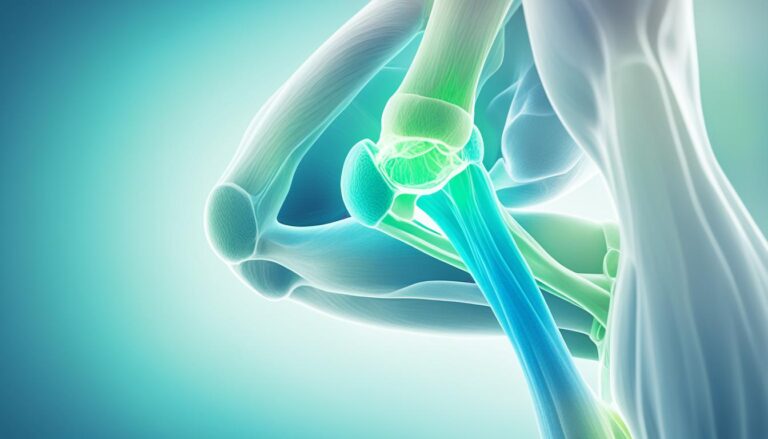
The patella, located at the front of the knee joint within the patellofemoral groove of the femur, is an essential component of the skeletal system. As we explore its triangular shape, superior and inferior aspects, and its articulation with other bones, we’ll gain a deeper understanding of how the patella enhances leg extension and protects the knee joint.
However, like any other body part, the patella is susceptible to certain conditions and disorders. We’ll uncover common patellar issues such as dislocation, subluxation, and their impact on knee health. We’ll also explore diagnostic tests and treatment options, providing valuable insights into managing and recovering from patella-related conditions.
Whether you’re an athlete, a fitness enthusiast, or simply curious about the intricate workings of the human body, this article will shed light on the fascinating world of patella anatomy and its crucial role in maintaining joint health.
Structure and Function of the Patella
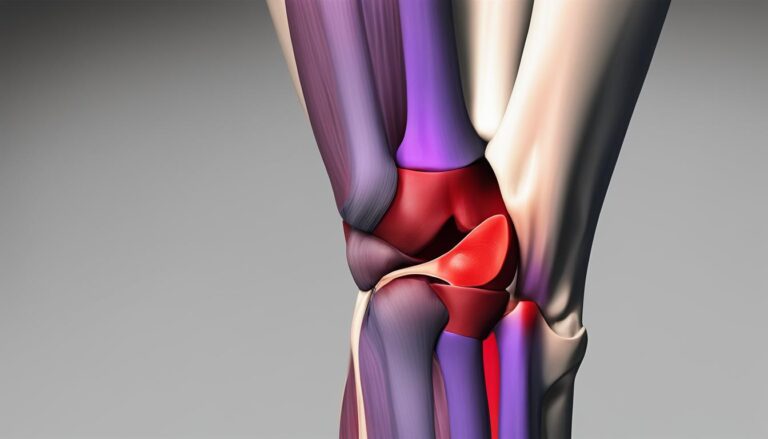
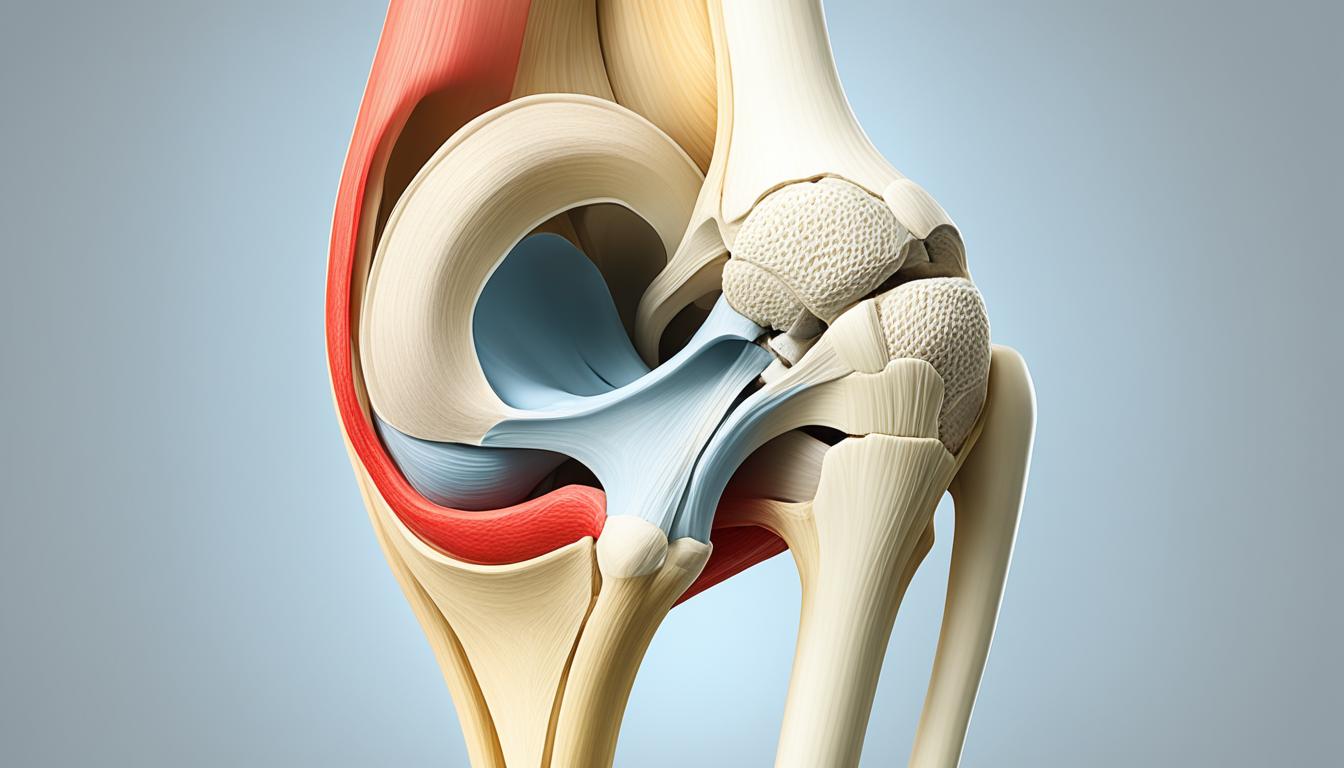
The patella, also known as the kneecap, is a bone with a distinctive triangular shape. It plays a crucial role in the bone structure of the knee joint. The patella has an anterior surface and a posterior surface. The apex of the patella is located at the inferior end, which is connected to the tibial tuberosity by the patellar ligament. On the other hand, the base forms the superior aspect of the bone and provides attachment for the quadriceps tendon, a major muscle group in the thigh.
The posterior surface of the patella articulates with the femur, the thigh bone, through its medial and lateral facets, allowing for smooth movement of the knee joint. This bone configuration enhances leg extension by increasing the leverage of the quadriceps tendon, allowing for powerful movements involved in activities like running, jumping, and walking. Moreover, the patella serves to protect the anterior aspect of the knee joint from physical trauma, such as blows or collisions.
The unique structure of the patella, with its triangular shape, anterior and posterior surfaces, apex, base, and medial and lateral facets, enables efficient movement and protects the knee joint. Understanding the structure and function of the patella helps us comprehend its vital role in leg extension and the overall stability and protection of the knee joint.

In the next section, we will explore common conditions and disorders that can affect the patella, leading to knee pain and discomfort. Understanding these conditions is essential for identifying potential issues and seeking appropriate treatment. Stay tuned!
Common Conditions and Disorders of the Patella
Several conditions and disorders can affect the patella, leading to knee pain and discomfort. Understanding these common issues is essential for proper diagnosis and treatment. The following are some of the most frequently encountered patellar conditions:
1. Patellar Dislocation
Patellar dislocation occurs when the patella completely or partially moves out of its normal position in the patellofemoral groove. This displacement can cause severe pain, swelling, and limited range of motion in the knee joint. Factors such as trauma, abnormal patellar alignment, or weak surrounding structures can contribute to this condition.
2. Patellar Subluxation
Patellar subluxation refers to a partial dislocation of the patella. It occurs when the patella momentarily slips out of its normal alignment but spontaneously returns to its original position. This can cause sudden pain, instability, and a feeling of the knee giving way. Frequent episodes may lead to increased joint laxity and further complications.
3. Osteoporosis
Osteoporosis is a condition characterized by reduced bone density and weakened bone structure. It can affect the patella and increase the risk of fractures. An important diagnostic tool for osteoporosis is a bone density test, which assesses the strength and density of the bones to determine the risk level and guide appropriate treatment options.
Other common conditions and disorders of the patella include patella fractures, tendonitis, and chondromalacia patella. These conditions may occur due to trauma, overuse, repetitive stress, or degenerative changes in the joint.
Treatments for patellar conditions vary depending on the specific condition and its severity. Here are some common treatment options:
- Use of immobilizing devices such as braces or splints to stabilize the knee and promote healing.
- Rest and avoiding activities that exacerbate the condition, allowing the patella to heal and reduce inflammation.
- Physical therapy exercises and stretches to strengthen the surrounding muscles and improve knee stability and function.
- Application of ice and heat, along with over-the-counter pain medications or anti-inflammatory drugs to alleviate pain and reduce swelling.
- In cases of patella fracture, immobilization with a cast or surgical intervention may be necessary to properly align and heal the bone.
- Treatment for osteoporosis includes a combination of exercise, dietary changes, calcium and vitamin D supplementation, and prescription medications to manage the condition and improve bone density.
Proper diagnosis and early intervention are crucial in effectively managing patellar conditions. If you experience persistent knee pain, swelling, or instability, seek medical attention to determine the underlying cause and receive appropriate treatment.
Tests and Diagnosis of Patella Conditions
When it comes to diagnosing patella conditions, healthcare professionals rely on a combination of tests to accurately assess the health and functionality of the kneecap. Understanding the root cause of pain or dysfunction in the patella is crucial in determining the appropriate treatment plan for patients. In this section, we will explore two essential diagnostic procedures: the patella reflex test and imaging tests.
Patella Reflex Test
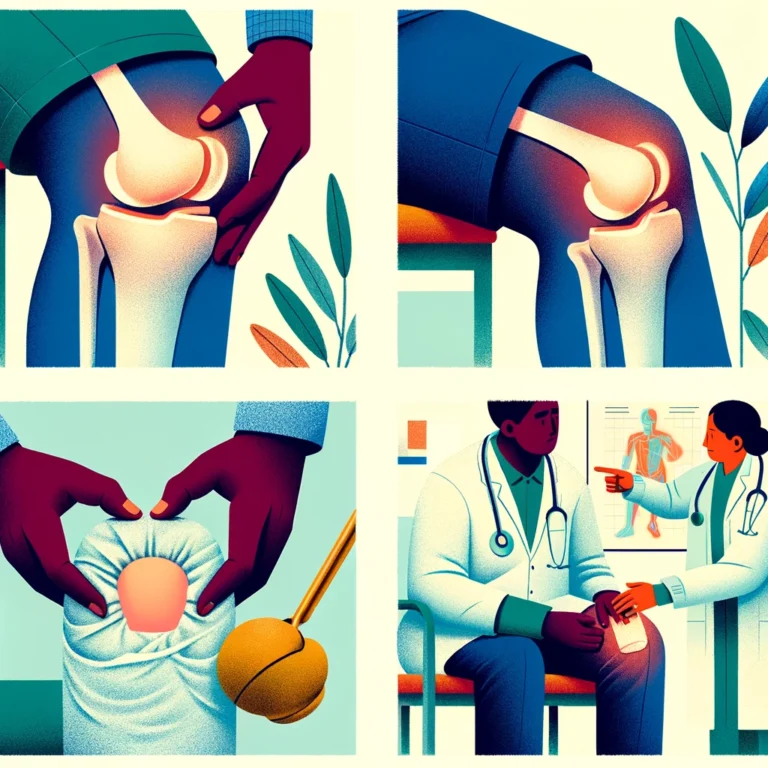
The patella reflex test is a simple yet effective examination that evaluates the integrity of the patellar reflex. This test involves tapping lightly below the patella with a reflex hammer, which stimulates a reflexive extension of the lower leg. The patella reflex, also known as the knee-jerk reflex, is mediated by the sensory and motor nerves in the patellar region.
During the patella reflex test, the examiner taps the patellar tendon, causing the quadriceps muscle to contract and the leg to extend. This reflex provides valuable information about the function of the nerves and muscles controlling the knee joint. A diminished or absent patellar reflex may indicate underlying nerve damage or dysfunction.
Imaging Tests
Imaging tests play a crucial role in diagnosing various patella conditions, including injuries and fractures. These tests provide detailed visual representations of the patella and surrounding structures, aiding healthcare professionals in accurate diagnosis and treatment planning.
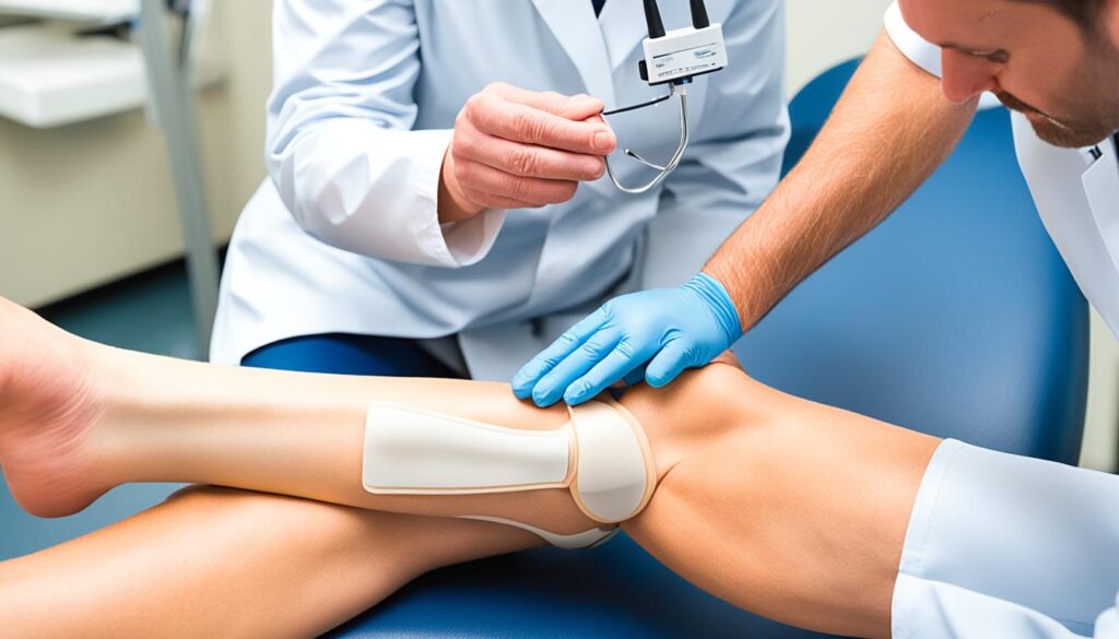
X-rays are commonly used to assess for patellar injuries, such as fractures or dislocations. X-ray images provide a clear view of the bone, enabling healthcare professionals to identify any abnormalities or structural damage. This imaging test is often performed as part of a routine examination or when a patellar injury is suspected.
| Imaging Tests | Use |
|---|---|
| X-rays | To diagnose patellar injuries and fractures |
| Magnetic Resonance Imaging (MRI) | To assess soft tissue issues, such as ligament tears or cartilage damage |
| Ultrasound | To evaluate soft tissue structures and identify any abnormalities or swelling |
Table: Imaging Tests for Patella Conditions
Additional imaging tests, such as magnetic resonance imaging (MRI) and ultrasound, may be employed to further investigate soft tissue structures and identify injuries or abnormalities not visible on X-rays alone. MRI is particularly useful in assessing ligament tears or cartilage damage, while ultrasound can provide real-time images, allowing for dynamic evaluation of the patella and surrounding structures.
These imaging tests, in conjunction with a thorough physical examination and medical history review, provide valuable insights into the underlying causes of patella conditions. By accurately diagnosing and understanding the specific issue affecting the patella, healthcare professionals can develop personalized treatment plans to promote optimal recovery and long-term joint health.
Treatment Options for Patella Conditions
When it comes to treating patella conditions, the approach may vary depending on the specific condition and its severity. Here are some common treatment options for patella-related issues:
1. Brace or Immobilizing Device: Wearing a brace or an immobilizing device can help provide stability and support to the knee, allowing it to heal properly. These devices limit the movement of the patella, reducing pain and preventing further damage.
2. Rest and Activity Modification: Resting the affected knee and avoiding activities that worsen the condition is vital in promoting healing and reducing pain. By giving the patella time to recover, you can prevent further strain and damage.
3. Physical Therapy: Physical therapy plays a crucial role in restoring strength, flexibility, and range of motion to the knee joint. A qualified physical therapist can design a personalized exercise program to strengthen the surrounding muscles and improve overall knee function.
4. At-Home Treatments: Implementing at-home treatments can help alleviate symptoms and support the healing process. Applying ice packs to the affected area can reduce inflammation and pain. Over-the-counter pain relievers, such as acetaminophen or nonsteroidal anti-inflammatory drugs (NSAIDs), can also provide temporary relief.
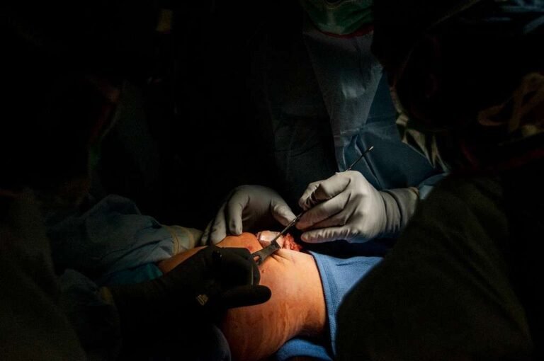
5. Specialized Interventions: In more severe cases, such as patella fractures or osteoporosis affecting the patella, specialized interventions may be necessary. For patella fractures, immobilization with a splint or cast may be required. In some instances, surgery may be needed to realign the fractured bones. Osteoporosis treatment focuses on improving overall bone health through exercise, supplements, and prescription medications.
| Treatment Options | Benefits |
|---|---|
| Brace or Immobilizing Device | Provides stability and support to the knee |
| Rest and Activity Modification | Promotes healing and reduces strain |
| Physical Therapy | Restores strength, flexibility, and range of motion |
| At-Home Treatments | Alleviates symptoms and supports healing |
| Specialized Interventions | Tailored treatments for severe cases |
By understanding the appropriate treatment options for patella conditions, you can work towards relieving pain, promoting healing, and improving overall knee function. It is important to consult with a healthcare professional for an accurate diagnosis and personalized treatment plan.
Clinical Relevance and Injuries to the Patella
The patella plays a crucial role in assessing and managing knee injuries, making it clinically relevant. One common injury involving the patella is patellar dislocation, which occurs when the patella is forced out of its normal position within the patellofemoral groove. This injury is often caused by high-force impacts or sudden twisting of the knee, commonly seen in sports injuries such as football, rugby, and ice hockey.
Another injury that can occur to the patella is a patellar fracture. This type of injury is typically the result of direct trauma to the patella or a sudden quadriceps muscle contraction. Patellar fractures are more prevalent in males and adults between the ages of 20 and 50.
| Injury | Cause | Common Occurrences |
|---|---|---|
| Patellar Dislocation | High-force impact Sudden twisting of the knee |
Sports injuries (football, rugby, ice hockey) |
| Patellar Fracture | Direct trauma to the patella Sudden quadriceps muscle contraction |
Males Adults between the ages of 20 and 50 |
Understanding the clinical relevance of the patella in these injuries is crucial for accurate diagnosis and effective treatment. Proper management of patellar injuries can help individuals recover and regain optimal knee function.
Conclusion
The patella, also known as the kneecap, is a vital bone in the human body’s skeletal system. By understanding its anatomy and function, we can better grasp its role in maintaining joint health. Various conditions and injuries can impact the patella, but with proper diagnosis and treatment, individuals can find relief from pain and improve their overall joint function.
It is crucial to seek medical attention when experiencing symptoms related to the patella, such as knee pain or instability. A healthcare professional can conduct appropriate tests and provide an accurate diagnosis. Following the recommended treatment plan, which may include rest, physical therapy, or even surgery in severe cases, is essential for optimal recovery.
By prioritizing joint health and taking prompt action, individuals can overcome patella-related conditions and injuries. Remember, early intervention is key in ensuring a swift and successful recovery. If you are experiencing any issues with your patella, don’t hesitate to consult a medical professional for proper evaluation and personalized treatment options.
FAQ
What is the patella?
The patella, also known as the kneecap, is a bone located at the front of the knee joint within the patellofemoral groove of the femur. It is the largest sesamoid bone in the body and plays a crucial role in the skeletal system.
What is the structure and function of the patella?
The patella has a triangular shape and consists of an anterior and posterior surface. It is connected to the tibial tuberosity by the patellar ligament and provides attachment for the quadriceps tendon. The main functions of the patella are to enhance leg extension by increasing the leverage of the quadriceps tendon and to protect the anterior aspect of the knee joint from physical trauma.
What are some common conditions and disorders of the patella?
Some common conditions and disorders of the patella include patellar dislocation, patellar subluxation, and osteoporosis. Patellar dislocation involves the complete or partial displacement of the patella from its normal position, while patellar subluxation refers to a partial dislocation. Osteoporosis is a condition characterized by weak and brittle bones that can also affect the patella.
How are patella conditions diagnosed?
Patella conditions can be diagnosed through various tests. The most common test is the patella reflex test, where the knee is tapped below the patella to elicit an involuntary leg extension reflex. Imaging tests such as X-rays may also be necessary to diagnose patellar injuries or fractures.
What are the treatment options for patella conditions?
The treatment for patella conditions depends on the specific condition and its severity. Conservative treatment options include wearing a brace or other immobilizing device, resting, undergoing physical therapy, and using at-home treatments such as icing and over-the-counter pain relievers. In cases of patella fracture or osteoporosis, more specialized interventions may be needed, including immobilization with a splint or cast and surgery to realign the bones.
How are injuries to the patella clinically relevant?
Injuries to the patella, such as patellar dislocation and fractures, are clinically relevant as they can affect joint health and function. Patellar dislocation often occurs due to high-force impacts or sudden twisting of the knee, commonly observed in sports like football, rugby, and ice hockey. Patellar fractures typically result from direct trauma to the bone or sudden contraction of the quadriceps muscle and are more prevalent in males and adults between the ages of 20 and 50.
Source Links
- https://my.clevelandclinic.org/health/body/25038-patella
- https://www.kenhub.com/en/library/anatomy/patella
- https://teachmeanatomy.info/lower-limb/bones/patella/