Understanding Patellar Ligament Attachment Points
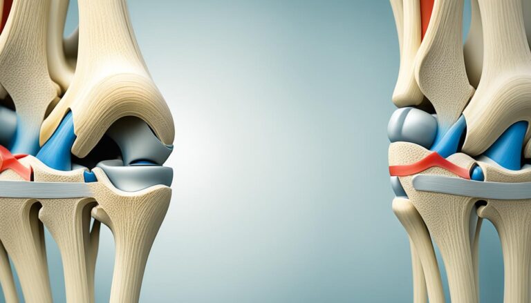
Did you know that the patellar ligament, also known as the attachment site of the patellar tendon, plays a crucial role in the stability and function of the knee joint? This small but mighty ligament connects the inferior third of the patella (kneecap) to the tibial tuberosity, anchoring the patella in place and allowing for efficient extension of the knee.
Located anterior to the knee joint, the patellar ligament acts as a bridge between the quadriceps muscle and the tibia, providing stability and support during movement. Without the proper attachment of the patellar ligament, the knee joint would be prone to instability and decreased functionality.
In this article, we will explore the anatomy and function of the patellar ligament, as well as its role in patellofemoral pain and pathology. We will also discuss the examination techniques used to assess the integrity of the patellar ligament and guide appropriate treatment strategies for conditions related to this important structure.
Join us as we delve into the fascinating world of the patellar ligament and discover how this seemingly small attachment point has a significant impact on our knee function and overall mobility.
Anatomy of the Patella and Knee Joint
The patella, also known as the kneecap, is a vital anatomical structure of the patella and plays a crucial role in knee joint anatomy. It is positioned deep to the fascia lata and rectus femoris tendon, anterior to the knee joint. With a triangular transverse cross-section, the patella articulates with the trochlear groove of the distal femur.
The patella possesses multiple articulating surfaces, including the medial and lateral facets, which facilitate smooth movement within the knee joint. Notably, the attachment points of the patellar ligament are of significant importance. The patellar ligament attaches to the tibial tuberosity, while the quadriceps tendon connects to the superior aspect of the patella.
The patellar ligament envelops the inferior third of the patella, offering stability to the knee joint during movement. This anatomy of the patella and the knee joint elucidates the interdependence of various structures in maintaining the functionality and integrity of the knee.
Function of the Patellar Ligament
The patellar ligament plays a crucial role in the function of the patellofemoral joint. It enhances the efficiency of the quadriceps muscle by increasing the moment arm of the extended knee.
The patella acts as a fulcrum for the quadriceps tendon, allowing for more effective torque generation on the tibia during knee extension. This increased torque is especially critical during the final 15° of knee extension, where twice as much torque is required compared to the initial phase of extension.
The patellar ligament helps to increase the moment arm during this phase, resulting in an additional 60% torque generation.

Furthermore, the patellar ligament contributes to the protection of the quadriceps tendon from frictional forces and acts as a bony shield for deeper structures in the knee joint.
Overall, the patellar ligament plays a significant role in optimizing quadriceps efficiency and maintaining stability within the patellofemoral joint.
Static and Dynamic Patellar Alignment
The alignment of the patella can vary between individuals and can significantly impact the function of the patellofemoral joint. Two important aspects to consider when assessing patellar alignment are static alignment and dynamic alignment.
Static Patellar Alignment
Static patellar alignment refers to the position of the patella in relation to the femur in a resting position. Ideally, the patella should be equidistant from the medial and lateral borders of the femur, ensuring optimal load distribution and minimizing excessive stress on any particular area. However, deviations from this ideal alignment can occur, leading to various issues and potential complications.
An anterior or posterior tilt of the patella refers to the forward or backward inclination of the patella, respectively. This tilt can result from muscular imbalances or connective tissue abnormalities. In contrast, an inferior or superior tilt refers to the downward or upward tilting of the patella, respectively. Lateral or medial tilting indicates deviations to the right or left, respectively.
These deviations from the optimal alignment can disrupt the smooth interaction between the patella and the femur, leading to patellofemoral pain, instability, and dysfunction. The precise assessment of static patellar alignment is crucial in diagnosing and managing patellofemoral issues.
Dynamic Patellar Alignment
Dynamic patellar alignment pertains to the movement of the patella during knee flexion and extension. As we move our knee joint, the patella can tilt and rotate. These dynamic adjustments help maintain optimal patellofemoral tracking and stability.
During knee flexion and extension, the patella can tilt forward or backward, as well as rotate medially or laterally. These movements allow the patella to maintain proper alignment and engage in smooth articulation with the trochlear groove of the femur.
Patella Alta and Patella Baja
Patella alta and patella baja are two specific alignment variations that can impact the function and stability of the patellofemoral joint.
Patella alta refers to a higher-riding patella, where the patella is positioned relatively higher in the knee joint. This anatomical discrepancy can result from factors such as a shallow trochlear groove or a patellar tendon that inserts higher on the patella. Patella alta can lead to patellar instability and an increased risk of patellar subluxation or dislocation.
On the other hand, patella baja refers to a lower-riding patella, where the patella is positioned relatively lower in the knee joint. This alignment variation can occur due to factors such as a deep trochlear groove or a patellar tendon that inserts lower on the patella. Patella baja can cause restrictions in knee flexion and increased patellofemoral joint stress.
Assessing static and dynamic patellar alignment is crucial in understanding and managing patellofemoral issues effectively. Proper alignment ensures optimal joint function, stability, and reduced risk of complications.
| Alignment | Description | Potential Complications |
|---|---|---|
| Anterior tilt | Forward inclination of the patella | Patellofemoral pain, instability |
| Posterior tilt | Backward inclination of the patella | Patellofemoral pain, instability |
| Inferior tilt | Downward tilting of the patella | Patellofemoral pain, restricted knee flexion |
| Superior tilt | Upward tilting of the patella | Patellofemoral pain, increased joint stress |
| Lateral tilt | Deviation to the right side | Patellofemoral pain, altered tracking |
| Medial tilt | Deviation to the left side | Patellofemoral pain, altered tracking |
| Patella alta | Higher-riding patella | Patellar instability, subluxation, dislocation |
| Patella baja | Lower-riding patella | Restricted knee flexion, increased joint stress |
Patellofemoral Pain and Pathology
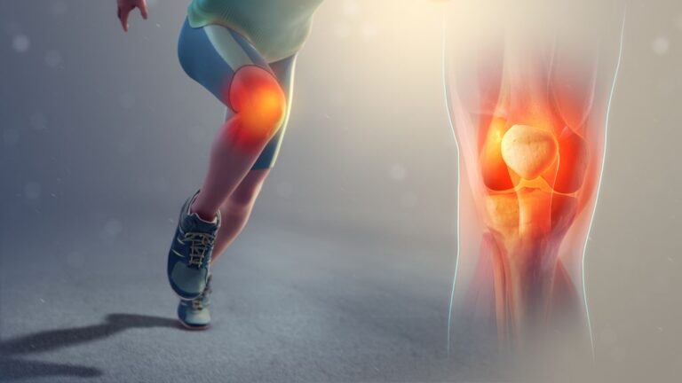
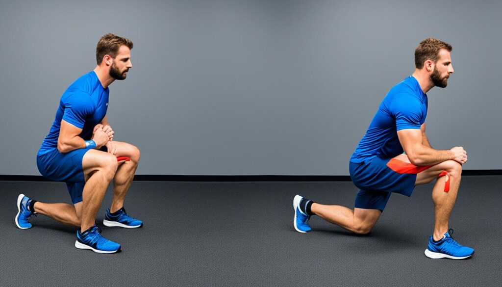
Patellofemoral pain is a common condition characterized by discomfort and pain in and around the patellofemoral joint, particularly during activities that involve lower-limb loading. This condition can be attributed to various factors, including patellar malalignment, overuse, muscle imbalances, and trauma. Individuals with patellofemoral pain may experience symptoms such as anterior knee pain, stiffness, and a sensation of grinding or popping in the knee.
Patellofemoral osteoarthritis is a degenerative condition that affects the patellofemoral joint, leading to pain, stiffness, and reduced mobility. It is characterized by the breakdown of cartilage in the patellofemoral joint, resulting in inflammation and joint degeneration. This condition can cause significant discomfort and impact the quality of life, particularly in individuals who engage in activities that place stress on the knee joint.
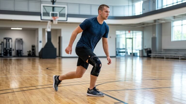
Patellar instability refers to the tendency of the patella to dislocate or subluxate from its normal position. This can occur as a result of patellar malalignment, ligament laxity, or trauma. Individuals with patellar instability may experience recurrent episodes of knee pain, swelling, and a feeling of instability in the knee joint. This condition can significantly impact daily activities and sports participation, requiring appropriate management and treatment.
Patellar tendon rupture is a rare but severe injury that can occur when the patellar tendon is completely severed from the patella or the tibial tuberosity. This injury often requires surgical repair to restore the function and stability of the patellofemoral joint. Individuals with a patellar tendon rupture may experience sudden, severe pain, an inability to straighten the knee, and swelling around the kneecap.
The patellar reflex is a neurological test used to assess the function of the patellar tendon and the reflex arc. It involves tapping the patellar tendon, which elicits a reflexive contraction of the quadriceps muscle and extension of the knee. This reflex provides information about the integrity and responsiveness of the neuromuscular pathways involved in knee extension.
| Condition | Symptoms | Treatment |
|---|---|---|
| Patellofemoral Pain | Anterior knee pain, stiffness, grinding or popping sensation in the knee | Physical therapy, bracing, pain management, muscle strengthening exercises |
| Patellofemoral Osteoarthritis | Pain, stiffness, reduced mobility | Non-surgical: medication, physical therapy, weight management; Surgical: arthroscopy, partial or total joint replacement |
| Patellar Instability | Recurrent dislocation, knee pain, instability | Physical therapy, bracing, surgical stabilization procedures |
| Patellar Tendon Rupture | Sudden, severe pain, inability to straighten the knee | Surgical repair, physical therapy, rehabilitation |
We encourage individuals experiencing any patellofemoral pain or pathology to seek proper medical evaluation and treatment. Early diagnosis and appropriate management can significantly improve outcomes and help individuals regain function and quality of life.
Examination of the Patellar Ligament
The examination of the patellar ligament is an essential component of assessing the alignment, function, and stability of the patellofemoral joint. By conducting a comprehensive physical examination, we can gather valuable information that guides proper treatment and rehabilitation strategies.
Different examination techniques can be employed to evaluate the patellar ligament in various positions, including standing, sitting, and supine. These examinations allow us to assess different aspects of the joint, providing a comprehensive understanding of its condition.
Standing Examination
The standing examination is performed to evaluate static alignment, patellar height, and leg-length discrepancies. By observing the patient in an upright position, we can assess the positioning of the patella in relation to the femur and tibia. Any deviations from the normal alignment can be indicative of underlying issues that may require further investigation.
Sitting Examination
The sitting examination provides valuable information about patellar tracking and potential patellofemoral pain. By observing the patient’s knee movement while seated, we can assess the smoothness and stability of the patella as it glides in the trochlear groove of the femur. This examination helps identify any abnormalities or imbalances in the patellar movement.
Supine Examination
The supine examination allows for the assessment of patellar mobility, stability, and the presence of any palpable abnormalities. By manipulating the patella while the patient lies flat on their back, we can evaluate the range of motion, joint stability, and detect any signs of tenderness or palpable abnormalities. This examination is crucial for identifying potential underlying issues and determining the overall health of the patellar ligament.
Special Tests for Patellar Ligament Assessment
In addition to the standard physical examination, special tests can be performed to assess patellar instability or patellofemoral pain. These tests involve specific maneuvers designed to reproduce symptoms and assess joint stability. Two common special tests include:
- The apprehension test: This test is used to assess for patellar instability. By applying lateral force to the patella, the examiner can evaluate the patient’s response and detect any signs of apprehension or discomfort.
- The Clarke’s test: This test helps assess for patellofemoral pain. By applying pressure to the patella while the patient performs a quadriceps contraction, the examiner can determine the presence of patellar maltracking or pain.
These special tests, along with the standard physical examination techniques, provide valuable insights into the condition of the patellar ligament and the overall health of the patellofemoral joint.
| Examination Technique | Purpose |
|---|---|
| Standing Examination | Assess static alignment, patellar height, and leg-length discrepancies |
| Sitting Examination | Evaluate patellar tracking and detect patellofemoral pain |
| Supine Examination | Assess patellar mobility, stability, and palpable abnormalities |
Conclusion
The patellar ligament plays a crucial role in the function and stability of the patellofemoral joint. It serves as an attachment point for the patella, allowing for efficient quadriceps muscle function and knee extension. Understanding the anatomy and function of the patellar ligament is essential for the diagnosis and management of patellofemoral pain and pathology.
A comprehensive examination of the patellar ligament provides valuable information about alignment, stability, and function, which guides appropriate treatment and rehabilitation strategies. By properly assessing the patellar ligament, we can contribute to enhanced mobility and function of the knee joint, leading to improved quality of life for individuals experiencing patellofemoral issues.
By focusing on the alignment, stability, and overall function of the patellar ligament, we can better address patellofemoral pain and pathology. This thorough understanding allows us to develop individualized treatment plans to optimize outcomes and restore optimal knee joint function for our patients. Together with other healthcare professionals, we can ensure that individuals experiencing patellofemoral pain receive the care they need.
FAQ
What is the patellar ligament attachment site?
The patellar ligament attaches the inferior third of the patella to the tibial tuberosity.
What is the function of the patellar ligament?
The patellar ligament plays a crucial role in the function of the patellofemoral joint, enhancing the efficiency of the quadriceps muscle and providing stability to the knee joint during movement.
How does patellar alignment affect the function of the patellofemoral joint?
Patellar alignment variations can impact patellofemoral tracking and stability, potentially leading to patellofemoral pain or dysfunction.
What are common patellofemoral pathologies?
Common patellofemoral pathologies include patellofemoral pain, patellofemoral osteoarthritis, patellar instability, and patellar tendon rupture.
How can the patellar ligament be examined?
The patellar ligament can be examined through a comprehensive physical examination, including standing, sitting, and supine assessments, as well as special tests to assess stability and function.
What is the significance of understanding the patellar ligament?
Understanding the anatomy and function of the patellar ligament is essential for diagnosing and managing patellofemoral pain and pathology, guiding appropriate treatment and rehabilitation strategies.
Source Links
- https://www.ncbi.nlm.nih.gov/books/NBK519534/
- https://www.physio-pedia.com/Patellofemoral_Joint
- https://www.kenhub.com/en/library/anatomy/patellar-tendon