Muscle strength and its improvement are important for everyone for their daily activities. Conventionally, muscle function can be assessed through physical performance tests and/or muscle strength measurements. However, these methods are not applicable to people who cannot undergo forceful muscle contractions, such as infants, the elderly, and patients with injuries or cognitive impairment (such as dementia).
A simple and popular alternative is the use of bioelectrical impedance analysis (BIA), which can quickly and non-invasively measure tissue resistance (which depends on the amount of water and electrolytes present in the tissue) and reactance (which depends on the integrity of the cell) can be measured. membrane). Phase angle (PhA), a measurement derived via BIA, is calculated using the resistance and reactance of the tissue. It is directly proportional to muscle cell mass and function. Many studies have linked PhA across the body to maximal muscle strength, but none associate PhA with knee extension strength or explosive muscle strength (the force required to make fast, powerful movements such as sprinting or standing up from a chair) in adults. Taking into account the importance of knee muscle strength – especially in the elderly, who need it for their independence, and athletes, who need strong knees for better performance – such a study was necessary.
In this spirit, a group of scientists, including Professor Ryota Akagi, from the College of Systems Engineering and Science, Shibaura Institute of Technology (SIT); Assistant Professor Kosuke Hirata from the Faculty of Sports Sciences, Waseda University; and researchers Yosuke Yamada and Tsukasa Yoshida from the Healthy Longevity Research Section, National Institute of Health and Nutrition, National Institutes of Biomedical Innovation, Health and Nutrition examined the association of PhA with the neuromuscular properties of knee extensor muscles in both young and older people. adults. Their findings were published in Part 13 of Frontiers in physiology on August 11, 2022. Dr. Akagi and Dr. Hirata, the corresponding authors of the paper, tell us: “We wanted to assess the association of PhA in the thigh with maximal muscle strength, explosive muscle strength, contractile properties and neuromuscular activity. and find out which of the two – whole body PhA or the thigh – was a better predictor of knee extensor strength.
The team measured the PhA of the whole body and thighs of 55 participants (23 young men and 32 older men) at 50 kHz. Participants were asked to perform a 4-second maximal voluntary isometric contraction (MVIC) to measure peak torque (PT).MVIC) and a 1 second MVIC to measure the rate of torque development (RTD) over a time difference of 0-200 milliseconds. The average mean value of these three measurements (i.e. PTMVIC and RTD) was used for further analysis. Contractile properties were also analyzed because they are significant markers of the mechanisms underlying force generation in the muscles. Finally, muscle activity was assessed by electromyography (EMG-RMS).
The study findings showed that both whole-body and thigh PhAs are associated with knee extensor muscle strength (with thigh PhA being the preferred predictor of knee extensor strength). However, this association was thought to be due to the contractile properties of the muscles and not to neural aspects. Thus, both measures could not predict neuromuscular activity or explosive muscle strength (which largely depends on neuromuscular control) of knee extensors.
This study is promising for current and future implications. Speaking of the present, being able to assess knee muscle strength is very important, especially for the elderly (where strong knee muscles mean greater independence in movement) and athletes (who need to maintain knee muscle strength to perform better ). . The above findings demonstrate a new way to assess muscle strength not only for healthy adults, but also for people suffering from orthopedic or cognitive disorders. And speaking about the future, Dr. Akagi shows us the implications of their work: “People can assess their muscle condition in just a few seconds using BIA. In the future, we may be able to build a system that uses a person’s BIA to provide them with advice to promote their health.”
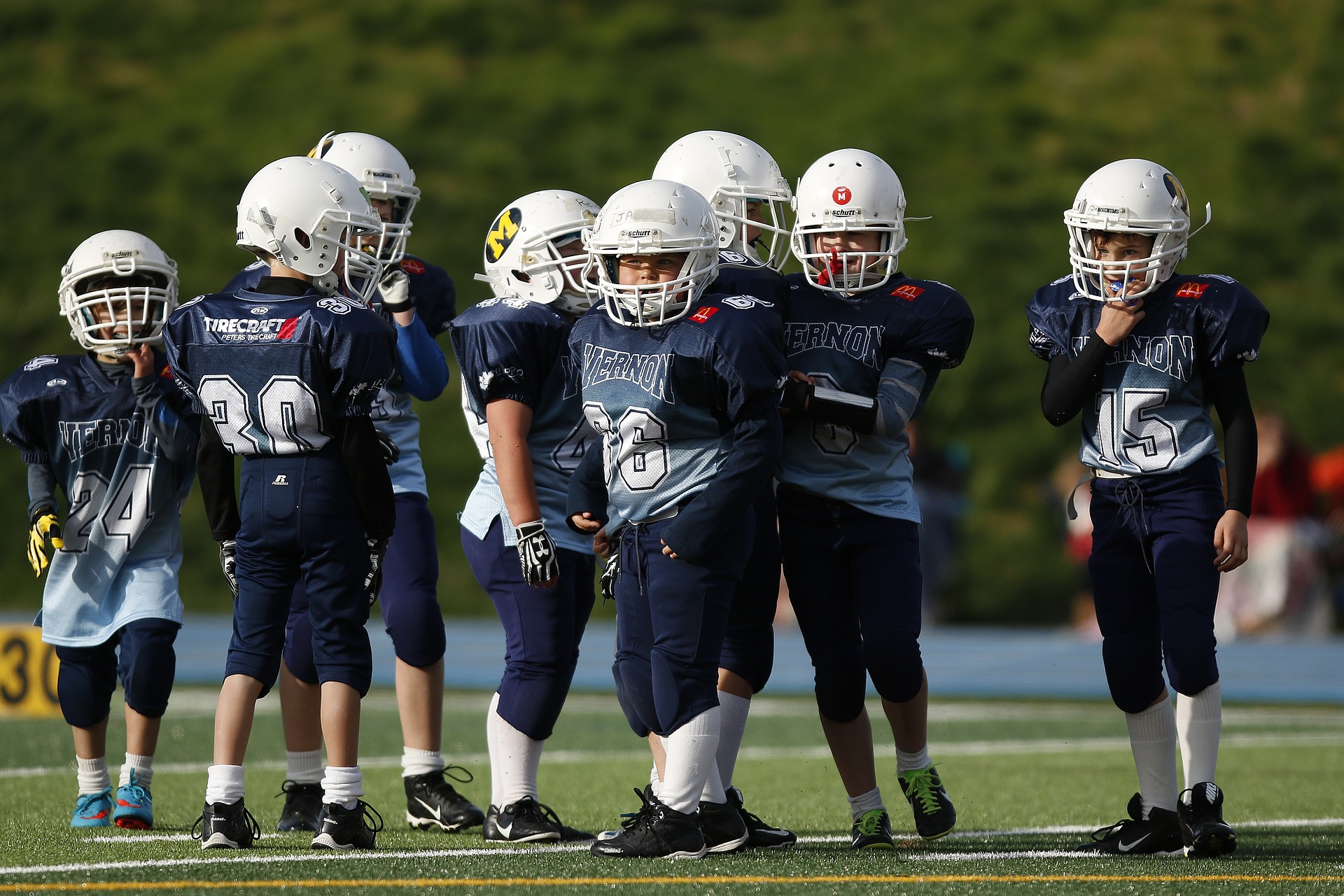
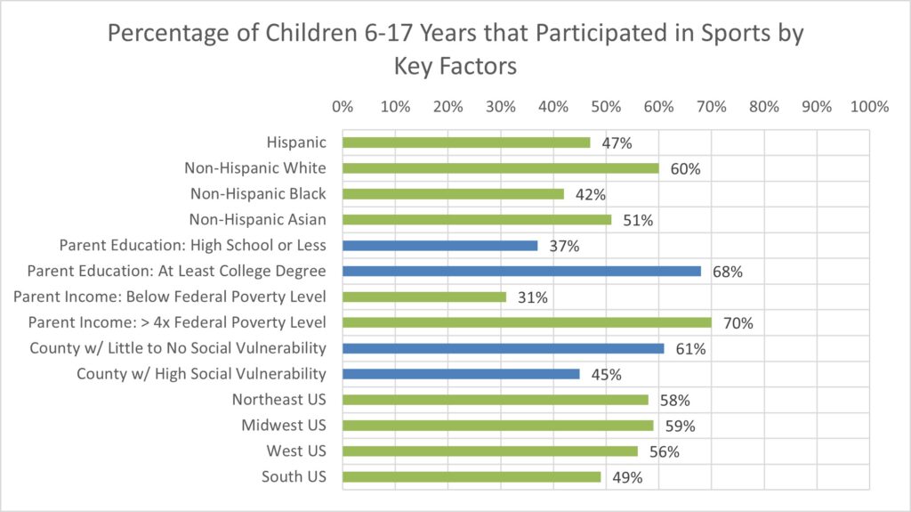
Organized sports participation among children aged 6–17 years: United States, 2020
Black LI, Terlizzi EP, Vahratian A. National Center for Health Statistics (US). Publication date: 08/11/2022. Series: data overview; No. 441
https://stacks.cdc.gov/view/cdc/119026
Take home message
Sports participation was lower among racial and ethnic minority children, children whose parents had lower education or income, children living in counties with greater social vulnerability, and children living in the southern United States.
Background
Youth participation in sports is associated with better physical fitness and mental health. Understanding the differences in sports participation can help inform strategies to promote active lifestyles among children and adolescents, which positively impacts their physical and mental health.
Study aim
Black and colleagues completed an analysis of data from the 2020 National Health Interview Survey to describe disparities in sports participation in the United States.
Methods
The authors analyzed data from the 2020 National Health Interview Survey. This survey is a nationally representative household survey conducted throughout the year. Parents reported whether their child participated in a sports team or club or took sports lessons at school or in the community in the previous 12 months. The authors then collected data on household income as a percentage of the federal poverty level, race and Hispanic origin, geographic region (Northeast US, Midwest US, Southern US, or Western US), social vulnerability of a county, and urbanicity ( urban region). -national classification) of a province.
Results
About half (54%) of children aged 6 to 17 have played sports in the past twelve months (boys: 56%; girls: 52%). The authors reported: “Participation levels were lower among children from racial and ethnic minority groups, children whose parents had lower levels of education and family income, children living in counties with greater social vulnerability, and children living in the South.”
Viewpoints
The results of this letter provide a surprising snapshot of sports participation in the United States. While it is established that early sports participation can have a positive impact on an individual, significant differences still exist. These findings indicate that sports participation is strongly associated with socioeconomic status. Ultimately, a higher socio-economic status can ensure children’s access to sports. This is particularly reflected in the fact that lower parental education, lower parental income and lower geographical area of social vulnerability are all associated with lower participation. These findings complement data from the Athletic Training Locations and Services Database, which shows that these communities also have less access to athletic training services.
This data provided a unique snapshot of 2020, when many sports shut down in the spring due to the COVID-19 pandemic. The survey asked about sports participation over the past year, so it is unclear whether the pandemic could influence these results. Furthermore, not every community is recovering from the pandemic in the same way. The authors acknowledge that it will be essential to replicate these analyzes with data from the 2022 survey to better understand patterns of sports participation after many of the restrictions associated with the pandemic are relaxed/removed.
Clinical implications
Sports medicine professionals should advocate for increased access to sports through local recreation organizations, schools, and other organizations. In regions with high social vulnerability, we should encourage policymakers that investments in youth sports and physical activity can help improve community health.
Questions for discussion
What can we do as doctors to positively influence participation in team sports?
Written by: Kyle Harris Review by: Jeffrey Driban
related posts
Asian Pacific Society of Cardiology Consensus Recommendations for Pre-Participation Screening in Young Competitive Athletes A lasting impression: youth sports participation and healthy habits as adults Previous participation in collision sports is associated with reduced quality of life Lower socioeconomic status is associated with less access to athletic training services
Kalamazoo, Mich., Nov. 2, 2023 (GLOBE NEWSWIRE) — Stryker (NYSE:SYK) reported operating results for the third quarter of 2023:
Third quarter Results
Reported net sales increased9.6% Unpleasant $4.9 billion
Organic net sales increased9.2%
Reported operating profit margin of 19.0%
Adjusted operating income margin(1)increased110 bps to23.4%
Reported earnings per share fell 15.9% to $1.80
Custom EPS(1)increased16.0% Unpleasant $2.46
Overview of net sales growth in the third quarter
Reported
Exchange foreign currencies
Constant currency
Acquisitions / divestments
Biological
MedSurg and neurotechnology
10.5
%
0.2
%
10.3
%
0.2
%
10.1
%
Orthopedics and spine
8.4
0.4
8.0
—
8.0
Total
9.6
%
0.3
%
9.3
%
0.1
%
9.2
%
“We delivered another quarter of strong organic revenue growth and continued margin expansion,” said Kevin A. Lobo, chairman and CEO. “The positive momentum in our business remains intact, including a strong procedural environment and our super cycle of innovation.”
Sales analysis
Consolidated net sales of $4.9 billion increased 9.6% in the quarter and 9.3% at constant exchange rates. Organic net sales increased by 9.2% in the quarter, of which 8.9% due to higher unit volume and 0.3% due to higher prices.
MedSurg and Neurotechnology net sales of $2.9 billion increased 10.5% in the quarter and 10.3% at constant exchange rates. Organic net sales increased by 10.1% in the quarter, of which 8.7% due to higher unit volume and 1.4% due to higher prices.
Orthopedics and Spine net sales of $2.1 billion increased 8.4% in the quarter and 8.0% at constant exchange rates. Organic net sales increased 8.0% in the quarter, of which 9.1% was due to higher unit volume, partially offset by 1.1% due to lower prices.
Revenue Analysis
Reported net income of $692 million fell 15.2% in the quarter. Reported net income per diluted share of $1.80 fell 15.9% in the quarter. Reported gross profit margin and reported operating profit margin were 64.3% and 19.0% in the quarter. The reported net profit includes certain items, such as costs for acquisition and integration-related activities, the amortization of purchased intangible assets, structural optimization and other special costs (including depreciation and impairment of assets), costs to comply with certain regulatory requirements medical devices, recall-related matters, regulatory and legal matters and tax matters. Excluding the above items, adjusted gross profit margin(1) was 64.7% in the quarter, and an adjusted operating income margin(1) amounted to 23.4% in the quarter. Adjusted net profit(1) of $944 million rose 16.5% in the quarter. Adjusted net income per diluted share(1) of $2.46 rose 16.0% in the quarter.
Outlook for 2023
Given our year-to-date performance, robust capital equipment backlog and continued positive procedural trends, we now expect organic net sales growth for full year 2023(2) between 10.0% and 10.5%, including slightly positive prices for the year. If exchange rates remain near their current levels, we expect net sales to be adversely affected by approximately 0.6% and adjusted net earnings per diluted share(2) will be adversely affected by $0.10 to $0.15 for the full year, both of which are included in our guidance. Based on our performance in the first nine months of the year, together with our strong sales momentum, we now expect adjusted net income per diluted share(2) are between $10.35 and $10.45.
(1) A reconciliation of the non-GAAP financial measures: adjusted gross profit margin, adjusted operating income and adjusted operating income margin, adjusted net income and adjusted net income per diluted share, with the most directly comparable GAAP measures: gross profit margin, operating income and operating income margin, net income and net income per diluted share, and other important information are attached to this press release.
(2) We are unable to provide a quantitative reconciliation between our expected net sales growth and expected organic net sales growth because we are unable to estimate with reasonable certainty and without unreasonable effort the impact and timing of acquisitions and divestitures and the impact of foreign currency exchange rates. We are unable to present a quantitative reconciliation between our expected net income per diluted share and expected adjusted net income per diluted share because we cannot estimate with reasonable certainty and without unreasonable effort the impact and timing of structural optimization and other special costs, acquisitions and predict takeovers. -related costs and adjustments to the fair value of inventories and the outcome of certain regulatory, legal and tax matters. The financial impact of these items is uncertain and depends on several factors, including timing, and could be material to our consolidated statements of operations.
Conference call enabled Thursday November 2, 2023
As previously announced, we are organizing a conference call Thursday November 2, 2023 at 4:30 p.m., Eastern Time, to discuss our operating results for the quarter ended September 30, 2023 and provide an operational update.
Register for this conference call at: https://www.veracast.com/webcasts/stryker/events/SYK3Q23.cfm. Once registered, an email confirmation will be sent, including dial-in information and unique conference call access codes required for call entry. Registration is open during the live call. To ensure that you are connected before the start of the call, we recommend that you register at least 15 minutes before the start of the call.
A simultaneous webcast of the call will be accessible via the Investor Relations page of our website at www.stryker.com. For those who do not intend to ask a management question, we recommend listening via the webcast. Please allow 15 minutes to register, download and install the necessary software.
A replay will be available on our website following the conference call for up to one year from the time of the earnings call.
Caution Regarding Forward-Looking Statements
This press release contains information that contains or is based on forward-looking statements within the meaning of the federal securities laws, which are subject to various risks and uncertainties that could cause our actual results to differ materially from those expressed or implied by such statements. Such factors include, but are not limited to: weakening of economic conditions, or anticipation thereof, which could adversely affect the level of demand for our products; pricing pressures generally, including cost control measures that could adversely affect the price of or demand for our products; changes in the foreign exchange markets; legislative and regulatory actions; unexpected issues arising in connection with clinical trials and otherwise affecting the approval of new products by the U.S. Food and Drug Administration; inflationary pressure; higher interest rates; supply chain disruptions; changes in reimbursement levels from third-party payers; a significant increase in product liability claims; the final total costs related to recall-related matters; the impact of investigative and legal proceedings and compliance risks; settlement of tax audits; changes in tax laws and regulations; the impact of federal legislation to reform the United States health care system; costs to comply with medical device regulations; changes in the financial markets; changes in our credit ratings; changes in the competitive environment; our ability to integrate and realize the expected benefits of acquisitions in full or within the expected time frames; our ability to achieve expected cost savings; potential negative impacts resulting from environmental, social and governance (ESG) and sustainability issues; the impact on our operations and financial results of a public health emergency and related policies and actions of governments or other third parties; and breaches or failures of our or our suppliers’ information technology systems or products, including through cyber-attacks, data breaches, unauthorized access or theft. Additional information about these and other factors is included in our filings with the U.S. Securities and Exchange Commission, including our Annual Report on Form 10-K and Quarterly Reports on Form 10-Q. We disclaim any intention or obligation to publicly update or revise any forward-looking statements to reflect any change in our expectations or in events, conditions or circumstances on which those expectations may be based or that could affect the likelihood that actual results will occur differ from the results. included in the forward-looking statements.
Stryker is one of the world’s leading medical technology companies, and together with our customers, we are passionate about making healthcare better. We provide innovative products and services in medical, surgical, neurotechnology, orthopedics and spine that help improve patient and healthcare outcomes. In addition to our customers around the world, Stryker impacts more than 130 million patients annually. More information is available at www.stryker.com.
For investor questions, please contact:
Jason Beach, Vice President, Investor Relations at 269-385-2600 or jason.beach@stryker.com
For media inquiries please contact:
Yin Becker, Vice President, Chief Corporate Affairs Officer at 269-385-2600 or yin.becker@stryker.com
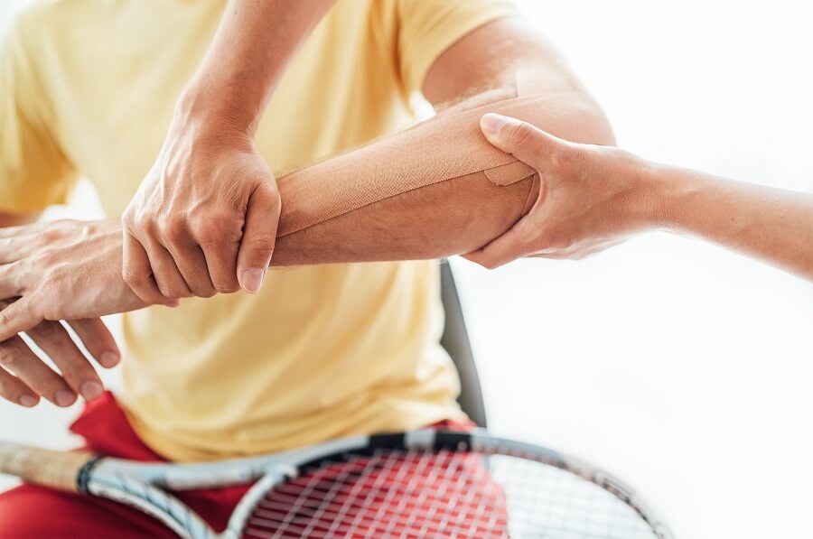
by Adam Halseth PT, DPT, SCS | Northeast Scottsdale
Tennis elbow or golfer’s elbow is an uncommon condition. It just has an impact about 1 to 3 percent of adults every year. But your chances of developing it increase as you get older. Although the name suggests it is a condition exclusive to these sports, it is possible to develop tennis or golfer’s elbow even if you have never played either game in your life.
Below you will find important information about this painful condition, including why it occurs, how to determine if you have the condition, and what treatment options are available.
Understanding tennis/golfer’s elbow
Tennis/golfer’s elbow is one of the most common causes of elbow pain. It is often persistent and painful enough to warrant physical therapy and/or a doctor’s visit. This condition occurs when the tendons that connect your forearm to the outside of the elbow bone become swollen and inflamed.
If this happens, it can lead to microscopic tears in the muscles and tendons. These tears can cause a lot of pain, even though they are very small.
Usually people don’t experience these symptoms unless they do a lot of repetitive movements. While the general population is not highly likely to experience this pain, as many as half of all players will experience it at some point in their lives.
What causes tennis/golfer’s elbow?
If you’re wondering what the causes are, the answer is: a lot of things! As mentioned, tennis/golfer’s elbow usually occurs as a result of repetitive movements of the elbow joint. Besides tennis and golf, some other activities that can lead to elbow pain and discomfort include:
Screens
Squash
Weightlifting
Racketball
Rake
Typing
To paint
Carpentry
To knit
Gardening
Swimming
Did some of the items in the list above surprise you? Fortunately, there are things you can do to reduce the chance of developing elbow pain or minimize symptoms if you already have it. Keep reading to learn more.
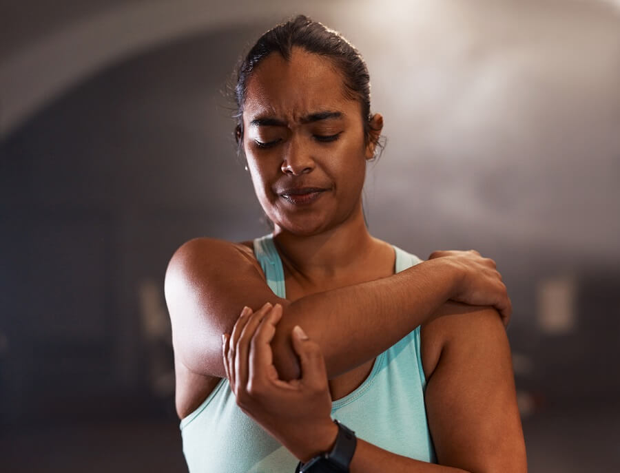
5 Symptoms of tennis/golfer’s elbow
Are you experiencing elbow pain that makes you wonder if you have tennis/golfer’s elbow or something else? Although a doctor or therapist should be consulted to make the most accurate diagnosis, these are five common symptoms:
Discomfort when you lift something
Weakness in your hand or forearm when making a fist or grabbing something
Recurring pain just below the bend in your elbow (on the outside of your forearm)
Pain that radiates from the elbow to the wrist
Discomfort when turning the forearm (for example, when opening a jar or a door)
As you can see, the primary symptom is pain in the elbow area. Conditions that mimic tennis elbow include:
Radiocapitellar arthritis
Osteochondritis dissecans
Intra-articular plica
Rotational instability
A visit to your PT can help rule out these conditions if you are unsure whether you have tennis/golfer’s elbow or another condition.
Prevention techniques
The best way to prevent developing this condition is to avoid doing repetitive movements too often. If you play a sport or other activity that requires repetitive use of your arm and muscles, make sure you take regular breaks. Stretch the muscles of your arms during your breaks.
It is also important to warm up the muscles of your arms before doing any physical activity that requires the use of your elbows and/or arms. When the muscles are warm, they can stretch and contract more easily without causing injury.
Once you’ve finished a sporting event or workout and stretched your arm muscles, consider applying ice to your elbows if you feel heat or inflammation in those areas. If you already have symptoms of tennis elbow despite your best efforts, consider getting physical therapy.
Physiotherapy treatment options
Physical therapy can help improve the flexibility and strength of your forearm muscles, making you less likely to develop tennis elbow. Physical therapy can also facilitate healing and reduce pain by stimulating blood flow to the affected tendons and muscles. Blood contains oxygen, which the muscles need to heal and function optimally.
Six common physical therapy treatment options recommended for tennis elbow:
Muscle stimulation
Ultrasound
Ice massage
Braces and tape to support the affected area
Specialized stretches and exercises
Nonsteroidal anti-inflammatory drugs
During your recovery from tennis/golfer’s elbow, it is important not to rush things. If you push your body before it is ready and your condition has healed enough, you can set yourself back. Before returning to your previous activity level, make sure that you can grasp objects without pain and that your elbow no longer looks or feels swollen. When you can bend and move your affected elbow without difficulty or discomfort, you can resume your normal activities.
Get your quality of life back
If you are ready to get your life back and fully recover from the symptoms of your tennis/golfer’s elbow, we are here to help. Contact Foothills Sports Medicine Physical Therapy today request your appointment.
The required sample size was calculated using G*Power (version 3.1, University of Düsseldorf, Germany) [21]. The a priori power analysis was calculated with an assumed power of 0.90, an alpha level of 0.01 and an effect size of Cohen’s f = 0.26 for pulmonary diffusion capacity (TL) [10]. The analysis showed that a total sample size of N=60 would be sufficient to detect a significant interaction effect by time. Accordingly, 80 participants were enrolled to account for possible dropouts due to injuries.
Eighty healthy prepubertal boys, aged 6 to 10 years, with normal lung volumes and regular flow-volume curves and no history of cardiovascular disease or allergies, volunteered to participate in the study. A respiratory functional examination was performed by a physician, including a health questionnaire to screen the participants’ medical history. The boys were randomly divided into two groups:
The football group (SG, n = 40) participated in four weekly football sessions of 75 minutes each over a period of 28 weeks.
The control group (CG, n = 40) matched to the SG in terms of age, body weight and height, participated only in regular physical education classes without any other extracurricular sports activities.
All participating boys went to the same school near the football training center. After providing written and verbal information about the risks and benefits of the study, written informed consent to participate in the study was obtained from all study participants and their parents or legal guardians before the start of the study. The ethics committee of Sousse Medical University (Tunisia) approved the study. The study was conducted in accordance with the latest version of the Declaration of Helsinki. The physical characteristics of the study participants at the time of inclusion are listed in Table 1.
Table 1 Characteristics of the participants and maximum exercise performance of the participating boys before the start of the study (pre: T1) and after completion of the study (post: T2)
Procedures
Baseline (Trial 1) anthropometric data (body height to the nearest 0.1 cm and body weight to the nearest 100 g) were collected using standard stadiometers (Seca™, Hamburg, Germany) and scales (Tefal, France). Maximum oxygen consumption (VO2max) and maximum aerobic power (MAP) were determined using standard protocols, with the extensive testing performed on a bicycle ergometer (Monark cycle). The bicycle ergometer was chosen for safety reasons, because the participants are boys aged 6 to 10 years and the treadmill is also more stressful. Participants cycled unloaded at 60-65 revolutions/min (rpm) for the first minute, after which the work rate was increased every minute according to the Cooper and Weiler-Ravell procedure. [22] to VO2the maximum had been reached. The examiners who performed the exercise test were blinded to group assignment.
Oxygen consumption (VO2) and carbon dioxide (VCO2) production were determined using a calibrated metabolic measurement system (MedGraphics CPX St Paul, MN, USA). The transfer of nitric oxide (NO) and carbon monoxide (CO) was measured simultaneously during a single breathing maneuver using an automated device (Medisoft, Dinant, Namur, Belgium), according to the latest ERS guidelines [23]. Each participant completed three validated transfer measurements [9, 10]: two at rest (before exercise) and one at the end of the maximum exercise test. After the test, the participants remained seated on the bicycle ergometer. An investigator attached the nose clip and mouthpiece to initiate the breathing maneuver under the experimenter’s guidance. Participants were informed about the importance of test standardization to achieve better test reproducibility. The validity of the test was visually checked by examining the trace showing the volume changes during the maneuver. In other words, the computer-generated trace should have no pause during the rapid inhalation, be flat during the breath-hold, and be continuous during the exhalation. The test was considered valid if these criteria were met. All participants were given three introductory trials to practice the maneuvers.
DM and Vc were determined from TLNO and T.LCO values as previously described by Dridi et al. [9]. Since the reactivity of NO and hemoglobin was considered very high and its inverse was negligible, TLNO was considered equivalent to DMNO. DMCO was determined using the coefficient of proportionality (a) and the DM values of the two gases (aDMNO = aDMCO = 1.97) according to “Graham’s law.
Same tests (maximum O2 consumption, maximum aerobic power and NO/CO transfer) were repeated 28 weeks later (Test 2). All tests and effort measurements were performed on the same equipment, calibrated using identical methods, and measured using identical laboratory techniques for the initial and follow-up tests.
Football training program
The training period was spread over 28 weeks (from October to April) (Table 2), with training taking place in four weekly sessions (between 5:00 PM and 6:15 PM on Tuesday, Wednesday, Friday and Sunday) (Table 3). During this time, only friendly matches were scheduled. Each training session included a 15-minute warm-up, followed by 20 minutes of physical work (jumping, wrapping, running) and 30 minutes of basic technical training (dribbling, juggling, passing, technical circuit), supplemented with active stretching exercises.
Table 2 Characteristics of the research design and sessions
Table 3 Illustrated microcycle of football training
The SG was exposed to seven months (i.e. 28 consecutive weeks) of systematic football training with three sessions per week. The training sessions were carried out on a synthetic football field and were led by three professional coaches. During the first 6 weeks, the coaches emphasized training physical fitness components such as endurance, linear speed and speed of change of direction, coordination including balance to provide a foundation for subsequent football-specific training. From weeks 7 to 12, the exercise program mainly included football-specific technical exercises (passing, dribbling, ball control, etc.).
From weeks 13 to 18, the coaches ensured the development of motor skills such as movement coordination, speed, joint flexibility, basic endurance through circuits, slaloms, races, jumps, etc. From weeks 19 to 28, the coaches focused on technical and tactical aspects of training including ball possession, recovering the ball, transition… and proposing simple situations of reduced squads by limiting the opposition (3 × 2; 4 × 3; 5 × 4…) in addition to fun games and small games (3 × 3 ; 4 × 4; 5 × 5 in small to medium-sized places 12 × 20 m; 16 × 24 m; 25 × 35 m). The last 10 weeks were reserved for technical development, maintenance of physical qualities and tactical placement (mainly in player position). The model applied by the coaches followed the model proposed by the Tunisian Football Federation.
Exercise intensity was regulated and monitored using smartwatches (H.Tang, Model F6, China) that allowed monitoring heart rate during continuous running (i.e. 50–70% of maximum heart rate [HRmax]), high-intensity interval training (i.e. 90-100% of HRmax), specific football training with and without the ball, tactical training, technical training, small matches and aerobic training. In addition, a football match was scheduled every week on Sunday. The participating team played against other regional clubs, using a 7 × 7 format on a relatively small field (30 ≠ 40 m). The match lasted 15 minutes at halftime. SG played 20 matches during the experimental period.
The participating boys were part of the same football training center in the city of Sousse (Tunisia). Nine of the forty children played football in a private training center before the start of the study (maximum two years). All other participants started playing football when they entered the study. During the same period, CG attended a one-hour weekly physical education course at their primary school, the content of which was fundamentally playful and based on educational games. Boys did not participate in any other physical training activities during the study. The children in both groups completed all aspects of the training programs. The participation rate in training for the experimental group (e.g. the football group) was 91%. No test- or training-related injuries occurred during the study.
static analysis
All results are presented as means and standard deviations (SDs) after the normality distribution of the data was assessed and confirmed using the Shapiro-Wilk test. The intraclass correlation coefficient (ICC) and the coefficients of variation (CV) were used to determine the consistency of the measurements and their variation (test-retest reliability). Based on the 95% CI of the ICC estimate, values less than 0.5, between 0.5 and 0.75, between 0.75 and 0.9 and greater than 0.90 were indicative of poor, moderate, good, respectively. and excellent agreement. Differences within and between groups were calculated using a two-way analysis of variance (ANOVA) for repeated measures. Bonferroni adjusted post hoc tests were calculated to assess any significant interactions by time period. Partial eta squared (η2p) are taken from the ANOVA output and Cohen’s d effect sizes (d) are calculated to quantify meaningful differences in the data [24, 25] with demarcations of trivial (<0.2), small (0.2–0.59), moderate (0.60–1.19), large (1.2–1.99) and very large (≥ 2, 0).
Pearson correlations were used to examine the relationship between variables. The magnitude of the correlations was determined using the modified scale proposed by Hopkins (2009). [26]. A stepwise multiple regression analysis was used to determine the best predictive independent variables. We attempted to use a stepwise regression between the VO2max, MAP) and the lung parameters (TLNOTLCOVA, Vc, DM).
All statistical analyzes were calculated using SPSS for Windows, version 16.0 (SPSS Inc., Chicago, IL, USA).
Too much screen time can slow children’s recovery after a concussion, but new research from UBC and the University of Calgary suggests banning screen time isn’t the answer.
The researchers looked for links between the self-reported screen time of more than 700 children aged 8 to 16 in the first 7 to 10 days after an injury, and the symptoms they and their caregivers reported over the next six months.
The children whose concussion symptoms resolved the quickest had used a moderate amount of screen time. “We call this the ‘Goldilocks’ group because it appears that spending too little or too much time on screens is not ideal for concussion recovery,” said Dr. Molly Cairncross, an assistant professor at Simon Fraser University who conducted the study while working as a postdoctoral researcher with Associate Professor Dr. Noah Silverberg in UBC’s psychology department. “Our findings show that the blanket recommendation to avoid smartphones, computers and televisions as much as possible may not be best for children.”
The study was part of a larger concussion project called Advancing Concussion Assessment in Pediatrics (A-CAP), led by psychology professor Dr. Keith Yeates at the University of Calgary and funded by the Canadian Institutes of Health Research. The data came from participants aged 8 to 16 who had suffered a concussion or orthopedic injury, such as a sprained ankle or a broken arm, and sought care at one of five emergency departments in Canada.
The purpose of including children with orthopedic injuries was to compare their recovery with the concussion group.
Patients in the concussion group tended to have relatively worse symptoms than their counterparts with orthopedic injuries inside in the concussion group, it was not simply a matter of symptoms worsening with increasing screen time. Children with minimal screen time also recovered more slowly.
“Children use smartphones and computers to stay connected with peers, so removing those screens completely can lead to feelings of disconnection, loneliness and not having social support,” said Dr. Cairncross. “These things are likely to have a negative impact on children’s mental health, which can make recovery take longer.”
The UBC/Cagary study differed from another study conducted in the US last year in that it tracked screen time and recovery over a longer period of time. The previous study found that screen time slowed recovery, but screen use was only measured in the first 48 hours and symptoms only for 10 days.
The longer timeline led to another interesting finding, described by Dr. Silverberg:
“The amount of time spent in front of screens during the early recovery period made little difference to long-term health outcomes.” he said. “After 30 days, children with a concussion or other type of injury reported similar symptoms regardless of their early screen use.”
The researchers also noted that screen time appeared to have less influence on symptoms than other factors such as the patient’s gender, age, sleep habits, physical activity, or pre-existing symptoms.
“Screen time didn’t make much of a difference compared to several other factors that we know can affect concussion recovery,” said Dr. Yeates. “Encouraging concussion patients to get good sleep and gradually engage in light physical activity will likely do much more for their recovery than keeping them off their smartphones.”
Ultimately, the findings suggest that blanket restrictions on screen time may not be helpful for children and adolescents with concussions. Instead, the researchers suggest using the same approach as with other activities, namely moderation. If symptoms flare up, screen time can always be limited.
Tired of unanswered questions about your healtheven after countless doctor visits?
Interested in the age-old wisdom of Ayurveda and its role in modern healthcare?
Would it be possible that the missing link in your osteoporosis journey has been around for thousands of years?
If you’re looking for answers, this episode is a must see!
Join me for an in-depth conversation with Amish Shah, the visionary behind the fascinating documentary ‘The Natural Law’. Together we explore powerful link between Ayurveda, celiac disease and osteoporosis. Join us as we dive deep into the wonders of holistic health and discover what this ancient remedy could be like the key to unlocking a stronger, healthier future.
Episode timeline
0:00 – Introducing the episode
2:25 – Introducing guest Amish Shah
3:09 – Amish’s personal health journey and exploration of Ayurveda
11:47 – Reflection on child nutrition and American influences
12:52 – Effects of the Panchakarma retreat and recurring health problems
13:33 – Historical background of Ayurveda and its proven nature
15:48 – The meaning of Ayurveda
16:07 – Differences between Ayurveda and Western medicine
16:47 – Explanation of the five elements in Ayurveda
21:13 – Introduction to the concept of Dosha in Ayurveda
23:48 – Discovery of genetic predisposition to celiac disease
26:31 – Commitment to using Ayurveda for healing
29:06 – The decision to complete The Natural Law
30:30 – The timelessness of Ayurveda knowledge
32:01 – Deep dive into celiac disease, its diagnosis and its connection to bone health and osteoporosis
35:26 – Information about Amish’s The Natural Law
37:05 – The importance of lymphatic health
37:28 – Suggestions for maintaining lymphatic health and the role it plays in bone health
41:01 – Where you can connect with Amish and learn more about his work
>> Natural Ayurvedic remedies for the 5 ways modern life is quietly making you sick. Click HERE to claim your free eBook now!
>> Connect with Amish Shah on Instagram here
What can you do to support your bone health and this podcast?
1. Press the “Subscribe” button on your respective podcast player (i.e. Apple, Google, Spotify, Stitcher, iHeart Radio and TuneIn). Never miss an episode that can help improve your bone health.
2. Leave a review. The more positive ratings and reviews and the more subscribers we have, the more people can find us and get the answers to the questions they need. Thank you! 🙂
3. Tell a friend about The Bone Coach Podcast or share via text, email or social. Do you know of a Facebook group where people can benefit from this information? Feel free to hit any of the share buttons below.
About Amish Shah:
Amish Shah is a successful entrepreneur who was highly driven at a young age and faced significant health issues that led him to the ancient sciences and the founding of Deep Origins. He has appeared on Discovery Channel, Travel Channel, Gaia, Dr. Oz, Virgin Unite and various social media channels. Shah’s most recent project is an award-winning documentary called “The Natural Law” focused on Ayurveda, accessible at thenaturallaw.com.
Medical disclaimer
The information shared above is for informational purposes only and is not intended as medical or nutritional therapy advice; it does not diagnose, treat or cure any disease or condition; it should not be used as a substitute or substitute for medical advice from physicians and trained medical professionals. If you are under the care of a healthcare professional or are currently taking prescription medications, you should discuss any changes in your diet and lifestyle or possible use of nutritional supplements with your doctor. You should not stop prescribed medications without first consulting your doctor.
Underrepresentation of female athletes in research informing influential consensus and position statements on concussion: a review and synthesis of evidence
D’Lauro C, Jones ER, Swope LM, Anderson MN, Broglio S, Schmidt JD. Br J Sports Med. July 18, 2022: bjsports-2021-105045. doi: 10.1136/bjsports-2021-105045.
Full text freely available
Take home message
Female athletes are underrepresented in the data used to inform concussion consensus and position papers.
Background
Medical professionals rely on concussion consensus and position statements to design their diagnosis and treatment protocols. These papers are based on evidence suggesting that men and women have similar presentations and recoveries; However, there may be clinically meaningful differences in concussion risk, presentation, and recovery among male, female, transgender, and non-binary athletes. Although this disparity is recognized, it remains unclear whether a gender imbalance in concussion research influences concussion consensus and position statements.
Study aim
The authors evaluated the three influential consensus and position papers to quantify the composition of the research data for clinical practice among female athletes.
Methods
The authors conducted a PubMed search in August 2021 using the terms “concussion position statement” and “concussion consensus statement.” The English language was the only limitation. The authors selected the International Conference on Concussion in Sport (ICCS, 2017), the National Athletic Training Association (NATA) Concussion Position Statement (2014), and the American Medical Society for Sports Medicine (AMSSM) Concussion Position Statement (2019). They selected these statements based on citation patterns, the publication of updated versions, and research into the use of these documents by physicians. The authors counted the total number of male and female participants in each study cited in the three documents that recorded or implied gender information. If the original authors failed to clearly state the ratio for sex or gender, the current authors excluded that study.
Results
Across all three statements, 375 citations were reviewed, including 171 articles with relevant gender and sex information (93 from NATA, 17 from ICCS and 68 from AMSSM). Eighteen were cited in two of the statements, while all three cited zero. Overall, the studies were ~80% male (NATA 80%, ICCS 88%, and AMSSM 79% male). Only two manuscripts contained an all-female sample, while 69 manuscripts contained an all-male sample.
Viewpoints
The information doctors receive about concussion care relies heavily on data among men. This is alarming because medical professionals rely on these expert-curated documents to guide their clinical decisions; however, the scientific evidence fails to adequately represent female and non-binary athletes. This disparity can lead to unequal treatment of these athletes who sustain a concussion. For example, men and women differ in responses to medications, substance abuse, and risk factors for other health problems. Therefore, this could indicate that female and non-binary athletes respond differently than men to concussion, both on a physiological and psychosocial level. It would be interesting to see if this trend also occurs in other consensus and position statements. Additionally, it would be helpful if future statements clearly describe how well the evidence represents the intended patient population (e.g., sex, gender, race/ethnicity).
Clinical implications
Consensus and position statements should be a starting point and not a shortcut. Medical professionals should seek research beyond consensus and position statements to guide their concussion practices toward female and non-binary athletes. Additionally, when educating a female or non-binary athlete about what to expect after a concussion, we should seek out relevant research or explain to the patient that their personal experience may differ from what they find online or hear from professionals who rely solely on consensus. or position statements.
Questions for discussion
Do you mainly get your medical data from position statements? Have you noticed any differences between gender, race/ethnicity, or gender? If so, what have you done to fill the knowledge gaps in the position statements?
related posts
Comparison of three new concussion guidelines
Do sex and contraception affect recovery from a concussion?
A Closer Look at Concussions 2016-202: Increasing Prevalence and Gender Differences
Bottom line: what role do age and gender play after a concussion?
Written by: Jane McDevitt Review by: Jeffrey Driban
Young elite athletes will benefit from a new screening tool with the potential to change clinical practice by ruling out serious heart disease that is often misdiagnosed. The research is thanks to a jointly led study between the Universities of Exeter and Bristol, working with emerging athletes from around the world.
Cardiomyopathy, a form of heart disease in the heart muscle, is a genetic condition in which the walls of the heart chambers no longer develop as they normally would, and can take many forms. This can lead to collapses on the field or affect the heart’s ability to pump blood around the body.
For one in twenty healthy adolescent athletes, training alone leads to changes in the appearance of the heart during ultrasound. This means that diagnosing a cardiomyopathy can often be difficult, and being told that a disease is even suspected can lead to a huge amount of psychological distress, often preventing them from participating in training and matches until a clear decision has been made.
In this major international study, more than 400 youth athletes from the Manchester United Youth Academy, Football Club Barcelona and the Qatar Aspire Academy were screened using new and non-invasive ultrasound techniques. These techniques, already used in the clinical diagnosis of patients, are an important step forward in the development of a new route for screening for cardiomyopathy in athletes. The team now hopes the new techniques will reduce the number of false diagnoses, potentially saving the NHS time and resources in providing follow-up tests to rule out the condition and prevent health problems.
Professor Craig Williams, Director of the Children’s Health & Exercise Research Center at the University of Exeter said: “Our results demonstrate the power of new ultrasound techniques in screening athletes suspected of having cardiomyopathy but who are otherwise completely healthy. .. The difference this makes is the more accurate diagnosis of cardiomyopathy, especially in young athletes, better protecting the athletes of tomorrow.”
Dan Dorobantu, PhD student in cardiology at the University of Exeter, added: “When screening athletes we often saw changes that could be due to disease, but equally to the way the heart adapts to training. Reaching a clear conclusion may involve more testing, follow-up visits and significant stress for the athletes. Any new technology that can help us better diagnose these cases would lead to improvements in the screening and care we provide to our athletes. “
Most high school athletic administrators reported having a written heat illness policy in place, but they were often missing key components. The presence of an athletic trainer helped create a written policy that included more components.
Background
Sports administrators play an important role in policy acceptance. Policy measures such as addressing acute heat illness are critical as improper management can lead to poor outcomes. Unfortunately, we know little about the factors that promote and hinder the implementation of heat illness policies.
Study aim
The authors surveyed high school athletic administrators in the United States to describe the adoption of exertional heat illness policies and to examine factors that influence the adoption of these policies.
Methods
During the 2018-2019 academic school year, the research team emailed nearly 7,000 athletic administrators inviting them to complete a questionnaire asking about 1) demographics, 2) exercise-related illness policies, 3) monitoring and modification of written policy, and 4) enablers and barriers to policy development. The researchers used the precautionary adoption process model to assess an athletics administrator’s willingness to adopt policies. The adoption model is based on 8 phases, from not being aware to maintaining a written policy for more than 6 months.
Results
Of 466 athletics administrators (~48 years old, 82% male, 77% worked in the field for more than 15 years), 78% reported having a written policy on the prevention and treatment of exertional heat illness. Only 6% adopted all eleven essential elements of an exertional heat illness policy. Almost half of the managers indicated that they adopted fewer than 5 essential elements.
Older athletics administrators, those who had previously dealt with heat illness, or those with an athletic trainer on their staff were more likely to have a written policy. Additionally, state mandates and having a medical professional were the most commonly cited facilitators for adopting policies on exertional illness prevention and use of a rectal thermometer. Similarly, the most commonly reported barrier to comprehensive heat illness management was the lack of a full-time athletic trainer (11). Administrators also recognized budget constraints that limited the use of a cold water immersion pool (23%), and the top barriers to using a rectal thermometer were discomfort using the thermometer (32%), parent/guardian resistance (30 %), resistance from parents/guardians (30%). coaches (30%) and liability issues (27%).
Viewpoints
Nearly 80% of athletics administrators surveyed reported that they had a written policy on exertional heat illness. Few integrated or were aware of all the necessary components to meet best clinical practices. The authors found that access to athletic training services was associated with better adoption of exercise health policies. This finding is consistent with it being an athletic trainer who would implement such a policy. It’s worth recognizing that only 7% of administrators contacted completed the survey. Therefore, these results may not accurately reflect what is happening in high schools across the country. One possibility is that people more interested in policy or heat illness completed the survey. So these results may show us the best-case scenario for written policies (78%) that include all components (6%) – which is a worrying sign.
Clinical implications
Encouraging state mandates and schools to hire athletic trainers can ensure that there are written policies to address heat illness. Clinicians may also want to consider strategies to educate coaches and parents/guardians about the reasons for this policy, such as rectal thermometers and cold water plunge pools.
Questions for discussion
Are you having trouble adding rectal temperature to your exercise heat illness protocol? Do you communicate with your athletics administrator regarding the approval, implementation and annual review/practice of your emergency policy?
related posts
Management of exertional heat stroke still leaves something to be desired
A little more education about heat stroke due to exertion could go a long way
Clinical Pearl: prevention and treatment of exertional heat stroke
Tag us! What do coaches know about heat stroke during exertion and the role of the athletic trainer?
Follow guidelines to prevent exertional heat illness? Let’s reconsider these guidelines
Written by Jane McDevitt Reviewed by Jeffrey Driban