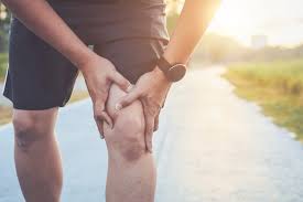
Strong knees are essential for maintaining mobility, independence, and quality of life as we age. For seniors, knee strength directly impacts the ability to walk, climb stairs, and perform daily activities without pain. Whether you’re dealing with arthritis, recovering from an injury, or simply want to maintain healthy joints, these exercises to strengthen knees for seniors can make a significant difference in your mobility and comfort.
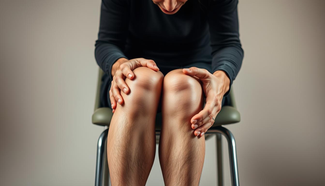
Proper form and support are essential when performing knee exercises
Safety First: Preparing for Knee Exercises
Before diving into any exercise routine, safety should be your top priority. These precautions will help ensure you strengthen your knees without risking injury:
- Consult your healthcare provider before starting any new exercise program, especially if you have existing knee pain, arthritis, or have had knee surgery.
- Start slowly with fewer repetitions and gradually increase as your strength improves.
- Use stable support like a sturdy chair or countertop when performing standing exercises.
- Warm up properly with 5-10 minutes of gentle walking or marching in place to increase blood flow to your muscles.
- Stop if you feel pain that goes beyond mild discomfort. Some muscle fatigue is normal, but sharp pain is not.
Need Professional Guidance?
If you’re unsure about which exercises are right for you, consider consulting with a physical therapist who can create a personalized program for your specific needs.
10 Effective Exercises to Strengthen Knees for Seniors
These exercises target the muscles that support your knees, including the quadriceps (front thigh), hamstrings (back thigh), and calf muscles. Strengthening these muscle groups helps stabilize your knee joints and improve overall function.
Seated exercises are excellent for beginners and those with balance concerns
1. Seated Knee Extensions
Seated knee extensions strengthen the quadriceps muscles, which are crucial for knee stability and support.
How to perform:
- Sit in a sturdy chair with your back straight and feet flat on the floor.
- Slowly extend your right leg until it’s as straight as possible without locking the knee.
- Hold for 3-5 seconds, focusing on tightening the thigh muscle.
- Slowly lower your leg back to the starting position.
- Repeat with the left leg.
Recommended: 8-10 repetitions per leg, 2-3 sets
Muscles targeted: Quadriceps (front thigh muscles)
Safety modifications:
- If extending your leg fully causes discomfort, only extend as far as feels comfortable.
- Place a rolled towel under your knee for support if needed.
- For added resistance as you progress, consider using light ankle weights (1-2 pounds).
2. Seated Marches
Seated marches improve hip flexor strength, which helps with knee alignment and stability during walking.

How to perform:
- Sit tall in a chair with your feet flat on the floor.
- Lift your right knee up toward your chest without leaning back.
- Lower your right foot back to the floor.
- Lift your left knee up toward your chest.
- Continue alternating legs in a marching motion.
Recommended: 10-15 repetitions per leg, 2 sets
Muscles targeted: Hip flexors, quadriceps, core muscles
Safety modifications:
- Hold onto the sides of the chair for added stability.
- Lift your knees only as high as is comfortable.
- Focus on maintaining good posture throughout the exercise.
3. Wall Slides
Wall slides (modified wall squats) strengthen multiple leg muscles while providing back support.

How to perform:
- Stand with your back against a wall, feet shoulder-width apart and about 12 inches from the wall.
- Slowly slide down the wall until your knees are bent at about a 30-45 degree angle (not a full squat).
- Hold this position for 5-10 seconds.
- Slowly slide back up to the starting position.
Recommended: 5-8 repetitions, 2 sets
Muscles targeted: Quadriceps, hamstrings, glutes
Safety modifications:
- Don’t slide down too far – a slight bend is sufficient to start.
- Keep your feet far enough from the wall so your knees don’t extend past your toes.
- Place a small exercise ball between your back and the wall for added comfort.
4. Calf Raises
Calf raises strengthen the lower leg muscles that help support the knee during walking and standing.

How to perform:
- Stand behind a sturdy chair or counter, holding on for balance.
- Slowly rise up onto your toes, lifting your heels off the ground.
- Hold the raised position for 2-3 seconds.
- Slowly lower your heels back to the floor.
Recommended: 10-12 repetitions, 2 sets
Muscles targeted: Calf muscles (gastrocnemius and soleus)
Safety modifications:
- If standing calf raises are too challenging, try seated calf raises.
- Rise only as high as is comfortable and stable.
- Ensure you have a sturdy support that won’t move during the exercise.
5. Hamstring Curls
Hamstring curls strengthen the muscles at the back of the thigh that help support and stabilize the knee joint.

How to perform:
- Stand behind a sturdy chair or counter, holding on for balance.
- Shift your weight to your left leg.
- Slowly bend your right knee, bringing your heel toward your buttocks.
- Hold for 2-3 seconds, then slowly lower your foot.
- Repeat with the left leg.
Recommended: 8-10 repetitions per leg, 2 sets
Muscles targeted: Hamstrings
Safety modifications:
- Keep a slight bend in your supporting leg.
- Don’t bend your knee beyond what’s comfortable.
- Focus on the muscle contraction rather than how high you can lift your heel.
Track Your Progress
Keeping a simple exercise journal can help you stay motivated and see your improvement over time. Note how many repetitions you complete and how your knees feel after each session.
6. Pillow Squeezes
Pillow squeezes strengthen the inner thigh muscles (adductors) which help stabilize the knee joint.

How to perform:
- Sit in a chair with good posture, feet flat on the floor.
- Place a small pillow or folded towel between your knees.
- Squeeze your knees together, compressing the pillow.
- Hold for 5-10 seconds, then relax without completely releasing pressure.
Recommended: 10-12 repetitions, 2 sets
Muscles targeted: Adductors (inner thigh muscles)
Safety modifications:
- Use a thinner pillow or folded towel if a regular pillow is too thick.
- Focus on gentle, controlled pressure rather than maximum force.
- Keep your back straight and avoid leaning forward during the squeeze.
7. Straight Leg Raises
Straight leg raises strengthen the quadriceps while minimizing knee joint stress.
How to perform:
- Lie on your back on a mat or firm bed with your left leg bent and foot flat.
- Keep your right leg straight and tighten the thigh muscle.
- Slowly raise your right leg to the height of your bent knee (about 12 inches).
- Hold for 3-5 seconds, then slowly lower.
- Repeat with the other leg.
Recommended: 8-10 repetitions per leg, 2 sets
Muscles targeted: Quadriceps, hip flexors
Safety modifications:
- Place a rolled towel under your lower back for support if needed.
- Keep the movement slow and controlled.
- If lying down is uncomfortable, try seated leg extensions instead.
8. Step-Ups
Step-ups strengthen multiple leg muscles while improving balance and coordination.

How to perform:
- Stand facing a sturdy step or stair (4-6 inches high).
- Hold onto a railing, wall, or sturdy furniture for balance.
- Step up with your right foot, then bring your left foot up to join it.
- Step back down with your right foot, then your left foot.
- Repeat, leading with your left foot.
Recommended: 6-8 repetitions per leg, 2 sets
Muscles targeted: Quadriceps, hamstrings, glutes, calves
Safety modifications:
- Use a lower step height if needed.
- Always use a sturdy support for balance.
- Focus on proper form rather than speed.
9. Seated Ankle Rotations
Seated ankle rotations improve ankle mobility, which helps with proper knee alignment during walking.

How to perform:
- Sit in a chair with good posture.
- Lift your right foot slightly off the floor.
- Rotate your ankle in a circular motion 10 times clockwise.
- Rotate your ankle 10 times counterclockwise.
- Repeat with the left ankle.
Recommended: 10 rotations in each direction, 2 sets per ankle
Muscles targeted: Ankle stabilizers, lower leg muscles
Safety modifications:
- If balance is a concern, keep your foot closer to the floor.
- Move slowly and gently, especially if you have ankle stiffness.
- Stop if you feel any joint pain (not just muscle fatigue).
10. Gentle Side Steps
Gentle side steps strengthen the hip abductors, which help stabilize the knee during walking and standing.

How to perform:
- Stand behind a sturdy chair or counter, holding on for balance.
- Step to the right with your right foot.
- Bring your left foot to join the right.
- Take 5-10 steps in one direction.
- Reverse direction, leading with your left foot.
Recommended: 5-10 steps in each direction, 2 sets
Muscles targeted: Hip abductors, adductors, quadriceps
Safety modifications:
- Take smaller steps if needed for stability.
- Always maintain a firm grip on your support.
- Keep a slight bend in your knees throughout the exercise.
Lifestyle Tips to Support Knee Health
Exercise is just one component of maintaining healthy knees. These additional recommendations can help maximize the benefits of your knee-strengthening routine:
Stay Hydrated
Proper hydration helps maintain the synovial fluid that lubricates your joints. Aim for 6-8 glasses of water daily, more if you’re active or it’s hot outside.
Maintain Healthy Weight
Extra weight puts additional pressure on your knees. Even a small weight reduction can significantly reduce knee stress and pain.
Wear Supportive Footwear
Proper shoes with good arch support and cushioning help align your legs correctly, reducing knee strain during daily activities.
Low-Impact Activities for Knee Health
Complement your strengthening exercises with these joint-friendly activities:
- Swimming or water aerobics – The water’s buoyancy reduces pressure on your knees
- Walking – Start with short distances on level surfaces
- Stationary cycling – Adjust the seat height for comfortable knee positioning
- Tai Chi – Gentle movements improve balance and joint mobility
Need Help Getting Started?
If you’re experiencing knee pain or unsure about which exercises are right for you, speaking with a healthcare professional can help you create a safe, effective routine.
Frequently Asked Questions About Knee Exercises for Seniors
How often should seniors do knee-strengthening exercises?
For best results, aim to perform these exercises 2-3 times per week with at least one day of rest between sessions. Consistency is more important than intensity, especially when starting out.
Is it normal to feel some discomfort when doing these exercises?
Mild muscle fatigue or a gentle stretching sensation is normal, but you should never experience sharp or severe pain. If an exercise causes pain, stop immediately and consult with your healthcare provider.
How long before I notice improvements in my knee strength?
Most people begin to notice improvements in strength and stability within 4-6 weeks of consistent exercise. However, individual results vary based on starting fitness level, age, and any existing conditions.
Can I do these exercises if I have arthritis in my knees?
Many people with arthritis benefit from gentle strengthening exercises. However, it’s essential to consult with your healthcare provider first and potentially work with a physical therapist to modify exercises for your specific condition.
Strengthening Your Knees, Enhancing Your Life
Regular knee-strengthening exercises offer seniors more than just physical benefits—they provide a pathway to greater independence and confidence in daily activities. By dedicating just a few minutes several times a week to these simple exercises, you can significantly improve your mobility, reduce pain, and enhance your overall quality of life.
Remember that consistency is key. Start slowly, listen to your body, and gradually increase the intensity as your strength improves. With patience and persistence, you’ll develop stronger knees that better support your active lifestyle for years to come.
Ready to Take the Next Step?
For personalized guidance on exercises to strengthen knees for seniors, consider consulting with a physical therapist who specializes in geriatric care.