Author: Mokhtar

  • What are the different types of traumatic knee injuries?

    What are the different types of traumatic knee injuries?

    834206db c5dd 45ef ab5a 3f07a60200b7 traumatic knee

    Types of Traumatic Knee Injuries: A Comprehensive Overview

    Traumatic knee injuries are a common occurrence in sports medicine and can cause significant pain and discomfort. Knee injuries involve trauma to one or more tissues that make up the knee joint, including bones, ligaments, cartilage, meniscus, and tendons. In many cases, injuries involve more than one structure in the knee.

    v2 3hgys ultna

    There are various types of traumatic knee injuries, and each injury requires a unique treatment approach. Some of the most common types of knee injuries include fractures, dislocations, tears of the anterior cruciate ligament (ACL), and tears of the meniscus. Knee injuries can occur due to a fall, forceful twisting of the knee, or high impact from a motor vehicle accident or another force.

    In this article, we will discuss the different types of traumatic knee injuries, their causes, symptoms, and treatment options. We will also provide tips for preventing knee injuries and maintaining healthy knee joints. Understanding the different types of knee injuries can help you take the necessary steps to prevent them and seek appropriate medical care if you experience knee pain or discomfort.

    Types of Knee Injuries

    When it comes to knee injuries, there are several types of traumatic knee injuries that can occur. Here, we will discuss some of the most common types of knee injuries and their subtypes:

    Ligament Injuries

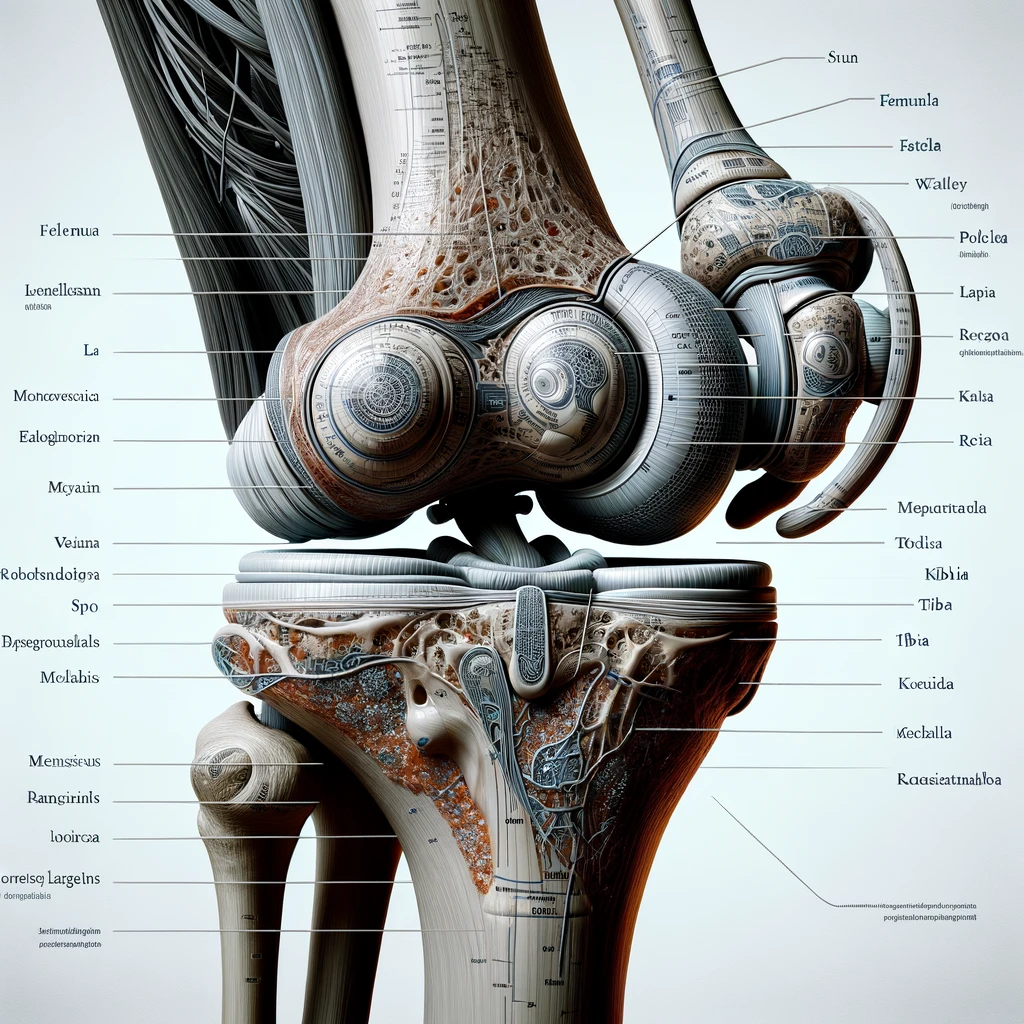
    The knee joint has four main ligaments: the anterior cruciate ligament (ACL), posterior cruciate ligament (PCL), medial collateral ligament (MCL), and lateral collateral ligament (LCL). These ligaments can be sprained or torn due to a fall, sports injury, or accident. Symptoms of a ligament injury may include pain, swelling, and instability in the knee joint.

    Meniscus Injuries

    The meniscus is a C-shaped piece of cartilage in the knee joint that acts as a shock absorber. Meniscal tears are a common type of knee injury that can happen due to a twisting motion or direct impact to the knee. Symptoms of a meniscal tear may include knee pain, swelling, and difficulty moving the knee joint.

    Tendon Injuries

    The knee joint also has two main tendons: the patellar tendon and the quadriceps tendon. These tendons can tear due to overuse or direct impact to the knee joint. Symptoms of a tendon tear may include inflammation, pain, and difficulty moving the knee joint.

    Fractures

    A knee fracture occurs when one or more of the bones that make up the knee joint (patella, femur, or tibia) break due to a fall, sports injury, or accident. Symptoms of a knee fracture may include severe pain, swelling, and difficulty moving the knee joint.

    Dislocations

    A knee dislocation occurs when the bones that make up the knee joint (patella, femur, or tibia) are forced out of their normal position. This can happen due to a fall, sports injury, or accident. Symptoms of a knee dislocation may include pain, swelling, and instability in the knee joint.

    In conclusion, knee injuries can be painful and debilitating. If you experience any symptoms of a knee injury, it is important to seek medical attention right away. Treatment options may include rest, physical therapy, or surgery depending on the severity of the injury.

    Diagnosis and Treatment

    Clinical Assessment

    When a patient presents with a traumatic knee injury, we first perform a clinical assessment to determine the extent and severity of the injury. This includes evaluating the patient’s medical history, performing a physical examination, and assessing the patient’s symptoms, such as swelling, pain, and stiffness.

    Imaging Techniques

    Imaging techniques, such as x-rays, MRI, and CT scans, may be used to further assess the injury and determine the appropriate treatment approach. X-rays are useful for evaluating bone fractures, while MRI and CT scans are better for assessing soft tissue injuries, such as ligament or cartilage tears.

    Treatment Approaches

    Treatment for traumatic knee injuries varies depending on the extent and severity of the injury. In some cases, rest, ice, compression, and elevation (RICE) may be sufficient to manage symptoms and promote healing. In more severe cases, surgery may be necessary to repair or reconstruct damaged ligaments, cartilage, or bones.

    Rehabilitation and Recovery

    Physical therapy is an important part of the recovery process for patients with traumatic knee injuries. Physical therapy can help reduce pain and swelling, improve range of motion, and strengthen the muscles around the knee joint. Patients may also benefit from exercises and stretching to improve flexibility and mobility.

    In summary, diagnosis and treatment of traumatic knee injuries requires a comprehensive approach that includes clinical assessment, imaging techniques, and appropriate treatment approaches. Rehabilitation and recovery through physical therapy and exercise are also important for promoting healing and returning to normal activities.

  • Inspired Spine unveils two Centers of Excellence for high BMI patients in rural Minnesota and Kansas

    Inspired Spine unveils two Centers of Excellence for high BMI patients in rural Minnesota and Kansas

    Inspired Spine CMO Dr. Hamid Abbasi performs one of more than 1,700 OLLIF procedures. The procedure has proven effective in hundreds of patients with a high BMI.

    BURNSVILLE, Minn., Nov. 29, 2023 /PRNewswire/ — Inspired Spine has announced the formation of two new Centers of Excellence focused on providing complex spine care, including fusion surgery, to patients with high Body Mass Index (BMI) in rural areas of Minnesota and Kansas, a groundbreaking turn in modern spine health management.

    In a remarkable display of dedication, Dr. Hamid Abbasi, a renowned neurosurgeon and the driving force behind Inspired Spine, recently performed an extraordinary feat by traveling to Hutchinson, Kansas to perform eleven spinal surgeries in just two days. Notably, five of these patients had a BMI over 40, with the highest BMIs being 54 and 64. All eleven operations were performed with unprecedented success and the patients were able to walk back and forth in the comfort of their own home within two days.

    Traditionally, patients with a BMI above 40 face significant challenges in seeking elective surgeries due to societal norms and medical recommendations against such procedures citing high risks. Inspired Spine’s innovative Oblique Lateral Lumbar Interbody Fusion (OLLIF) technology and comprehensive surgical protocols have dramatically minimized associated risks for patients requiring spinal fusion surgery. Many of them have become non-ambulatory due to spinal problems and have no alternative. The benefits of OLLIF now impressively outweigh the risks.

    This advancement comes at a crucial time for patients with high BMI, who often face a debilitating cycle of weight gain and deteriorating health due to the lack of mobility and associated comorbidities, such as cardiovascular decline. In response, Inspired Spine has established specialty medical centers accessible to patients from across the United States that provide advanced and life-changing care.

    With a ten-year track record of successfully treating patients with a high BMI with the OLLIF procedure. This breakthrough has opened new doors to patient care that many thought were permanently closed. Despite the complex nature of these operations, patients are typically discharged only one or two nights after surgery and can return home within a few days – undeniable proof of the high efficiency and effectiveness of the treatment.

    To underline this success, the publication of Inspired Spine’s OLLIF high BMI paper shows the transformative effects of this procedure. “We are proud to bring our innovation to rural Kansas,” said Dr. Abbasi. “The impact is already being felt with a series of successful surgeries performed in environments that would not normally be suitable for traditional spine surgery. Many of these patients require higher levels of care such as ICU or blood transfusions and intensive medical care due to the invasiveness of traditional surgeries.”

    The human dimension of these successes is vividly reflected in patient testimonials. A recent patient from North Carolina who traveled to Inspired Spine of Kansas for the OLLIF procedure shares her experience: “I never thought relief was possible until I found Inspired Spine. The OLLIF procedure not only gave me back my mobility, but also gave me a new lease on life. The care I received in Kansas was exceptional, and I am grateful for the team at Inspired Spine.” Her story, along with her moving testimony and images, underscores the critical importance and positive results of the Centers of Excellence at Inspired Spine.

    Inspired Spine is a total spine care provider dedicated to improving patients’ quality of life with revolutionary procedures and technologies. Our commitment to advancing spine health, grounded in innovation, has made us leaders in the field of minimally invasive spine surgery.

    For more information about the OLLIF procedure and to read inspiring patient stories, visit www.inspiredspine.com or contact Amanda Armagost at aarmagost@islife.us

    SOURCE Inspired Spine

    rt

    Source link

  • Orthofix announces publication in The Spine Journal of five-year data for the M6-C artificial cervical disc

    Orthofix announces publication in The Spine Journal of five-year data for the M6-C artificial cervical disc


    With more than 100,000 implants worldwide, the M6-C and M6-L drives continue to demonstrate strong clinical performance, safety and patient benefits

    The Orthofix M6-C™ artificial intervertebral disc is a next-generation artificial intervertebral disc designed to replace an intervertebral disc damaged by degeneration of the cervical intervertebral discs. The M6-C disc is designed to restore spinal motion and is an alternative to cervical fusion.

    LEWISVILLE, Texas, November 29, 2023–(BUSINESS WIRE)–Orthofix Medical Inc. (NASDAQ:OFIX), a leading global spine and orthopedics company, today announced the release of the five-year results of its US clinical trial comparing the M6 ​​-C artificial cervical disc with anterior cervical discectomy and fusion (ACDF). Published in The Spine diarypatients treated with the M6-C disc demonstrated superior clinical success at 60 months compared to ACDF patients. Secondary findings indicated significant improvements in neck and arm pain, function and quality of life. The M6-C patient group maintained flexion-extension and lateral bending motion as reported at previous time points. The release of these data coincides with the recognition that more than 100,000 implantations of the M6-C artificial cervical disc and the M6-L artificial lumbar disc (available outside the US only) have been performed worldwide since the product was first introduced in 2006.

    “Publication of these data is important as it validates the strong clinical performance observed in the five-year data from the US IDE study,” said lead author Dr. Frank Phillips, professor of orthopedic surgery at Rush University Medical Center in Chicago and the director investigator for the FDA clinical trial. “Artificial intervertebral disc replacement is becoming the gold standard of care for indicated patients who might otherwise experience intervertebral disc fusion. Data from this study demonstrate that the M6-C artificial disc demonstrated superior clinical success over five years compared to ACDF controls. Furthermore, significantly more subjects in the M6-C group reported improved pain and physical functioning scores than observed in ACDF subjects, with no difference in reoperation rates or safety outcomes.”

    A prospective, nonrandomized, concurrently controlled clinical trial, the M6-C IDE study, was conducted at 23 sites in the United States with a mean patient age of 44 years. The study evaluated the safety and effectiveness of the M6-C artificial cervical disc compared to ACDF for the treatment of symptomatic single-level cervical radiculopathy with or without umbilical cord compression. The overall success rate for the protocol-specified primary endpoint for the M6-C disk patients was 82.3 percent, compared to 67.0 percent in the control group. The rates of subsequent surgical intervention (SSI) on the M6-C drive were 3.1 percent, device or procedure related serious adverse events (SAE) were 3.1 percent and were comparable to ACDF rates of SSI of 5.3 percent and SAE failure of 4.8 percent. The M6-C drive received U.S. Food and Drug Administration (FDA) approval in February 2019 based on the two-year results of this study.

    “To date, there have been more than 100,000 implants of M6 artificial disc technology in 20 countries around the world,” said Kevin Kenny, president of Orthofix Global Spine. “We are proud of this life-changing technology that has helped so many people enjoy their lives again.”

    About the M6-C artificial cervical disc

    The M6-C artificial cervical disc is designed to maintain the natural behavior of a functional spinal unit by replicating the biomechanical characteristics of the original disc and is indicated as an alternative to cervical fusion. The unique design of the M6-C disc features a compressible viscoelastic core core and annular structure construction that allows shock absorption at the implanted level, providing a controlled range of motion as the spine transitions into its combined complex movements.

    Orthofix invites attendees of the Cervical Spine Research Society (CSRS) annual meeting in Las Vegas, November 29 through December 2, 2023, to visit booth #109 to learn more about the company’s full portfolio of cervical solutions .

    About Orthofix

    Orthofix is ​​a leading global spine and orthopedics company with a comprehensive portfolio of biologics, innovative spine hardware, bone growth therapies, specialty orthopedic solutions and a leading surgical navigation system. The products are distributed in approximately 68 countries around the world.

    The company is headquartered in Lewisville, Texas, where it conducts general business, product development, medical education and manufacturing, and corporate offices in Carlsbad, CA, with a focus on spine and biologics product innovation and surgeon education, and Verona, Italy. with an emphasis on product innovation, production and medical education for orthopedics. The combined company’s global R&D, commercial and manufacturing footprint also includes facilities and offices in Irvine, CA, Toronto, Canada, Sunnyvale, CA, Wayne, PA, Olive Branch, MS, Maidenhead, UK, Munich, Germany, Paris, France and São Paulo, Brazil. For more information, visit Orthofix.com.

    Forward-Looking Statements

    This press release may contain forward-looking statements within the meaning of Section 21E of the Securities Exchange Act of 1934, as amended, and Section 27A of the Securities Act of 1933, as amended. In some cases, you can identify forward-looking statements by terminology such as “may,” “will,” “should,” “expects,” “plans,” “anticipates,” “believes,” “estimates,” “projects” , ‘intends’, ‘predicts’, ‘potential’, ‘continue’ or other similar terminology. Orthofix cautions you that statements in this press release that are not descriptions of historical facts are forward-looking statements based on the company’s current expectations and assumptions. Any forward-looking statement contained in this press release is subject to risks and uncertainties that could cause actual results to differ materially from those expressed or implied by such statement. Applicable risks and uncertainties include, but are not limited to: the ability of newly launched products to perform as designed and intended and to meet the needs of surgeons and patients, including due to the lack of robust clinical validation; and the risks identified under the heading “Risk Factors” in Orthofix Medical Inc.’s annual report. on Form 10-K for the fiscal year ended December 31, 2022, which was filed with the Securities and Exchange Commission (SEC) on March 6. , 2023. The Company’s public filings with the Securities and Exchange Commission are available at www.sec.gov. You are cautioned not to place undue reliance on forward-looking statements, which speak only as of the date on which they are made. Orthofix does not intend to revise or update any forward-looking statement contained in this press release to reflect events or circumstances occurring after the date of this press release, except as may be required by law.

    Contacts

    Media relations
    Denise Landry
    DeniseLandry@orthofix.com
    214.937.2529

    Investor Relations
    Louisa Smith, Gilmartin Group
    IR@orthofix.com

    Source link

  • Athletic trainers can assist with communication between coach and athlete

    Athletic trainers can assist with communication between coach and athlete


    Provision of concussion information by coaches and attendance of athletic trainers: Findings from the 2021 YouthStyles Survey

    Daugherty J, Waltzman D, Sarmiento KJ Athl Trein. January 16, 2023. doi: 10.4085/1062-6050-0454.22. E-publishing prior to printing. PMID: 36645830.

    Full text freely available

    Take home message

    More than 40% of adolescents participating in sports reported that their coach had not provided them with concussion information in the past year. This lack of communication may be linked to a lack of access to athletic trainers during games and practices.

    Background

    Coaches can provide concussion education and make a positive impact by translating concussion knowledge into healthy behaviors, such as reporting suspected concussions. Coaches who complete concussion training and relay that information to their athletes positively impact an athlete’s perspective on concussions. However, it is unknown how often coaches discuss concussion education with the athletes and whether the presence of an athletic trainer helps with better communication between coach and athlete.

    Study aim

    The authors used survey data to examine how often coaches provided concussion safety information to their athletes and whether this varied based on the presence of athletic trainers at games and practices.

    Methods

    In June 2021, adolescents living with Ipsos Knowledge Panel member parents were invited to complete the annual YouthStyle Web-based survey. Approximately 48% of invited adolescents completed survey questions about history of concussions, information about concussion safety, how concerned they think their coach is about concussion safety, and the frequency of athletic trainers at games and practices in the past year . The authors took statistical steps to ensure that the data represented adolescents across the United States.

    Results

    Of the 829 adolescents, 39% reported having participated in sports in the past twelve months: 1) only in school sports (19%), 2) only in sports competitions (13%), or both (7%). The most commonly reported sports were basketball and football. Nearly half (47%) of youth athletes reported that their coach discussed concussions. Additionally, 32% reported receiving a handout, and 23% said their coach had sent them an email or had them watch a video in the past 12 months. Overall, 58% of adolescent athletes reported that their coach discussed or provided information about concussions. More youth athletes who participated in school sports (65%) reported receiving this information, compared to athletes who only participated in non-school sports competitions (39%). About half of youth athletes indicate that they have an athletic trainer during training (55%) or competitions (55%). Youth athletes who participated in school sports were more likely to report having access to athletic trainers (72%) than those who participated on non-school sports teams (49%). Of youth athletes who always/sometimes had an athletic trainer, 63% reported that the coach talked to them about concussions, compared to 24% of youth athletes who rarely or never had access to athletic trainers.

    Viewpoints

    Four in 10 athletes report that their coaches do not discuss concussions or provide concussion information. The authors found that access to an athletic trainer can improve communication between coaches and youth athletes. It would be interesting to know if the athletic trainer increases the amount of communication as they provide resources for the coaches to share with athletes. It would be helpful to understand how athletic trainers can improve a coach’s concussion communication with athletes. Furthermore, this study focused on whether the coach provided information, and not on the quality of that information. It would be interesting to know how well the education worked. Coaches without access to athletic trainers may provide concussion information to the athletes, but the athlete does not remember it because it was not helpful.

    Clinical implications

    We must encourage coaches to consistently educate athletes about concussions to improve reporting behavior among youth athletes. In addition, sports trainers have a positive influence on communication between coach and youth. So when we encourage administrators to hire full-time athletic trainers, we can remind them that athletic trainers can help coaches better communicate about concussions and other injuries with their athletes.

    Questions for discussion

    Are you trying to get coaches to talk to the athletes about concussions? If so, have you seen better results in communication, knowledge and reporting behavior?

    related posts

    1. Improving Concussion Education: Consensus from the NCAA Department of Defense Mind Matters Research and Education Grand Challenge
    2. CDC Heads Up program increases concussion knowledge and injury communication
    3. Peer-led concussion education can improve concussion knowledge and reporting behavior
    4. Concussion Education Videos. Viewing once does not help with Info Stick
    5. Coaches are provided with information about concussions with a five-minute fact sheet
    6. Concussion knowledge is getting better, but concussion reporting is getting worse
    7. Better attitudes can improve reporting habits
    8. Center for Concussion Education and Research – Peer Concussion Education

    Written by Jane McDevitt
    Reviewed by Jeffrey Driban

    Evidence-based assessment of concussion course - 5 EBP CEUs

    Source link

  • Tyber Medical takes home a competitive award for workplace safety

    Tyber Medical takes home a competitive award for workplace safety

    BETHLEHEM, Pa., Nov. 28, 2023 /PRNewswire/ — Tyber Medical has been selected to receive the Governor’s Award for Safety Excellence (GASE) – a highly competitive program that recognizes exceptional workplace safety and prevention initiatives, as well as the effective and successful management of these initiatives by its employees.

    “Tyber Medical is committed to developing global products to help people recover from trauma and degenerative diseases. In order for our team to create these great products, the company must provide a safe, efficient workplace that keeps our ‘Work Hard, Play Hard’ mentality on track,” said Jeff Tyber, CEO and President of Tyber Medical . “The GASE Award recognizes that spirit and our commitment to maintaining an industry-leading approach to safety and performance.”

    The eligibility criteria for the GASE is extensive, starting with a nomination process followed by an initial evaluation of each company by an assessment committee. To become a semifinalist, companies must undergo a multifaceted assessment of accident and illness rates, safety program innovation, and strategic safety policy development and approach. They are then compared to industry-wide rates and standards for those conditions.

    Only after being approved by PA Labor & Industry Department officials will the semifinalists have the opportunity to host members of the GASE Review Committee for an on-site review. These reports help determine the finalists, who are then recommended to the Secretary of Labor and Industry for final review and decision-making.

    Among the impressive factors noted by the review committee were Tyber Medical’s comprehensive safety policies and procedures, as well as their virtual reality training and online safety video reference – all accessible via a QR code. With these measures, employees have worked an outstanding total of 280,000 hours in 2021 and 2022 and 330,000 hours in 2022 without incident.

    “I am so grateful to Tyber Medical, which puts employee well-being at the forefront of its mission. The team won this award by empowering its members to use creativity and technology in new ways to create the safest and most fun work environment I have ever been a part of,” said Toby Borcoman, Head of Culture and Vice President of Human Resources at Tyber. Medical. “Our culture promotes creativity and the implementation of new ideas. The company is willing to try out new ideas every day!”

    Tyber Medical received the award from representatives of the Department of Labor and Industry at the Governor’s Occupational Safety and Health (GOSH) Conference last October, and the safety practices used at Tyber Medical facilities are now shared across the state as a model for reference for companies within the industry.

    “Building a safe work environment is not a one-person endeavor,” Tyber noted in response to the recognition. “It will take a team of leaders, such as our safety officers and champions, and a corporate commitment not only to implement this policy, but also to train our people on the policy and ensure everyone can benefit from its positive impacts .”

    About Tyber Medical LLC

    Tyber Medical LLC is a leading orthopedic device manufacturer providing rapid access to FDA-cleared and CE-marked private label, portfolio-expanding, regulatory-approved orthopedic implants for the spine, limb and trauma markets. Tyber Medical offers customers a fast and seamless path to market. Since its founding in 2012, the company has introduced more than 50 spine, extremity and trauma systems. Tyber Medical strives to develop and utilize differentiated technologies to create advanced orthopedic implants.

    Contact person: Toby Borcoman, Tyber Medical
    Email: tborcoman@tybermed.com

    SOURCE Tyber Medical

    rt

    Source link

  • Longitudinal changes in youth baseball batting based on body rotation and separation |  BMC Sports sciences, medicine and rehabilitation

    Longitudinal changes in youth baseball batting based on body rotation and separation | BMC Sports sciences, medicine and rehabilitation

    Attendees

    We initially recruited 230 junior baseball players from six teams in Tokyo, Japan in April 2018. The inclusion criteria were males between 6 and 12 years old. The exclusion criteria were injury and illness that prevented the participant from taking measurements. Participants were divided into age groups during the baseball season according to little league eligibility rules – players were classified by age as of July 31 of a given year. Subsequently, the period up to and including July 31 of the second year of primary school was defined as Under 8 (U8), and then the period was divided by year up to and including U13. They played and practiced baseball at least twice a week (Saturday and Sunday) for 3 to 6 hours. Before the study, all participants completed a data questionnaire that asked for the following information: birth, age when they first started playing baseball, and the side they hit. In addition, all participants and their guardians were given a detailed explanation of the experimental procedures and risks of the study before any measurements were taken. In addition, written informed consent was obtained from all participants and their guardians who agreed to the study. This study was approved by the Ethics Committee of Waseda University (No. 2018 − 208).

    Batting procedure

    Testing was conducted between 9:00 AM and 4:00 PM on an outdoor baseball field maintained under standard environmental conditions. In addition, testing took place between January and March, spread over 4 to 6 days per season. First, we measured the participant’s height and weight while wearing clothes. Then, without shoes on, height was measured to the nearest 0.1 cm without shoes on using a stadiometer (YG200DN, Yagami Co., Nagoya, Japan); and weight was measured to the nearest 0.1 kg using a digital scale (BC622, TANITA Co., Tokyo, Japan). The hitting test was then performed after simple warm-up exercises including dynamic stretching, jogging, light throwing and swinging for approximately 20 minutes. Each participant received unreflected white markers on the top of the head, both lateral acromion points, and the anterior and superior iliac spines. The trial involved toss batting with an automatic toss machine (FTM-240; Field Force Company, China). The toss machine was placed 0.7 m from the center of home plate on the opposite side of the batter and 1.1 m toward the pitcher; it was positioned to launch diagonally in front of the batter. The height of the throwing machine was adjusted to the participant’s height as follows: 45% of the height minus 52.5 cm. Then, after one practice trial, actual testing was performed twice and the hitting motion was recorded at 240 Hz with three high-speed cameras (Ex-100PRO, Casio Co., Tokyo Japan) placed on the side, back, and front of the hitter (oblique). The environment of the impact test environment was shown in Figure 1.

    figure 1
    Figure 1

    Environment of batting test (in case of left-handed batsman)

    In addition, swing speed, a component of hitting performance, was measured using a Zepp sensor (ZEP-BT-000002; Zepp Company, Cupertino, California, USA), which has been shown to have high reliability (ICC, 0. 88). [13]and indicated that it correlates moderately to strongly with data analyzed by 3D motion tracking [14]. Participants were allowed to retry the hitting test if they missed the ball while swinging or made timing errors. During the batting test, participants used the bat they would normally use in baseball practice and games, and consistently used the same bat throughout their trials. Data were collected from the highest swing speed test.

    Variables

    The rotation angles of the head, trunk, pelvis and arm direction in the horizontal plane during the stroke movement and the separation angle between each segment, the amount of head movement and the step width were analyzed by manual digitizing using a motion analysis. system (Frame-Dias V; DKH, Tokyo, Japan). In addition, we visualized the body markers attached to the head, both the lateral acromion points, the anterior and superior iliac spines, the nose, the toes and the midpoint between both hands on the screen using a digital format. Then, three-dimensional coordinates were obtained using the direct linear transformation method [15], and the right orthogonal reference frame was defined as the X-axis, Y-axis, and Z-axis. The Y axis was directed from the pitcher’s mound to home plate, and the Z axis indicated a vertical direction (bottom to top). Furthermore, the X-axis was defined as the cross product of the Y-axis and the Z-axis. For calibration, posts with nine markers (from 0 to 2.0 m at 25 cm intervals) were placed vertically in a 4 x 4 grid at 40 cm intervals (the standard errors were as follows: x = 0.22 cm; y = 0.28 cm; z = 0.34 centimeter). From the beginning to the end of the at bat, a recording of the calibration points was performed using the three high-speed cameras. The analysis data was collected at five points: stance, load, foot contact, front swing and ball contact. Stance and foot contact were defined as the point of the toe of the stepping leg on the Z axis at which the Z axis value began to increase in a positive direction. In addition, load and foreswing were defined as the midpoints between stance and foot contact and between foot contact and ball contact, respectively.

    All rotation angles were calculated using values ​​corresponding to spaces in global coordinates, because batting is an operation initiated by responding to a thrown ball and is defined as the projected angle on the horizontal plane relative to the X axis ( Fig. 2). Additionally, the rotation angles were set as positive/negative relative to the pitcher/catcher.

    Fig. 2
    Figure 2

    Definitions of rotation and separation variables

    The variables analyzed in this study and their definitions are as follows:

    • Head rotation: the angle between the head vector (top of the head to the nose) and the X-axis.

    • Upper torso rotation: the angles between the upper torso vector (through the center of both acromions and perpendicular to the line joining both points) and the X-axis.

    • Arm direction: the angle between the hand vector (center of both acromions to a point between both hands) and the X-axis.

    In addition, the separation angle was expressed as the difference between each rotation angle, and the separation between head and upper trunk was calculated by subtracting the head rotation from the upper trunk rotation. In addition, the separation between the torso and arms was calculated by subtracting the rotation of the upper torso from the arm direction. The upper to pelvic separation was calculated by subtracting the rotation of the upper torso from the rotation of the pelvis. The linear head movement distance (head movement) from stance to foot contact and foot contact to ball contact was calculated as the resulting displacement of the top of the head. Finally, stance widths during stance and foot contact were calculated as the distance between the toes.

    static analysis

    Statistical power analysis was performed to estimate the sample size. For this study, we needed more than twelve players to perform a comparison between the three groups with 80% power, an alpha of 0.05. and a partial η of 0.14. Seventy-seven baseball players who met inclusion criteria completed three measurements over three seasons. Of these, 17 players formed group 1 (U8 to U10) and 13 players formed group 2 (U11 to U13) (Fig. 3).

    Fig. 3
    figure 3

    Flowchart of exclusion criteria and final participants

    Descriptive statistics (mean ± standard deviation) were performed. After confirming that all data were normally distributed using the Kolmogorov-Smirnov test and confirming homoscedasticity using the Levene test, we performed a one-way analysis of variance (ANOVA) to determine chronological age, height, body weight, years of competition, rotation, and separation comparable. angles, swing speed, head movement and step width at stances, load, foot contact, pre-swing and ball contact between the initial, second and final measurements over three seasons. Additionally, we performed multiple comparisons of the means of the controlled variables using the Bonferroni test. Partial η2 was calculated for the effect size of the one-way ANOVA, with values ​​of ≥ 0.01 to < 0.06, ≥ 0.06 to < 0.14, and ≥ 0.14, indicating small, medium, and large effects, respectively [16]. Finally, the alpha level was set at 0.05 and all statistical analyzes were performed using SPSS Statistics 27.0 (IBM, Armonk, New York, USA).

    Source link

  • Monogram Orthopedics delivers first surgical robot

    Monogram Orthopedics delivers first surgical robot

    – First sale to major global distributor represents major revenue milestone and paves way for broader commercialization –

    AUSTIN, TX / ACCESS WIRE / November 28, 2023 / Monogram Orthopedics Inc. (NASDAQ:MGRM) (“Monogram” or the “Company”), a medical technology company focused on reconstructive joint procedures and surgical robotics, has delivered its first surgical robot to one of the world’s largest global robotics distributors.

    “Delivering our first robot and achieving our first commercial revenues validates our technology and represents a critical milestone for our strategic roadmap,” said Ben Sexson, Chief Executive Officer of Monogram Orthopedics. “Our system performs at an extremely high level. We now look forward to seeing how our robot competes and scales in the real world. We hope that the mBôs robot will contribute to improving the standard of care for orthopedic patients worldwide.”

    Monogram aims to improve the standard of care in orthopedic joint replacement surgery with advanced, next-generation active surgical robotics. About 88% of knee replacement surgeries (the company’s first target group) are still performed manually. The mBôs robot aims to combine safety, ease of use, streamlined costs, novel implant design, broad clinical functionality and speed to help drive the next wave of robot adoption in orthopedics.

    About Monogram Orthopedics

    Monogram Orthopedics (NASDAQ: MGRM) is developing a product solution architecture with the long-term goal of enabling patient-optimized orthopedic implants at scale by coupling 3D printing and robotics with advanced pre-operative imaging. The company has a robot prototype that can autonomously perform optimized paths for high-precision insertion of implants into synthetic bone specimens. Monogram plans to manufacture and market robotic surgical equipment and related software, orthopedic implants, tissue ablation devices, navigation consumables and other miscellaneous instruments required for reconstructive joint replacement procedures. The company has not yet filed a 510(k) premarket notification or obtained 510(k) approvals for its robotic products. Marketing these products requires FDA approval, and the Company has not obtained FDA approval for any of its robotic products, and it cannot estimate the timing or guarantee the ability to obtain such approvals.

    Monogram Orthopedics is working to improve the way orthopedic surgery is performed. Our system is being developed to combine personalized knee implants with precision robotic surgical assistants to hopefully provide patients with a more appropriate knee replacement with minimally invasive surgery. One hundred thousand knee replacements that fail each year in a $19.4 billion market represent a huge opportunity for us.

    For more information, visit www.monogramorthopedics.com.

    Forward-Looking Statements

    This press release may contain “forward-looking statements.” To the extent that the information presented in this presentation discusses financial projections, information or expectations about Monogram Orthopedics Inc.’s business plans, results of operations, products or markets, or otherwise makes statements regarding future events, such statements are forward-looking. . Such forward-looking statements can be identified by the use of words such as “should”, “may”, “intend”, “anticipate”, “believe”, “estimate”, “project”, “predict”, “expect” , ”planning” and ”making proposals”.

    Although Monogram Orthopedics Inc. believes that the expectations reflected in these forward-looking statements are based on reasonable assumptions, there are a number of risks and uncertainties that could cause actual results to differ materially from such forward-looking statements. You are urged to carefully review and consider all cautionary statements and other disclosures, including the statements under the heading “Risk Factors” and elsewhere in the offering statement filed with the SEC. Forward-looking statements speak only as of the date of the document in which they appear, and Monogram Orthopedics Inc. assumes no obligation to update any forward-looking statements, except as may be required by law.

    Investor Relations

    Chris Tyson
    Executive Vice President
    MZ North America
    Direct: 949-491-8235
    MGRM@mzgroup.us

    SOURCE: MONOGRAM ORTHOPEDICS INC

    Source link

  • New Shotel™ medical ankle arthrodesis device significantly improves patient quality of life

    New Shotel™ medical ankle arthrodesis device significantly improves patient quality of life

    Reduces time to weight bearing to two weeks from the current six to eight weeks

    ATLANTA , Nov. 28, 2023 /PRNewswire/ — Shotel Medical today announced its Shotel™ Ankle Arthrodesis Nail System, which utilizes a novel device design for the treatment of end-stage ankle arthritis. It was used for the first time in Florida last month during a procedure in Delray Beach. Two weeks postoperatively, the patient was weight-bearing with the use of a CAM walking shoe, a significant improvement over the six- to eight-week time to weight-bearing offered by traditional ankle arthrodesis systems.

    The patented Shotel Nail System, manufactured and distributed by BioPro Implants, differs from traditional ankle arthrodesis devices because its unique curved design allows for a minimally invasive approach with very small incisions. It is designed to achieve fusion at the tibiotalar joint while allowing unrestricted motion at all other joints. This benefits the patient with faster healing, faster recovery and faster loading. End-stage ankle arthritis is becoming increasingly common and can lead to significant physical disability. There are an estimated 50,000 new cases of ankle arthritis in the US each year1

    “We designed our ankle fusion nail system so that patients have smaller incisions, allowing them to heal faster and function much earlier, giving them a better quality of life. The improvement in time to weight bearing for the patient is meaningful,” said Christopher Weathers, Chief Commercial Officer. “We are pleased to see our Shotel nailing system gaining traction among surgeons across the country. So far, 24 operations have been performed with it.”

    Kevin Palmer, DPM, dual board certified podiatrist from Boca Raton and Delray Beach, FL, who performed the procedure, said, “The patient presented with progressively worsening ankle disease and pre-existing structurally compromised hardware. The new design of the Shotel Nail System gave me confidence that it would be stable and would hold up over the long term. During the procedure, after I removed the patient’s current hardware, the Shotel Nail System was inserted smoothly, with a smaller incision and a much less invasive approach than traditional ankle fusion systems. I also like that it provides multiple layers of compression, which is needed at the fusion site to speed healing.

    “I cannot emphasize enough how important it is that the patient started walking with a CAM walking shoe within two weeks,” added Dr. Palmer added. “This will be a game changer – and life changing – for many patients in the future.”

    For more information about the Shotel™ Ankle Arthrodesis Nailing System or for surgical training, visit www.shotelmedical.com.

    About Shotel Medical
    Shotel Medical is the developer of the Shotel™ Ankle Arthrodesis Nail System, an entirely new device that has the potential to transform patient care and have a significant impact on the healthcare landscape. Developed in collaboration with a team of biomechanical engineers, orthopedic surgeons and industry thought leaders, the device addresses the needs of patients with end-stage ankle arthritis. The unique design allows patients to heal faster, function sooner and improve quality of life compared to current treatment options. Founded in 2017, the company has offices in New Orleans and Atlanta.

    About BioPro
    For more than three decades, BioPro has been at the forefront of orthopedic innovation, focused on improving the lives of patients suffering from orthopedic conditions. The company is committed to developing advanced implants and surgical devices that reduce pain and restore function, providing patients and surgeons with a diverse portfolio of established solutions and emerging technology. For more information, visit https://bioproimplants.com/.

    Media contact:
    Barbara Bikkel
    305-215-2121
    369140@email4pr.com

    ___________________________________
    1 Smyth, Niall A. MD; Dawkins, Brody J.BA; Goldstein, Joshua P.B.S.; Kaplan, Jonathan R. MD; Schon, Lew C. MD; Aiyer, Amiethab A. MD. Consumer prices for surgical treatment of ankle arthritis: limited availability and high variability. JAAOS: Global Research and Reviews 3(7):p e011, July 2019. | DOI: 10.5435/JAAOSGlobal-D-19-00011

    SOURCE Shotel Medical

    rt

    Source link

  • DISC Surgery Center in Newport Beach is now enrolling patients in a clinical trial of artificial disc replacement

    DISC Surgery Center in Newport Beach is now enrolling patients in a clinical trial of artificial disc replacement

    Drs. Ali H. Mesiwala and Grant D. Shifflett participate in the pivotal two-level study of the safety and effectiveness of the Orthofix M6-C™ artificial cervical disc compared to anterior cervical discectomy and fusion.

    NEWPORT BEACH, Calif., Nov. 28, 2023 /PRNewswire/ – DISC Surgery Center of Newport Beach (“DISC”), committed to supporting research vital to the field of spine care, has announced its participation in a clinical trial to artificial disc replacement and is now enrolling qualified patients. Candidates may include patients between the ages of 18 and 75 who have been told they need cervical (neck) surgery at two consecutive levels (among other requirements).

    Drs. Ali H. Mesiwala and Grant D. Shifflett – both board-certified, fellowship-trained spine surgeons at DISC – join other experts at approximately 30 clinical sites participating in this study across the country. The primary objective of the study is to evaluate the safety and effectiveness of the Orthofix M6-C™ artificial cervical disc (already FDA-approved for single-level surgery) in patients with contiguous symptomatic bilevel cervical radiculopathy, with or without umbilical cord compression. The results will then be compared with those in patients undergoing anterior cervical discectomy and fusion (ACDF).

    Artificial disc replacement surgery is very similar to ACDF surgery. Both procedures remove the damaged disk. However, when the damaged disc is removed to replace the disc, the space between the vertebrae is filled with a specialized implant called an artificial disc, instead of a bone graft. The artificial disc is designed to restore the distance between the vertebrae while still allowing some movement.

    With its track record as a national leader in outpatient spine and its team’s extensive experience performing advanced motion maintenance techniques, DISC is ideally positioned to participate in such research.

    “The DISC Surgery Center in Newport Beach has placed more than 3,000 cervical discs since 2018 and we are well versed in the motion-preserving qualities of the surgery,” said Dr. Mesiwala. “I think it is important to stay actively involved in research to improve patient care, so I wanted to be part of a study that will improve our learnings and discoveries.”

    Dr. Shifflett added, “As pioneers in minimally invasive spine surgery and techniques, we always welcome the opportunity to advance the field while informing the treatment options available to patients.”

    Patients who would like to know if they are eligible for this study can visit https://www.discmdgroup.com/adr-clinical-study/.

    About DISC Surgery Center in Newport Beach
    DISC Surgery Center in Newport Beach is a purpose-built outpatient clinic focused on providing patients with the safest, most advanced minimally invasive spine surgery, orthopedic sports medicine, total joint replacement and pain management. DISC is a subsidiary of TriasMD, a portfolio company of Chicago Pacific Founders, and also an official partner of Red Bull Athlete Performance Center. For more information, call 949-988-7800, visit www.discmdgroup.com or follow @DISCMD on Instagram.

    Media contact:
    Kristien Brada-Thompson
    760-274-6393
    369218@email4pr.com

    SOURCE DISC Sports and Spine Center

    rt

    Source link

  • Non-arthritic hip pain with Keelan Enseki

    Non-arthritic hip pain with Keelan Enseki

    Hip pain is common in athletes, especially in sports such as ice hockey.

    Non-arthritic hip pain includes a variety of intra-articular diagnoses commonly seen in these athletes that are not directly related to osteoarthritis. These include dysplasia, bony changes, femoroacetabular impingement, labral tears, and more.

    A recent clinical practice guideline on this topic was published in JOSPT to help us. In this episode, I talk to the lead author, Keelan Enseki, about the CPG findings.

    Show notes

    Keelan works at the Rooney Center for Sports Medicine at the University of Pittsburgh Medical Center. He currently serves as director of clinical practice innovation and administrative director of physical therapy residency programs. Keelan is also an adjunct professor at the University of Pittsburgh. In these roles, he divides time between clinical practice, teaching, research/writing, and clinical program design. His clinical interests focus on the nonoperative and postoperative care of individuals with injuries to the hip region. These interests are also represented through collaboration with the Orthopedic and Sports Physiotherapy Academies and the International Society of Hip Arthroscopy (ISHA).

    Social tools for COS:
    – Instagram: @keelan_enseki


    Source link