Category: Blog

Your blog category

  • What is that pain under my kneecap?  Understanding Patellofemoral Pain Syndrome – ACL Strong

    What is that pain under my kneecap? Understanding Patellofemoral Pain Syndrome – ACL Strong


    Patellofemoral pain syndrome

    That dull, aching, throbbing, sometimes sharp pain around the knee or kneecap that comes and goes based on activity may be a condition known as Patellofemoral Pain Syndrome. Sometimes associated with cracking and popping, patellofemoral pain can range from not so bad to disabling discomfort causing people of all ages to avoid activities such as walking, jogging or participating in a sport.

    What causes patellofemoral pain?

    With patellofemoral pain syndrome, discomfort in or around the front of the knee is often due to irritation of the cartilage that lines the underside of the kneecap in the joint. Stress on the knee can cause the cartilage to become inflamed, which can ultimately lead to thinning and fraying of the tissue over time. In some cases, athletes begin to notice symptoms after years of competition and training, even without immediate injury. In other cases, people only report this pain days, weeks, or months after an increase or change in activity. If the causes of patellofemoral pain syndrome are not treated, they can eventually lead to tissue degeneration in the knee, such as osteoarthritis.

    patellofemoral pain800 800x426 1 800x426 1

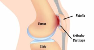
    The patella, or kneecap, is a floating bone connected to the femur and shin bone by tendons and ligaments. The bottom, or back part, of the patella is covered with a layer of cartilage, which helps it slide smoothly over the thigh bone when you bend and straighten your knee. There should be sufficient joint space between the patella and the underlying femur, although this joint space sometimes narrows, increasing the chance of contact or friction between the bony surfaces.

    A variety of factors including anatomy, soft tissue mobility, biomechanics (movement quality), and physical activity may contribute to a higher risk of patellofemoral syndrome. For example…

    • The anatomical resting position of the patella may be slightly higher than normal, also called ‘patella alta’, which can make the patella more sensitive to gradual wear.
    • A patella that is hypermobile or has too much mobility can bump and rub against the underlying bone, irritating the cartilage and causing pain. Conversely, a patella that is hypomobile or limited in mobility may become painful due to too much force being placed on a particular part of the patella.
    • In terms of movement, the patella moves over the femur, but the femur also moves under the patella. The biomechanics of the entire leg must be addressed to understand what is happening at the knee. The hip and foot play an important role in what the knee experiences. For example, someone with overpronated feet or weakness in the hip muscles may experience knee pain due to altered mechanics and resulting pressure on the cartilage lining of the patella. Abnormal biomechanics can be corrected through exercises prescribed by a physical therapist.
    • Physical activity also contributes to patellofemoral pain. The intensity or load of the activity can place more force on the knees than the body can safely tolerate. Activity modification may be necessary to control symptoms. For example, exercises such as running can be avoided for a while to calm symptoms such as swelling and pain.

    If the contributing factors are not addressed, the friction and friction between the surfaces can worsen, leading to thinning and fraying of the cartilage, wear and tear of the joint, and ultimately bone-on-bone osteoarthritis.

    What is the best treatment for patellofemoral pain syndrome?

    The recommended treatment for patellofemoral pain generally focuses on strengthening the muscles of the leg, from the hip and trunk to the foot, along with maintaining or increasing flexibility to reduce stress on the patella. Normalizing strength, flexibility and biomechanics through the limbs will help reduce pressure on specific areas of the cartilage that have become irritated and painful, allowing activity and exercise to be comfortable again.

    In addition to strengthening, ice and anti-inflammatory medications, such as ibuprofen, are often recommended to reduce swelling and relieve pain during the acute phase. Reducing painful activities such as climbing stairs can be helpful in recovering from patellofemoral pain syndrome. Although you limit painful exercises, you can still be active by incorporating alternative, non-painful exercises that will strengthen the knee around the knee and reduce irritation to the cartilage and other structures that support the patella. For example, a runner with patellofemoral pain can substitute swimming a few times a week to stay active without aggravating the knee.

    Have I waited too long? Is it too late for me?

    No matter how long you have been suffering from knee pain, it is never too late to build strength. Focusing on correcting your mechanics during exercise and exercise can help reduce your pain over time. Strengthening the muscles around the knees allows you to move with better control and precision, which also improves performance. Increasing body control provides more stability in the patellofemoral joint, which can take pressure off the patella and reduce the risk of further cartilage breakdown.

    Can it be cured?

    Patellofemoral pain syndrome is one of the most common causes of knee pain and one of the most common conditions resolved with physical therapy. A supervised exercise program to address the underlying causes is the best treatment strategy to relieve patellofemoral pain.

    Our courses at ACL Strong are designed and programmed by physiotherapists to give you complete confidence that the exercises you perform are both safe and effective.

    Many of our members experience an unexpected benefit when they enroll in an ACL Strong course… their patellofemoral pain improved because they trained smarter and reduced the strain on their knees.

    “I can’t believe I’m skiing without pain for the first time in years!”

    “The knee pain I used to have while playing football is no longer there!”

    “One of the biggest benefits we have seen in our athletes through ACL Strong is that it helps them resolve old injuries that have been bothering them.”

    When you take part in the ACL Strong Snow Course or Classic Course, you will learn how to care for your knees in the long term, so you can be as active or competitive as you want, and for as long as you want.

    👉 Start strengthening and training smarter today!

    Source link

  • “If you’re trying to get pregnant, you should actually have sex”

    “If you’re trying to get pregnant, you should actually have sex”


    Screen Shot 2021 03 14 at 2.19.43 PM

    When I first started thinking about getting pregnant after being diagnosed with rheumatoid arthritis, I was eager to hear from other women who had succeeded in this goal. At the time there wasn’t much social media, but I did find a great book called Arthritis, Pregnancy and the Path to Parenthood. It contained quotes and advice from real women, and I found it invaluable.

    But I also discovered a problem: the book started by talking about the possibility of changing some of your medications before getting pregnant, and then skipped straight to being pregnant. But wait! If I stop taking my meds, won’t I flare up? And if I’m in pain, how can I ever get pregnant? Is there a chapter missing from this book about trying to get pregnant while living with arthritis?

    That’s the question I asked Iris Zink, a rheumatology nurse who recently wrote a book with Jenny Thorn Palter about intimacy and chronic illness. (The book is called “Sex – Interrupted” and you can read my review of it here!) Their book recommends many alternatives to intercourse – which I think is generally good advice for maintaining intimacy in a relationship while dealing with a chronic illness! But what if you want to start a family? If you’re trying to get pregnant, you actually have to have sex!

    Iris and I decided to have a discussion about a topic we haven’t seen anyone else talk about: the challenges many women face when actually trying to conceive while living with a chronic illness. I share my personal experiences, and Iris shares her expert advice this video!

    Source link

  • 4 Major Types of ACL Prehab Exercises

    4 Major Types of ACL Prehab Exercises

    You will notice that your range of motion is significantly limited immediately after an ACL injury. In most cases, simple range-of-motion exercises are recommended early on with the aim of restoring normal range of motion. You may hear terms like “heel slides” and “quad sets,” two very simple, low-load exercises designed to help you regain flexion (ability to bend the knee) and extension (ability to straighten the knee). to get.

    When performing these types of exercises, keep in mind that the goal of prehab is to reduce the guarding mechanisms the body has developed as a result of the injury. Try to only work within a range of motion that does not increase pain levels above 2/10 and breathe well. If you are in a lot of pain and holding your breath, you may find that these exercises are not as effective.

    Immediately after an ACL injury occurs, the brain changes the way it uses the muscles in the affected area. Over a short period of time, these muscles begin to lose strength. When muscles lose strength, they are unable to create and absorb force as efficiently, putting joints, tendons and ligaments at greater risk for injury. Therefore, strengthening exercises are necessary to build strength in the affected muscles.

    In general, strengthening exercises involve moving a resistance load through a range of motion. You can use a number of items for resistance, including your own body weight (think a simple squat pattern), resistance band, a set of weights, or even the preferred form of resistance for Accelerate ACL, electricity.

    Source link

  • Cerapedics Reinforces Commitment to Level 1 Evidence with Long-Term Results from Six-Year Central IDE Cervix Study Showing 98.6% of Spinal Fusions Occurring in Patients Treated with i-FACTOR®

    Cerapedics Reinforces Commitment to Level 1 Evidence with Long-Term Results from Six-Year Central IDE Cervix Study Showing 98.6% of Spinal Fusions Occurring in Patients Treated with i-FACTOR®

    WESTMINSTER, Colo., Oct. 17, 2023 /PRNewswire/ — Cerapedics Inc., a commercial-stage orthopedic company committed to redefining the standard of care for bone repair, today announced long-term follow-up data from its pivotal IDE study of i-FACTOR Peptide-Enhanced Bone Graft. Published data demonstrated single-level anterior cervical discectomy and fusion (ACDF) fusion rates of 98.6% after six years in patients treated with i-FACTOR and reinforce Cerapedics’ commitment to accumulating long-term clinical evidence. In particular, the results support the safety and efficacy profile of i-FACTOR at one and two years, as published in Spine And Neurosurgery.

    i-FACTOR has demonstrated statistical superiority over local autograft in overall success* at one- and two-year endpoints for single-level ACDF. i-FACTOR further demonstrated spine fusion rates of 89.7%, 97.3% and 98.6% at one, two and six years, respectively, versus 85.8%, 95.8% and 97.3% for local autograft, with results published in Spine And Neurosurgery. 220 of the original 319 patients were observed for six years or 72 months.

    The six-year results, published earlier this year in Neurosurgery, were based on a single-blinded, randomized, controlled prospective study of i-FACTOR compared to local autograft in single-level ACDF, which enrolled a total of 220 patients, including 106 in the i-FACTOR group and 114 in the local autograft control arm. Of the 22 locations from the original IO study, 17 participated in this six-year post-approval study.

    “Physicians are looking for long-term safety and efficacy data to support decision-making about the safest and most effective spine technologies,” said Professor Paul Arnold, CARLE Illinois College of Medicine, principal investigator and author of the 12-, 24- and publications from 72 months. “In this unique long-term follow-up data from a pivotal IDE study, i-FACTOR met all four FDA-mandated non-inferiority success criteria and demonstrated safety and efficacy in single-level anterior cervical discectomy and fusion compared to autograft at 12 years of age age. , 24 and 72 months.”

    “These results clearly demonstrate the efficacy rates and safety profile of i-FACTOR over time and also reflect Cerapedics’ commitment to producing the highest quality evidence on the use of our products,” said Valeska Schroeder, Chief Executive Officer of Cerapedics. “Combined with the results of the IDE study, these data contribute to the clinical understanding of the efficacy and safety of i-FACTOR. While there are numerous other bone grafting options, many have not undergone the same level of testing. This data has a significant impact on insurance payers looking for Level 1 evidence when making coverage decisions.”

    i-FACTOR is available in the US through pre-market approval. It is the most stringent type of marketing application required by the FDA and involves the process of scientific and regulatory review to evaluate the safety and effectiveness of Class III medical devices based on Level 1 clinical trials. Cerapedics is currently conducting its second clinical randomized controlled trial for an indication for transforaminal lumbar interbody fusion (TLIF) involving 290 subjects, which is registered onclinicaltrials.gov.

    About i-FACTOR Peptide-enhanced bone graft
    i-FACTOR is a Class III FDA-approved bone graft supported by rigorous Level 1 human clinical data from an IDE study published in Spine And Neurosurgery. It is the only spinal bone graft powered by P-15 osteogenic cell-binding peptide, with a precise bone-building mechanism.** i-FACTOR has a demonstrated safety profile and is as safe as local autograft in single-level ACDF with proven statistical superiority overall success* after one and two years.

    i-FACTOR Peptide Enhanced Bone Graft is indicated for use in skeletally mature patients for the reconstruction of a degenerated cervical intervertebral disc at one level from C3-C4 to C6-C7. i-FACTOR Peptide Enhanced Bone Graft should be used in an allograft bone ring and with additional anterior plate fixation. For more information, full description of the indication for use, contraindications, safety warnings etc. about i-FACTOR, please visit our website: https://www.cerapedics.com/sites/default/files/2023-08/40002 – 07-4%20Putty%20USA.pdf

    About Cerapedia
    Cerapedics is a global commercial-stage orthopedic company that aims to redefine the standard of care for bone repair by healing bones faster and faster, without compromising safety, so patients can live their healthiest lives. Bone grafts, including Cerapedics products, are used in more than four million spine, orthopedics, trauma and interventional procedures worldwide each year. i-FACTOR is a Class III FDA-approved product indicated for single-level ACDF. Cerapedics’ new “P-15 Bone Graft” product is currently being evaluated for use in lumbar interbody fusion through ASPIRE, a pivotal clinical trial, and has received Breakthrough Device Designation from the FDA. Cerapedics is headquartered in Westminster, CO.

    For more information, please visit us at www.cerapedics.com.

    Media contact:
    Ten Bridge communications
    TBCCerapedics@tenbridgecommunications.com

    *Defined as meeting the Fusion, Function (Neck Disability Index), Neurological, and Safety endpoints
    **Surface-based mode of action

    SOURCE Cerapedics Inc.

    rt

    Source link

  • You Don’t Look Sick – Living With Rheumatoid Arthritis: DAY 4 IN YOSEMITE

    The day started so well. I got up extra early to get to Yosemite before the Saturday crowds. I went to Tunnel View where I thought I was supposed to be, then I saw a text message with a different location so I went to that location. Turns out I was supposed to be at Tunnel View. Emily, my guide, and I finally connected.

    Since Tunnel View was fogged up, we headed to Glacier Point. It was also fogged up. To say it was foggy is an understatement. It was foggy in San Francisco. We couldn’t see any mountains.

    AVvXsEhKk2qKiZ2axvQzdcDUtY 6NEB5ACV6GBiJWDghH5 F9gYJMluhxK mzNjxU5fRbLLwB9olW6kjV0XNeFXVcoz31cE fbKTchl2WszwJ9vEGH2H9gcF 2tyZaLIp8Dr2V6waQXfJLYipaDEFMnn3oi64nyjv


    We hiked at Glacier Point.

    AVvXsEgtvV axvQK8sevA5561N0i9k9vLrxp4UJ7832KpJMh8ebS NODITkccYxUVx95pPiTdpepgo0ocWp8GhfuLLJtGUU57o8EeEa3joWPAliDQBhSKWDQvE1oU0TJD9wKrJ5rhYbtUL0P2AjjWy6oqd2oMRY


    AVvXsEjBe7MU1e5hyJzFlQZ1ZzE2JnMx8dcQaEaNMkP6 RU9y5h0AxnCSTdf6lpGDIaK83CH4tx5q1m92e8SHvAfPrv68gXyzwqkI PixL241CuARFk14Gh0vcu0tElJHB2 mTrzhBOzgDtWZ6nnm8yKnnOP5FwjRLHoks7FMYRdDXgnbyo5KyJXyLUEce0H3E

    AVvXsEgjU5XZEjsclFjfioW3DRbGlj9jInxajE0fhd7aXftPvNgrPtfdTAFaNOdAVzw 9ZbPBhfr4 2m7tHMK


    Then we walked to Taft Point. We had some lunch during the walk. It started sprinkling every now and then. We climbed to the top of Taft Point and right as we were getting to the top it started raining heavily. We were scrambling to get down when the heavy rain came. I put one foot down and slipped, so I put my other foot down, twisted both feet and fell down. I think I screamed. I stood up and couldn’t put weight on my right foot. The guide and I slowly got off the rock because it started to snow! It’s too early for snow in Yosemite. Once off the rock, the guide sat me down and took off my shoes. She bandaged it and I tried to put weight on it, but it didn’t work. I also fell on my camera so I have to look at it to see if I broke it. 😫

    Meanwhile, these four students were hanging out. They came to me and asked if I needed help. We said yes. They were students from Spain. There were three boys and a girl. The four of them, along with the guide, helped me down the hill and to my car. Before you say the big problem. The terrain was amazing. Rocks, puddles, slopes and it was snowing heavily. None of us were dressed for snow.

    AVvXsEhEa1vUJ5l2a0 Oi3HEF1CL4uVRCrUzvZNYsOb9vUq 29D5K1lOggff2eQxIHXAOTqWLqGy eMKSkP2LxvsSscJW dF1jtAtIt7njMoJAf255DLbxTcQyHayifa72pZ77C6LPKCjH x2 KZO6oYiBELk QYC5EP c4aUtLGA5ZdKNTcHChWNGJk6VPdV0

    AVvXsEj5nLCarI D23Q0OdbH0QAYGPstSP1s IT BL4DLpqrrdVo8yWvgOaYKEcr wyHASIk848i2PqKFunT yMc4YdEvbon2y6 f8iRLm pn1uYN2UjOJ2UQiOcrqftgLXgSl8uUQFYbfQUG


    It took me about an hour to get to the parking lot. We had to go down and then immediately up again to get to the parking lot. About halfway through, a couple from another country asked if we needed help. They ended up carrying everyone’s backpacks.

    We finally go to the parking lot where the students parked. Our cars were further away, so the guide ran to get her car to take me to my car or the hospital. I chose my car because I didn’t know how much snow the park would get and I didn’t want my car to get stuck.

    Before we all parted ways, we took a group photo of the rescuers. They have to tell their friends a story about the lady they saved from a mountain in Yosemite.

    I could drive, but my foot really hurt when I hit the brakes. I had to get from my car to my cabin. I did it, but I’m not going to eat. I ate a lot of things I brought with me.

    Here are some photos:

    Source link

  • Irma Jennings’ Bone Health Test Results (DXA) 2020

    Irma Jennings’ Bone Health Test Results (DXA) 2020

    DXA 2022

    My latest DXA and TBS report has arrived. As a reminder, in 2005 I was diagnosed with osteopenia and given a script for Fosamax. I refused. I was in my fifties. Now I’m in my seventies. My intention was to implement my 7 pillars of bone health for better results:

    1. Calcium and mineral rich foods
    2. Supplements (Cal Citrate, Vit. D., Vit K2 – Mk7, Dr. McCormick’s Collagen and Mineral Whey)
    3. Bioidentical hormones
    4. Weight-bearing exercise
    5. Stress reduction
    6. Sleep hygiene
    7. Brings joy to my daily life

    At that time, Fosamax was a long-term drug. I would have been a long-term user. I am grateful for the work of Dr. Schneider and who has revealed her terrible story below. Because of her work, Fosamax users are now taking a drug holiday.

    The story of Dr. Jennifer Schneider

    Dr. Jennifer Schneider was standing on a subway in New York City when the train jolted and Schneider felt a kink in her right leg. It was her femur, the strongest bone in the body.

    “I shifted to the right to keep my balance and felt a large crack. I felt it in my thigh. There was no doubt that I had broken my femur,” says Schneider, a semi-retired internist from Tucson, whose The fracture was in the upper third of her thigh.

    “It’s extremely unusual to break that part of the femur,” she said. “It just doesn’t happen that way.”

    Schneider’s femur fracture occurred in October 2001, but it wasn’t until 2005 that she began to suspect the cause of the strange injury: long-term use of the drug Fosamax, which ironically is prescribed to treat and prevent the bone-thinning disease osteoporosis.

    Schneider had been taking the drug since 1995, when she was diagnosed with osteopenia, a condition considered by many doctors to be a precursor to osteoporosis.

    “The drug suppresses bone loss and in some people, who knows, it may be doing its job too well. It suppresses too much,” Schneider said.

    Fosamax drugs from New Jersey-based Merck say there is no evidence of a link between the drug and femur fractures. But in the interest of patient safety, Merck says it voluntarily amended the Fosamax label in July 2009 to include “low-energy femoral shaft and subtrochanteric fractures” in the side effects section of the label. [1]

    IMPORTANT:

    In June 2023 I will open my:

    Pilot program: Strong Bones – Healthy You
    A comprehensive bone health program – the whole body approach.

    Click HERE to schedule a free 30 minute session.

    This conversation will determine if my program is right for you.

    DXA from IRMA

    My ninety-minute pilgrimage to the University of Pennsylvania hospital yielded an excellent DXA.

    Below are my results

    Great results for my hips and spine – everything is moving in the right direction over time.

    Not so much my forearm.

    Note the colors in the report. We are looking for green.

    VEGETABLE

    The color chart below will help you understand the ranges.

    T-score less than -1 = Normal | COLOR GREEN

    T-score between -1 to -2.5 = osteopenia | COLOR YELLOW

    T-score greater than -2.5 = Osteoporosis | COLOR RED

    DXA AND TBS

    TBS L1-L4 = 1.393 (normal is 1.35 and higher)

    Below you will find the Trabecular Bone Score or TBS report of my spine. Simply put, the TBS measures the inner bones, while DXA measures the outer bones. Color falls into the vegetable normal area. My inner bones look good.

    Trabecular bone score

    DXA spine – Normal range L1-L4 = T-score 0.0

    Below is my DXA for my spine. It also falls into the vegetable (normal) part.

    DXA Spine 2022
    Measurement of the spine at DXA 2022

    DXA hip –

    Femoral neck -1.8 Total left hip -1.1

    YELLOW – Osteopenia

    Also measures in the normal yellow range. Note the DOT in the yellow section.

    DXA to measure bone density of the hip

    Forearm

    RED – Osteoporosis

    Radius 33% = -2.7

    Radius 33% is the size used for the forearm.

    DXA Forearm 2022

    Irma’s tracking schedule

    Click on the image for a better view.

    For my readers who like to track their DXA, below is my tracking chart. The diagram shows the type of machine used. Each machine has a serial number. When looking at a DXA comparison, it is essential to know the DXA machine and its serial number. That serial number can usually be found in the lower right corner of your DXA report. All my tests were on GE Lunar Prodigy with the same serial number.

    DXA Spreadsheet 2022 page 1
    DXA 2022 spreadsheet pg 2

    How to get a good DXA

    Hips

    The legs are turned inward, allowing a good DXA measurement.

    Hip rotation

    Spine

    The box places my back flat on the table

    Spine Position DXA small

    Forearm

    Forearm DXA 1

    Next steps

    My annual CMP (Comprehensive Metabolic Panel) blood test next year. This test measures the calcium levels that I monitor.

    Dr. McCormick uses blood tests to delve deeper into bone health.

    Also, my friend, Dr. Lani Simpson, DC, CCD, who has been in the osteoporosis trenches for decades, before I would get and follow bone markers:

    • CTX (full name Carboxyterminal cross-linking telopeptide or bone collagen) is a bone resorption marker.
    • My CTx was:
    • 346 (27-10-22 – Lab Corp)
    • 304 (2/22/22) Quest Labs (within the range offered in my Bone Marker presentation
    • P1NP (full name Procollagen type 1 N propeptide) measures bone formation. If my doctor does not write a script for this test, I will receive the script through Evexia or Life Extension.
    • My previous P1NP was 34
    • Vitamin D: I test twice a year – after summer and after winter
    • My last test showed 44.9 ng/ml

    Dr. Lani Simpson suggested an x-ray of my spine because arthritis makes a DXA look better than it is. My x-ray was taken by Dr. Kim Zambito, who reported mild arthritis in my spine and wrist.

    It takes a community.

    Forearm

    Why is the non-dominant forearm added to the DXA/TBS report?

    The spine and hip may have arthritis, but the DXA shows stronger results. The forearm can be a possible indication of problems with the parathyroid gland.

    Your forearm consists of 80 to 100% cortical bone. When a person has hyperparathyroidism, too much parathyroid hormone is pumped out. What does that degrade?

    That degrades cortical bone more than trabecular bone. And that’s why when you look at someone with hyperparathyroidism, their forearms often have low bone density.

    • “Primary hyperparathyroidism: Although this is a systemic disease that affects the entire skeleton, the lowest BMD is often the forearm, which may be the only site where the BMD is less than -2.5. In this case, the result should influence the decision regarding the surgical treatment of primary hyperparathyroidism (PHPT).” [i]

    Tests show negative for hyperparathyroidism. Bones are complicated.

    We are all different

    Several members of my Bones Tribe are on medication because it was deemed necessary based on a complete and comprehensive intake and blood work reviewed by their bone doctor. My Bones Tribe members seek other opinions before making a decision about medication based on knowledge versus fear of a decision.

    TIPS

    My DXA/TBS score was run on:

    The Hospital of the University of Pennsylvania

    3400 Civic Center Blvd. – Ground floor

    Philadelphia, PA

    phone: 215-662-3000 to book a DXA (ask for radiology to book your DXA).

    My script reads:

    • DXA bone density, axial CPT 77080
    • DXA bone density/peripheral CPT: 77081
    • Includes TBS and peripheral forearm.

    I asked for one full colored copy of my reports Before I left.

    I have a good feeling about the results.

    MEMORY:

    I open my:

    Pilot program: Stronger bones – Healthy you

    My 8-week Comprehensive Bone Health Program – The Whole Body Approach promises to educate you on every aspect of bone health. From DXA/TBS, bone markers, blood tests, calcium rich foods, exercise. You gain knowledge, a deep understanding of the terminology and move on to empowerment.

    Click HERE for a free 30 minute consultation to confirm that my program is right for you.

    I hope my shared experience is helpful to you and your beautiful bones.

    From my loving bones to yours,

    Irma Jennings, INHC, holistic bone coach

    e-mail: [email protected]

    [1] https://www.drug-injury.com/drug_injury/2010/09/doctor-files-suit-says-bone-drug-leads-to-breaks.html


    Let me support your bones

    Receive an in-depth, tailor-made private session

    Book a private coaching with Irma now


    Join our amazing Bone Tribe community

    Get the support and information you need to live fearlessly with your bone diagnosis

    30 Essential Foods for Bone Health

    Source link

  • Low back pain and spinal stenosis

    Low back pain and spinal stenosis

    Spinal stenosis is a narrowing of the spinal canal such that the nerve roots running through the spinal canal are compressed by a variety of different structures, such as bulging discs, bulges, ligaments, and/or arthritic enlarged facet joints. Joints. The area of ​​the lumbar spine that becomes narrowed often determines the type of symptoms patients will experience. Patients often experience back pain, hip pain, buttock pain, such as pain, numbness and tingling in the legs or feet, or a combination of all these symptoms. It often occurs in patients over 40 years of age, but can also occur in younger people.

    Other conditions, such as cardiovascular constriction of the blood vessels in the legs, should be considered as another possible cause for the patient’s symptoms.

    Spinal stenosis is often a condition that can become a chronic source of disability for people. It can seriously affect their quality of life, making activities such as walking, sports and hobbies very difficult.

    Spinal stenosis can be treated in several ways, usually with a combination of exercise and physical therapy. When therapy no longer leads to improvement in a patient’s symptoms, spinal injections may become an option. If all else fails, simple decompression to clear the space for the spinal canal by removing the bony arthritis, thick and ligament, and/or bulging disc is performed using minimally invasive techniques to correct the spinal stenosis. to light up.

    Dr. Shukairy, minimally invasive spine surgeon

    Read more, or contact us at www.orthopedicdoctors.com

    Source link

  • What does my KOOS score mean and is my KOOS score normal?

    What does my KOOS score mean and is my KOOS score normal?

    If you weigh yourself on a scale and receive a number, how do you understand the value? How do you know if you are overweight, underweight or normal? Who do you compare yourself to?

    To determine what’s normal and understand your own score, you’ll likely compare your weight to other people who have similar characteristics to you, such as gender, age, and height. There would be no point in comparing yourself to someone with different characteristics, since what is normal for him or her is likely different for you. This same concept applies to normative values.

    Normative data identify what is common or typical and describe observed characteristics of a specific population at a specific time.[1] Using normative values ​​allows you to give meaning to your test scores by comparing your score to scores of people with similar characteristics to you.[2]

    As explained in part 1 of the KOOS blog series, the Knee Injury and Osteoarthritis Score (KOOS) is a questionnaire specifically designed for people with various knee conditions.[3] By completing the KOOS you will gain insight into the course of your knee injury and you and your healthcare provider can monitor the effects of the treatment over time.[3] While comparing your own preoperative and postoperative KOOS scores provides insight into your recovery process, you can also compare your scores to normative KOOS values ​​to determine your degree of disability or your level of progress compared to people who have had a similar injury, surgery, or have undergone treatment. .

    View the normal KOOS scoring blog for populations that have suffered an ACL injury,[4][5][6] total knee replacement (TKR) surgery, [7][8][9] as well as those who have knee osteoarthritis,[10] and populations without known knee disorders [11] in part 2 of the KOOS blog series.

    If you have had a knee injury or surgery, try our Curovate app for your daily recovery. Curovate offers video-guided daily exercises, progress tracking, the ability to measure the range of motion of your knee and hip, and the ability to complete the KOOS outcome measurement, all within the app.

    If you need more tailored help during your surgery or recovery from your injury, check out our Virtual Physiotherapy page to book your 1-on-1 video session with a physiotherapist.

    Learn more about the KOOS and what normative values ​​are. Also learn how to interpret your KOOS score in this YouTube video presented by Joey Wong, kinesiologist.

    acl knee physical therapy 1080x1080 2
    Download it on Google Play


    Other recommended blogs


    References

    Source link

  • Research Fellow – Rubin Institute for Advanced Orthopedics (RIAO) at Sinai Hospital in Baltimore


    This new position is intended for medical students who wish to strengthen their competitive position to obtain a residency position in the field of orthopedic surgery. Research assistants will work with national and international leaders in the field of total joint replacement to assist in preparing manuscripts for publication in peer-reviewed journals, to collect and analyze data, to attend and present your work at regional and national conferences, and to assist with FDA investigations of new drugs and devices. This is a large-scale research center where numerous projects have not only been published in PubMed-indexed journals, but have also been selected to present at national orthopedic meetings, namely the AAOS and AAHKS.

    Research assistants have the opportunity to attend daily morning conferences at the orthopedic surgery residency, led by program director Dr. Jack Ingari, MD. This is an excellent opportunity to strengthen your resume and increase your chances of matching with an orthopedic surgery residency.

    Student researchers will work under the guidance of:

    -Ronald E. Delanois, MD

    -Michael A. Mont, MD

    -James Nace, DO, MPT

    GOALS:

    – Research into basic scientific and clinical aspects of total joint arthroplasty and preservation.

    – Learn to create and extract clinical data from institutional and national databases

    – Develop a better appreciation for biostatistics in the orthopedics setting

    – Prepare manuscripts and textbook chapters.

    – Present at research meetings, seminars, journal clubs, symposia and professional association meetings.

    EDUCATIONAL LEVEL REQUIREMENT OR REASONS: All candidates who have completed their third year and passed their USMLE Step 1 and/or COMLEX Level 1 board exams. It is preferable that USMLE Step 2 be completed before beginning the research program. Experience in writing and biostatistics is a plus.

    APPLICATION: Attach the following materials This email address is being protected from spambots. You need JavaScript enabled to view it.:

    – Application letter OR letter of interest

    – PDF of Curriculum Vitae

    – All available USMLE AND/OR COMLEX score reports

    – ≥ 1 letters of recommendation

    RIAO website: www.lifebridgehealth.org/RIAO/RIAO.aspx

    Twitter handle: @RIAOResearch Instagram handle: @RIAOorthopedics

    US/Canadian MD/DO preferred. US IMGs will be considered on a case-by-case basis. J-1 visa is possible.

    Source link

  • The histone acetyltransferases CBP and p300 regulate stress response pathways in synovial fibroblasts at transcriptional and functional levels


    Hypoxia, the formation of reactive oxygen species (ROS) and subsequent oxidative stress in synovial tissues are key events in the pathogenesis of RA1. We provide evidence here that CBP and p300 are critical regulators of the adaptive response of SF that integrate the transcriptional and functional regulation of stress response pathways throughout the cell.

    CBP and p300 are HATs and H3K27ac mark writers, which activate post-translational histone modifications present in enhancers and promoters20. Among the CBP/p300 target proteins are several non-histone proteins in addition to histones, including many transcription factors and signaling effectors16.21. Despite their high protein sequence homology, CBP and p300 have distinct individual functions identified by our own and other studies12,13,14.

    In SF, persistent H3K27ac in inflammatory gene promoters was associated with long-lasting and persistent expression of the corresponding genes22. We recently demonstrated that p300 is the major HAT in SF that exerts a pro- and anti-inflammatory role. This is in contrast to CBP, which exerted anti-inflammatory effects upon silencing, and specifically regulated TNF-induced interferon signature gene expression14. Although we have ruled out that silencing p300 additionally reduced CBP and vice versa, we cannot completely rule out other potential off-target effects after our silencing approach. Krosel etc already. have shown that inhibitors targeting the HAT or bromodomain of CBP/p300 closely resemble the effects of p300 silencing, including increased expression of TNF-induced proinflammatory gene expression14.

    Our RNAseq data provide evidence that the number of p300-regulated target genes is greater than that of CBP-regulated target genes, also in terms of stress response. While the cellular response to oxidative stress and autophagy was co-regulated by CBP and p300, genes associated with response to oxygen levels, hypoxia, and pathways associated with proteasome regulation and function were specifically enriched upon knockdown of p300. In pathways co-regulated by CBP and p300, we identified several genes that were regulated in opposite directions. These results point to individual functions of the two enzymes at the level of target genes, similar to what we have already observed for many inflammatory genes14. Furthermore, a small number of measured CBP and p300 target genes, namely BCL2, SOD3 and HDAC6, could be regulated in a joint-specific manner, as indicated by our Real-time PCR results in a limited number of samples for each joint site. Frank-Bertoncelj et al. have previously shown that H3K27ac is one of the mechanisms controlling the joint-specific expression of homeobox (HOX) transcription factors in SF from different locations7. To draw a definitive conclusion whether stress-associated target genes are regulated in a joint-specific manner, larger numbers of SF from different joints would be needed, together with H3K27ac ChIPseq data in unstimulated and TNF-stimulated SF from different joints.

    In addition to the differential roles of CBP and p300 in regulating target gene expression, we showed here a differential regulation of CBP and p300 by stimulating SF with 4-HNE and TNF (Fig. 6). These factors are present in the synovial microenvironment in RA and mimic the oxidative stress and inflammation, respectively. While 4-HNE and TNF, similar to H2O2suppressed the expression of p300, CBP was not affected. 4-HNE is a lipid peroxidation product generated at elevated levels of ROS. Levels of 4-HNE are elevated in serum, synovial fluids, and synovial tissues of RA patients, and serum levels of 4-HNE correlate with structural damage such as erosions in the early stage of RA23,24. As mimicked by our silencing approach, the TNF- and 4-HNE-mediated suppression of p300 expression in the synovial RA microenvironment has fundamental consequences for SF behavior. Our datasets from the previous one14 and the present study indicates that reduced expression of p300 was associated with increased expression of many inflammatory cytokines, chemokines matrix metalloproteinases and stress response genes in SF. Among these genes were HK2, a marker indicating the metabolic switch from SF to glycolysis, and VEGF, a pro-angiogenic factor secreted to overcome hypoxia.1.

    Figure 6
    figure 6

    Summary of CBP- and p300-regulated pathways in SF. The expression of p300 but not CBP is down-regulated in synovial fibroblasts after exposure to TNF and oxidative stress. The effects of p300 and CBP silencing are demonstrated based on findings from this and a previous study14. Downward arrows indicate suppressed expression or function, upward arrows indicate increased expression or function. The figure was created by BioRender.com.

    TNF stimulation of SF markers of endoplasmic reticulum (ER) was shown to induce stress and autophagy25. Our data suggest that CBP and p300 regulate autophagy at the transcriptional level and influence autophagic flux. The assessment of autophagy in the presence of the lysosomal inhibitor bafilomycin A1 indicated that CBP and p300 regulate autophagy function at different stages within the autophagic process. CBP silencing affected autophagosome synthesis. In contrast, knockdown of p300 induced autophagy in unstimulated SF, and induced a late-stage block of autophagy in TNF-stimulated SF, a condition in which polyubiquitinated proteins in SF accumulated. Accordingly, knocking down p300 only increased cell death in the presence of TNF, as previously indicated14. Kato etc already. have previously shown that autophagy induction partially compensated for reduced clearance of polyubiquitinated proteins in SF after blocking proteasome function, indicating a protective effect of autophagy induction in SF under such conditions5. Here we observed a similar compensatory mechanism after p300 knockdown, which was associated with a suppression of proteasome enzymatic activities and an induction of autophagy. This finding is consistent with a previous study in HeLa cells in which p300 knockdown was associated with reduced acetylation of autophagy-related proteins and increased levels of autophagy.26.

    Acetylation and deacetylation of components of the autophagy machinery control all steps of this catabolic process, from autophagosome initiation to LC3 conjugation, cargo assembly, and autophagosome-lysosome fusion27,28. Several classes of acetyltransferases, including CBP and p300, and deacetylases, including sirtuin1, HDAC4 and HDAC6 are involved in the regulation of autophagy27.29. Furthermore, the function of autophagy-related transcription factors, such as transcription factor EB (TFEB), Foxo1 and Foxo3, is regulated by deacetylation28.30. Recently, the increased translation of FOXO3 mRNA has been described to facilitate the initiation of autophagy31. We showed here that in unstimulated SF, FOXO3 mRNA increased after p300 knockdown, a condition in which autophagic flux increased. HDAC6 binds to polyubiquitinated proteins in SF29and promotes autophagy by facilitating autophagosome-lysosome fusion27. On the other hand, HDAC6 was also shown to suppress autophagy by deacetylating TFEB and Foxo130. This could explain the inverse regulation of HDAC6 and ATG5 and ATG16L1 in SF.

    A global analysis of the CBP/p300-dependent acetylome in mouse embryonic fibroblasts (MEF) suggested that proteasome functions might also be regulated by these enzymes.21. The majority of proteasome machinery components showed numerous CBP/p300-dependent acetylation sites in regulatory and enzymatic subunits in MEF (http://p300db.choudharylab.org). Furthermore, ATG5 and ATG16L1, two proteins essential for autophagosome assembly, showed CBP/p300-dependent acetylation sites21. Whether proteasome and autophagy components are acetylated in a CBP- and p300-dependent manner in SF remains to be functionally evaluated21. Because CBP/p300-dependent acetylation sites in MEF were analyzed after a combinatorial knockout of both enzymes, it is not clear which of them is the key enzyme in regulating the post-translational acetylation of proteins involved in the regulation of autophagy and proteasome activities.

    In summary, we have identified CBP and p300 in particular as critical regulators of stress response pathways in SF, with overlapping and distinct functions within specific pathways. The downregulation of p300 by TNF and oxidative stress provides a mechanism underlying SF activation in the synovial microenvironment.

    Source link