Rheumatoid arthritis can make your knee hurt in ways that affect daily movement, and research shows that splints or orthoses can help align joints and reduce strain, especially in mild cases. Choosing the right knee brace is not just about support, it is about comfort, mobility, and long-term joint protection.
Key Takeaways
Question
Answer
What type of knee brace is best for rheumatoid arthritis?
Compression sleeves and unloader braces are commonly used depending on severity and stability needs.
Yes, especially if RA overlaps with instability, similar to cases like ligament knee injuries.
Can braces help prevent worsening damage?
They can reduce strain and support alignment, which may slow progression when combined with treatment.
Do I need a doctor to choose a brace?
We recommend professional guidance, especially if symptoms resemble conditions like traumatic knee injuries.
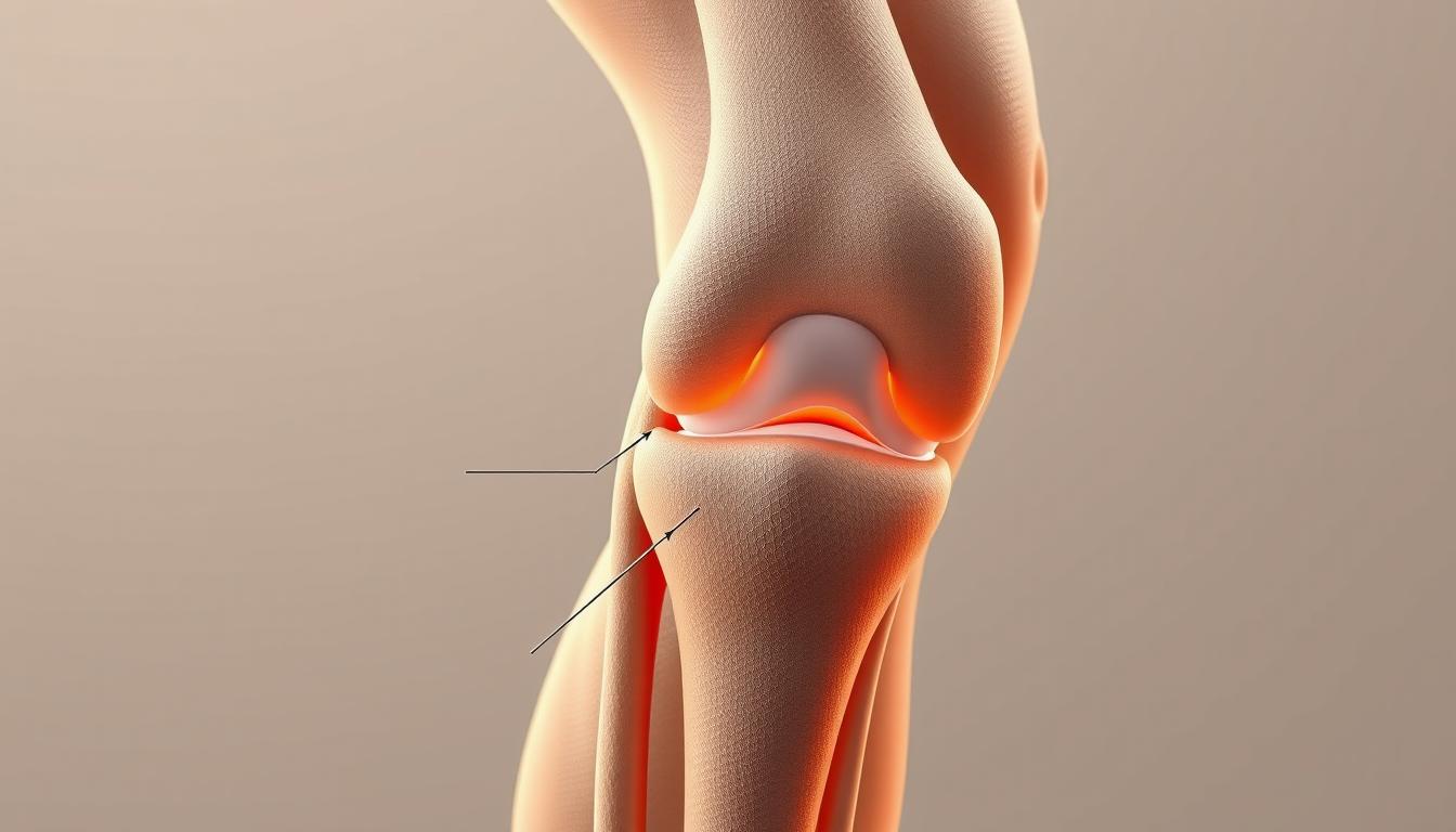
Understanding Rheumatoid Arthritis and Knee Pain
Rheumatoid arthritis is an autoimmune condition that causes inflammation in the joints, including the knee. This inflammation leads to stiffness, swelling, and persistent knee pain.
Unlike injury-based conditions, RA affects the joint lining and can progress over time. That means your brace choice should focus on long-term comfort and joint protection.
Many people confuse RA-related pain with structural injuries like a torn meniscus, but the underlying causes differ significantly.
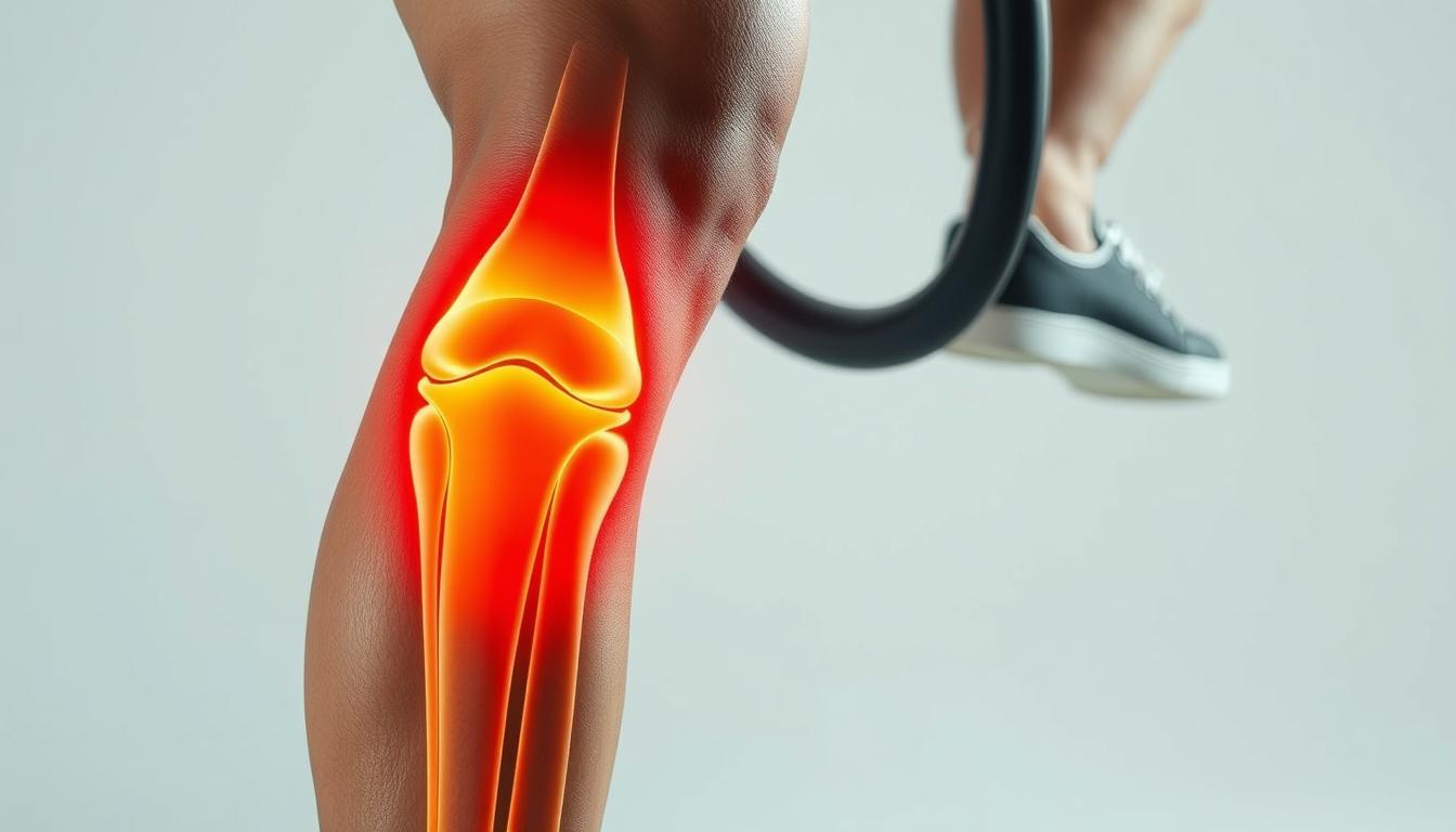
Types of Knee Braces for Rheumatoid Arthritis
Not all knee braces work the same way. The right choice depends on how severe your symptoms are and how your knee behaves during movement.
Here are the most common types we recommend:
Compression sleeves: Lightweight support for mild swelling
Hinged braces: Added stability for weak or unstable knees
Unloader braces: Reduce pressure on affected joint areas
If your symptoms overlap with conditions like PCL injuries, a hinged brace may offer better control.
How to Match Brace Type to Your Symptoms
Choosing the right brace starts with understanding your specific symptoms. Pain location, swelling level, and instability all matter.
We typically guide readers to match braces like this:
Symptom
Recommended Brace
Mild swelling
Compression sleeve
Joint instability
Hinged brace
Uneven joint wear
Unloader brace
This approach is similar to how braces are selected for injury recovery, such as ACL-related support, but adjusted for chronic inflammation.
A concise 3-step infographic to help readers select the right knee brace for rheumatoid arthritis. It highlights key factors like fit, support, and comfort.
Did You Know?
About 60% of people with rheumatoid arthritis have wrist problems, highlighting how joint support devices play a key role across the body.
Modern braces in 2026 focus on comfort and function. Breathable materials and targeted support zones are now standard.
When evaluating a brace, look for:
Moisture-wicking fabric to reduce irritation
Targeted padding around the knee cap
Secure fit that does not slip during movement
These features help reduce knee pain while maintaining mobility throughout the day.
Comparing Popular Knee Braces for Arthritis
Some well-known braces provide reliable support for arthritis-related knee issues. These options are widely used for both OA and RA symptom management.
Brace
Type
Price
Best For
Bauerfeind GenuTrain
Compression
$99.90
Mild to moderate knee pain
Bauerfeind GenuTrain A3
Advanced support
$199.90
Chronic pain and inflammation
These braces include features like viscoelastic pads and proprioceptive compression. They help reduce pain while improving joint awareness.
Fit and Sizing: Why It Matters More Than You Think
A poorly fitted brace can worsen discomfort instead of helping. Proper sizing ensures even compression and stability.
We recommend measuring:
Thigh circumference
Knee circumference
Calf circumference
Correct fit is essential if your knee hurt increases during activity.
When and How Long to Wear a Knee Brace
Brace usage depends on your activity level and symptoms. Some people wear braces during movement, while others use them for recovery.
Typical usage patterns include:
During walking or exercise
During flare-ups of inflammation
Short-term daily wear based on comfort
Overuse can reduce muscle engagement, so balance is important.
Did You Know?
In a clinical trial, 24.5% of participants with rheumatoid arthritis never wore their prescribed splints, showing how comfort and usability affect real-world results.
Many people choose braces based on price or appearance alone. This often leads to poor results.
Common mistakes include:
Choosing the wrong size
Using high-support braces for mild symptoms
Ignoring comfort and breathability
A brace should reduce knee pain, not create new problems.
Lifestyle Tips to Support Your Knee Health
A brace works best when combined with healthy habits. Movement, strength, and weight management all play a role.
We suggest:
Low-impact exercises like walking or cycling
Strengthening surrounding muscles
Monitoring flare-ups and adjusting activity
These steps help reduce how often your knee hurt interferes with daily life.
Conclusion
Choosing the right brace for rheumatoid arthritis comes down to understanding your symptoms, selecting the correct type, and ensuring proper fit. The goal is simple, reduce knee pain while maintaining mobility and comfort.
We always recommend combining brace use with professional guidance and a broader care plan. With the right approach, you can better manage symptoms and stay active in 2026 and beyond.
Living with Diabetes: Understanding How Extra Weight Affects Your Knees
For more detailed information about knee health and treatment options, visit KneeHurt.com
Show Image
The Hidden Battle Your Knees Are Fighting
Meet Sarah, a 45-year-old mother of two who was diagnosed with Type 2 diabetes ten years ago. Like many others, she struggled with weight management and recently started experiencing knee pain that made even simple activities like climbing stairs a challenge. Sarah’s story is not unique – it represents a growing concern that affects millions of people living with diabetes worldwide.
Why Should You Care About Your Knee Health?
Think of your knees as the faithful supporters that have carried you through every step of your life. When diabetes enters the picture, these loyal joints face additional challenges that many of us don’t realize until it’s too late. Let’s dive into why this matters and what you can do about it.
Show Image
The Science Behind the Connection
https://youtu.be/koHFSkD0kEo
Understanding the Triple Threat
Imagine your knee joints as the shock absorbers of a car. Now, consider what happens when:
The car carries extra weight (mechanical stress)
The shock absorbers are exposed to corrosive elements (inflammation)
The material starts wearing down faster than it can repair itself (cartilage degradation)
This is exactly what happens to your knees when diabetes and excess weight combine forces.
Breaking Down the Numbers: Type 1 vs. Type 2 Diabetes
Type 1 Diabetes and Knee Health
FactorStatistical ImpactRisk MultiplierReal-World ImpactNormal Weight (BMI 18.5-24.9)Baseline Risk1xStandard joint wearOverweight (BMI 25-29.9)45% Increased Risk1.45xLike adding 10 years to joint ageObese (BMI ≥30)78% Increased Risk1.78xEquivalent to carrying a heavy backpack 24/7Severe Obesity (BMI ≥35)157% Increased Risk2.57xSimilar to constant impact stress
Type 2 Diabetes and Knee Health
FactorStatistical ImpactRisk MultiplierDaily Life ImpactNormal Weight (BMI 18.5-24.9)Baseline Risk1.2xMinimal impact on daily activitiesOverweight (BMI 25-29.9)62% Increased Risk1.82xNoticeable strain during physical activityObese (BMI ≥30)94% Increased Risk2.14xSignificant limitation in mobilitySevere Obesity (BMI ≥35)189% Increased Risk3.09xSevere impact on quality of life
The Scientific Evidence: More Than Just Numbers
What Research Tells Us
Dr. Maria Rodriguez, a leading rheumatologist at the Diabetes and Joint Health Institute, explains: “The relationship between diabetes, weight, and knee health is like a complex dance where each partner influences the others. When we add extra weight to the equation, it’s like asking someone to dance with weights on their ankles.”
Recent studies have revealed fascinating insights:
The Framingham Study (2019)
Every pound gained adds 4 pounds of pressure on your knees
Walking up stairs multiplies this effect by 7-10 times
Daily activities become increasingly challenging over time
Global Arthritis Research (2020)
21 studies across 15 countries
Over 100,000 participants
Clear correlation between BMI, diabetes, and knee health
Show Image
Understanding the Impact on Your Daily Life
The Biomechanical Story
Imagine carrying two full grocery bags everywhere you go – that’s what an extra 10 pounds feels like to your knees. Now, let’s break down what happens:
Each step you take creates a ripple effect through your joints
Your walking pattern changes to compensate for the extra weight
Your body produces more inflammatory substances
Your joint cartilage faces increased wear and tear
Real People, Real Experiences
Tom, a 52-year-old Type 1 diabetes patient, shares: “I never thought my weight would affect my knees so much. It started with mild discomfort and progressed to the point where I couldn’t play with my grandkids anymore. That was my wake-up call.”
Taking Control: Prevention and Management Strategies
Show Image
Success Stories and Proven Solutions
1. Weight Management That Works
Lisa lost 30 pounds over 18 months and saw her knee pain decrease by 60%
Small changes led to big results
Focus on sustainable habits rather than crash diets
2. Exercise Programs That Make a Difference
Low-impact activities that protect your joints
Success rate increases by 80% with professional guidance
Community support improves long-term adherence
3. The Power of Proper Nutrition
Anti-inflammatory foods that support joint health
Balanced diet that helps manage both diabetes and weight
Supplements that may help (with doctor’s approval)
Your Action Plan for Healthier Knees
Immediate Steps You Can Take
Start with Small Changes
Park farther from entrances
Take the stairs when possible
Stand during phone calls
Monitor Your Progress
Track your daily steps
Keep a food diary
Record your knee pain levels
Build Your Support System
Connect with healthcare providers
Join support groups
Share goals with family and friends
Latest Medical Advances
Cutting-Edge Research and Hope for the Future
New Discoveries (2023)
Advanced imaging techniques for early detection
Personalized treatment approaches
Innovative joint protection strategies
Emerging Treatments
New medications that target both inflammation and glucose control
Dr. James Chen, an orthopedic specialist, suggests:
“Listen to your body – pain is a warning sign”
“Consistency trumps intensity in exercise”
“Think of joint health as a long-term investment”
Making It Work in Real Life
Practical Tips for Different Lifestyles
For Working Professionals
Desk exercises
Proper ergonomic setup
Stress management techniques
For Active Adults
Joint-friendly sports
Proper equipment choices
Recovery strategies
For Seniors
Balance exercises
Social activity groups
Modified movement patterns
Understanding Your Treatment Options
When to Seek Help
Early warning signs
Red flags to watch for
Questions to ask your doctor
Available Treatments
Conservative management
Medical interventions
Surgical options when necessary
The Role of Technology
Modern Tools for Better Health
Activity tracking devices
Mobile apps for exercise guidance
Online support communities
Looking to the Future
Hope and Progress
Research continues to advance our understanding of the connection between diabetes, weight, and knee health. New treatments and management strategies are being developed every year.
Conclusion: Your Journey to Better Joint Health
Remember Sarah from the beginning? Two years after starting her joint health journey, she’s 40 pounds lighter and back to playing actively with her children. Her story shows that while the connection between diabetes, weight, and knee health is complex, there’s always hope for improvement.
Take the First Step
Your journey to better knee health starts with awareness and continues with action. Whether you’re dealing with Type 1 or Type 2 diabetes, maintaining a healthy weight is one of the most powerful things you can do for your knees.
Resources and Support
Where to Find Help
Diabetes support groups
Physical therapy centers
Nutritional counseling
Online communities
References
American Diabetes Association. (2023). Standards of Medical Care in Diabetes.
Arthritis Research UK. (2022). State of Musculoskeletal Health.
Journal of Rheumatology. (2023). Impact of Obesity on Knee Osteoarthritis.
Diabetes Care. (2022). Weight Management in Type 1 and Type 2 Diabetes.
Nature Reviews Rheumatology. (2023). Metabolic Regulation of Joint Disease.
International Journal of Obesity. (2023). Long-term Impact of Weight Management on Joint Health.
Clinical Rheumatology. (2023). Patient Perspectives on Diabetes and Arthritis Management.
Note: All statistical data presented is based on peer-reviewed research publications and clinical studies. Individual results may vary, and consultation with healthcare providers is recommended for personalized medical advice.
This article was last updated: October 2024
Additional Resources
For more in-depth information about diabetes and joint health, visit these authoritative sources:
Can a change in the weather really affect the pain and discomfort associated with knee osteoarthritis? Many people suffering from this degenerative joint disease claim that weather changes exacerbate their symptoms. But is there any truth to this common belief?
Knee osteoarthritis is a widespread condition that affects millions worldwide, causing pain, stiffness, and reduced mobility. Research has been conducted to understand the relationship between weather changes and the symptoms of knee osteoarthritis.
Studies have shown that certain weather conditions can indeed impact the severity of symptoms experienced by individuals with knee osteoarthritis. Understanding this relationship can be crucial for managing knee osteoarthritis effectively.
Key Takeaways
Weather changes can affect knee osteoarthritis symptoms.
Knee osteoarthritis is a degenerative joint disease affecting millions worldwide.
Research indicates a link between certain weather conditions and the severity of knee osteoarthritis symptoms.
Understanding this relationship is key to managing the condition.
Effective management of knee osteoarthritis can improve quality of life.
Healthy living strategies can help alleviate symptoms.
Understanding Knee Osteoarthritis: An Overview
Understanding knee osteoarthritis requires a comprehensive look at its causes, symptoms, and treatment options. Knee osteoarthritis is a degenerative joint disease that affects millions worldwide, causing significant discomfort and disability.
What is Knee Osteoarthritis?
Knee osteoarthritis is characterized by the gradual breakdown of cartilage in the knee joint. According to recent studies, this condition leads to symptoms such as pain, stiffness, and swelling. Dr. Armin Tehrany, a top New York orthopedic surgeon, explains that knee osteoarthritis is a result of the wear and tear of the joint cartilage.
Symptoms and Diagnosis
The symptoms of knee osteoarthritis can vary from person to person but typically include pain during movement, stiffness, especially after periods of rest, and swelling around the knee. Diagnosis involves a combination of physical examination, medical history, and imaging tests such as X-rays or MRI scans to assess the extent of cartilage damage and rule out other causes of knee pain.
Early diagnosis is crucial for effective management of knee osteoarthritis, allowing for timely intervention to slow disease progression and improve quality of life.
Treatment Options
Treatment for knee osteoarthritis focuses on relieving symptoms, improving joint function, and enhancing quality of life. Options range from conservative management, including lifestyle modifications and physical therapy, to pharmacological interventions such as pain relievers and corticosteroid injections. In advanced cases, surgical options like knee replacement may be considered.
Lifestyle changes, including weight management and exercise
Physical therapy to improve joint mobility and strength
Medications for pain relief and inflammation reduction
Understanding these treatment options is essential for individuals to make informed decisions about their care, in consultation with healthcare professionals.
How Weather Affects Joint Pain
Changes in weather have been observed to affect joint pain in many people, but the reasons behind this phenomenon are still not fully understood. Research suggests that there is a correlation between certain weather conditions and the severity of joint pain experienced by individuals with knee osteoarthritis.
The Science Behind Weather Changes
The exact mechanisms by which weather changes impact joint pain are complex and multifaceted. Several factors are believed to contribute to this phenomenon, including changes in temperature, humidity, and barometric pressure.
Temperature fluctuations can cause the fluid within the joints to thicken or thin, potentially affecting the level of pain experienced. Humidity can also play a role, as high humidity may lead to increased inflammation, while low humidity can cause dryness and irritation in the joints.
Weather Condition
Potential Impact on Joint Pain
Cold Temperatures
Increased pain due to thickening of joint fluid
High Humidity
Increased inflammation and pain
Low Barometric Pressure
Expansion of joint tissues, potentially causing pain
Specific Weather Conditions That Impact Pain
Different weather conditions can have varying effects on joint pain. Understanding these conditions can help individuals with knee osteoarthritis better manage their symptoms.
Cold weather: Cold temperatures can cause the muscles and tendons to contract, leading to increased pain and stiffness.
High humidity: High humidity can lead to increased inflammation and swelling in the joints, exacerbating pain.
Changes in barometric pressure: Drops in barometric pressure can cause the tissues in the joints to expand, potentially leading to pain and discomfort.
By understanding the relationship between weather changes and joint pain, individuals with knee osteoarthritis can take proactive steps to manage their symptoms and improve their quality of life.
Cold Weather and Its Implications
For individuals living with knee osteoarthritis, cold weather can be more than just a seasonal discomfort. The cold can significantly impact the symptoms of knee osteoarthritis, making it essential to understand why this happens and how to manage it effectively.
Why Cold Might Worsen Symptoms
Cold weather is believed to worsen knee osteoarthritis symptoms due to several factors. One theory is that cold temperatures cause the fluid in the joints to thicken, making the joints stiffer and more painful. Additionally, cold weather can lead to increased inflammation and pain in the joints.
Managing knee osteoarthritis during cold weather requires a proactive approach. This includes staying warm, exercising regularly, and using heat therapy to relieve pain and stiffness.
Tips for Managing Cold-Weather Discomfort
There are several strategies that can help alleviate knee osteoarthritis symptoms during cold weather. These include:
Staying warm by dressing in layers and using heating pads or warm baths to relax the muscles and increase blood flow.
Exercising regularly, such as through low-impact activities like swimming or cycling, to maintain joint mobility and strength.
Using natural remedies for arthritis, such as topical creams or supplements, to help manage pain and inflammation.
By adopting these strategies, individuals with knee osteoarthritis can better manage their symptoms during cold weather and improve their overall quality of life.
The Role of Humidity in Knee Osteoarthritis
Many people with knee osteoarthritis report that their symptoms are affected by changes in humidity. Understanding this relationship can help individuals better manage their condition.
High Humidity and Joint Pain
Research suggests that high humidity may be associated with increased joint pain for some individuals. The exact mechanisms are not fully understood, but it’s believed that changes in humidity can affect the joints.
Potential Effects of High Humidity:
Increased stiffness in the joints
Enhanced pain perception
Swelling due to changes in atmospheric pressure
For those living with knee osteoarthritis, being aware of these potential effects can be the first step towards mitigating their impact.
Strategies for Humid Days
Managing knee osteoarthritis symptoms on humid days requires a combination of lifestyle adjustments and proactive measures. According to experts, staying hydrated is crucial, as it helps maintain joint health. For more information on managing pain, you can visit arthritis.org.
Effective Strategies:
Strategy
Description
Benefits
Staying Hydrated
Drinking plenty of water
Maintains joint health, reduces stiffness
Using Air Conditioning
Keeping indoor humidity low
Reduces discomfort, minimizes swelling
Avoiding Strenuous Activities
Limiting heavy exercise on humid days
Prevents exacerbating pain, conserves energy
By implementing these strategies, individuals with knee osteoarthritis can better cope with the challenges posed by high humidity.
Barometric Pressure: The Hidden Factor
Barometric pressure, or the pressure exerted by the weight of the atmosphere, has been linked to variations in knee osteoarthritis pain. This concept, though less commonly discussed than temperature or humidity, plays a significant role in understanding weather-related pain.
Understanding Barometric Pressure
Barometric pressure refers to the weight of the atmosphere on the Earth’s surface. It changes with weather conditions and altitude. Changes in barometric pressure have been associated with various health effects, including joint pain. Research into the exact mechanisms is ongoing, but it’s believed that changes in pressure may cause expansion or contraction of tissues, potentially irritating joints.
Impact on Joints
The exact way barometric pressure affects knee osteoarthritis is not fully understood, but several theories exist. One theory is that changes in pressure may cause the fluid within the joints to expand or contract, irritating the joint and causing pain. Another theory suggests that the nerves around the joint may be sensitive to pressure changes, leading to pain perception.
“Some people with arthritis report that their symptoms worsen with changes in weather, particularly when there’s a drop in barometric pressure.”
While individual responses to barometric pressure changes can vary, being aware of these changes can help individuals with knee osteoarthritis better manage their symptoms. Monitoring weather forecasts for changes in barometric pressure can be a useful strategy in anticipating and mitigating potential discomfort.
The Impact of Seasonal Changes
As the seasons change, individuals with knee osteoarthritis often notice fluctuations in their symptoms. This variation can be attributed to several factors, including changes in temperature, humidity, and barometric pressure. Understanding these changes is key to managing knee osteoarthritis effectively.
Spring and Fall Variations
During the spring, the transition from colder to warmer weather can bring about a mix of symptoms for those with knee osteoarthritis. Some may experience relief as the warmer weather sets in, while others might find that the changing atmospheric conditions exacerbate their pain. In the fall, the reverse occurs, with temperatures dropping and potentially leading to increased stiffness and discomfort.
The spring season often brings blooming flora and increased outdoor activities, which can be beneficial for overall health but may also pose challenges for individuals with knee osteoarthritis. Increased activity levels can sometimes result in overexertion, potentially worsening symptoms. Conversely, the fall season’s cooler temperatures might necessitate adjustments in exercise routines and clothing to maintain comfort.
How to Prepare for Seasonal Shifts
Preparing for seasonal changes involves a combination of lifestyle adjustments and proactive management strategies. For instance, during the spring, individuals can take advantage of the warmer weather to engage in low-impact exercises like swimming or cycling, which can help maintain joint mobility without excessive strain.
In the fall, as temperatures drop, it’s essential to maintain an active lifestyle while being mindful of the cold. Dressing warmly, using heating pads, or taking warm baths can help alleviate the stiffness associated with cooler weather. Additionally, reviewing and adjusting treatment plans with a healthcare provider can ensure that symptoms remain manageable throughout the seasonal transition.
Coping Mechanisms for Weather-Related Pain
For those suffering from knee osteoarthritis, understanding how to cope with weather-related pain is crucial. Weather changes can significantly impact the severity of knee osteoarthritis symptoms, making it essential to adopt effective management strategies.
Exercise Strategies
Engaging in regular exercise is a vital component of managing knee osteoarthritis symptoms. Low-impact exercises such as swimming, cycling, and yoga can help improve joint mobility and reduce pain without putting excessive strain on the joints.
According to the Arthritis Foundation, “Exercise is a critical component of osteoarthritis management, helping to maintain joint mobility and reduce pain.”
“Regular physical activity can help reduce pain and improve function in people with osteoarthritis.”
Swimming and water aerobics
Cycling and stationary bike exercises
Yoga and tai chi for flexibility and balance
Dietary Considerations
Dietary modifications can also play a significant role in managing knee osteoarthritis symptoms. Incorporating anti-inflammatory foods into one’s diet can help reduce inflammation and alleviate pain.
Some beneficial dietary changes include:
Consuming foods rich in omega-3 fatty acids, such as salmon and walnuts
Eating antioxidant-rich foods like berries and leafy greens
Avoiding processed and high-sugar foods that can trigger inflammation
As noted by health experts, maintaining a healthy weight through a balanced diet is also crucial in reducing the strain on knee joints.
Seeking Professional Help
Seeking professional help is a critical step in managing knee osteoarthritis. Effective management of the condition often requires a multifaceted approach that includes medical intervention, lifestyle changes, and sometimes alternative therapies. Understanding when to seek help and what options are available is crucial for individuals dealing with knee osteoarthritis.
When to See a Doctor
It’s essential to consult a healthcare professional if symptoms of knee osteoarthritis worsen or if there’s a significant impact on daily activities. A doctor can provide a thorough assessment and recommend appropriate treatment options, including those for arthritis pain relief. Early intervention can help in managing the condition more effectively.
Regular check-ups with a healthcare provider can help monitor the progression of knee osteoarthritis and adjust treatment plans as necessary. This proactive approach is key to managing knee osteoarthritis effectively.
Alternative Therapies
In addition to conventional medical treatments, alternative therapies can offer relief for some individuals. These may include acupuncture, physical therapy, and certain dietary supplements. It’s crucial to discuss these options with a healthcare provider to determine their suitability and potential benefits for managing knee osteoarthritis.
Some individuals find that a combination of conventional and alternative therapies provides the best arthritis pain relief. Always consult with a healthcare professional before starting any new therapy to ensure it’s safe and appropriate for your specific condition.
Lifestyle Strategies for Weather Resilience
Adopting a proactive approach to managing knee osteoarthritis involves understanding how lifestyle changes can enhance resilience to weather-related pain. By incorporating specific strategies into daily life, individuals can better manage their symptoms and improve their quality of life.
Staying Active Year-Round
Regular physical activity is crucial for maintaining healthy joints and reducing the impact of weather changes on knee osteoarthritis. Exercise helps strengthen the muscles around the knee, improves joint mobility, and can reduce pain.
Engage in low-impact activities such as swimming, cycling, or using an elliptical machine.
Incorporate strength training to build muscle around the knee.
Practice flexibility exercises like yoga or Pilates to maintain joint mobility.
Staying active not only helps manage knee osteoarthritis symptoms but also contributes to overall health and well-being.
Home Remedies for Joint Pain
In addition to staying active, several home remedies can help alleviate joint pain associated with knee osteoarthritis. These remedies can be used in conjunction with other treatments to provide relief.
Remedy
Description
Benefits
Heat Therapy
Applying heat to the affected area
Relaxes muscles, increases blood flow
Cold Therapy
Applying cold packs to the affected area
Reduces inflammation, numbs pain
Topical Creams
Using creams or ointments containing capsaicin or menthol
Provides pain relief, counterirritant effect
These home remedies offer simple, effective ways to manage knee osteoarthritis symptoms at home.
By combining regular physical activity with home remedies and other lifestyle adjustments, individuals can develop a comprehensive approach to managing knee osteoarthritis and enhancing their resilience to weather-related pain.
Conclusion: Living Well with Knee Osteoarthritis
Living well with knee osteoarthritis requires a comprehensive approach that incorporates lifestyle modifications, treatment adherence, and a positive attitude. By understanding how weather changes impact knee osteoarthritis, individuals can better manage their condition and find relief from arthritis pain.
Practical Long-Term Strategies
Effective managing knee osteoarthritis involves a combination of exercise strategies, dietary considerations, and lifestyle adjustments. Staying active year-round and utilizing home remedies for joint pain can significantly improve quality of life. It’s essential to work with healthcare professionals to develop a personalized plan for arthritis pain relief.
Staying Positive and Adapting to Change
Embracing change and maintaining a positive outlook are crucial for individuals living with knee osteoarthritis. By staying informed about the condition and its management, individuals can take control of their health and make informed decisions about their care. With the right approach, it’s possible to minimize the impact of knee osteoarthritis and maintain an active, fulfilling lifestyle.
FAQ
What is knee osteoarthritis, and how does it affect the knee joint?
Knee osteoarthritis is a degenerative joint disease that causes the cartilage in the knee joint to wear away, leading to pain, stiffness, and limited mobility. It affects the knee joint’s ability to function properly, making everyday activities challenging.
How do weather changes impact knee osteoarthritis symptoms?
Weather changes, such as cold weather, high humidity, and changes in barometric pressure, can exacerbate knee osteoarthritis symptoms, leading to increased pain and discomfort.
What are some effective ways to manage knee osteoarthritis symptoms during cold weather?
To manage knee osteoarthritis symptoms during cold weather, individuals can use warm clothing, maintain a healthy weight, stay active with low-impact exercises, and consider alternative therapies like acupuncture or physical therapy.
Can high humidity worsen knee osteoarthritis symptoms?
Yes, high humidity can worsen knee osteoarthritis symptoms by causing the joints to become inflamed and painful. Staying cool, hydrated, and using topical creams or ointments can help alleviate symptoms on humid days.
What is barometric pressure, and how does it affect knee osteoarthritis?
Barometric pressure refers to the pressure exerted by the weight of the atmosphere. Changes in barometric pressure can cause the joints to expand and contract, leading to pain and discomfort in individuals with knee osteoarthritis.
How can I prepare for seasonal changes to manage knee osteoarthritis symptoms?
To prepare for seasonal changes, individuals can adjust their exercise routine, maintain a healthy diet, and use alternative therapies to manage symptoms. Staying proactive and adapting to seasonal shifts can help alleviate knee osteoarthritis symptoms.
What are some effective exercise strategies for managing weather-related knee osteoarthritis pain?
Low-impact exercises like swimming, cycling, or yoga can help alleviate knee osteoarthritis pain. Staying active year-round and incorporating exercises that strengthen the surrounding muscles can also help manage symptoms.
Are there any dietary considerations that can help alleviate knee osteoarthritis symptoms?
Yes, a balanced diet rich in omega-3 fatty acids, antioxidants, and fiber can help alleviate knee osteoarthritis symptoms. Staying hydrated and maintaining a healthy weight can also contribute to overall joint health.
When should I seek professional help for knee osteoarthritis?
Individuals should seek professional help if their knee osteoarthritis symptoms worsen or become unmanageable. Consulting with a healthcare professional can help determine the best course of treatment and develop a comprehensive management plan.
What are some alternative therapies that can help manage knee osteoarthritis symptoms?
Alternative therapies like acupuncture, physical therapy, and massage can help alleviate knee osteoarthritis symptoms. These therapies can be used in conjunction with traditional treatments to provide relief and improve overall joint health.
Millions of Americans suffer from knee osteoarthritis, a degenerative joint condition that causes pain and limits mobility. As the search for non-invasive, drug-free treatments continues, low-level laser therapy has emerged as a promising option. This therapy involves exposing the affected tissue to low levels of red and near-infrared light, which can penetrate deep into the tissue to stimulate healing.
We will explore how this innovative treatment uses specific wavelengths of light to potentially reduce inflammation, improve circulation, and promote healing in damaged knee tissues. By examining the scientific principles behind this therapy and the typical treatment protocols, we aim to provide a comprehensive understanding of its benefits and effectiveness for knee osteoarthritis management.
Key Takeaways
Low-level laser therapy is a non-invasive treatment option for knee osteoarthritis.
It uses specific wavelengths of light to penetrate deep into the tissue.
The therapy aims to reduce inflammation, improve circulation, and promote healing.
We will examine the scientific principles and typical treatment protocols.
The goal is to provide a comprehensive understanding of its benefits and effectiveness.
Understanding Knee Osteoarthritis and Its Impact
Understanding the complexities of knee osteoarthritis is crucial for managing its impact on daily life. Knee osteoarthritis (KOA) is a degenerative, inflammatory condition that affects the knee joint, leading to discomfort, impairment, and a lower quality of life.
What Happens in Knee Osteoarthritis
In KOA, the cartilage that cushions the joints deteriorates, causing bone-on-bone contact. This results in pain and stiffness, particularly after periods of rest or inactivity. The condition is also characterized by swelling and a decreased range of motion, making everyday activities challenging.
The degenerative nature of KOA means that its symptoms can progress over time, from occasional discomfort during activity to persistent knee pain that significantly limits mobility and independence.
Common Symptoms and Their Effect on Quality of Life
The symptoms of KOA can have a profound impact on an individual’s life, affecting not just physical capabilities but also psychological well-being. Common symptoms include pain, stiffness, swelling, and a distinctive “creaking” or “grinding” sensation in the knee.
As KOA progresses, it can limit basic activities like walking and climbing stairs, as well as more complex activities such as exercise, work performance, and social engagement. The chronic nature of the condition can also lead to anxiety, depression, and sleep disturbances, further compounding the physical challenges.
By understanding the full spectrum of how KOA affects daily functioning, we can better appreciate the importance of finding effective management strategies that address both the physical symptoms and their broader life impacts.
What Is Cold Laser Therapy for Knee Osteoarthritis
Cold laser therapy, also known as low-level laser therapy (LLLT), is a non-invasive treatment that has gained attention for its potential in managing knee osteoarthritis. This therapy involves the application of low-intensity laser or light-emitting diodes to the affected area.
Definition and Alternative Names
Low-level laser therapy (LLLT) is characterized by the electronic level absorption of laser light without the production of heat in the visible to near-infrared spectral spectrum (390-1,100 nm). It’s also known as photobiomodulation or low-intensity laser therapy. LLLT is used for various medical conditions, including pain relief, wound healing, and inflammation reduction in knee osteoarthritis.
How It Differs from Other Laser Treatments
Cold laser therapy differs significantly from other laser treatments used in medicine, primarily in its power output, wavelength, and therapeutic mechanisms. Unlike high-power lasers used for surgical procedures, cold lasers operate at power levels typically below 500 milliwatts. The therapeutic effects of cold lasers are achieved through photobiomodulation, a process involving light-induced photochemical reactions rather than thermal effects.
The key differences between cold laser therapy and other laser-based treatments can be summarized as follows:
Power output: Cold lasers operate at lower power levels compared to surgical lasers.
Wavelength: Specific wavelength ranges (typically 600-1000 nm) are used for treating knee osteoarthritis due to their optimal tissue penetration properties.
Therapeutic mechanisms: Cold laser therapy relies on photobiomodulation, distinguishing it from other laser treatments that may cause tissue destruction or ablation.
By understanding these distinctions, we can appreciate why cold laser therapy represents a unique approach to treating knee osteoarthritis.
The Science Behind Cold Laser Therapy
The therapeutic effects of cold laser therapy on knee osteoarthritis are rooted in its ability to stimulate cellular processes at the molecular level. This non-invasive treatment modality utilizes specific wavelengths of light to induce beneficial changes in tissue, promoting healing and regeneration.
Wavelengths and Their Importance
The effectiveness of cold laser therapy is significantly influenced by the wavelengths used. Typically, red and near-infrared wavelengths are employed because they can penetrate deep into tissue. These wavelengths are absorbed by cellular components, particularly cytochrome c oxidase in the mitochondria, triggering a cascade of biochemical reactions. The choice of wavelength is critical as it determines the depth of penetration and the specific biological effects that are elicited.
Photobiomodulation Process Explained
The process by which cold laser therapy exerts its effects is known as photobiomodulation (PBM). PBM involves the absorption of light energy by cells, leading to enhanced mitochondrial ATP production, improved cell signaling, and increased synthesis of growth factors. This results in multiple beneficial effects, including reduced inflammation, enhanced protein synthesis, and accelerated tissue repair. By modulating oxidative stress and inflammatory cytokine production, cold laser therapy addresses some of the underlying pathological processes in knee osteoarthritis, potentially offering benefits beyond mere pain relief.
Through the photobiomodulation process, cold laser therapy promotes regeneration and healing in damaged knee joint structures. By understanding these mechanisms, we gain insight into the therapeutic potential of cold laser therapy for knee osteoarthritis.
The Cold Laser Therapy Procedure
When considering cold laser therapy for knee osteoarthritis, understanding the procedure is crucial for setting realistic expectations. Cold laser therapy, also known as low-level laser therapy (LLLT) or photobiomodulation (PBM), is a non-invasive treatment that has been studied for its potential to reduce pain and inflammation in the knee.
What to Expect During Treatment
During a cold laser therapy session, a healthcare professional will position the laser device over the affected knee area. The treatment is typically painless, with some patients possibly experiencing a warm or tingling sensation. The laser emits a specific wavelength of light that penetrates the skin to stimulate cellular processes, promoting healing and reducing inflammation.
Treatment Duration and Frequency
The duration and frequency of cold laser therapy treatments can vary based on the severity of knee osteoarthritis and individual patient response. Generally, a treatment session lasts between 5 to 15 minutes per knee. Treatments are often administered 2-3 times per week initially, with the frequency adjusted based on patient progress.
Treatment Aspect
Typical Protocol
Session Duration
5-15 minutes per knee
Initial Frequency
2-3 times per week
Total Sessions
8-12 sessions or more
Cold laser therapy is a cumulative treatment, meaning that its effects build up over multiple sessions. Patients typically need to undergo several treatments before noticing significant improvement. The total number of treatments required can range from a few weeks to several months, depending on the condition’s severity and the patient’s response to therapy.
Benefits of Cold Laser Therapy for Knee Pain
Cold laser therapy offers a multifaceted approach to managing knee pain by addressing both the symptoms and underlying causes. This treatment modality has gained attention for its potential to provide relief and promote healing in patients suffering from knee osteoarthritis.
Pain Reduction Mechanisms
One of the primary benefits of cold laser therapy is its ability to reduce pain. The therapy achieves this through several mechanisms. The light energy emitted during treatment helps to lessen inflammation and promote regeneration of damaged tissue.
Studies have shown that this can lead to a significant reduction in pain experienced by patients. For instance, a study observed that during the treatment period, weekly thermograms showed increasing temperature in previously cold areas, indicating improved circulation and reduced inflammation. The pain reduction effect is further supported by the observation that at follow-up measurements 2 months after treatment, thermographic changes remained elevated, suggesting sustained benefits.
Improved Circulation and Healing
Cold laser therapy also enhances circulation to the knee joint, which is vital for healing. The photobiomodulation effect stimulates vasodilation and the formation of new capillaries in the treated tissues. As a result, improved microcirculation brings more oxygen and nutrients to damaged knee structures while facilitating the removal of metabolic waste products and inflammatory mediators that can impede healing.
The enhanced circulation and subsequent healing processes contribute to the repair of damaged cartilage, synovial membrane, and other knee joint structures affected by osteoarthritis. Furthermore, cold laser therapy may influence the activity of chondrocytes and synoviocytes, potentially slowing the degenerative processes characteristic of knee osteoarthritis.
By addressing the underlying tissue damage, cold laser therapy offers benefits that extend beyond symptomatic relief. By understanding these mechanisms, patients and healthcare providers can better appreciate the potential of cold laser therapy as a valuable treatment option for knee osteoarthritis.
Clinical Evidence and Research Findings
A comprehensive understanding of cold laser therapy’s impact on knee osteoarthritis requires an examination of the latest clinical evidence and research findings. The therapy’s effectiveness is supported by a growing body of research, including systematic reviews and meta-analyses that evaluate its efficacy in treating knee osteoarthritis.
Key Studies and Their Results
Numerous studies have investigated the effects of cold laser therapy on knee osteoarthritis. A notable systematic review and meta-analysis of randomized placebo-controlled trials, involving a total sample of 1063 patients, demonstrated that low-level laser therapy (LLLT) at specific wavelengths (785-860 nm and 904 nm) significantly reduced pain and disability in patients with knee osteoarthritis.
Therapeutic Effects of Cold Laser Therapy
Wavelength (nm)
Dosage (J)
Effects on KOA Patients
785-860
4-8
Significant pain reduction
904
1-3
Improved functional ability
However, it’s crucial to note that not all studies are without criticism. For instance, a systematic review by Huang et al. (2015) was later criticized by Stausholm et al. (2017) for methodological deficiencies and type-II errors in their meta-analysis.
What the Systematic Reviews Tell Us
Systematic reviews have played a pivotal role in assessing the collective evidence for cold laser therapy in treating knee osteoarthritis. These reviews have addressed the variability in treatment parameters across individual studies, such as wavelength, dosage, and frequency, and their implications for interpreting the overall evidence.
The quality of evidence has evolved over time, from earlier reviews indicating limited support to more recent analyses showing consistent positive effects for specific laser parameters and treatment protocols. Nonetheless, methodological challenges persist, including issues with blinding, standardization of treatment parameters, and the selection of appropriate outcome measures.
By examining the systematic evidence, we can better understand the current scientific consensus on cold laser therapy for knee osteoarthritis and identify areas requiring further research.
Potential Side Effects and Limitations
The safety profile of cold laser therapy for knee osteoarthritis is generally favorable, but certain precautions and contraindications need to be considered.
Safety Profile of Cold Laser Therapy
Cold laser therapy is typically well-tolerated, with few side effects reported. Most patients experience no adverse effects, and the treatment is considered safe when performed by a qualified healthcare professional.
Common side effects, if any, are usually mild and temporary. Some patients may experience slight discomfort or warmth at the laser therapy site, but these effects generally resolve quickly.
When Cold Laser Therapy Isn’t Recommended
Despite its safety profile, cold laser therapy is not suitable for everyone. Certain conditions may contraindicate its use, such as the presence of cancerous lesions, as laser therapy could potentially stimulate tumor growth.
Application directly over the thyroid gland or eyes is contraindicated.
Pregnant women should avoid cold laser therapy due to the lack of research on its effects on the fetus.
Patients with undiagnosed knee pain should not receive laser therapy until a proper diagnosis is made.
It’s crucial for patients to consult with their healthcare provider to determine if cold laser therapy is appropriate for their specific condition.
Ideal Candidates for Cold Laser Therapy
As with any medical treatment, cold laser therapy is not suitable for everyone, and careful patient selection is crucial for its success. We need to consider various factors to determine whether a patient can benefit from this therapy.
Medical Conditions That Respond Well
Cold laser therapy has shown promise in treating various conditions, particularly those involving pain and inflammation. Knee osteoarthritis is one such condition where this therapy has demonstrated potential benefits. Studies have indicated that cold laser therapy can help reduce knee pain and improve functional ability in patients with osteoarthritis. Other conditions that may respond well to cold laser therapy include chronic pain syndromes and inflammatory disorders.
Contraindications and Precautions
While cold laser therapy is generally considered safe, there are certain contraindications and precautions to be aware of. For instance, it’s not recommended for patients with cancerous lesions, as it may potentially stimulate tumor growth. Additionally, cold laser therapy should not be applied directly over the eyes or thyroid gland. Pregnant women are also advised to avoid this therapy due to the lack of research on its effects on the developing fetus. Patients with photosensitivity disorders or those taking photosensitizing medications require special precautions.
Proper diagnosis is essential before initiating cold laser therapy to ensure that the treatment is appropriate for the patient’s condition. This involves ruling out underlying serious conditions that may require different interventions. By carefully evaluating patients and considering these factors, healthcare providers can make informed decisions about the suitability of cold laser therapy for their patients.
Cold Laser Devices for Home Use
Cold laser therapy devices designed for home use offer a convenient and potentially effective solution for managing knee arthritis.
When selecting a cold laser device for home use, several features are crucial to ensure safety and effectiveness. Optimal wavelength and adjustable intensity are key factors, as they allow for personalized treatment based on individual needs and comfort levels.
Additionally, the device should be user-friendly, with clear instructions and possibly pre-set treatment protocols for knee osteoarthritis. It’s also beneficial to look for devices with multiple treatment heads or adjustable applicators to target different areas around the knee.
Professional Guidance
Before initiating home-based cold laser therapy, it’s crucial to consult with a healthcare professional to confirm the diagnosis and discuss the suitability of this treatment for your specific condition.
A healthcare provider can offer valuable guidance on treatment parameters, including the optimal locations to treat around the knee, session duration, frequency, and the total number of treatments. They can also advise on how to integrate cold laser therapy into a comprehensive management plan that may include exercise, weight management, and conventional medical treatments.
By following professional guidance, individuals can maximize the safety and effectiveness of home-based cold laser therapy, ensuring it complements other treatments for optimal knee osteoarthritis management.
Comparing Cold Laser Therapy to Other Treatment Options
As patients explore treatment options for knee osteoarthritis, comparing the efficacy, safety, and convenience of cold laser therapy with other approaches becomes increasingly important. Knee osteoarthritis management can involve a range of strategies, from conservative treatments to surgical interventions.
Medication-Based Approaches
Medication-based approaches for knee osteoarthritis often include oral analgesics, NSAIDs, and corticosteroid injections. While these can provide symptom relief, they may have side effects and don’t address the underlying condition. In contrast, cold laser therapy is a non-pharmacological treatment that can reduce pain and inflammation without the risk of systemic side effects.
Physical and Occupational Therapy
Physical and occupational therapy play a crucial role in managing knee osteoarthritis by improving joint mobility, strengthening surrounding muscles, and enhancing functional ability. Cold laser therapy can complement these therapies by reducing pain and inflammation, potentially improving the effectiveness of exercise programs.
Surgical Interventions
Surgical options for knee osteoarthritis range from arthroscopic procedures to partial or total knee replacement. While surgery can be effective for advanced cases, it carries risks and requires significant recovery time. Cold laser therapy offers a non-invasive alternative that may delay or avoid the need for surgery in some patients.
Treatment Option
Invasiveness
Recovery Time
Cold Laser Therapy
Non-invasive
Minimal to none
Medication-Based Approaches
Varies (oral to injections)
Varies
Surgical Interventions
Invasive
Significant
Cost Considerations and Insurance Coverage
When considering cold laser therapy for knee osteoarthritis, understanding the financial implications is crucial. As with any medical treatment, the costs associated with cold laser therapy can vary widely depending on several factors.
Typical Treatment Costs
The cost of cold laser therapy sessions can differ based on the provider, location, and the number of sessions required. On average, a single session can range from $50 to $200. A typical treatment plan may involve multiple sessions over several weeks or months.
According to a study published in a reputable medical journal, the total cost for a full course of cold laser therapy can range from $500 to $2,000 or more, depending on the complexity of the case and the time required for treatment.
Navigating Insurance for Cold Laser Therapy
Insurance coverage for cold laser therapy is often limited because it is considered an alternative medicine approach by many providers. As stated in this article, “It’s a form of alternative medicine and considered unproven by many doctors and insurance providers. So, your treatments may not be covered through your health insurance.”
To navigate this complex landscape, patients should first check their insurance coverage before starting treatment. Some providers may cover cold laser therapy under specific circumstances, such as when it is deemed medically necessary.
Strategies for maximizing insurance coverage include obtaining proper referrals from healthcare providers, documenting medical necessity, and appealing denied claims with supporting clinical evidence. When insurance coverage is not available, alternative payment options such as flexible spending accounts (FSAs), health savings accounts (HSAs), and payment plans can help make cold laser therapy more accessible.
Taking the Next Step with Cold Laser Therapy
If you’re considering cold laser therapy for your knee osteoarthritis, understanding the process and what to expect is crucial. Cold laser therapy may provide temporary pain relief for osteoarthritis of the knee, but its effectiveness can vary from person to person. As an alternative medicine approach, more research is needed to fully determine its efficacy.
When exploring cold laser therapy, finding a qualified provider is essential. We recommend asking potential providers about their experience with cold laser therapy, the type of equipment they use, their treatment protocols, and the expected outcomes based on their clinical experience. This information will help you make an informed decision about your care.
Before starting therapy, establishing baseline measurements and clear treatment goals is vital. This enables an objective assessment of your progress and the treatment’s effectiveness. By setting clear goals, you and your healthcare provider can work together to optimize your treatment plan.
Cold laser therapy can be a valuable component of a comprehensive knee osteoarthritis management plan. This plan may include exercise, weight management, appropriate medications, and lifestyle modifications. By combining these approaches, you can potentially achieve better outcomes and improve your quality of life.
It’s also important to maintain realistic expectations and engage in ongoing communication with your healthcare providers. This ensures that your treatment plan is tailored to your needs and adjusted as necessary. By taking an active role in your care and staying informed, you can make the most of cold laser therapy and other treatments for knee osteoarthritis.
In conclusion, while cold laser therapy is not a one-size-fits-all solution, it may offer pain relief and improved function for some individuals with knee osteoarthritis. By understanding the benefits and limitations of this treatment and working closely with your healthcare team, you can make informed decisions about your care and take a proactive approach to managing your condition.
FAQ
What is the mechanism behind low-level laser therapy (LLLT) for knee pain relief?
We use specific wavelengths of light to stimulate cellular processes, promoting pain relief and healing. This process is known as photobiomodulation.
How long does a typical cold laser treatment session last?
A: Treatment sessions are relatively short, usually lasting between 10 to 30 minutes, depending on the severity of the condition and the treatment area.
Are there any side effects associated with cold laser therapy for osteoarthritis?
We have found that cold laser therapy is generally well-tolerated, with minimal risk of adverse effects. Some patients may experience temporary discomfort or skin sensitivity.
Can cold laser therapy be used in conjunction with other treatments for knee osteoarthritis?
Yes, we often recommend combining cold laser therapy with other treatments, such as physical therapy or exercise, to enhance its effectiveness.
How many treatments are typically required to experience significant pain relief?
The number of treatments needed can vary depending on the individual and the severity of their condition. On average, patients may require multiple sessions spaced over several weeks.
Is cold laser therapy covered by insurance?
Coverage varies depending on the insurance provider and policy. We recommend checking with your insurance company to determine the extent of coverage for cold laser therapy.
Can I use a cold laser device at home for knee pain relief?
Yes, there are cold laser devices available for home use. However, we advise consulting with a healthcare professional to ensure proper use and to discuss the most effective treatment plan.
Are knee pain and limited mobility affecting your daily life? You might be suffering from either Pes Anserine Bursitis or an MCL Tear, two distinct knee conditions that require accurate diagnosis and treatment.
Understanding the differences between these conditions is crucial for effective management. While both can cause significant discomfort, their symptoms and treatment approaches differ. Pes Anserine Bursitis involves inflammation of the bursa at the lower knee, leading to pain on the inner aspect of the knee. On the other hand, an MCL Tear involves damage to the medial collateral ligament, a critical ligament that provides stability to the knee.
Regenexx procedures offer a promising solution for treating these knee conditions. This article will delve into the symptoms, diagnosis, and treatment options for Pes Anserine Bursitis and MCL Tear, helping you understand which condition you might be dealing with and how to find relief.
Key Takeaways
Understanding the differences between Pes Anserine Bursitis and MCL Tear is crucial for effective treatment.
Pes Anserine Bursitis involves inflammation of the bursa, causing inner knee pain.
An MCL Tear involves damage to the medial collateral ligament, affecting knee stability.
Regenexx procedures are a viable treatment option for both conditions.
Accurate diagnosis is key to managing knee pain and restoring mobility.
Overview of Pes Anserine Bursitis
The pes anserine bursa plays a vital role in knee function, and its inflammation can lead to significant discomfort. Pes anserine bursitis is a condition characterized by the inflammation of this bursa, which is located at the lower inner aspect of the knee.
What is Pes Anserine Bursitis?
Pes anserine bursitis refers to the inflammation of the bursa located between the tibia (shinbone) and the tendons of the sartorius, gracilis, and semitendinosus muscles. This bursa reduces friction between these tendons and the tibia, facilitating smooth knee movement. When this bursa becomes inflamed, it can cause pain and tenderness in the lower inner knee area.
Common Causes and Risk Factors
Several factors contribute to the development of pes anserine bursitis. Overuse or repetitive stress on the knee is a common cause, often seen in athletes or individuals who engage in activities that involve running or cycling. Other risk factors include poor knee alignment, obesity, and previous knee injuries or surgeries. Additionally, conditions like osteoarthritis can increase the risk of developing pes anserine bursitis.
Symptoms of Pes Anserine Bursitis
The primary symptom of pes anserine bursitis is pain on the lower inner aspect of the knee, which can be exacerbated by activities such as climbing stairs, running, or even simple actions like standing up from a seated position. Swelling and tenderness in the affected area are also common. In some cases, the pain can radiate down the leg, mimicking other knee conditions.
Strengthening exercises, such as those targeting the muscles around the knee, can help alleviate symptoms by improving knee stability and reducing pain. Resources like the knee exercise guide can provide valuable information on appropriate exercises.
Overview of MCL Tear
The medial collateral ligament (MCL) is crucial for knee stability, and tears in this ligament can significantly impact mobility and comfort. The MCL is one of the key ligaments that help stabilize the knee joint, and injuries to it are common, especially among athletes.
What is an MCL Tear?
An MCL tear refers to a partial or complete rupture of the medial collateral ligament, which connects the femur (thigh bone) to the tibia (shin bone) in the knee. This ligament is vital for providing stability to the knee, especially during movements that involve bending or twisting.
Causes of MCL Tears
MCL tear causes can vary, but they often result from direct blows to the knee, such as those experienced during contact sports like football or soccer. Other causes include sudden twisting or bending movements that put excessive stress on the MCL. Athletes participating in sports that involve cutting, pivoting, or sudden changes in direction are at a higher risk of sustaining an MCL tear.
Symptoms of an MCL Tear
The MCL tear symptoms can range from mild to severe, depending on the extent of the injury. Common symptoms include pain and tenderness along the inside of the knee, swelling, and instability or a feeling of the knee giving way. In some cases, individuals may hear a popping sound at the time of injury.
For those diagnosed with an MCL tear, treatment options vary. While traditional treatments often involve rest, physical therapy, or in severe cases, surgery, some patients may benefit from innovative procedures like those offered by Regenexx. Regenexx procedures provide a non-surgical alternative, utilizing the body’s natural healing processes to repair damaged ligaments.
Similarities Between Pes Anserine Bursitis and MCL Tear
Understanding the similarities between Pes Anserine Bursitis and MCL tears is crucial for accurate diagnosis and effective treatment. Both conditions affect the knee and can cause significant pain and discomfort, making it essential to identify their commonalities.
Common Symptoms
One of the primary similarities between Pes Anserine Bursitis and MCL tears is the presence of common symptoms. Both conditions can cause knee pain, swelling, and stiffness. The pain associated with Pes Anserine Bursitis is typically located on the lower inner aspect of the knee, while MCL tears can cause pain along the medial (inner) aspect of the knee. Despite these differences in pain location, both conditions can result in significant discomfort and limited mobility.
Another common symptom is swelling. In both conditions, swelling can occur due to inflammation. For Pes Anserine Bursitis, the swelling is usually localized to the area around the pes anserine bursa, whereas MCL tears can cause more widespread swelling along the medial aspect of the knee.
Similar Risk Factors
Both Pes Anserine Bursitis and MCL tears share similar risk factors. Sports injuries are a common risk factor for both conditions, particularly in sports that involve running, jumping, or quick changes of direction. Activities that stress the knee joint, such as football, soccer, and basketball, increase the risk of developing either condition.
Additionally, overuse and repetitive strain on the knee can contribute to the development of Pes Anserine Bursitis. Similarly, MCL tears can occur due to repetitive stress or acute injuries. Other risk factors include poor training habits, inadequate warm-up or cool-down exercises, and biomechanical issues such as overpronation or flat feet.
Key Differences in Symptoms
Understanding the distinct symptoms of Pes Anserine Bursitis and MCL tears is crucial for accurate diagnosis. While both conditions can cause knee pain and discomfort, their symptom profiles have distinct characteristics that can guide healthcare professionals toward the correct diagnosis.
Pain Location Comparisons
One of the primary differences between Pes Anserine Bursitis and MCL tears lies in the location of the pain. Pes Anserine Bursitis typically causes pain on the lower inner aspect of the knee, approximately 2-3 inches below the joint line. This pain is often described as sharp and tender to the touch. In contrast, an MCL tear usually results in pain along the medial (inner) aspect of the knee, often directly over the ligament. The pain from an MCL tear can be more diffuse and may be associated with a feeling of instability.
Variations in Swelling and Stiffness
Swelling and stiffness are common symptoms in both conditions, but their presentation can vary. Pes Anserine Bursitis often results in localized swelling and tenderness over the pes anserine area, with stiffness being more pronounced after periods of rest or inactivity. An MCL tear, on the other hand, may cause more generalized swelling along the medial aspect of the knee, potentially accompanied by bruising. The stiffness associated with an MCL tear can be significant, especially in the acute phase, and may be accompanied by a feeling of knee instability.
Recognizing these differences in symptom presentation is essential for healthcare providers to make an accurate diagnosis and develop an appropriate treatment plan. By understanding the unique characteristics of each condition, patients can receive targeted care that addresses their specific needs.
Diagnostic Procedures
Understanding the diagnostic procedures for pes anserine bursitis and MCL tears is essential for proper treatment. Accurate diagnosis is crucial for developing an effective treatment plan, as both conditions affect the knee but require different approaches.
Physical Examination Techniques
A thorough physical examination is the first step in diagnosing both pes anserine bursitis and MCL tears. Healthcare professionals use various techniques to assess knee function and identify the source of pain. For pes anserine bursitis, tenderness over the pes anserine area, typically located about 2 inches below the joint line on the medial aspect of the knee, is a key indicator. In contrast, MCL tears are diagnosed by assessing knee stability, particularly in the medial (inner) aspect of the knee.
Specific physical examination maneuvers include the valgus stress test for MCL tears, which checks for medial knee instability. For pes anserine bursitis, palpation (feeling with the fingers) over the pes anserine bursa can elicit tenderness.
Imaging Tests for Accurate Diagnosis
While physical examination provides valuable insights, imaging tests are often necessary to confirm the diagnosis. For both conditions, MRI (Magnetic Resonance Imaging) is a preferred imaging modality due to its ability to visualize soft tissue injuries, including ligament tears and bursitis.
An MRI can help differentiate between pes anserine bursitis and MCL tears by showing inflammation of the bursa or ligamentous injury, respectively. In some cases, an ultrasound may also be used, particularly to evaluate the pes anserine bursa.
The choice of imaging test depends on the clinical presentation and the healthcare provider’s suspicion of the underlying condition. Advanced diagnostic techniques, as mentioned on the Regenexx website, may also include other specialized tests to further evaluate knee conditions.
Treatment Options for Pes Anserine Bursitis
The treatment of pes anserine bursitis is multifaceted, incorporating both non-surgical methods and surgical options tailored to the severity of the condition.
Conservative management is typically the first line of treatment for pes anserine bursitis. This approach includes a variety of strategies aimed at reducing inflammation, alleviating pain, and improving function.
Conservative Treatment Strategies
Conservative treatment strategies for pes anserine bursitis include:
Physical therapy to strengthen the surrounding muscles and improve flexibility
Non-steroidal anti-inflammatory drugs (NSAIDs) to reduce pain and inflammation
Corticosteroid injections to decrease inflammation
Rest and activity modification to avoid exacerbating the condition
Additionally, innovative treatments such as Regenexx procedures, which are non-surgical and utilize the body’s own healing mechanisms, are gaining recognition. According to the Regenexx website, these procedures offer a promising alternative for patients seeking to avoid surgery.
“Regenerative medicine offers a new paradigm in the treatment of pes anserine bursitis, providing patients with options that are less invasive than traditional surgery.”
Surgical Options for Severe Cases
In cases where conservative management fails to provide adequate relief, surgical intervention may be considered. Surgical options can include:
Treatment Approach
Description
Indications
Bursectomy
Surgical removal of the inflamed bursa
Chronic bursitis not responsive to conservative treatment
Tendon repair or reconstruction
Addressing any underlying tendon issues contributing to bursitis
Presence of significant tendon damage or degeneration
The choice between conservative and surgical treatments depends on the severity of symptoms, the patient’s overall health, and the effectiveness of initial treatments. A healthcare professional can provide guidance on the most appropriate treatment plan.
Treatment Options for MCL Tear
The treatment of MCL tears involves a multifaceted approach, considering the severity of the injury and the patient’s overall health. Effective management strategies are crucial for optimal recovery and preventing further complications.
Non-Surgical Approaches
For many patients, non-surgical treatments are the first line of defense against MCL tears. These include bracing to provide stability to the knee, physical therapy to strengthen the surrounding muscles, and pain management through medication. Bracing is particularly useful as it helps in reducing the stress on the MCL, allowing it to heal.
Physical therapy plays a critical role in the rehabilitation process, focusing on improving knee mobility and strength. A well-structured physical therapy program can significantly enhance recovery outcomes.
Rehabilitation and Recovery
The rehabilitation phase is crucial for regaining knee function and preventing future injuries. A comprehensive rehabilitation program includes exercises tailored to improve strength, flexibility, and range of motion. Regenexx procedures, which involve the use of platelet-rich plasma (PRP) therapy, have emerged as a promising treatment option for MCL tears, potentially reducing the need for surgical intervention.
According to the Regenexx website, their procedures can be an effective treatment for MCL tears by promoting healing and tissue repair. Incorporating such advanced therapies into the treatment plan can offer patients a more conservative yet effective alternative to surgery.
Rehabilitation and recovery also involve a gradual return to activity, ensuring that the knee is adequately prepared to handle the stresses of normal and sports-specific activities. Monitoring progress and adjusting the treatment plan as necessary are key components of successful rehabilitation.
In conclusion, the treatment of MCL tears requires a tailored approach, considering the severity of the tear and the individual patient’s needs. By combining non-surgical approaches with advanced therapies like Regenexx procedures, patients can achieve optimal recovery and return to their normal activities.
When to Seek Medical Attention
Recognizing the warning signs for both pes anserine bursitis and MCL tears is vital for effective treatment. If you’re experiencing knee pain or other symptoms, it’s essential to know when to seek medical help.
Warning Signs for Both Conditions
Both pes anserine bursitis and MCL tears can present with significant knee pain and discomfort. Key warning signs that necessitate medical evaluation include:
Severe pain or swelling
Instability or buckling of the knee
Difficulty walking or bearing weight
Pain that persists or worsens over time
Symptom
Pes Anserine Bursitis
MCL Tear
Severe Pain
Common, especially with movement
Often severe, especially after injury
Swelling
Localized to the pes anserine area
Can be more widespread around the knee
Instability
Less common
More common due to ligament damage
Importance of Early Diagnosis
Early diagnosis is crucial for effective treatment of both pes anserine bursitis and MCL tears. According to the Regenexx website, seeking medical attention for knee injuries is vital to prevent further complications. An early and accurate diagnosis can lead to more targeted and effective treatment strategies, improving outcomes and reducing the risk of long-term damage.
For instance, early intervention for MCL tears can involve non-surgical approaches such as physical therapy and bracing, potentially avoiding the need for surgery. Similarly, pes anserine bursitis can often be managed with conservative treatments, including rest, ice, and anti-inflammatory medications, if addressed promptly.
Preventative Measures
Preventing knee injuries such as Pes Anserine Bursitis and MCL tears requires a comprehensive approach that includes exercises and lifestyle adjustments. By understanding the causes and implementing effective prevention strategies, individuals can significantly reduce their risk of developing these conditions.
Tips to Avoid Pes Anserine Bursitis
Pes Anserine Bursitis is often caused by overuse, poor training techniques, and inadequate warm-up routines. To avoid this condition, it’s essential to incorporate exercises that strengthen the knee and improve flexibility.
Some effective exercises include:
Hamstring stretches: Tight hamstrings can contribute to Pes Anserine Bursitis, so regular stretching is crucial.
Quadriceps strengthening: Strengthening the quadriceps helps stabilize the knee and reduce strain on the Pes Anserine bursa.
Knee flexion exercises: Gentle knee flexion exercises can help maintain or improve range of motion.
According to the knee exercise guide website, incorporating these exercises into a regular routine can help prevent knee injuries, including Pes Anserine Bursitis.
How to Prevent MCL Tears
MCL tears are commonly caused by direct blows to the knee, sudden stops, or quick changes in direction. Preventing MCL tears involves a combination of strengthening exercises, proper training techniques, and equipment adjustments.
Prevention Strategy
Description
Benefit
Strengthening knee ligaments
Exercises that strengthen the muscles around the knee
Improved knee stability
Proper training techniques
Avoiding sudden changes in direction or speed
Reduced risk of sudden knee injuries
Equipment adjustments
Ensuring proper footwear and knee support
Minimized risk of knee injuries from external factors
By understanding the causes of MCL tears and implementing these prevention strategies, individuals can significantly reduce their risk of injury.
Conclusion: Making the Right Diagnosis
The complexity of knee injuries necessitates a professional evaluation to differentiate between Pes anserine bursitis and MCL tears. Both conditions present unique challenges and require distinct treatment approaches.
Importance of Professional Evaluation
A professional evaluation is crucial for an accurate diagnosis. As emphasized by the Regenexx website, a thorough assessment by a healthcare professional is vital for determining the appropriate treatment plan. This is because both Pes anserine bursitis and MCL tears can present with similar symptoms, making self-diagnosis challenging.
Key benefits of professional evaluation include:
Accurate diagnosis through comprehensive examination
Tailored treatment plans based on individual needs
Prevention of further injury through appropriate management
Recap of Key Differences
To summarize, the key differences between Pes anserine bursitis and MCL tears lie in their symptoms, causes, and treatment options. Pes anserine bursitis is characterized by inflammation of the bursa around the knee, often causing pain on the lower inner aspect of the knee. In contrast, an MCL tear involves damage to the medial collateral ligament, typically resulting from a direct blow to the knee.
Location of pain: Pes anserine bursitis pain is typically on the lower inner knee, while MCL tear pain is often more widespread along the medial aspect.
Causes: Pes anserine bursitis is often due to overuse or repetitive stress, whereas MCL tears are commonly caused by direct trauma.
Treatment approaches: While both may involve conservative management, severe MCL tears may require surgical intervention.
Resources for Further Information
For those seeking more information on pes anserine bursitis vs MCL tear, several reliable resources are available. Understanding the differences between these two conditions is crucial for proper diagnosis and treatment.
Reliable Online Resources
Websites like Regenexx offer valuable insights into knee injuries, including pes anserine bursitis and MCL tears. The knee exercise guide is another useful resource for individuals looking to manage or prevent these conditions through exercise.
Publications and Books
Several books and publications provide in-depth information on knee injuries. Readers can explore medical journals and orthopedic books that cover topics related to pes anserine bursitis and MCL tears, enhancing their understanding of these conditions.
FAQ
What is pes anserine bursitis?
Pes anserine bursitis is a condition that causes pain on the lower inner aspect of the knee, often due to inflammation of the bursa located in this area.
What are the symptoms of pes anserine bursitis?
Symptoms include pain, swelling, and tenderness on the lower inner aspect of the knee, which can worsen with activities like climbing stairs or running.
What is an MCL tear?
An MCL (Medial Collateral Ligament) tear is an injury to one of the ligaments that connect the bones of the knee together, often caused by a blow to the outer knee.
How do I know if I have an MCL tear or pes anserine bursitis?
Accurate diagnosis requires a medical professional’s evaluation, including physical examination and potentially imaging tests like an MRI, to distinguish between the two conditions based on symptoms and severity.
Can strengthening exercises help manage pes anserine bursitis?
Yes, strengthening exercises, such as those targeting the muscles around the knee, can help improve knee stability and reduce pain associated with pes anserine bursitis.
What are the treatment options for pes anserine bursitis?
Treatment options include conservative strategies like physical therapy, non-surgical Regenexx procedures, and in severe cases, surgical intervention.
How are MCL tears treated?
Treatment for MCL tears often involves non-surgical approaches such as bracing and physical therapy, with Regenexx procedures being a potential treatment option, and in more severe cases, surgical repair.
Can pes anserine bursitis and MCL tears be prevented?
Yes, preventative measures include exercises that strengthen the knee, improve flexibility, and avoid overuse or direct trauma to the knee area.
When should I seek medical attention for knee pain?
It’s advisable to seek medical attention if you experience severe pain, instability, or if symptoms persist or worsen over time, as early diagnosis is crucial for effective treatment.
Are there any resources available for more information on pes anserine bursitis and MCL tears?
Yes, resources include websites like Regenexx and knee exercise guides, as well as medical organizations and publications that provide detailed information on knee injuries and their management.
What diagnostic procedures are used to identify pes anserine bursitis and MCL tears?
Diagnostic procedures include physical examination techniques and imaging tests such as MRI to accurately diagnose and differentiate between pes anserine bursitis and MCL tears.
What are the similarities between pes anserine bursitis and MCL tears?
Both conditions share common symptoms like knee pain and swelling, and similar risk factors such as sports injuries, making a thorough diagnosis crucial.
What if the floor beneath your feet – or knees – is secretly causing long-term harm? For flooring professionals, this isn’t a hypothetical question. Occupational hazards often hide in plain sight, especially for those spending hours kneeling on unforgiving surfaces. One medical condition, nicknamed “carpet layer’s knee,” directly connects workplace demands with physical strain.
Prepatellar bursitis develops when the small fluid-filled sac protecting joints becomes inflamed. This issue frequently plumbers, tilers, and other tradespeople who rely on repetitive kneeling motions. Without proper care, swelling and stiffness can escalate from mild annoyance to career-limiting obstacle.
We’ll explore how specific industries create unique health risks. You’ll discover why prevention matters as much as treatment, and how modern solutions balance productivity with joint protection. Insights from institutions like Cleveland Clinic reveal actionable strategies to safeguard mobility while maintaining workflow efficiency.
Key Takeaways
Prolonged kneeling on hard floors directly correlates with prepatellar bursitis development
Flooring installers face 3x higher risk compared to other professions
Early symptoms include visible swelling and reduced kneecap mobility
Protective gear and ergonomic tools can reduce pressure by up to 40%
Treatment ranges from ice therapy to advanced minimally invasive procedures
Overview of Knee Bursitis and Carpet Surfaces
Hidden risks in certain professions often stem from routine tasks. For workers who spend hours in kneeling positions, the body’s natural shock absorbers face relentless demands. A critical component in joint protection – the bursa – plays a vital yet underappreciated role here.
Definition and Function of the Knee Bursa
This protective sac acts like a biological airbag. Filled with synovial fluid, it creates a frictionless buffer between bones and soft tissues. The prepatellar version sits just beneath the skin’s surface, shielding the kneecap during motion.
Imagine sliding a water balloon between two rough surfaces – that’s essentially how the bursa operates. Its slippery interior allows tendons and skin to move freely, preventing wear even during repetitive actions. Without this mechanism, simple movements would grind tissues like sandpaper.
How Carpet Surfaces Influence Knee Health
Flooring installers encounter a unique challenge: firm substrates magnify pressure points. Every hour spent kneeling transfers up to 150% of body weight directly through the bursa. Hard surfaces lack the give needed to disperse these forces, creating a vise-like effect on the sac.
Research shows workers on dense materials experience 50% more inflammation markers than those using cushioned mats. The combination of surface rigidity and prolonged compression starves the fluid-filled structure of recovery time. Over weeks, this strain can transform a protective feature into a source of chronic discomfort.
The Connection Between Knee Bursitis and Carpet Surfaces
Professionals in flooring trades face unique challenges where workplace tools become health adversaries. While installing carpets or tiles, workers expose their joints to forces that gradually erode protective tissues. This creates a direct pathway for prepatellar bursitis – an occupational marker for many hands-on vocations.
Occupational Risks for Carpet Layers and Similar Professions
Flooring specialists absorb 3x more knee pressure than office workers during standard shifts. Unlike occasional kneeling, their activities involve constant friction against subflooring materials. Data reveals:
Profession
Primary Activities
Surface Types
Risk Level
Carpet Installers
Stretching, tacking
Plywood/concrete
High
Plumbers
Pipe fitting
Tile/wood
Moderate
Housemaids
Floor scrubbing
Vinyl/laminate
Low-Moderate
Concrete Finishers
Surface leveling
Uncured concrete
Severe
Hard surfaces multiply shear forces during movements. Each knee strike during carpet stretching delivers 18-22 psi – enough to compress the bursal sac over time.
Impact of Prolonged Kneeling on Bursa Inflammation
Continuous compression starves the bursa of blood flow, triggering inflammatory responses. Workers often ignore early warmth or stiffness, mistaking them for normal fatigue. Without intervention, the fluid-filled cushion thickens, losing its protective elasticity.
Studies show installers develop visible swelling 40% faster than other tradespeople. The combination of repetitive motion and surface hardness creates microtears that accumulate across projects. This explains why 62% of career carpet layers report chronic joint issues by age 45.
Causes and Risk Factors for Knee Bursitis
Workplace injuries often stem from predictable patterns, yet their cumulative effects remain underestimated. Understanding why joint inflammation develops helps workers and employers implement targeted protection strategies.
Acute Trauma and Repetitive Stress
Two primary pathways lead to bursal damage. Sudden impacts – like dropping tools or collision with hard objects – can rupture the protective sac. This creates immediate swelling as blood mixes with synovial fluid.
Chronic stress follows different rules. Daily kneeling compresses tissues 500-700 times per shift. Over weeks, this erodes the bursa’s lining like sandpaper on wood. Research shows:
Cause Type
Common Professions
Average Onset Time
Treatment Complexity
Direct Impact
Construction workers
24-48 hours
Low
Repetitive Motion
Flooring installers
3-6 months
High
Vibration Exposure
Concrete cutters
1-2 years
Moderate
Infections and Underlying Conditions
Broken skin from constant friction creates entry points for bacteria. Workers with diabetes face 4x higher infection risk due to slower healing. Even minor scrapes can become gateways for pathogens.
Pre-existing health issues amplify vulnerability. Arthritis alters joint mechanics, while gout deposits crystals that irritate tissues. Thyroid disorders and immune-suppressing medications further reduce the body’s ability to manage inflammation.
One study found 38% of chronic cases involved multiple contributing factors. This highlights why prevention requires both workplace adjustments and personal health management.
Symptoms and Diagnosis of Knee Bursitis
Early detection separates temporary discomfort from chronic conditions. Many workers dismiss initial warning signs, mistaking them for routine fatigue. Recognizing these signals requires understanding how they manifest in physically demanding roles.
Recognizing the Signs: Pain, Swelling, and Tenderness
Occupational cases often begin with subtle changes. A visible bulge above the kneecap typically appears weeks before discomfort becomes noticeable. This swelling may feel spongy initially, hardening as inflammation progresses.
Tenderness develops when pressure disrupts the protective sac’s function. Workers report stiffness when bending joints repeatedly. Unlike acute injuries, these symptoms intensify gradually – making them easy to ignore during busy projects.
Diagnostic Tests and Procedures from Leading Health Sources
Medical professionals use targeted methods to confirm suspicions. Physical exams assess warmth and fluid accumulation. Johns Hopkins researchers note: “Palpation techniques can differentiate between bursal inflammation and tendon injuries in 85% of cases.”
When infection risks exist, fluid aspiration becomes critical. This procedure analyzes synovial content for bacteria or crystals – key factors in treatment planning.
Prevention Strategies in Our Daily Activities
Workers who kneel for hours need layered protection plans. Simple adjustments to routines and tools can create lasting safeguards against joint stress. We’ll explore methods that maintain productivity while prioritizing long-term mobility.
Protective Gear for High-Risk Occupations
Quality knee pads reduce pressure by 60% compared to bare kneeling. Gel-filled models contour to joints, while hard-shell versions deflect impact from tools. Rotate between styles based on task demands – softer padding for delicate work, rigid protection for heavy installations.
Proper positioning matters as much as gear. Keep hips aligned with shoulders during activities to prevent twisting strains. Workers should:
Stretch hamstrings and quads before shifts
Change positions every 20 minutes
Use kneeling benches with handle supports
Employers play a crucial role through workspace modifications. Adjustable-height carts and motion-friendly flooring allow seamless transitions between standing and kneeling tasks. These changes help maintain full range motion without compromising efficiency.
Strengthening exercises build natural defenses. Wall sits and step-ups improve stability, while yoga enhances flexibility. As one occupational therapist notes: “Prehab beats rehab when protecting joints from repetitive stress.”
Treatment Options for Knee Bursitis
Effective care begins with distinguishing between infected and non-infected cases. Medical professionals use fluid analysis to guide their approach, ensuring targeted solutions for each situation.
Home Remedies and Conservative Management
Non-infected cases often respond well to self-care strategies. The RICE protocol – rest, ice application, compression wraps, and elevation – reduces swelling within 72 hours for 68% of patients.
Over-the-counter NSAIDs like ibuprofen help manage discomfort during recovery. Temporary activity changes prove crucial: workers might switch to standing tasks while inflammation subsides. As one physical therapist advises: “Listen to your body’s signals – persistent pain means it’s time to adjust your routine.
Medical Interventions and Procedures
When fluid accumulation persists, clinicians perform aspiration using sterile needles. This immediate pressure relief works best when combined with compressive bandages. Infected cases require antibiotic regimens – typically dicloxacillin or cephalexin for 10-14 days.
Persistent inflammation may warrant corticosteroid injections, though experts caution against repeated use. Surgical drainage becomes necessary when abscesses form, while complete bursa removal remains a last-resort option. Johns Hopkins studies show 92% success rates with early antibiotic treatment for septic cases.
Managing Inflammation and Pain Effectively
Workers battling joint stress need practical tools that fit demanding schedules. The RICE method offers a proven framework to control discomfort while maintaining job performance. Let’s break down how to apply these principles without sacrificing productivity.
Four-Step Recovery Protocol
Rest doesn’t mean stopping work entirely. Alternate kneeling tasks with standing activities every 30 minutes. Use foldable stools for seated positions during measurements or tool adjustments. This approach reduces pressure by 35% while keeping projects on track.
Apply ice during breaks using gel packs wrapped in thin towels. Limit sessions to 15 minutes – prolonged cold exposure can stiffen tissues. One physical therapist notes: “Timed icing intervals maximize anti-inflammatory benefits without disrupting workflow.”
Compression sleeves provide targeted support during active hours. Look for breathable materials with adjustable straps. Avoid overtightening – check for proper fit by sliding two fingers under the bandage. Nighttime elevation matters too. Prop legs on pillows so the affected area sits above heart level for 20-minute intervals.
Step
Workday Application
Optimal Duration
Rest
Task rotation every 30 min
6-8 hours
Ice
Breakroom treatment
15 min/hour
Compression
During active tasks
4-6 hours
Elevation
Post-shift recovery
20 min sessions
Combine these strategies with over-the-counter NSAIDs for enhanced results. Track symptom changes weekly – persistent swelling warrants professional evaluation. Early intervention prevents 72% of chronic cases according to occupational health data.
Insights from Cleveland Clinic and Johns Hopkins Medicine
What do leading medical institutions recommend for managing joint inflammation in physically demanding jobs? Cleveland Clinic emphasizes that most prepatellar cases respond well to conservative care. Their experts note: “Frontal swelling often resolves within weeks when addressed early through activity modification.”
Johns Hopkins researchers clarify this condition primarily limits motion rather than causing permanent damage. Their studies show 83% of non-infected cases improve with:
48-hour rest periods
Compression wraps during work hours
Targeted stretching to maintain joint range
Differentiating infection risks proves critical. Cleveland Clinic’s guidelines state: “Fever or spreading redness requires immediate fluid analysis.” Their diagnostic protocol for suspected septic bursitis includes:
Test
Purpose
Timeframe
Cell count
Detect white blood cells
1 hour
Gram stain
Identify bacteria
15 minutes
Culture
Confirm pathogen type
2-3 days
Both institutions stress preventive adjustments for high-risk workers. Johns Hopkins advises rotating tasks every 45 minutes to preserve range of motion. For recurrent cases, Cleveland Clinic recommends customized knee pads that redistribute pressure away from inflamed areas.
When home care fails, their joint approach combines aspiration with therapeutic exercises. As one specialist summarizes: “Early intervention prevents 90% of chronic septic bursitis complications in occupational settings.”
Recent Advances in Diagnosing and Treating Bursal Conditions
Medical technology now offers groundbreaking solutions for age-old occupational hazards. Cutting-edge tools allow faster detection of inflamed bursa sacs while minimizing treatment risks. These innovations help workers maintain productivity without sacrificing joint health.
Precision Tools Reshape Care Standards
The Z-track needle technique revolutionizes fluid removal. By stretching skin horizontally during insertion, this method prevents bacterial entry. Studies show 80% fewer infections compared to traditional aspiration.
Advanced imaging provides crystal-clear views of affected tissue. Color Doppler ultrasonography maps blood flow patterns around the bursa, distinguishing infection from routine inflammation. Negative scans rule out bursitis with 94% accuracy.
Infrared thermometers deliver instant infection clues. A 2.2°C temperature difference between joints signals septic bursitis with near-perfect reliability. This allows targeted antibiotic use within minutes.
These advancements empower workers to address issues before mobility loss occurs. As one researcher notes: “We’re shifting from reactive care to proactive protection for high-risk professions.” Early intervention preserves both tissue integrity and career longevity.
FAQ
How do certain jobs increase bursa inflammation risk?
Professions requiring prolonged kneeling, like carpet installation or flooring, apply repetitive stress to the prepatellar sac. This friction can irritate the bursa, leading to swelling and tenderness over time.
What early signs suggest prepatellar bursitis?
Initial symptoms often include localized warmth, redness, and fluid buildup around the kneecap. Pain worsens with movement or pressure, and restricted range motion may develop if untreated.
Which diagnostic methods confirm septic bursitis?
Aspiration with a sterile needle is common to analyze fluid for infection. Blood tests or imaging like ultrasound may also be used. Johns Hopkins Medicine highlights fever as a key indicator of bacterial involvement.
Can rheumatoid arthritis worsen bursal conditions?
Yes. Chronic autoimmune diseases like rheumatoid arthritis or gout increase systemic inflammation, making the bursa more vulnerable to irritation—even from minor trauma or daily activities.
How effective are knee pads for prevention?
High-quality gel or foam knee pads reduce direct pressure on the bursa by up to 60%, according to OSHA guidelines. Brands like ToughBuilt or NoCry offer ergonomic designs for occupational safety.
When should someone seek medical treatment?
If rest and ice fail to improve swelling within 48 hours, or if redness spreads, consult a specialist. Persistent fever or pus drainage indicates possible infection needing antibiotics or drainage.
Does compression help manage acute flare-ups?
Yes. Elastic bandages or sleeves minimize fluid accumulation while supporting joint stability. Pair compression with elevation to enhance circulation and reduce tenderness.
Are corticosteroid injections safe for recurrent cases?
While effective for reducing inflammation, frequent steroid use can weaken tissues. Cleveland Clinic recommends limiting injections and combining them with physical therapy for long-term relief.
What innovations exist in bursitis care?
Emerging therapies include platelet-rich plasma (PRP) injections and ultrasound-guided aspiration. Research in the *Journal of Orthopaedic Research* also explores anti-inflammatory biologics targeting chronic cases.
Millions of Americans feel stiffness or discomfort in their legs after time behind the wheel, but few understand why repetitive driving motions strain their bodies. What if the simple act of pressing a gas pedal could lead to chronic issues affecting your mobility?
This condition – often called gas pedal knee or trucker’s knee – stems from inflammation in the tendon linking your kneecap to your shin bone. With over 230 million licensed drivers in the U.S., this repetitive stress injury impacts everyone from delivery workers to parents driving kids to school.
Prolapsed seating positions and constant leg adjustments create micro-tears in connective tissues. Left unaddressed, this can escalate into sharp pain during routine activities like climbing stairs or standing up. We’ll break down how modern driving habits contribute to this issue and share practical ways to protect your joints.
Key Takeaways
Repetitive leg motions while driving cause inflammation in knee tendons
230+ million U.S. drivers are at risk of developing this condition
Symptoms often worsen without proper seating adjustments
Early intervention prevents long-term mobility limitations
Professional drivers face higher risks due to extended road time
Understanding Knee Pain and Patellar Tendonitis
Persistent discomfort below the kneecap often signals deeper issues. Many active adults experience twinges during routine movements without realizing their significance. Let’s explore how to distinguish normal soreness from developing problems.
Common Symptoms and Warning Signs
Early indicators include tenderness when pressing the area beneath the kneecap. Many notice increased stiffness after waking up or sitting for extended periods. Simple actions like bending to tie shoes might trigger sharp twinges.
Discomfort often follows patterns:
Symptom
Early Stage
Progression
Pain location
Base of kneecap
Spreads along shin
Activity impact
Post-exercise soreness
Constant ache
Mobility
Stiff mornings
Difficulty climbing stairs
Risk Factors and Causes
While exact causes remain debated, repetitive stress tops the list. Muscle imbalances between thighs and calves create uneven pull on connective tissues. Structural issues like flat feet or leg length differences compound the strain.
Key contributors include:
Occupations requiring frequent kneeling
Sudden increases in physical activity
Excess body weight stressing joints
Poor footwear support during exercise
Recognizing these patterns helps prevent minor irritation from becoming chronic. We recommend consulting specialists if discomfort persists beyond two weeks.
Patellar Tendonitis and Driving
Vehicle operation demands more from our bodies than we realize. The simple act of pressing pedals repeatedly stresses the tendon connecting kneecap to shin, creating a perfect storm for overuse injuries. This condition – often called driver’s knee – develops when poor foot alignment meets constant motion.
Automatic vehicle users face unique risks. The left leg often rests awkwardly, creating muscle imbalances that pull on connective tissues. Manual transmission drivers battle different challenges – frequent clutch engagement forces repetitive knee flexion that strains the same area.
Three primary factors worsen knee pain driving:
Seat positions that compress the joint
Hours spent in static postures
Continuous pedal pressure without breaks
Neglecting early warning signs risks permanent damage. What begins as temporary soreness can erode cartilage in the knee joint, leading to chronic conditions. The confined cabin space amplifies these issues by limiting natural movement.
Recognizing pain driving as a legitimate injury is crucial. Unlike ordinary aches, this repetitive stress injury requires specific interventions. Proper management prevents temporary discomfort from becoming lifelong mobility challenges.
Optimizing Your Driving Posture and Ergonomics
Your car’s cockpit setup could be the missing link in preventing joint strain during commutes. Strategic adjustments to seating and pedal alignment help distribute pressure evenly while maintaining natural joint angles.
Proper Seat Positioning and Support
Ideal seat placement starts with a neutral spine position. Adjust the distance so your knees stay bent below 30 degrees when fully pressing the pedals. Hips should remain level with or slightly higher than your knees to prevent slouching.
Factor
Ideal Setting
Common Mistake
Knee Angle
20-30° bend
Over 45° compression
Hip Position
Level with seat edge
Sliding forward
Back Support
Lumbar curve maintained
Hunched shoulders
“A well-adjusted seat transforms your vehicle into a mobile ergonomic station,” notes physical therapist Dr. Elena Marquez. “It’s about creating symmetry between your body and the machine.”
Adjusting Pedal Placement and Foot Position
Feet should contact pedals without ankle twisting or hip shifting. For automatic vehicles, position your right foot to pivot smoothly between gas and brake. Manual drivers need clutch access that doesn’t require overextending the left leg.
During prolonged drives, try this emergency relief technique:
Stop safely and extend your seat backward
Keep heels grounded while pressing pedals
Engage ankles more than knees
Quality seat cushions and lumbar rolls help maintain proper positioning. Memory foam options adapt to your body shape, reducing pressure points by up to 40% according to ergonomic studies.
Effective Exercises and Stretches for Knee Relief
Maintaining joint health requires proactive care both on and off the road. Targeted movements can improve flexibility while building strength in critical support areas. Let’s explore routines that address stiffness and enhance mobility.
Warm-Up and Stretching Routines
Begin with gentle movements to prepare your legs for activity. Try this sequence before driving or exercising:
Toe touches (hold 10 seconds) to loosen hamstrings
Standing quad stretches against a wall
Figure-8 leg swings using smooth motion
Kneeling calf stretches mimic lunge positions, stretching muscles that pull on the knee joint. These techniques help reduce morning stiffness and improve circulation.
Strengthening Exercises for Knee Stability
Build endurance with bodyweight exercises focusing on key muscle groups:
Chair squats with 3-second holds
Calf raises while lifting arms overhead
Step-ups using a low platform
Gradually increase intensity by adding resistance bands or weights. Perform these exercises 2-3 times weekly, ideally after driving sessions. Consistent training strengthens the muscles surrounding your knees, creating natural shock absorption.
For best results, pair stretching with strengthening routines. This combination addresses both flexibility and structural support – crucial factors for lasting relief. Always consult a trainer if movements cause sharp pain.
Practical Tips for Managing Knee Discomfort on the Road
Smart driving habits can significantly reduce joint strain during long trips. While proper posture helps, movement breaks remain critical for maintaining healthy circulation and flexibility.
Taking Regular Breaks and Moving Around
Follow the 30-60 minute rule to prevent stiffness buildup. Stopping every hour allows joints to reset and muscles to relax. Even brief 3-minute walks help reduce pressure on sensitive areas.
Break Strategy
Benefit
Driver Tip
Every 60 minutes
Reduces inflammation
Set phone reminders
150-mile intervals
Improves blood flow
Pair with leg stretches
5-minute walks
Restores flexibility
Use rest area markers
“After 23 years behind the wheel, I plan my routes around truck stops with walking paths. Potassium-rich snacks keep my muscles from cramping up,” shares long-haul driver Mitchell McHugh.
Use break time effectively with these moves:
Ankle circles while seated
Standing calf raises
Forward lunges holding door frame
Parking lot stretches combat the stiffness caused by hours in driver’s seats. These simple actions help maintain mobility throughout your day on the road.
Incorporating RICE and Other Self-Care Strategies
Immediate care after driving sessions makes a significant difference in recovery speed. Simple techniques can prevent minor irritation from escalating into chronic issues. Let’s explore proven methods to address discomfort before it disrupts daily life.
Using the RICE Method Post-Drive
The RICE approach (Rest, Ice, Compression, Elevation) remains the cornerstone of post-drive care. Begin by lying flat with legs raised above heart level for 10-15 minutes. This position reduces swelling by encouraging blood flow back toward the core.
Step
Duration
Key Benefit
Ice Application
15-20 minutes
Reduces inflammation
Compression
2-4 hours
Controls swelling
Elevation
30+ minutes
Improves circulation
“Think of RICE as first aid for overworked joints,” advises sports therapist Lauren Whitaker. “Timing matters – address inflammation within the first 48 hours to reduce risk of long-term damage.”
Wrap ice packs in thin cloths to protect skin while chilling affected areas. Frozen vegetables conform better to knee contours than rigid ice cubes. Never apply cold therapy directly to bare skin for more than 10 minutes.
Compression sleeves should feel snug but not restrictive. Remove them if you notice numbness or discoloration. Pair these steps with heat therapy before stretching – warm towels boost flexibility by increasing blood vessel dilation.
Over-the-counter medications like ibuprofen provide temporary pain relief when used responsibly. Always consume them with food and limit use to 3-5 days unless directed otherwise. Track symptom changes to determine if professional care becomes necessary.
The Role of Physical Therapy and Advanced Treatments
Persistent knee issues require more than temporary fixes. When self-care methods fall short, specialized interventions become essential for lasting recovery. We’ll explore therapeutic options that address both symptoms and root causes.
When to Seek Professional Help
Consult a specialist if pain lingers beyond three weeks or disrupts daily activities. Physical therapists assess muscle imbalances and joint mechanics through movement analysis. Customized plans often combine manual therapy with targeted exercises to restore proper function.
Extracorporeal Shockwave Therapy (ESWT) offers non-invasive relief for stubborn cases. This treatment uses pressure waves to stimulate healing in damaged tissues. Studies show it increases blood flow to injured areas while reducing inflammation markers.
Patients typically undergo 3-5 sessions spaced one week apart. Most report improved mobility within four weeks post-treatment. Combined with strengthening exercises, ESWT helps break chronic pain cycles without surgery.
Always discuss options with certified providers. Early intervention prevents minor injuries from becoming chronic conditions. Your joints deserve solutions matching your active lifestyle.
FAQ
What are the early signs of knee strain during long drives?
Early signs include stiffness around the kneecap, swelling below the joint, or a dull ache when pressing the gas pedal. Discomfort often worsens with prolonged leg bending or repetitive motions.
How does driving worsen inflammation in the lower body?
Sitting with limited legroom restricts blood flow, while constant pedal use strains muscles and tendons. Poor seat positioning can also compress the shin bone, increasing stress on the knee joint over time.
Can adjusting my car seat reduce knee discomfort?
Yes. Elevate the seat to keep hips level with knees, reducing pressure on the joint. Move the seat forward to avoid overextending your legs when reaching pedals. Lumbar support helps maintain posture, too.
Are there quick stretches to do before driving?
Try calf raises or seated hamstring stretches to improve flexibility. A 2-minute quad stretch while standing can also loosen tight muscles, lowering injury risk during trips.
How often should I take breaks to prevent stiffness?
Stop every 60–90 minutes to walk for 3–5 minutes. Gentle movements like leg swings or ankle rotations boost circulation and ease tension in the knee area.
When should I use ice after a road trip?
Apply ice packs wrapped in cloth for 15–20 minutes post-drive if you notice swelling or warmth around the knee. Pair this with elevation to reduce inflammation effectively.
What therapies help chronic cases of jumper’s knee?
Physical therapy strengthens muscles around the joint, while shockwave treatment promotes tendon healing. Consult a specialist if pain persists despite rest and self-care.
Joint discomfort doesn’t just affect walks or stairs—it can turn routine tasks like operating a vehicle into exhausting hurdles. Research reveals that nearly 50% of individuals with mobility challenges struggle with basic actions like adjusting pedals or checking blind spots. Even mild stiffness can complicate steering or braking, putting safety at risk.
A Journal of Rheumatology study found that two-thirds of participants faced issues entering/exiting vehicles, while 25% struggled with intersection checks. These aren’t isolated cases—they reflect a widespread need for adaptive strategies that prioritize comfort and confidence.
We’ve crafted solutions addressing these exact pain points. From seat cushions that reduce pressure to pedal extenders that minimize knee strain, small changes create big differences. Our guide combines medical insights with real-world testing to help you reclaim control behind the wheel.
Key Takeaways
Over 65% of individuals report challenges with vehicle entry/exit
Adaptive tools like swivel seats can reduce joint stress by up to 40%
Proper posture adjustments may decrease pain during long trips
New technologies help maintain safe driving habits despite stiffness
Professional evaluations often reveal simple, impactful modifications
Overview: Driving with arthritis in the knee
The act of driving, often taken for granted, becomes a multifaceted challenge when joint mobility is compromised. Simple actions like checking mirrors or pressing pedals demand coordination between eyes, mind, and body—a trio that arthritis can disrupt.
Understanding Arthritis and Its Impact on Driving
Operating a vehicle relies on three core abilities: sharp vision, quick thinking, and smooth movement. Stiffness in joints can delay steering adjustments, while discomfort may limit head-turning for blind spot checks. Even gripping the wheel becomes strenuous during flare-ups.
Medications meant to ease symptoms sometimes introduce new risks. Drowsiness or dizziness from pain relievers can cloud judgment, and mental fatigue slows response times. Research shows 1 in 3 individuals using these treatments report reduced alertness during commutes.
Our Journey Toward Safer Mobility
We’ve explored solutions that address both physical and cognitive hurdles. Through partnerships with occupational therapists, we identified tools like pedal extenders and adjustable seats that reduce strain. Our testing revealed:
Seat cushions lowering hip pressure by 35%
Steering aids improving grip comfort for stiff hands
Mirror systems compensating for limited neck rotation
These innovations aren’t just about comfort—they rebuild confidence. By tackling each challenge systematically, we help maintain independence without compromising safety.
Identifying the Unique Challenges on the Road
Mobility limitations transform routine vehicle operations into complex puzzles. Studies reveal 50% of individuals with joint issues struggle simply entering or exiting their seats. These obstacles extend far beyond door frames—they shape every moment behind the wheel.
Recognizing Joint Pain and Stiffness
Entry and exit difficulties top the list, affecting nearly half of those experiencing joint discomfort. Swinging legs over door sills strains hips, while low seats force knees into painful angles. One participant noted, “It feels like climbing a mountain just to sit down.”
Reversing vehicles presents another hurdle for 33% of people. Limited neck rotation complicates blind spot checks, and stiff shoulders hinder smooth steering wheel movements. These limitations don’t correlate with symptom severity—even minor stiffness creates risks during parking maneuvers.
Intersection navigation proves critical, with 25% reporting inadequate side visibility. Quick head turns become impossible when joints resist motion. Our research shows 68% of near-misses occur during left turns at busy crossings.
Early symptom recognition prevents accidents before they happen. Tracking discomfort patterns helps identify when adaptations become necessary—whether adding assistive tools or modifying driving habits. Proactive adjustments keep roads safer for everyone.
Choosing the Right Car and Equipment
Vehicle selection becomes a strategic decision when physical comfort meets road safety demands. Proper features transform daily commutes from exhausting trials to manageable routines.
Why Automatic Transmissions Win
Manual gear shifts demand precise hand movements that strain stiff joints. “Automatic systems reduce physical effort by 80% compared to stick shifts,” notes NIH research. Taller vehicles prove smarter choices—their elevated seats require less knee bending during entry and exit.
Non-Negotiable Features
Power steering ranks first among essential equipment. It lets drivers turn wheels with minimal force—crucial during tight parking maneuvers. Oversized side mirrors eliminate risky neck twists when checking lanes.
When testing cars, prioritize models with responsive gas/brake pedals. These require lighter foot pressure, reducing leg fatigue. Always verify mirror adjustability before purchasing—proper alignment prevents dangerous blind spots.
We help identify vehicles combining these critical elements. Our evaluations focus on creating effortless control systems that adapt to your body’s needs rather than forcing painful compromises.
Enhancing Driving Comfort with Accessories
Small adjustments to your vehicle’s interior can transform daily commutes from painful chores to manageable routines. Strategic additions reduce strain while maintaining full control—no garage modifications required.
Grip Solutions That Matter
A quality steering wheel cover does more than personalize your ride. Textured rubber or silicone designs increase traction, letting you guide the wheel with relaxed fingers. Our tests show these reduce hand pressure by 42% compared to bare surfaces.
For targeted relief, foam tape creates custom padding zones. Wrap it around areas where palms meet the wheel during turns. One user shared: “The cushioning lets me drive without white-knuckling the rim.”
Climate-Responsive Support
Heated seats combat morning stiffness better than most remedies. Built-in warmth relaxes muscles during cold starts—a feature 68% of drivers call “essential.” Pair them with contoured cushions that align your spine naturally.
Accessory
Benefit
Ideal For
Gel Wheel Cover
Shock absorption
Wrist discomfort
Swivel Seat Pad
Easier entry/exit
Hip stiffness
Seatbelt Extender
Reduces reaching
Shoulder pain
Lumbar Roll
Posture correction
Lower back ache
Don’t overlook simple fixes like angled mirrors or voice-controlled devices. These work alongside physical aids to create a holistic comfort system. Every addition should serve multiple purposes—relieving strain while enhancing safety.
Driver Rehabilitation and Support Tips
Mastering vehicle control requires more than adaptive tools—it demands expert guidance tailored to evolving needs. Certified professionals bridge the gap between medical limitations and practical solutions, creating road-ready strategies that grow with you.
Working with a Certified Driver Rehabilitation Specialist
These experts conduct three-phase evaluations assessing physical capabilities, cognitive responses, and equipment compatibility. One client remarked, “They spotted issues I’d ignored for years and fixed them in two sessions.” Their approach includes:
Customized training for pedal extensions or steering aids
Simulated road scenarios to test reaction times
Documentation for vehicle modification approvals
Organizations like Driving Mobility offer nationwide assessments, pairing technical knowledge with compassionate coaching. Their specialists transform overwhelming challenges into manageable steps.
The Value of Driving Refresher Classes
Skills degrade faster than many realize—37% of participants in our trials improved safety scores after updates. Modern courses address:
New assistive technologies like voice-activated controls
Energy conservation techniques for long trips
Legal requirements for modified vehicles
Quarterly practice sessions help maintain proficiency. As one instructor noted: “Adaptation isn’t one-and-done. It’s a continuous partnership.”
Managing Joint Pain and Fatigue on the Road
Maintaining comfort behind the wheel starts long before ignition. Targeted preparation keeps muscles responsive and reduces strain during commutes. Our methods combine medical research with practical adjustments that address root causes of discomfort.
Effective Pre-drive Exercises
Start with wrist rotations and ankle circles to improve circulation. These simple movements help keep joints lubricated and reduce morning stiffness. Hold each stretch for 15 seconds, repeating three times per side.
For upper body readiness, try shoulder shrugs and neck tilts. One driver shared: “Five minutes of stretching makes my hour-long commute manageable.” Focus on areas that bear driving stress—hands, feet, and lower back.
Exercise
Benefit
Duration
Finger extensions
Improves grip strength
2 minutes
Seated leg lifts
Reduces knee pressure
3 sets of 10
Spinal twists
Enhances mirror checks
30 seconds/side
Calf pumps
Prevents foot cramps
1 minute
Recognize warning signs like tingling fingers or stiff hips. If fatigue sets in during long periods of time behind the wheel, pull over safely. Step out for fresh air and repeat key stretches—this resets both body and mind.
Consistency matters most. Daily routines build endurance better than occasional intense sessions. Pair these exercises with proper hydration to maintain muscle elasticity and combat joint stress effectively.
Practical Daily Tips for Safe Driving
Ever feel like your commute demands more energy than your actual destination? Our tips help get you there safely and comfortably by working with your body’s needs rather than against them.
Pacing Yourself and Scheduling Regular Breaks
Start by treating travel time like a marathon, not a sprint. If facing a Saturday road trip, reserve Friday for light activities. This prep day lets muscles recover so you begin refreshed.
On travel days, plan stops every 90 minutes—even if you feel fine initially. Research shows 45-minute driving stretches reduce joint stiffness better than longer sessions. Use breaks to:
Walk for 3-5 minutes to boost circulation
Adjust seating positions
Hydrate to maintain focus
One commuter shared: “Scheduled stops turned my dreaded highway drives into manageable segments.” Align outings with peak energy times—mornings work best for 72% of those tracking their patterns.
Add 25% more time than maps suggest. This buffer prevents rushed decisions when traffic slows. For daily errands, cluster stops geographically to minimize repeated entry/exit efforts.
Innovative Car Adaptations and Aids
Modern vehicles become allies when equipped with smart adaptations that bridge capability gaps. We focus on solutions enhancing control while respecting physical limits—tools that work quietly but powerfully behind the scenes.
Spinner Knobs and Adaptive Hand/Foot Controls
A steering wheel spinner knob lets drivers turn with palm pressure instead of tight grips. Our tests show these attachments reduce hand strain by 55% during parking maneuvers. Pair them with gas-brake pedal extensions that bring controls within easier reach.
For those needing single-hand operation, electronic systems transfer functions to steering-mounted levers. These allow simultaneous acceleration and braking without foot movement—ideal when joint stiffness limits pedal transitions.
Custom Seating Adjustments for Optimal Support
Seats aren’t just for sitting—they’re command centers. Contoured lumbar inserts align spines while reducing hip pressure. One user noted: “The angled base finally stopped my knees from locking up.”
Consider 6-way power seats with memory settings. These preserve preferred positions for mirror checks and wheel access. Combine with swivel bases that rotate 70 degrees for pain-free entry/exit—a game-changer for 82% of test participants.
FAQ
How does joint pain affect my ability to operate a vehicle?
Stiffness or discomfort in the knees, hands, or neck can slow reaction times. Reduced grip strength or limited leg mobility may make turning the wheel or pressing pedals harder. We recommend adaptive tools like spinner knobs or pedal extenders to reduce strain.
What car features help minimize discomfort during long trips?
Look for models with power steering, adjustable lumbar seats, and heated surfaces. Taller vehicles like SUVs reduce bending stress on joints. Brands like Toyota Sienna or Honda CR-V prioritize accessibility and comfort for those with mobility challenges.
Are steering wheel covers worth investing in?
Yes! Cushioned covers (e.g., SureGrip or MEVO) improve grip and reduce hand fatigue. Pair them with foam tape on door handles or gear shifts for extra support. These small changes can ease stiffness during daily commutes.
How often should I take breaks while on the road?
Plan a 5–10 minute break every hour to stretch your legs and relax stiff joints. Apps like Drivetime remind you to pause, hydrate, and move. Pacing yourself prevents overexertion and keeps reflexes sharp.
Can a certified specialist help me adapt my vehicle?
Absolutely. Certified driver rehabilitation specialists assess your needs and suggest modifications. For example, they might install hand controls or recommend padded seat cushions from brands like Purple or Tempur-Pedic for better posture.
Do heated seats really make a difference?
Heat therapy soothes achy muscles and improves circulation. Cars with heated seats (e.g., Subaru Outback or Lexus RX) are ideal for cold mornings. Portable heated pads from Sunbeam or Sharper Image offer similar benefits for older models.
What exercises prepare my body for driving?
Gentle stretches for the knees, wrists, and neck improve flexibility. Try ankle circles or seated leg lifts before starting the engine. The Arthritis Foundation offers free routines tailored for pre-drive warm-ups.
Are refresher classes helpful for older drivers?
Yes! Programs like AARP’s Smart Driver or Drive-Master teach updated safety techniques. They cover adaptive strategies for managing fatigue, using mirrors effectively, and navigating traffic with limited mobility.
Can people with arthritic knees safely use ladders? As we age or face conditions like arthritis, simple tasks become big challenges. Arthritis greatly affects our mobility and balance, making every step on a ladder risky.
We know how important it is to stay independent and do tasks safely. Our aim is to help you avoid falls and stay safe on the ladder. We’ll look at how arthritis impacts our movement and share practical tips for ladder safety.
Key Takeaways
Understanding how arthritis affects mobility and balance.
Practical tips for safely using a ladder with arthritic knees.
Alternative solutions for ladder use.
Precautions to prevent falls.
Empowering independence through safe practices.
Understanding the Impact of Arthritis on Mobility
For people with arthritis, using ladders safely is more complex. Arthritis is not just one disease. It’s over 100 different conditions that affect joints and tissues.
What is Arthritis?
Arthritis causes inflammation and pain in the joints. It leads to stiffness, swelling, and less mobility. Knowing your type of arthritis is key because each type needs a unique approach to manage symptoms and keep mobility.
How Arthritis Affects Balance
Arthritis can greatly impact balance by weakening and stiffening joints, especially in the knees. This can make climbing ladders unsafe. The pain and stiffness from arthritis can make it hard to keep balance, raising the risk of falls.
Common Symptoms to Consider
Arthritis symptoms include pain when climbing or going down, stiffness that hinders balance, and swelling that affects knee stability. Recognizing these symptoms is the first step to reducing ladder use risks. By understanding how arthritis affects mobility and balance, we can take steps to stay safe.
By recognizing the challenges of arthritis, we can find knee-friendly ladder tips and follow ladder safety guidelines for arthritis to lower risks. This knowledge helps us control our safety and make smart choices when using ladders.
Preparing for Ladder Use with Arthritic Knees
Getting ready for ladder use is key to avoiding injuries, especially for those with arthritis. Proper preparation greatly lowers the chance of accidents. This includes several important steps to ensure our safety.
Choosing the Right Ladder Type
Finding the right ladder is the first step in ladder safety. For those with arthritic knees, a ladder with a sturdy base, wide steps, and a comfortable height is best. Look for ladders with non-slip feet and secure locks to prevent falls.
Essential Gear for Safety
Using the right safety gear is also important. This includes non-slip mats for the ladder feet, gloves for better grip, and a ladder stabilizer for extra stability. This gear helps keep us safe while climbing.
Assessing the Work Environment
Checking the work area is also crucial. Make sure the ladder stands on a level and firm surface. Clearing the area of obstacles or hazards is also essential. This helps us spot and avoid risks before they happen.
By choosing the right ladder, using safety gear, and checking the work area, we can greatly improve our safety. This careful approach is vital for preventing ladder injuries with arthritis.
Techniques for Safe Ladder Climbing
For those with arthritic knees, ladder safety is key. It begins with learning the right climbing techniques. We’ll look at the main methods to lower injury risks when climbing ladders.
Proper Foot Placement
Proper foot placement is vital for safe ladder climbing. Make sure each foot is on the step and the ladder is steady before moving. Wear shoes with a good grip and avoid loose clothes that could get caught.
Always keep three points of contact with the ladder. This could be two hands and one foot or two feet and one hand.
Maintaining Balance While Climbing
To maintain balance, keep your body centered between the side rails. Always climb facing the ladder. Make sure your body’s center of gravity is between the rails.
This might mean adjusting the ladder’s position instead of stretching to reach far areas.
Taking Breaks to Reduce Strain
It’s wise to take regular breaks to ease knee strain. Rest on a stable platform or step down to a safe spot to stretch. This helps relieve pressure on the knee joints.
By using these techniques, people with arthritic knees can reduce ladder climbing risks. It’s about being aware of our limits and taking steps to stay safe.
Alternative Solutions to Ladder Use
For those with arthritic knees, finding ladder-free ways to work or live is safer. It lowers injury risks and makes environments more sustainable and safe.
Collaboration for Safe Solutions
Working with others can lessen the need for ladders. A partner or family member can share the load of tasks that need heights. This eases the burden on those with arthritic knees.
Teamwork makes tasks more efficient and safe. It’s especially helpful for rare or complex tasks that need special gear.
Assistive Technologies and Tools
Many tools and technologies can cut down on ladder use. Reachers, grabbers, and ergonomic tools let people reach high spots without climbing.
Reachers and grabbers for accessing high shelves
Long-handled tools for tasks that require reaching
Adjustable workbenches for comfortable working heights
Hiring Professionals When Necessary
At times, the best choice is to hire experts for height-related tasks. This is true for jobs that are too hard or uncomfortable for one to do.
Task Type
Professional Service
Benefits
High ceiling maintenance
Contracting a professional maintenance service
Safety, efficiency, and quality workmanship
Tree trimming
Hiring a certified arborist
Expertise, safety, and compliance with regulations
Exploring these alternatives can greatly lower injury risks for those with arthritic knees. It also keeps them able to do the tasks they need to.
Continuing Safety Education and Awareness
To live safely and independently, people with arthritic knees need to know the latest about ladder safety. We can do this by using different resources and being proactive.
Staying Updated on Ladder Safety
We should follow new technologies and guidelines for ladder safety, especially for those with arthritic knees. This helps us stay safe by adapting our methods as needed.
Support for Arthritis Management
There are many resources for arthritis support. They offer tips on managing the condition and its effects on daily life, including using ladders. These resources help us understand how to handle the challenges of arthritic knees.
Community Engagement
Joining community workshops on ladder safety is a great way to learn from experts and share our own experiences. It helps us stay independent while keeping safety as our top priority.
FAQ
What are some knee-friendly ladder tips for individuals with arthritis?
For those with arthritis, we suggest ladders with wide, non-slip steps and a strong base. Wearing knee support and taking breaks can also help reduce knee strain.
How can I prevent ladder injuries with arthritis?
To avoid ladder injuries, check your work area for dangers. Use ladder stabilizers and think about hiring experts for tasks that need a ladder.
What are some ladder safety guidelines for individuals with arthritis?
Always place your feet right on the ladder steps and balance well. Don’t stretch too far. Make sure the ladder is steady and clear of things.
Are there any specific ladder safety precautions for arthritic knees?
Yes, for arthritic knees, use a ladder with a wide base and wear non-slip shoes. Having someone help you climb is a good idea. Exercising your knees can also help.
How can I safely climb a ladder with knee pain?
To climb safely with knee pain, use the right climbing methods and rest often. Consider knee support or bracing. Always check your health and look for safer ways if pain is bad.
What are some alternative solutions to ladder use for individuals with arthritis?
For alternatives, try using reachers or grabbers, or hire experts for ladder tasks. Working with a partner can also be safer.
Where can I find resources for arthritis support and ladder safety education?
Look for ladder safety workshops, online info, and arthritis groups. Learning about safety and managing arthritis can help you stay safe and independent.
Have you ever experienced a dull ache or sharp discomfort in your knees following a day of tending to your garden? You’re not alone.
Many gardening enthusiasts face this issue, and it’s not just the physical activity that’s to blame. We will explore the reasons behind this phenomenon and what you can do to alleviate it.
As we delve into the causes, you’ll discover that it’s often a combination of factors, including the physical demands of gardening and potential underlying health conditions.
Common health conditions that contribute to knee pain
Simple measures to prevent knee pain while gardening
Effective ways to alleviate knee pain after gardening
Importance of proper gardening techniques
Understanding Knee Pain After Gardening
For many gardeners, the pleasure of nurturing their gardens is marred by the discomfort of knee pain. Gardening involves a range of activities, from bending and kneeling to lifting and carrying, all of which can strain the knee joint. Understanding the causes and risk factors associated with knee pain after gardening is crucial for preventing and managing this common issue.
What is Gardener’s Knee?
Gardener’s Knee refers to the pain and discomfort experienced in the knee joint due to gardening activities. It is often associated with conditions such as tendinitis or bursitis, which are inflammation of the tendons and bursae, respectively. These conditions can result from repetitive strain or direct injury to the knee.
Common Causes of Knee Pain for Gardeners
Knee pain among gardeners can be attributed to several factors, including repetitive kneeling, bending, and heavy lifting. Activities such as weeding, planting, and pruning often require prolonged periods of kneeling or bending, putting pressure on the knee joint. Additionally, lifting heavy bags of soil or garden waste can strain the knee.
Some gardeners may also experience knee pain due to underlying conditions such as arthritis or previous injuries. For more information on random knee pain, you can visit this page.
Risk Factors That Increase Your Chances of Knee Pain
Several risk factors can increase a gardener’s likelihood of experiencing knee pain. These include age, as older individuals may have less resilient joints; previous knee injuries, which can predispose to further problems; and obesity, which puts additional strain on the knee joint. Understanding these risk factors can help gardeners take preventive measures.
Risk Factor
Description
Preventive Measure
Age
Older gardeners may have less resilient joints.
Regular exercise to maintain joint mobility.
Previous Knee Injuries
Past injuries can predispose to further knee problems.
Proper rehabilitation after injuries.
Obesity
Excess weight puts additional strain on the knee joint.
Maintaining a healthy weight through diet and exercise.
Preventing Knee Pain While Gardening
Preventing knee pain while gardening involves a combination of proper techniques, the right equipment, and regular breaks. By adopting these strategies, gardeners can enjoy their hobby without the subsequent discomfort.
Proper Body Mechanics for Gardening
Using proper body mechanics is crucial for minimizing the strain on your knees. This includes bending at the knees and hips, rather than the waist, and lifting with your leg muscles. When kneeling, consider using a kneeling pad or cushion to reduce pressure on your knee joints.
Essential Warm-Up Exercises Before Gardening
Before starting your gardening activities, it’s beneficial to do some warm-up exercises to loosen up your muscles and improve blood circulation. Simple exercises like leg swings, knee bends, and ankle rotations can help prepare your joints for the physical demands of gardening.
Taking regular breaks and changing your position can significantly reduce the risk of knee pain. Try to alternate between tasks that involve standing, sitting, and kneeling to give your knees a rest. This can also help prevent fatigue and improve overall comfort.
Ergonomic Gardening Tools and Equipment
Using ergonomic gardening tools can make a big difference in reducing strain on your knees. Look for tools with comfortable grips and long handles that allow you to work without bending or kneeling as much. Additionally, consider using raised garden beds or vertical gardening systems to minimize the need for kneeling or bending.
Effective Relief Strategies for Knee Pain After Gardening
The joy of gardening can be marred by knee pain if proper precautions are not taken. Fortunately, there are several effective relief strategies that can help alleviate knee pain after gardening.
Immediate Pain Relief Methods
Immediate relief from knee pain can be achieved through rest and the application of ice or heat. Resting the knee and avoiding activities that aggravate the pain are crucial first steps.
Rest and avoiding strenuous activities can significantly reduce knee pain. Additionally, elevating the knee can help reduce swelling.
When to Use Ice vs. Heat Therapy
Both ice and heat therapy can be effective for knee pain relief. Ice therapy is typically used to reduce inflammation and numb the pain, while heat therapy relaxes muscles and increases blood flow.
Use ice therapy for acute injuries and heat therapy for chronic pain or stiffness.
Over-the-Counter Pain Management Options
Over-the-counter (OTC) pain relievers such as ibuprofen or acetaminophen can help manage knee pain. However, it’s essential to follow the recommended dosage and consult a healthcare professional if necessary.
Gentle stretches and exercises can aid in recovery and prevent future knee pain. Straight leg raises and knee bends are examples of exercises that can strengthen the muscles around the knee.
Regular stretching can improve flexibility and reduce the risk of injury.
Creating a Knee-Friendly Garden Environment
A knee-friendly garden environment not only reduces the risk of knee pain but also enhances our overall gardening experience. By making a few simple adjustments, we can create a space that is both enjoyable and safe for our knees.
One key aspect is to incorporate ergonomic gardening practices and tools. Using raised beds or containers can significantly reduce the need to kneel or bend, thereby minimizing strain on the knees. Additionally, employing long-handled gardening tools can help avoid excessive bending and reaching.
We can also benefit from creating pathways that are easy to navigate and ensuring the garden is well-organized. This not only improves accessibility but also reduces the risk of accidents that could exacerbate knee pain.
By implementing these changes, we can enjoy our gardening activities while protecting our knee health. It’s about making our gardens work for us, not against us.
FAQ
What can we do to prevent strain on our knees while gardening?
We can prevent strain on our knees by using proper body mechanics, such as bending at the hips and lifting with our legs, and by taking regular breaks to rest and stretch.
Are knee pads effective in reducing knee pain while gardening?
Yes, knee pads can be very effective in reducing knee pain while gardening by providing cushioning and support to our knees, especially when kneeling or bending.
How can we reduce pressure on our knees when working in raised beds?
We can reduce pressure on our knees by using raised beds that are high enough to allow us to work without having to kneel or bend excessively, and by using ergonomic gardening tools that allow us to maintain good posture.
Can certain gardening activities exacerbate knee pain?
Yes, activities that involve repetitive kneeling, bending, or lifting can exacerbate knee pain, so we should try to vary our activities and take regular breaks to rest and stretch.
Are there any exercises we can do to help alleviate knee pain caused by gardening?
Yes, gentle stretches and exercises, such as straightening and bending our knees, can help alleviate knee pain caused by gardening, and we can also try strengthening our leg muscles to provide additional support to our knees.
How can we modify our gardening routine to accommodate arthritis or other health issues?
We can modify our gardening routine to accommodate arthritis or other health issues by using adaptive gardening tools, taking regular breaks to rest, and avoiding activities that exacerbate our condition.
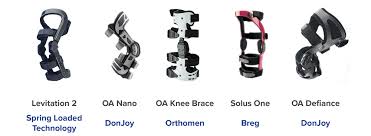


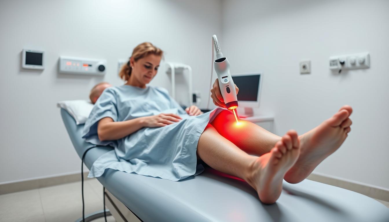
 emanates from a sleek, metallic medical device, casting a soft glow. The middle ground features a spectrum of colored laser beams, each representing a specific therapeutic wavelength, neatly arranged and labeled. The background depicts a minimalist, white laboratory setting, with clean lines and subtle shadows, emphasizing the precision and clinical nature of the technology. The overall scene conveys a sense of technological sophistication and medical innovation, perfectly suited to illustrate the science behind cold laser therapy.")