Image by South_agency from Getty Images Signature from Canva Pro
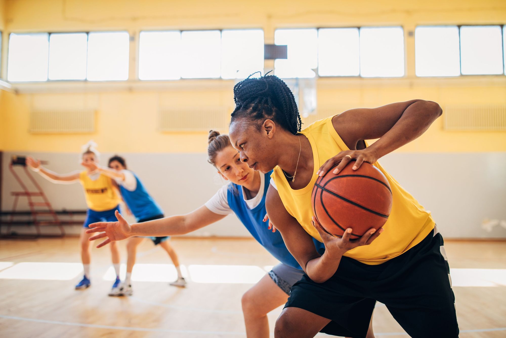
The greatest risk of anterior cruciate ligament injuries or ACL injuries is for people who participate in sports that involve a lot of changing direction, twisting and jumping.1 Females are 2-8 times more likely to injure their ACL compared to males. Read this blog to learn more about the issue that makes women more likely to tear their ACL. Most ACL injuries occur between the ages of 15 and 45.1
In which sports do most ACL injuries occur?
Studies consistently report basketball and football as sports that cause ACL injuries.1,2 However, it is possible to injure the ACL in other sports that require a lot of twisting, cutting (changing direction quickly), jumping and contact sports.1 Female athletes are generally more at risk for ACL injuries.1
Using data from the National Collegiate Athletics Association (NCAA) Injury Surveillance System (ISS), 5,000 ACL injuries were recorded between 1988 and 2004. Based on this data, sports are ranked below based on the number of ACL injuries. The left side of this table shows the number of ACL injuries that have occurred as a percentage compared to all other injuries in a sport. On the right side of this table the number of ACL injuries registered per 1000 matches and training (also called exposures).
ACL injuries as a percentage compared to all injuries3
Injury rate per 1000 exposures3
1. Women’s Basketball (tie)
1. Men’s Spring Football (tie)
1. Women’s gymnastics (tie)
1. Women’s gymnastics (tie)
2. Women’s Lacrosse
2. Women’s football
3. Women’s football
3. Women’s basketball
4. Men’s Spring Football
4. Men’s football
5. Men’s football
5. Men’s football
6. Women’s Softball
6. Men’s wrestling
7. Women’s volleyball
7. Men’s wrestling
8. Women’s hockey
8. Men’s football
9. Men’s wrestling
9. Women’s volleyball
10. Men’s Basketball
10. Women’s Softball
11. Men’s football
11. Women’s hockey
12. Men’s Ice Hockey
12. Men’s Basketball
13. Women’s Ice Hockey
13. Men’s Ice Hockey
14. Men’s Baseball
14. Women’s Ice Hockey
If you have suffered an ACL tear from playing any of the above sports, there is an app, Curovate, to help you with your daily recovery. Curovate offers recovery exercises for ACL injuries and ACL surgery to help you return to the sport you love. Curovate provides your daily physiotherapy exercises, tracks your daily training progress, has in-app chat with a physiotherapist to answer your questions and allows you to measure your knee range of motion with the app. Download Curovate via the links below.
If you need more tailored help during your ACL injury or ACL surgery, check out our Virtual Physiotherapy page to book your 1-on-1 video session with a physiotherapist.
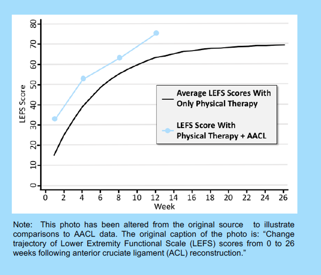
For example, twelve weeks later one of our clients, a 39-year-old CrossFit athlete named Josh, scored a 75/80 on the LEFS. Comparing this result to the graph, he is almost 20% ahead of the average ACL recovery.
His PT was extremely impressed with his progress and offered the following thoughts:
“By combining my physical therapy care with Accelerate ACL protocols at home, my patient experienced an increase in range of motion, increase in strength, and an overall decrease in pain. Three months postoperatively, his daily functional activity scores were 94%. He has set a new standard for our ACL recovery.”
Remember, this is just one example. Other athletes may find that they fall a little below the curve. That’s okay – keep working at it and you’ll get there. Ultimately, we hope you can use this information to start a conversation with your doctor, PT, or trainer about where you are in the ACL recovery process. As is the case with most things in life, try not to be ‘too high’ or ‘too low’ about what the score is telling you.
No matter what happens, your goal for tomorrow is the same. Be consistent, stay disciplined in your approach and work hard.
After anterior cruciate ligament surgery or ACL surgery, you experience loss of muscle mass. You may have spent a few weeks or months building up your muscles after your ACL injury, so you may have assumed that your muscles would be strong and healthy after surgery. However, there are many reasons why you may lose muscle mass after ACL surgery. When you first wake up after your ACL surgery, you may notice that the muscles around your surgical leg already look smaller! The main reasons for this are that your activity level has generally decreased, your nervous system reduces the contraction of the muscles around the knee, and new swelling occurs in your knee joint as a result of the surgery. Read this blog for more information about muscle loss after ACL surgery.
Listen to Andrew Veley, physiotherapist, explain why muscle loss occurs after ACL surgery.
After watching the video above, you may be wondering if there is a way to track your progress as you regain muscle mass after ACL surgery. A simple method called “thigh circumference measurements”, which you can learn more about by reading this blog on how to measure the difference between your two legs by taking a thigh circumference measurement. If you need a video explanation of what thigh circumference measurements are and how they can be used to track your progress, watch this short video.
Here is Andrew’s full blog on muscle loss after ACL surgery.
Read all 6 Andrew’s blogs here!
If you are recovering from an ACL injury or ACL surgery and want to regain your muscle mass, download our Curovate physiotherapy app from the links below. Curovate offers daily video-guided strength exercises, the ability to measure knee range of motion, in-app chat with a physical therapist to answer your questions about surgery and recovery, and educational blogs and webinars.
If you need more tailored help during your ACL recovery, check out our Virtual Physiotherapy page to book your 1-on-1 video session with a physiotherapist.
Document
Other blogs related to ACL surgery and rehabilitation
Do you know how many people walk around with a torn meniscus without knowing they have a tear?
💥About 5% of young people and up to 67% of older people have asymptomatic (non-painful) meniscus tears! 💥
How is that possible?
Depending on the type of injury and how it is treated, the symptoms of a meniscus tear may resolve within days to weeks or months; and you may not even know the tear exists!
The initial injury may not even have been severe enough to warrant a visit to an orthopedist or ordering an X-ray or MRI, especially if the knee only hurt for a few days before the pain went away.
Here’s what you need to know about meniscus injuries so they don’t hinder your favorite activities…
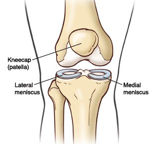
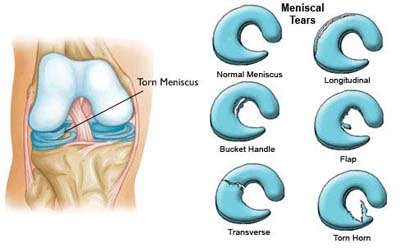
Where is the meniscus?
The meniscus is a “C”-shaped disk made of cartilage that is located in the knee, between the shin bone and the femur. It sits on the weight-bearing surface of the tibia, or tibia, in the joint capsule.
There are two menisci, one on the lateral (outside) of the knee and one on the medial (inside). A healthy meniscus provides space, cushion and lubrication to the joint so that the knee can bend, straighten and bear weight comfortably.
The meniscus is located deep in the knee, along with the anterior cruciate ligament (ACL) and the posterior cruciate ligament (PCL). Due to their deep location, the menisci do not have a very good blood supply, so certain areas are not likely to heal from an injury.
💥 🤯 However, the fact that the meniscus does not heal properly on its own does not mean that a tear always needs to be repaired!
What does the meniscus do and why is it so important?
Shock absorption: The meniscus acts as a shock absorber for shocks that occur during daily activities, such as walking, running, jumping and sports. As we move, force travels through the foot and through the shin bone to the knee, where the meniscus absorbs and disperses that force through the knee, so the impact is spread out and not concentrated in one area.
Stability: The meniscus also acts as a wedge that supports the front and back of your knee, keeping the knee from going beyond a certain range of motion, such as hyperextension, which puts you at risk for other injuries.
How does the meniscus become injured?
The meniscus is most commonly injured during twisting of the knee when the foot is planted on the ground. Rotational stress or “shearing” can cause the femur and tibia to rotate beyond what the meniscus can handle, resulting in a tear in the cartilage.
Meniscal tears are common in athletes who exert a large amount of force on one leg and twist their body in a different direction, such as football 🏈, soccer ⚽️, lacrosse 🥍, tennis 🎾, or basketball 🏀. Field and field athletes often put their feet down to stop and change direction; and in a split second the knee rotates with enough force to cause a meniscus tear.
Snow sports, such as skiing ⛷ and snowboarding 🏂, also carry a high risk of meniscus injuries. The edge of a ski or board can unexpectedly hit the snow, causing a sudden rotation of the leg. The long lever arm can quickly generate a high-speed rotational force, strong enough to cause meniscus injury.
Meniscal tears occur in people of all ages, including athletes and non-athletes. In middle-aged and older adults, meniscus tears can occur due to wear and tear on the knee joint. As the body ages, degeneration of the cartilage tissue occurs as the joints are exposed to force for years.
Osteoarthritis, a condition in which cartilage wears away over time, is common in older adults and is often accompanied by a torn meniscus. A meniscus tear earlier in life can also make the knee more susceptible to earlier-onset osteoarthritis. That’s why it’s so important to learn the right moves and exercises to protect your knees!
I have a meniscus tear. What now?
Depending on the type of meniscus tear, you may experience symptoms such as pain, swelling, locking, or locking of the knee. Although a meniscal injury cannot be detected on X-rays, it can be diagnosed through a careful examination or an MRI.
Most meniscal tears are initially treated conservatively (without surgery); although an orthopedic doctor may recommend surgery if symptoms persist and daily activities are limited.
Meniscectomy: The most common surgery for a meniscus tear is called a menisectomy, in which the surgeon removes the torn piece of cartilage and cleans up the surrounding ragged tissue. Although it is a surgical procedure, recovery after a meniscectomy is relatively short. Most people can resume daily activities within a few weeks and exercise within a few months.
It is important to add that everyone responds to surgery differently; and a ‘minor’ surgery for one person may be a more stressful experience and longer rehabilitation for someone else.
Meniscus repair: An alternative surgery is a meniscus repair, which is performed when the orthopedic surgeon believes the torn cartilage structure can be sewn together, saving cartilage to prevent osteoarthritis in the future. This procedure is typically performed more in younger people to preserve the cartilage rather than remove it. Recovery from a meniscus repair takes longer than a menisectomy and involves close monitoring of range of motion, weight bearing, and activity progression during rehabilitation.
Non-surgical treatment A meniscus tear is very common and results in great results in most cases! A diagnosis of a meniscus tear does NOT mean that surgery is always necessary!
Although a meniscus tear will not heal itself, you can strengthen the knee and improve movement patterns to disperse forces and reduce stress at the site of the tear, which will help resolve symptoms.
💪 With the right exercises and learning to move properly, most meniscus tears can be treated conservatively WITHOUT surgical intervention.
💥It’s important to remember that you are not your MRI result; and a meniscus tear should not determine the rest of your life.
How can you treat a meniscus injury without surgery?
A meniscus injury must be properly guided through the healing phases. Pain and inflammation are likely to be early symptoms, while stiffness and weakness may be the main complaints after a few weeks. Dealing with injuries with “PEACE & LOVE” is a good reference for dealing with acute injuries.
To return to normal activities, follow the guidance of your physical therapist, who will develop a care plan for strengthening, balance training, improving mobility, and returning to exercise.
👉Doing the right exercises with good form and consistency will significantly increase your ability to recover from a meniscus tear and prevent future injuries.
If you’re ready to start strengthening at home, ACL Strong has courses that include all the necessary components of exercise and movement to train your body and brain to protect your knees now and for the long term.
Whether you have a torn meniscus or not, doing the right exercises the right way will keep your knees strong and stable, so you’ll be less prone to further injury and better equipped to maintain an active lifestyle for as long as you want. to lead!
A few weeks ago I worked virtually with a client who was 10 weeks post-surgery. We had started working together around week 3 and so far he had absolutely dominated the recovery process.
In addition to his physical therapy appointments, he would meet with me virtually once a week to determine the best plan of attack for his workouts with the Neubie… and then he would get after it. By incorporating our recommended workouts with the Neubie 3x per day into his routine, he regained full strength and range of motion before 8 weeks post-surgery.
But around week 10, they started increasing the load at his physical therapy appointments. In particular, they started trying to increase the weight of his exercise on the knee extension machine, which led to a bit of pain in the front of the knee. Each day he returned to do the knee extensions, the pain in the front of his knee increased.
As I mentioned earlier, this client met with me once a week, and by the time we met again, he had reached a point where the pain continued even in his daily activities. He no longer had the ability to fully extend the knee without pain.
Despite doing everything right and botching the first eight weeks of the recovery process, he experienced a setback in week 10.
Setbacks can be difficult, but with the right strategies you can overcome them quickly and emerge stronger.
It is usually normal for your knee to feel numb and tingly after ACL surgery. This is because some small nerves are cut during the surgical procedure. Don’t worry, we’re not talking about the really important nerves that allow you to bend and straighten your knee, but the nerves that allow you to feel things around your knee. This video below explains all the reasons why your knee may feel numb and tingly after ACL surgery. For more information, read this blog about 5 tips to help you recover after ACL surgery.
Lauren Youssef, a physiotherapy student at the University of Toronto, explains why your knee may feel numb and tingly after ACL surgery.
Read Lauren’s blog: Why is my knee numb and tingling after ACL surgery? | Curovat
There are also 5 great blogs written by Lauren on our site where you can learn more about surgery and injury recovery.
If you have had an ACL injury or surgery, download Curovate from the links below. Curovate is a physical therapy app that offers daily video-guided exercises, progress tracking, in-app chat with a physical therapist to answer your questions about surgery or injury, and the ability to measure your knee’s range of motion using just your phone.
If you need more tailored help during your surgery or recovery from your injury, check out our Virtual Physiotherapy page to book your 1-on-1 video session with a physiotherapist.
Other blogs related to ACL injuries:
References
1. Horteur, C., Cavalié, G., Gaulin, B., Cohen Bacry, M., Morin, V., Cavaignac, E., & Pailhé, R. (2020). Saphenous nerve damage after anterior cruciate ligament reconstruction: Decreased area of numbness after ligamentoplasty using the quadriceps tendon compared to the hamstring tendon. The Knee, 27(4), 1151–1157.
2. Inderhaug, E., Strand, T., and Solheim, E. (2015). The impact of sensory deficits after hamstring autograft harvesting for ACL reconstruction. Knee surgery, sports traumatology, arthroscopy: Official Journal of the ESSKA, 23(4), 1060–1064.
3. Kjaergaard, J., Faunø, L. Z., & Faunø, P. (2008). Loss of sensation after ACL reconstruction with hamstring graft. International Journal of Sports Medicine, 29(6), 507–511.
4. Nerve Blocks for Surgery – Yale Medicine. (2020, July 02). Retrieved from https://www.yalemedicine.org/conditions/nerve-blocks-for-surgery
5. Ochiai, S., Hagino, T., Senga, S., Yamashita, T., Oda, K., & Haro, H. (2017). Damage to the infrapatellar branch of the saphenous nerve in anterior cruciate ligament reconstruction using vertical skin incision for hamstring harvesting: risk factors and influence on treatment outcome. Journal of Orthopedic Surgery and Research, 12(1), 101.
6. Sanders, B., Rolf, R., McClelland, W., and Xerogeanes, J. (2007). Prevalence of saphenous nerve damage after autogenous hamstring harvest: an anatomical and clinical study of tailor branch injury. Arthroscopy: The Journal of Arthroscopic and Related Surgery: Official Publication of the Arthroscopy Association of North America and the International Arthroscopy Association, 23(9), 956–963.
7. Wang, H. D., Zhang, H., Wang, T. R., Zhang, W. F., Wang, F. S., & Zhang, Y. Z. (2018). Comparison of clinical outcomes after anterior cruciate ligament reconstruction with hamstring tendon autograft versus soft tissue allograft: a meta-analysis of randomized controlled trials. International Journal of Surgery (London, England), 56, 174–183.
If you’re working your way through the ACL prehab process, you may have heard a few ACL prehab goals to aim for before your doctor’s surgery. These goals often include goals such as:
Perform 20 straight leg movements without deceleration
Minimal effusion/swelling
Achieving these goals WILL put you in a much better position than if you avoided prehab altogether. However, much more can be accomplished during the prehab phase. The overarching goal we recommend is to get as close to 100% as possible before surgery (achieving the goals listed above is certainly not an indication that you are 100%). Doing so will increase your chances of optimal posterior recovery, increase your confidence toward surgery (which also plays a role in recovery), and potentially allow you to have a conversation with your doctor about ask whether or not you need surgery. (another blog post will go into that in more detail).
The two most common areas where a surgeon can take a graft to replace your ACL are the patellar tendon and the hamstring tendon. If this is new information to you, please refer to our previous blog on where the most common areas for an ACL graft are. Also keep in mind that the patellar tendon and hamstring tendon are the most common but not the only sources for an ACL graft.
As you read the differences below, it is important to remember that your surgeon will choose the graft site for your ACL surgery. In some cases, due to a previous injury or surgery, one or more of the graft sites may not be feasible. The good news is that our ACL rehabilitation app, Curovate, has a protocol for hamstring tendon, patellar tendon, quadriceps tendon, allograft, and non-surgical ACL rehabilitation.
Overall, the patellar tendon is the more stable ACL graft and has a lower re-injury rate compared to the hamstring tendon graft. Read the advantages and disadvantages of each graft site in the table below.
Hamstring Graft Surgery (HT)
Patellar tendon surgery, also called bone-patellar-bone surgery (BTB).
1. The surgery takes longer and the tourniquette, a device used during surgery to restrict blood flow in the leg, stays on longer2
1. The knee is stiffer after surgery2
2. The knee moves more freely forward and backward when tested with a KT-1000 Arthrometer machine2a device that measures the forward and backward movement of your lower leg compared to your thigh.
2. Increased knee pain when kneeling on the ground3
3. During a period of 15 years after surgery, more reinjury was reported.3
3. Over a period of 15 years after surgery, increased stiffness and difficulty straightening the knee were reported3.
4. The need to perform another surgery to address problems with the first surgery is called revision surgery. The revision rate for hamstring operations increases by 0.65% within 1 year and by 4.45% within 5 years4.
4. The 1-year revision rate is 0.16% and the 5-year revision rate is 3.03% for patellar tendon graft surgery4.
5. Hamstring muscle strength reduced by 15%1. Muscle strength is the maximum force you can exert in the shortest possible time.
6. Muscle strength decreases by 11% when stretching the knee2. Muscle strength is the amount of force a muscle can produce during maximum effort.
7. There is increased instability in the knee when force is applied to it2. Instability is unwanted movement in a joint.
Conclusion
Based on the research reviewed in this blog, the patellar tendon has a lower re-injury rate and is more stable at 1 and 5 years after ACL surgery. However, the location of the graft should be discussed with your surgeon and ultimately the surgeon will decide the most suitable area for the ACL graft.
If you have had ACL surgery and want clear daily, weekly and monthly guidance for your knee extension and daily exercises for your recovery, try our Cuorvate app. Curovate offers video-guided daily exercises, weekly range of motion goals and exercises, progress tracking, the ability to measure your knee and hip range of motion, and in-app chat with a physical therapist.
If you need more tailored help during your ACL recovery, check out our Virtual Physiotherapy page to book your 1-on-1 video session with a physiotherapist.
Other recommended blogs
References
Ageberg, E., Roos, HP, Silbernagel, KG, Thomeé, R., Roos, EM, akademin, S., Gothenburg University. (2009). Knee extension and flexion muscle strength after anterior cruciate ligament reconstruction with patellar tendon graft or hamstring tendon graft: a cross-sectional comparison 3 years after surgery. Knee Surgery, Sports Traumatology, Arthroscopy, 17(2), 162-169.
Beynnon, B.D., Johnson, R.J., Fleming, B.C., Kannus, P., Kaplan, M., Samani, J., and Per Renstrom. (2002). Anterior cruciate ligament replacement: comparison of bone-patellar tendon-bone grafts with two-strand hamstring grafts A prospective, randomized study. The Journal of Bone & Joint Surgery, 84(9), 1503-1513.
Leys, T., Salmon, L., Waller, A., Linklater, J., and Pinczewski, L. (2012). Clinical outcomes and risk factors for re-injury 15 years after anterior cruciate ligament reconstruction: a prospective study of hamstring and patellar tendon grafts. The American Journal of Sports Medicine, 40(3), 595-605.
Rahr-Wagner, L., Thillemann, T.M., Pedersen, A.B., and Lind, M. (2014). Comparison of hamstring tendon and patellar tendon grafts in anterior cruciate ligament reconstruction in a nationwide population-based cohort study: results from the Danish Knee Ligament Reconstruction Register. The American Journal of Sports Medicine, 42(2), 278-284.
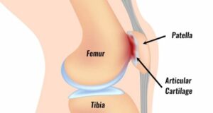
That dull, aching, throbbing, sometimes sharp pain around the knee or kneecap that comes and goes based on activity may be a condition known as Patellofemoral Pain Syndrome. Sometimes associated with cracking and popping, patellofemoral pain can range from not so bad to disabling discomfort causing people of all ages to avoid activities such as walking, jogging or participating in a sport.
What causes patellofemoral pain?
With patellofemoral pain syndrome, discomfort in or around the front of the knee is often due to irritation of the cartilage that lines the underside of the kneecap in the joint. Stress on the knee can cause the cartilage to become inflamed, which can ultimately lead to thinning and fraying of the tissue over time. In some cases, athletes begin to notice symptoms after years of competition and training, even without immediate injury. In other cases, people only report this pain days, weeks, or months after an increase or change in activity. If the causes of patellofemoral pain syndrome are not treated, they can eventually lead to tissue degeneration in the knee, such as osteoarthritis.
The patella, or kneecap, is a floating bone connected to the femur and shin bone by tendons and ligaments. The bottom, or back part, of the patella is covered with a layer of cartilage, which helps it slide smoothly over the thigh bone when you bend and straighten your knee. There should be sufficient joint space between the patella and the underlying femur, although this joint space sometimes narrows, increasing the chance of contact or friction between the bony surfaces.
A variety of factors including anatomy, soft tissue mobility, biomechanics (movement quality), and physical activity may contribute to a higher risk of patellofemoral syndrome. For example…
The anatomical resting position of the patella may be slightly higher than normal, also called ‘patella alta’, which can make the patella more sensitive to gradual wear.
A patella that is hypermobile or has too much mobility can bump and rub against the underlying bone, irritating the cartilage and causing pain. Conversely, a patella that is hypomobile or limited in mobility may become painful due to too much force being placed on a particular part of the patella.
In terms of movement, the patella moves over the femur, but the femur also moves under the patella. The biomechanics of the entire leg must be addressed to understand what is happening at the knee. The hip and foot play an important role in what the knee experiences. For example, someone with overpronated feet or weakness in the hip muscles may experience knee pain due to altered mechanics and resulting pressure on the cartilage lining of the patella. Abnormal biomechanics can be corrected through exercises prescribed by a physical therapist.
Physical activity also contributes to patellofemoral pain. The intensity or load of the activity can place more force on the knees than the body can safely tolerate. Activity modification may be necessary to control symptoms. For example, exercises such as running can be avoided for a while to calm symptoms such as swelling and pain.
If the contributing factors are not addressed, the friction and friction between the surfaces can worsen, leading to thinning and fraying of the cartilage, wear and tear of the joint, and ultimately bone-on-bone osteoarthritis.
What is the best treatment for patellofemoral pain syndrome?
The recommended treatment for patellofemoral pain generally focuses on strengthening the muscles of the leg, from the hip and trunk to the foot, along with maintaining or increasing flexibility to reduce stress on the patella. Normalizing strength, flexibility and biomechanics through the limbs will help reduce pressure on specific areas of the cartilage that have become irritated and painful, allowing activity and exercise to be comfortable again.
In addition to strengthening, ice and anti-inflammatory medications, such as ibuprofen, are often recommended to reduce swelling and relieve pain during the acute phase. Reducing painful activities such as climbing stairs can be helpful in recovering from patellofemoral pain syndrome. Although you limit painful exercises, you can still be active by incorporating alternative, non-painful exercises that will strengthen the knee around the knee and reduce irritation to the cartilage and other structures that support the patella. For example, a runner with patellofemoral pain can substitute swimming a few times a week to stay active without aggravating the knee.
Have I waited too long? Is it too late for me?
No matter how long you have been suffering from knee pain, it is never too late to build strength. Focusing on correcting your mechanics during exercise and exercise can help reduce your pain over time. Strengthening the muscles around the knees allows you to move with better control and precision, which also improves performance. Increasing body control provides more stability in the patellofemoral joint, which can take pressure off the patella and reduce the risk of further cartilage breakdown.
Can it be cured?
Patellofemoral pain syndrome is one of the most common causes of knee pain and one of the most common conditions resolved with physical therapy. A supervised exercise program to address the underlying causes is the best treatment strategy to relieve patellofemoral pain.
Our courses at ACL Strong are designed and programmed by physiotherapists to give you complete confidence that the exercises you perform are both safe and effective.
Many of our members experience an unexpected benefit when they enroll in an ACL Strong course… their patellofemoral pain improved because they trained smarter and reduced the strain on their knees.
“I can’t believe I’m skiing without pain for the first time in years!”
“The knee pain I used to have while playing football is no longer there!”
“One of the biggest benefits we have seen in our athletes through ACL Strong is that it helps them resolve old injuries that have been bothering them.”
When you take part in the ACL Strong Snow Course or Classic Course, you will learn how to care for your knees in the long term, so you can be as active or competitive as you want, and for as long as you want.
You will notice that your range of motion is significantly limited immediately after an ACL injury. In most cases, simple range-of-motion exercises are recommended early on with the aim of restoring normal range of motion. You may hear terms like “heel slides” and “quad sets,” two very simple, low-load exercises designed to help you regain flexion (ability to bend the knee) and extension (ability to straighten the knee). to get.
When performing these types of exercises, keep in mind that the goal of prehab is to reduce the guarding mechanisms the body has developed as a result of the injury. Try to only work within a range of motion that does not increase pain levels above 2/10 and breathe well. If you are in a lot of pain and holding your breath, you may find that these exercises are not as effective.
Immediately after an ACL injury occurs, the brain changes the way it uses the muscles in the affected area. Over a short period of time, these muscles begin to lose strength. When muscles lose strength, they are unable to create and absorb force as efficiently, putting joints, tendons and ligaments at greater risk for injury. Therefore, strengthening exercises are necessary to build strength in the affected muscles.
In general, strengthening exercises involve moving a resistance load through a range of motion. You can use a number of items for resistance, including your own body weight (think a simple squat pattern), resistance band, a set of weights, or even the preferred form of resistance for Accelerate ACL, electricity.