Have you ever wondered why certain physical discomforts seem to intensify when the sun goes down? For many women, the later stages of pregnancy bring unexpected challenges that peak during nighttime hours. While the body adapts to support new life, shifts in posture, hormonal activity, and added pressure on joints can create unique hurdles.
During the gestational period, weight distribution changes dramatically. This alters how muscles and ligaments support the frame. A key hormone called relaxin increases flexibility in pelvic areas but may also reduce stability in other joints. Combined with fluid retention and gravity’s effects after long days, these factors often amplify sensations of stiffness or tenderness.
Evening hours tend to highlight these adjustments. Lying down removes the subtle support of daytime movement, letting accumulated stress on the knees become more noticeable. Understanding these connections helps explain why restful nights sometimes feel elusive – and how targeted strategies can make a difference.
Key Takeaways
- Hormonal shifts and weight redistribution impact joint stability
- Relaxin increases ligament flexibility but may reduce joint support
- Postural changes affect pressure points during rest
- Daytime activity patterns influence nighttime discomfort
- Gravity and fluid retention contribute to evening symptoms
Introduction to Knee Pain During Pregnancy
The journey of pregnancy involves remarkable bodily adjustments that affect daily comfort. As our center of gravity shifts, muscles and ligaments work harder to maintain balance. This creates new pressures on areas like the hips, pelvis, and joints.
Our Body Changes and Weight Shifts
Extra pounds from weight gain place direct strain on load-bearing areas. Research shows the average person gains 25-35 pounds during gestation. This redistributed mass alters posture, often tilting the pelvis forward. Knees then absorb more pressure during standing or walking.
The Impact of Hormones on Our Joints
Hormones like relaxin loosen connective tissues to prepare for childbirth. While essential, this increased flexibility reduces stability in areas beyond the pelvis. Ankles and wrists may feel less supported, while spinal alignment shifts subtly.
| Low-Impact Exercise | Benefits | Frequency |
|---|---|---|
| Swimming | Reduces joint pressure | 3x/week |
| Prenatal Yoga | Improves balance | 2x/week |
| Stationary Cycling | Strengthens legs | 4x/week |
Regular exercise helps counteract these effects. Water aerobics or chair-based stretches allow movement without overloading sensitive areas. Always consult healthcare providers before starting new routines.
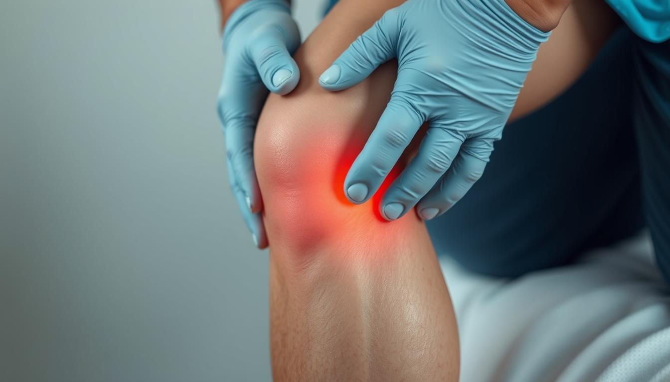
Knee pain only at night during pregnancy
As daylight fades, many expecting mothers notice their bodies respond differently to the day’s accumulated stresses. While daytime activity distracts from minor aches, stillness allows these sensations to surface. This shift highlights how posture and fluid dynamics evolve as the body prepares for new life.
Understanding the Unique Nighttime Experience
When we lie down, gravitational forces redistribute fluids retained during the day. This can lead to swelling around joints, making them feel stiffer. Reduced movement also limits blood flow, amplifying sensations that daytime motion might mask.

How Gravity and Body Changes Affect Our Joints
Weight gain across trimesters shifts our center of gravity, altering how pressure distributes through the legs. By the third trimester, the uterus’s expansion pushes downward, intensifying strain on lower-body structures. Lying flat may inadvertently compress sensitive areas, especially without proper cushioning.
Consulting a doctor becomes crucial if discomfort disrupts sleep patterns. They can assess whether adjustments to sleeping positions or supportive gear might help. Simple changes—like using pillows between the legs—often ease tension by aligning hips and reducing rotational stress.
Though these changes support a growing baby, they demand careful attention to our physical limits. Later sections will explore practical methods to manage these shifts while maintaining comfort through all stages of gestation.
Preventative Measures and Daily Habits for Knee Health
Simple choices in our daily routine can shape how our bodies adapt to pregnancy’s physical demands. Strategic adjustments to footwear and posture create lasting impacts on joint stability throughout gestation periods.
Foundation First: Shoes That Support
Supportive footwear acts as a shock absorber against gravity’s pull. Look for shoes with arch support and cushioning to distribute weight evenly. A study of 150 expectant mothers found those wearing orthopedic inserts reported 40% less joint discomfort during daily activities.
| Feature | Benefit | Ideal Trimester |
|---|---|---|
| Wide Toe Box | Reduces swelling pressure | First to Third |
| Low Heel (1-2″) | Improves spinal alignment | Second & Third |
| Adjustable Straps | Accommodates fluid retention | Third |
Posture Perfected
Slouching shifts up to 50% more weight onto lower-body joints. When sitting, keep feet flat and use a lumbar pillow. Standing? Engage core muscles and avoid locking knees. These micro-adjustments help relieve strain accumulated over time.
Morning stretches and evening elevation routines make significant differences. Try placing a pillow under ankles while resting to improve circulation. Consistency with these habits supports mobility during joint pain pregnancy challenges while building resilience for postpartum recovery.
Managing Activity and Exercise Through Trimesters
As our bodies evolve during gestation, movement patterns require thoughtful adaptation. Tailoring physical activity to each phase helps maintain strength while respecting biological changes. Strategic adjustments protect vulnerable areas like ligaments and hips as relaxin levels rise.
First and Second Trimester Adjustments
Early stages focus on building foundational strength. Low-impact exercises like swimming or prenatal yoga improve stability. “Focus on alignment first – proper posture reduces strain before weight increases become significant,” advises a maternal health specialist.
Gentle massage techniques enhance circulation around joints. This helps counteract stiffness caused by hormonal shifts. Many women find foam rolling quads and calves eases tension in connected ligaments.
Third Trimester Activity Modifications
Final months demand reduced intensity. Shorter walks replace jogging, while seated resistance bands protect overstretched ligaments. Avoid movements requiring sudden direction changes to prevent injury.
Supportive gear becomes crucial. Maternity belts redistribute weight from hips, while compression sleeves stabilize knees. Always prioritize rest days – recovery periods let relaxin-softened tissues rebuild resilience.
Consulting healthcare providers ensures routines match individual needs. Custom plans account for ligament flexibility, weight distribution, and energy levels. This personalized approach helps women stay active safely through all gestational phases.
Expert Guidance and Physical Therapy Options
What separates temporary discomfort from signals needing expert attention? Recognizing when to seek professional help ensures both safety and comfort during this transformative period. Specialized care teams assess how hormonal shifts and physical changes interact, offering strategies tailored to individual needs.
When to Consult Our Healthcare Provider
Persistent stiffness that limits walking or daily activities warrants a discussion with a doctor specializing in prenatal care. Sudden swelling, redness, or warmth around joints could indicate underlying risks requiring immediate evaluation. Professionals analyze posture adjustments and recommend support tools like braces or compression sleeves.
Benefits of Tailored Physical Therapy
Customized therapy plans address hormone-driven ligament flexibility while strengthening muscles around the center of gravity. A 2023 study showed 68% of participants reduced discomfort through targeted exercises improving hip stability. Therapists also teach safe movement patterns for activities like lifting or climbing stairs.
Continuous monitoring ensures alignment adapts as the body changes. Open communication with providers helps balance rest and activity. For deeper insights, explore this comprehensive prenatal health guide detailing collaborative care approaches.
Nighttime Strategies for Restful Relief and Sleep
Finding comfort after sunset becomes a priority as our bodies seek recovery from daily demands. Strategic adjustments to sleep positions and supportive routines can transform restless nights into rejuvenating rest. These methods address both immediate discomfort and long-term musculoskeletal balance.
Optimizing Sleep Position and Support
Side-lying positions with proper cushioning protect bones and joints. Placing a firm pillow between the legs maintains hip alignment, reducing rotational stress on the lower body. Adding support under the belly prevents spinal twisting that contributes to back pain.
- Elevate legs slightly to improve circulation
- Use wedge pillows to minimize pressure points
- Rotate sides periodically to prevent stiffness
Safe Home Remedies for Reducing Discomfort
Warm compresses applied before bed may help relax tense muscles around joints. Epsom salt baths provide magnesium absorption through the skin, which studies suggest aids muscle recovery. Gentle stretching routines approved by physical therapy professionals enhance flexibility without overexertion.
Always discuss new remedies with healthcare providers. What works for one person might not suit another’s unique needs. Combining these approaches often yields better results than single solutions.
Conclusion
Navigating physical changes while expecting requires awareness and adaptable strategies. We’ve explored how hormonal shifts, managing extra weight, and postural adaptations impact comfort levels. These changes body dynamics create unique challenges that often peak during rest periods.
Strategic habits like supportive footwear and alignment-focused movement help counterbalance shifts in center gravity. Combining daytime prevention with nighttime relief methods – from elevation techniques to therapeutic heat – creates cumulative benefits. Consulting specialists ensures personalized solutions for individual needs.
By addressing changes body effects holistically, we reduce strain caused by extra weight distribution. Small adjustments to daily routines and sleep setups help stabilize the center gravity while promoting restorative rest. Every proactive choice supports both immediate comfort and long-term mobility.
We encourage expecting mothers to blend these approaches while maintaining open communication with healthcare teams. With mindful attention to physical transformations, improved comfort becomes an achievable goal throughout this transformative journey.
FAQ
Why does joint discomfort worsen at night while expecting?
Fluid retention increases after prolonged standing or sitting, adding pressure to joints. Reduced movement during rest periods also allows stiffness to set in, amplifying sensations once we lie down.
How do hormones contribute to weakened joints in pregnancy?
Hormones like relaxin loosen ligaments to prepare our bodies for birth. This natural process can reduce stability in areas like hips and knees, making them more vulnerable to strain from weight shifts.
Are certain sleeping positions better for reducing pressure?
Side-lying with a pillow between legs aligns hips and spine, easing tension on joints. Elevating legs slightly with cushions improves circulation and minimizes swelling that contributes to nighttime aches.
Can footwear choices really impact joint health during pregnancy?
Yes! Supportive shoes with arch cushioning distribute weight evenly, reducing stress on knees. Avoid flat soles or high heels, which alter posture and increase strain on already taxed muscles.
What low-impact exercises are safest as our bodies change?
Swimming, prenatal yoga, and stationary cycling build strength without jarring movements. Focus on controlled motions that maintain flexibility while respecting our shifting center of gravity.
When should we seek professional guidance for persistent pain?
Contact your OB-GYN or physical therapist if discomfort interferes with daily tasks, includes swelling/redness, or feels sharp. Early intervention prevents minor issues from escalating.
Are warm compresses safe for managing soreness at home?
Generally yes, but limit heat sessions to 15 minutes and avoid abdominal areas. Pair gentle massage with approved topical creams (like arnica) for targeted relief before bedtime routines.
How does physical therapy adapt to different trimesters?
Therapists modify techniques as pregnancy progresses—focusing on pelvic stability early on, then introducing seated stretches or aquatic therapy in later stages to accommodate our growing bodies safely.