Category: Knee injury

  • Who participates in youth sports?

    Who participates in youth sports?


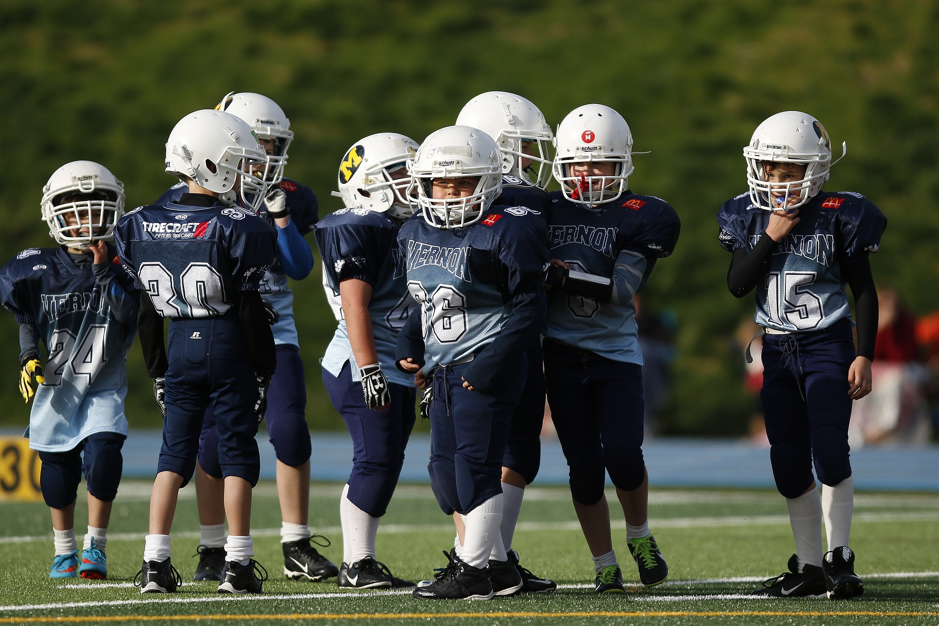
    Organized sports participation among children aged 6–17 years: United States, 2020

    Black LI, Terlizzi EP, Vahratian A. National Center for Health Statistics (US). Publication date: 08/11/2022. Series: data overview; No. 441

    https://stacks.cdc.gov/view/cdc/119026

    Take home message

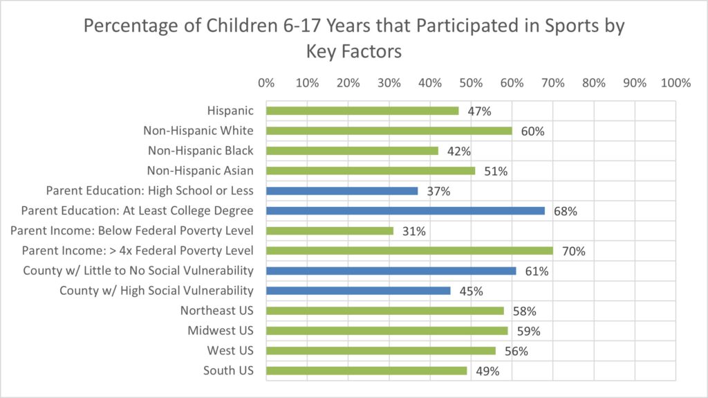
    Sports participation was lower among racial and ethnic minority children, children whose parents had lower education or income, children living in counties with greater social vulnerability, and children living in the southern United States.

    Background

    Youth participation in sports is associated with better physical fitness and mental health. Understanding the differences in sports participation can help inform strategies to promote active lifestyles among children and adolescents, which positively impacts their physical and mental health.

    Study aim

    Black and colleagues completed an analysis of data from the 2020 National Health Interview Survey to describe disparities in sports participation in the United States.

    Methods

    The authors analyzed data from the 2020 National Health Interview Survey. This survey is a nationally representative household survey conducted throughout the year. Parents reported whether their child participated in a sports team or club or took sports lessons at school or in the community in the previous 12 months. The authors then collected data on household income as a percentage of the federal poverty level, race and Hispanic origin, geographic region (Northeast US, Midwest US, Southern US, or Western US), social vulnerability of a county, and urbanicity ( urban region). -national classification) of a province.

    Results

    About half (54%) of children aged 6 to 17 have played sports in the past twelve months (boys: 56%; girls: 52%). The authors reported: “Participation levels were lower among children from racial and ethnic minority groups, children whose parents had lower levels of education and family income, children living in counties with greater social vulnerability, and children living in the South.”

    Black Graph

    Viewpoints

    The results of this letter provide a surprising snapshot of sports participation in the United States. While it is established that early sports participation can have a positive impact on an individual, significant differences still exist. These findings indicate that sports participation is strongly associated with socioeconomic status. Ultimately, a higher socio-economic status can ensure children’s access to sports. This is particularly reflected in the fact that lower parental education, lower parental income and lower geographical area of ​​social vulnerability are all associated with lower participation. These findings complement data from the Athletic Training Locations and Services Database, which shows that these communities also have less access to athletic training services.

    This data provided a unique snapshot of 2020, when many sports shut down in the spring due to the COVID-19 pandemic. The survey asked about sports participation over the past year, so it is unclear whether the pandemic could influence these results. Furthermore, not every community is recovering from the pandemic in the same way. The authors acknowledge that it will be essential to replicate these analyzes with data from the 2022 survey to better understand patterns of sports participation after many of the restrictions associated with the pandemic are relaxed/removed.

    Clinical implications

    Sports medicine professionals should advocate for increased access to sports through local recreation organizations, schools, and other organizations. In regions with high social vulnerability, we should encourage policymakers that investments in youth sports and physical activity can help improve community health.

    Questions for discussion

    What can we do as doctors to positively influence participation in team sports?

    Written by: Kyle Harris
    Review by: Jeffrey Driban

    related posts

    Asian Pacific Society of Cardiology Consensus Recommendations for Pre-Participation Screening in Young Competitive Athletes
    A lasting impression: youth sports participation and healthy habits as adults
    Previous participation in collision sports is associated with reduced quality of life
    Lower socioeconomic status is associated with less access to athletic training services

    9 EBP CEU courses

    Source link

  • Tennis Elbow: What It Is and How to Treat It |  Foothills Physical Therapy and Sports Medicine

    Tennis Elbow: What It Is and How to Treat It | Foothills Physical Therapy and Sports Medicine


    by Adam Halseth PT, DPT, SCS | Northeast Scottsdale

    Tennis elbow or golfer’s elbow is an uncommon condition. It just has an impact about 1 to 3 percent of adults every year. But your chances of developing it increase as you get older. Although the name suggests it is a condition exclusive to these sports, it is possible to develop tennis or golfer’s elbow even if you have never played either game in your life.

    Below you will find important information about this painful condition, including why it occurs, how to determine if you have the condition, and what treatment options are available.

    Young woman experiencing pain while exercising in a gym.

    Understanding tennis/golfer’s elbow

    Tennis/golfer’s elbow is one of the most common causes of elbow pain. It is often persistent and painful enough to warrant physical therapy and/or a doctor’s visit. This condition occurs when the tendons that connect your forearm to the outside of the elbow bone become swollen and inflamed.

    If this happens, it can lead to microscopic tears in the muscles and tendons. These tears can cause a lot of pain, even though they are very small.

    Usually people don’t experience these symptoms unless they do a lot of repetitive movements. While the general population is not highly likely to experience this pain, as many as half of all players will experience it at some point in their lives.

    What causes tennis/golfer’s elbow?

    If you’re wondering what the causes are, the answer is: a lot of things! As mentioned, tennis/golfer’s elbow usually occurs as a result of repetitive movements of the elbow joint. Besides tennis and golf, some other activities that can lead to elbow pain and discomfort include:

    • Screens
    • Squash
    • Weightlifting
    • Racketball
    • Rake
    • Typing
    • To paint
    • Carpentry
    • To knit
    • Gardening
    • Swimming

    Did some of the items in the list above surprise you? Fortunately, there are things you can do to reduce the chance of developing elbow pain or minimize symptoms if you already have it. Keep reading to learn more.

    Man with elbow pain while playing tennis

    5 Symptoms of tennis/golfer’s elbow

    Are you experiencing elbow pain that makes you wonder if you have tennis/golfer’s elbow or something else? Although a doctor or therapist should be consulted to make the most accurate diagnosis, these are five common symptoms:

    1. Discomfort when you lift something
    2. Weakness in your hand or forearm when making a fist or grabbing something
    3. Recurring pain just below the bend in your elbow (on the outside of your forearm)
    4. Pain that radiates from the elbow to the wrist
    5. Discomfort when turning the forearm (for example, when opening a jar or a door)

    As you can see, the primary symptom is pain in the elbow area. Conditions that mimic tennis elbow include:

    • Radiocapitellar arthritis
    • Osteochondritis dissecans
    • Intra-articular plica
    • Rotational instability

    A visit to your PT can help rule out these conditions if you are unsure whether you have tennis/golfer’s elbow or another condition.

    FH Shin Splits Blog 1

    Prevention techniques

    The best way to prevent developing this condition is to avoid doing repetitive movements too often. If you play a sport or other activity that requires repetitive use of your arm and muscles, make sure you take regular breaks. Stretch the muscles of your arms during your breaks.

    It is also important to warm up the muscles of your arms before doing any physical activity that requires the use of your elbows and/or arms. When the muscles are warm, they can stretch and contract more easily without causing injury.

    Once you’ve finished a sporting event or workout and stretched your arm muscles, consider applying ice to your elbows if you feel heat or inflammation in those areas. If you already have symptoms of tennis elbow despite your best efforts, consider getting physical therapy.

    Physiotherapy treatment options

    Physical therapy can help improve the flexibility and strength of your forearm muscles, making you less likely to develop tennis elbow. Physical therapy can also facilitate healing and reduce pain by stimulating blood flow to the affected tendons and muscles. Blood contains oxygen, which the muscles need to heal and function optimally.

    Six common physical therapy treatment options recommended for tennis elbow:

    1. Muscle stimulation
    2. Ultrasound
    3. Ice massage
    4. Braces and tape to support the affected area
    5. Specialized stretches and exercises
    6. Nonsteroidal anti-inflammatory drugs

    During your recovery from tennis/golfer’s elbow, it is important not to rush things. If you push your body before it is ready and your condition has healed enough, you can set yourself back. Before returning to your previous activity level, make sure that you can grasp objects without pain and that your elbow no longer looks or feels swollen. When you can bend and move your affected elbow without difficulty or discomfort, you can resume your normal activities.

    Get your quality of life back

    If you are ready to get your life back and fully recover from the symptoms of your tennis/golfer’s elbow, we are here to help. Contact Foothills Sports Medicine Physical Therapy today request your appointment.

    Source link

  • Corin Group Achieves 510(k) Clearance for Apollo™ Platform: Revolutionary Robot-Assisted Total Knee Arthroplasty

    Corin Group Achieves 510(k) Clearance for Apollo™ Platform: Revolutionary Robot-Assisted Total Knee Arthroplasty

    CIRENCESTER, UNITED KINGDOM, November 1, 2023 / EINPresswire.com / — Corin Group, an innovator in orthopedic procedures, is proud to announce 510(k) clearance for its Apollo™ Robotic-Assisted Surgical platform and ApolloKnee™ software application. This marks a major industry milestone in Corin’s continued commitment to helping surgeons increase satisfaction and successful outcomes for their patients when performing total joint replacement surgeries.

    Dr. Jim Pierrepont, Global Franchise Lead at Corin Group said: “The Apollo™ platform is the result of the combination of objective planning, precision implementation and continuous learning in the pursuit of forgotten total joint replacement. The Apollo™ platform goes beyond personalized knee alignment during surgery. Corin’s patented BalanceBot™ technology is the first and only system in the world that can achieve personalized dynamic balance for every patient, every time.”

    Corin’s experience in this exciting area of ​​robot-assisted total knee arthroplasty (TKA) began with his OMNIBotics® technology, which has gained worldwide recognition for its surgical precision, efficiency and its crucial role in defining dynamic joint balance. The Apollo™ platform, with BalanceBot™ technology, builds on a decade of clinical success to target future improvements in patient outcomes while delivering a more intuitive experience for surgeons.

    This approval opens an exciting new chapter in Corin’s innovation journey as the Apollo™ platform technology expands its capabilities into additional clinical applications in joint arthroplasty in the coming years. With the Apollo™ platform leading the way, Corin is uniquely positioned to increase total joint replacement patient satisfaction through objective planning, precision implantation and data-driven continuous learning.

    Corin Group
    Headquartered in Cirencester, UK, with offices around the world, Corin is a fast-growing global company with a vision to advance orthopedics by delivering technology-driven procedures and personalized dynamic balance for any total joint replacement. The unique combination of advanced robotic and AI technologies to plan, implement and learn, along with clinically proven implants, is intended to deliver better outcomes and maximize the value of healthcare for patients, surgeons and healthcare providers.

    The Corinium Center Cirencester Gloucestershire, GL7 1YJ
    For more information about Corin Group, please visit www.coringroup.com and follow us on X and LinkedIn.

    Kalena Lee
    Corin Group
    email us here

    Source link

  • Exactech announces first surgeries using Activit-E™ for knee replacement

    Exactech announces first surgeries using Activit-E™ for knee replacement


    GAINESVILLE, Fla., November 1, 2023–(BUSINESS WIRE)–Exactech, a developer and manufacturer of innovative implants, instrumentation and smart technologies for joint replacement surgery, announced the successful first surgery using its new, advanced Activit-E™ polyethylene for the Truliant® Knee Replacement System.

    Hany Bedair, MD, performed the procedure at Massachusetts General Hospital (MGH) in Boston, where the material was developed by noted polyethylene expert Orhun Muratoglu, Ph.D., director of the Harris Orthopedic Laboratory at MGH. Dr. Muratoglu was also present at the knee replacement case.

    “It’s a profound moment when the inventor who developed our advanced polyethylene is carried into the case at the hospital where it was developed,” said Adam Hayden, CMO and SVP, Large Joints Business Unit at Exactech. “Exactech is extremely proud to offer this next generation of highly cross-linked polyethylene with vitamin E antioxidant.”

    Activit-E provides an optimized balance between material strength and toughness through chemically cross-linked polyethylene, while eliminating the need for gamma radiation technology used in previous generations. It provides active oxidative resistance and a long-lasting, high-quality polyethylene bearing, ultimately providing greater strength and active stabilization.

    This new generation of polyethylene is the latest, unique innovation from Dr. Muratoglu and his team, including Ebru Oral, Ph.D., director of biomaterials research. Muratoglu invented the first cross-linked polyethylene, and the first of several generations of vitamin E antioxidant polyethylene for leading orthopedic companies.

    “It is exciting to perform the first surgical procedure with an implant material both conceived and developed at our hospital,” said Dr. Bedair. “With this advancement, we expect better results compared to knees with traditional, highly cross-linked polyethylene.”

    Activit-E recently received 510(k) clearance from the Food and Drug Administration for Exactech knee and ankle replacements. Activit-E will be on display at the 2023 AAHKS Annual Meeting in Dallas, Texas, November 2-5.

    References:

    Knight, J, Rodgers III, William P, Freedman, Jordan. “Mixed and Hot Irradiated Versus Infused: Mechanical Properties of Vitamin E Cross-linked UHMWPEs.” ORS Annual Meeting 2017. Poster no. 2034.

    Muratoglu, O, Asik, M, Nepple, C, Wannomae, K, Micheli, B, Connolly, R, Oral, E. “Dicumyl peroxide cross-linked UHMWPE/vitamin E blend for total joint arthroplasty implants.” Journal of Orthopedic Research. DOI: 10.1002/jor.25679.

    About Exactech

    Exactech is a global medical device company that develops and markets orthopedic implant devices, related surgical instruments and Active Intelligence® platform of smart technologies for hospitals and doctors. Headquartered in Gainesville, Florida, Exactech markets its products in the United States, in addition to more than 30 markets in Europe, Latin America, Asia and the Pacific. Visit www.exac.com for more information and connect with us on LinkedIn, VuMedi, YouTube, Tweet and Instagram. With Exactech by your side you have EXACTLY what you need.

    Contacts

    Morgan Lee
    Senior Manager Marketing Communications
    media@exac.com

    Source link

  • Good news for athletes who are slow to recover from a concussion

    Good news for athletes who are slow to recover from a concussion


    A new study suggests that athletes who recover more slowly from a concussion may be able to return to play with an additional month of recovery beyond the usual recovery time, according to a new study published in the Jan. 18, 2023, online issue. Neurology®, the medical journal of the American Academy of Neurology. Slow recovery was defined as taking more than 14 days to resolve symptoms or taking more than 24 days to return to play, both of which are considered typical recovery times for approximately 80% of athletes with a concussion.

    “Although an athlete may experience a slow or delayed recovery, there is reason to believe that recovery is achievable with additional time and injury management,” says study author Thomas W. McAllister, MD, of the Indiana University School of Medicine in Indianapolis. “This is an encouraging message that can help remove some of the discouragement athletes may feel when trying to return to their sport. While some athletes took longer than 24 days to return to play, we found that three-quarters of them were able to return to the sport if you were given just a month to recover.”

    The study examined 1,751 college athletes diagnosed with a concussion by a team physician. Of the athletes, 63% were male and 37% female. Male athletes mainly competed in football, soccer and basketball. Female athletes mainly competed in football, volleyball and basketball.

    Participants were evaluated five times: within six hours of their injury, one to two days later, as soon as they were free of complaints, as soon as they were allowed to play again and after six months.

    Participants reported symptoms to medical staff daily for up to 14 days post-injury and then weekly if they had not yet returned to play.

    A total of 399 athletes, or 23%, experienced a slow recovery.

    Researchers found that among athletes who took longer than 24 days to return to play, more than three-quarters, or 78%, were able to return to play within 60 days of injury, and four-fifths, or 83%, were able to resume. to return to play within 90 days of the injury. Only 11% had not returned to play six months after the injury.

    For the slow-recovery group, the average time to return to play was 35 days post-injury, compared to 13 days in the overall group.

    “The results of this study provide useful information for athletes and medical teams to consider when evaluating expectations and making difficult decisions about medical disqualification and the value of continuing with their sport,” McAllister said.

    A limitation of the study is that the participants were all collegiate varsity athletes and may not be representative of other age groups or sport levels, and the results may not apply to other types of mild brain injuries.

    The study was supported by the Grand Alliance Concussion Assessment, Research, and Education Consortium, National Collegiate Athletic Association, and the Department of Defense.

    Source link

  • Leaving it allows the ACL to heal

    Leaving it allows the ACL to heal


    Evidence of ACL healing on MRI after ACL rupture treated with rehabilitation alone may be associated with better patient-reported outcomes: a secondary analysis from the KANON study.

    Filbay SR, Roemer FW, Lohmander LS, et. already. Br J Sports Med. Doi 2022:10.1136/bjsports-2022-105473. E-publishing prior to printing.

    Full text freely available

    Take home message

    Nearly one in three participants offered supervised exercise therapy with optional delayed anterior cruciate ligament (ACL) reconstruction had MRI evidence of spontaneous ACL healing two years after ACL rupture. Those who are healed are more likely to report better patient-reported outcomes than participants without healing or who underwent early or delayed ACL reconstruction.

    Background

    Many assume that a torn ACL cannot heal spontaneously. If we better understood how often a torn ACL heals and how ACL healing relates to patient-reported outcomes, we could identify the best treatment options for certain patients.

    Study aim

    Filbay and colleagues completed a secondary analysis of data from the KANON study to report how often the ACL heals within the first five years after an ACL injury and to compare 2- and 5-year outcomes among those who do and do not heal are.

    Methods

    The KANON randomized controlled trial compared results between 62 participants who received early reconstruction and 59 participants who received supervised exercise therapy with optional delayed ACL reconstruction. A blinded radiologist assessed each knee 2 and 5 years after injury for ACL healing (on MRI) and osteoarthritis. The primary outcomes of interest were ACL healing, the Knee Injury and Osteoarthritis Outcome Score (KOOS), Tegner Activity Scale, measures of passive knee laxity, and osteoarthritis at 2 and 5 years postinjury.

    Results

    Two years after injury, 30% (16 of 54) of participants assigned to rehabilitation with the option of delayed ACL reconstruction had evidence of ACL healing. More specifically, 53% of participants (16 of 30) treated with rehabilitation alone had evidence of ACL healing. Participants who demonstrated ACL healing reported better KOOS scores at the two-year follow-up than the nonunion, delayed ACL reconstruction, and early ACL reconstruction groups. At the five-year follow-up, KOOS scores were more comparable between groups.

    Viewpoints

    The results of this study show that spontaneous ACL healing occurs in approximately 30% of people with ACL rupture. This finding of spontaneous healing is consistent with previous research. Participants with ACL healing reported better knee symptoms than peers who did not heal or received ACL reconstruction (early or delayed). Hopefully, this study will lead to new research with larger cohorts to help us identify who will experience and benefit from spontaneous ACL healing.

    Questions for discussion

    Do you feel that this research will have an impact on your clinical practice regarding ACL ruptures and their treatment?

    Written by: Kyle Harris
    Review by: Jeffrey Driban

    related posts

    Why patients treated nonoperatively after an ACL injury decide to undergo surgery
    Treatment after acute injury of the anterior cruciate ligament (ACL), part 2: treatment of the patient with an ACL injury
    Coach the coach, make ACL injury prevention programs stick!
    BEAR: a bridge to better psychological preparedness after ACL injury
    Do patients check all the boxes after ACL reconstruction?

    ACL EBP CEUs

    Source link

  • Combined treatment with steroids and statins could reduce ‘accelerated aging’ in premature babies, study in rats suggests

    Combined treatment with steroids and statins could reduce ‘accelerated aging’ in premature babies, study in rats suggests


    Potentially life-saving steroids commonly given to premature babies also increase the risk of long-term cardiovascular problems, but a new study in rats has found that when given in combination with statins, their positive effects persist, while the potential negative side effects ‘weeded out’.

    Cambridge scientists gave newborn rats, which are naturally born prematurely, a combination of glucocorticoid steroids and statin therapy. The results, published today in Hypertension, show that the combined treatment led to the elimination of the negative effects of steroids on the cardiovascular system, while maintaining their positive effects on the developing respiratory system.

    Premature birth (before 37 weeks) is one of the leading causes of death in perinatal medicine today. In high-income countries, one in ten babies are born prematurely; this can rise to almost 40% in low- and middle-income countries.

    Premature babies are extremely vulnerable because they miss a crucial final stage of development in which the hormone cortisol is exponentially produced and released into the unborn baby’s blood. Cortisol is essential for the maturation of organs and systems necessary to keep the baby alive after birth.

    In the lungs, for example, cortisol ensures that they become more elastic. This allows the lungs to expand so that the baby can take his first breath. Without cortisol, newborns’ lungs would be too stiff, which leads to Respiratory Distress Syndrome (RDS) and can be fatal.

    The established clinical treatment for any pregnancy at risk of preterm delivery is glucocorticoid therapy, administered via the mother before the birth of the baby and/or directly to the baby after birth. These synthetic steroids mimic natural cortisol by accelerating the development of organs – including the lungs – meaning the premature baby has a much greater chance of survival.

    Lead author Professor Dino Giussani from the Department of Physiology, Development and Neuroscience at the University of Cambridge said: “Glucocorticoids are clearly a lifesaver, but the problem with steroids is that they accelerate steroid maturation. all organs. This is beneficial for the baby’s lungs, but it can be harmful to the heart and circulatory system; it looks like accelerated aging.”

    An earlier clinical study by Professor Paul Leeson’s laboratory at the University of Oxford found that people exposed to glucocorticoid therapy as unborn babies, through their mothers, showed measures of cardiovascular health typical of people ten years older.

    Cambridge researcher Dr. Andrew Kane, involved in the rat research, thought that this accelerated aging could be the result of steroids causing oxidative stress. Steroids lead to an imbalance of molecules known as free radicals, resulting in a reduction in nitric oxide. Nitric oxide is very beneficial for the cardiovascular system: it increases blood flow and has antioxidant and anti-inflammatory properties.

    To test whether a nitric oxide deficiency could be the cause of the adverse cardiovascular side effects associated with glucocorticoid therapy, the researchers combined steroid treatment with statins, which are commonly used to lower cholesterol and are known to increase nitric oxide.

    Researchers gave the synthetic steroid dexamethasone, combined with the statin pravastatin, to rat pups. There were three other groups: one received only dexamethasone, one received only pravastatin, and a control group received saline. Measurements of respiratory and cardiovascular function were then taken when the rats had grown into ‘infancy’.

    The Cambridge scientists found that steroids had adverse effects on the heart and blood vessels, and on molecular indices linked to cardiovascular problems. But when statins were given at the same time, the rats were protected from these effects. Crucially, the statins had no effect whatsoever on the beneficial effects of steroids on the respiratory system.

    “Our discovery suggests that combined treatment with glucocorticoids and statins may be safer than glucocorticoids alone for the treatment of premature infants,” said Professor Giussani.

    “Goods not say to stop using glucocorticoids because they are clearly a life-saving treatment. We say against that improve this therapy – to refine it — we could combine it with statins. This gives us the best of both worlds: we can retain the benefits of steroids on the developing lungs, but eliminate the adverse side effects on the developing heart and circulatory system, making therapy much safer for the treatment of preterm labor. .”

    The team plans to replicate the experiment in sheep, which have similar physiology to humans, before conducting clinical trials on humans.

    The research was funded by the British Heart Foundation and the Research Council for Biotechnology and Biological Sciences (BBSRC). Dr. Andrew Kane was supported by the Frank Edward Elmore Fund and the James Baird Fund.

    Source link

  • Delicious spiced okra |  GF, DF |  BoneCoach™ Recipes – BoneCoach™

    Delicious spiced okra | GF, DF | BoneCoach™ Recipes – BoneCoach™


    Looking for a way to enjoy okra, that’s it not fried?

    Try our twist on this southern staple!

    Our recipe for Delicious Seasoned Okra, seasoned with coconut aminos, sesame oil and pepper, is perfect gluten-free, dairy-free and keto-friendly addition to every meal.

    Additionally, okra is naturally rich in dietary fiber, inflammation-fighting antioxidants, and… bone supporting nutrients such as vitamin C, vitamin K, folic acid and magnesium.

    Make our Delicious Seasoned Okra recipe this week!

    Bone Coach Recipes | Delicious spiced okra | Bone loss Bone Healthy diet Nutrients Osteoporosis

    SERVES: 2

    TOTAL TIME: 10 minutes

    Ingredients

    227 g okra, small or medium, ends trimmed

    1/2 teaspoon (2 ml) sea salt

    2 teaspoons (10 ml) coconut aminos (or gluten-free tamari)

    1 teaspoon (5 ml) toasted sesame oil (or freshly pressed extra-virgin olive oil)

    pinch of ground white pepper (optional)

    Directions

    1) Bring a large pot of water to the boil. Once boiled, salt water and add okra. Blanch for 2 minutes. Using tongs, place in a bowl of ice water and let cool. Drain and place in a mixing bowl.

    2) Cut each okra in half diagonally.

    3) Mix the okra with coconut aminos, sesame oil and pepper. Serve at room temperature or chilled as a side dish. Garnish with roasted sesame seeds for an extra crunch!

    Recipe created by BoneCoach™ Team Dietitian Amanda Natividad-Li, RD & Chef.

    Medical disclaimer

    The information shared above is for informational purposes only and is not intended as medical or nutritional therapy advice; it does not diagnose, treat or cure any disease or condition; it should not be used as a substitute or substitute for medical advice from physicians and trained medical professionals. If you are under the care of a healthcare professional or are currently taking prescription medications, you should discuss any changes in your diet and lifestyle or possible use of nutritional supplements with your doctor. You should not stop taking prescribed medications without first consulting your doctor.

    Source link

  • Injury prevention warm-up programs, one size fits some?

    Injury prevention warm-up programs, one size fits some?


    Who doesn’t respond to warm-up programs for injury prevention? A secondary analysis of pilot data from neuromuscular training programs in youth basketball, football, and physical education.

    Anu M. Raisänen, Jean-Michel Galarneau, Carla Van Den Berg, Paul Eliason, Lauren C. Benson, Oluwatoyosi BA Owoeye, Kati Pasanen, Brent Hagel and Carolyn A. Emery. Journal of orthopedic and sports physiotherapy 0 0:0, 1-28

    Full article available for free

    Take home message

    Although neuromuscular training warm-up programs for injury prevention are effective in reducing one’s risk of injury, not everyone reaps the same benefits. Women and youth with a history of injury in the previous year are more likely to not respond to these programs.

    Background

    Preventive neuromuscular training programs can effectively reduce an active person’s risk of injury. However, these programs can be more effective if they are tailored to a person’s physical capabilities and characteristics.

    Study aim

    Räisänen and colleagues conducted a secondary analysis of data from young active individuals (11–18 years old) who performed a neuromuscular training warm-up program during one of four previous clinical trials. They examined whether differences in adherence were associated with non-response to the program (injury) and which factors were associated with non-response.

    Methods

    The authors used data collected across four studies (1793 participants), including high school basketball (18-week intervention), community football (20 weeks), and two separate high school physical education studies (12-14 weeks). The authors examined age, gender, body mass index (BMI), height, weight, lower limb dominance and balance. They also monitored participants throughout the season for injuries, number of warm-up sessions completed, number of weeks of warm-up completed, and sports participation or exposure. The warm-up programs all lasted 15 minutes and were tailored to the sport or class. All warm-ups include an aerobic and balance component. However, basketball included static and dynamic stretches, while the other two used only dynamic stretches. In addition, the soccer and gym classes included various strengthening exercises, and soccer was the only warm-up that included agility.

    Results

    The authors found that women and participants with a history of injury in the previous 12 months were more likely to fail to respond to a neuromuscular training warm-up. Furthermore, greater weekly compliance with warm-up reduced the risk of injury, only in football.

    Viewpoints

    Fifteen-minute neuromuscular training warm-up programs reduce the risk of injury in organized sports activities and general physical education courses. Consistent with existing literature, the authors demonstrated the importance of adherence (performing the program more times per week) in reducing the risk of injury in football players. However, adherence may be less relevant in high school physical education and basketball classes. The authors also found that women and people with a previous injury were more likely to be non-responders than men and people without a previous injury, respectively. We should avoid suggesting that women or people with a history of injury won’t benefit from these programs, because they could – just less so than their peers. Instead, we must find ways to optimize these programs for these populations. We should also note that the authors examined all injuries, not just lower extremity injuries, as these programs are typically designed. So it is possible that a ‘non-responder’ in the study was a ‘lower extremity responder’ if he or she had suffered an upper extremity or other injury.

    Clinical implications

    Clinicians should encourage the use of neuromuscular training programs and consider how these can be better tailored to women or those with a history of injury.

    Questions for discussion

    Would an ‘a la carte’ approach work for those identified as high risk? This means that a general warm-up can be created with a few additional movements added specifically to the participants and considered high risk?

    Written by Shelly Fetchen DiCesaro
    Reviewed by Jeffrey Driban

    related posts

    Coach the coach, make ACL injury prevention programs stick!|
    Back-to-school injury prevention programs
    Forget about 7 minutes of Abs. What about the 10-minute lower extremity injury prevention program?

    Source link

  • Can PRP help reduce the risk of rotator cuff re-injury?

    Can PRP help reduce the risk of rotator cuff re-injury?


    Platelet-rich plasma does not improve clinical outcomes in patients with rotator cuff disease, but reduces relapse rates. A systematic review and meta-analysis

    Feltri P, Gonalba GC, Boffa A, Candrian C, Menon A, Randelli PS, Filardo G. Knee Surgery Sports Traumatol Arthrosc. December 11, 2022. doi: 10.1007/s00167-022-07223-9. E-publishing prior to printing. PMID: 36496450.

    Full text freely available

    Take home message

    Including platelet-rich plasma (PRP) with rotator cuff surgery reduced the number of relapses compared to surgery without PRP. However, PRP had no other apparent clinical benefits during conservative or surgical treatment.

    Background

    Rotator cuff injuries are common injuries that can be treated with numerous conservative or surgical treatment options; However, these options are not always effective and can lead to persistent symptoms or retreats. Platelet-rich plasma (PRP) has attracted interest due to its high concentration of proteins involved in the healing process and low rate of side effects. There is mixed evidence regarding the effectiveness of PRP in reducing pain, improving function, and reducing the risk of hernia. Therefore, a systematic review may shed light on the role of PRP in the treatment of people with rotator cuff disorders.

    Study aim

    The authors conducted a systematic review and meta-analysis to evaluate the efficacy of PRP in terms of functional improvement, pain reduction, and retrauma after conservative or surgical treatment of rotator cuff disorders.

    Methods

    An initial database search yielded 1,229 articles, and the authors subsequently identified 36 clinical trials eligible for the systematic review. The authors then used 18 of those studies for the meta-analysis. The inclusion criteria required a randomized trial (published in English) that included specific outcomes (functional scores, pain scores and retear rate) and the use of PRP among people receiving conservative or surgical treatment of rotator cuff disease. The authors extracted 1) publication information (e.g. authors, journal), 2) population characteristics (age, gender, type of lesion, tendon involved), 3) tear characteristics (type of lesion, tendon(s) involved), 4) PRP characteristics, 5) repair technique (if applicable), 5) follow-up [split into short term (<6.5 months) and long term (>12 months)]and 6) clinical outcomes.

    Results

    The 36 clinical trials included 2,443 participants (~49% female, ~54 years old, ~16 months follow-up). Of the 36 articles from the randomized control trial, 16 examined PRP within the context of conservative treatment, and 20 examined PRP within the context of surgical treatment. The authors could not perform a meta-analysis among the studies with conservative management because they varied too much in outcomes, number of PRP injections, PRP volume used, and alternative treatments offered (e.g., corticosteroids, saline, exercise, or physiotherapy). Overall, PRP had mixed results in pain and function compared with other conservative treatments. Nineteen of the twenty surgical studies were included in the meta-analysis. The authors found no differences in self-reported pain or function between participants who underwent surgery with or without PRP. The patients who received PRP and surgery had a recurrence rate of ~7%, while the control group had a recurrence rate of ~14%.

    Viewpoints

    PRP can be an effective treatment for rotator cuff surgery to reduce the risk of re-tear compared to surgery without PRP. Interestingly, the PRP failed to provide better self-reported pain or function than surgery alone. Unfortunately, it remains unclear whether PRP reduces pain and improves function in people treated conservatively. When a doctor reviews the various studies among people managed conservatively, the doctor must take into account what the other treatment group received, the PRP protocol, and follow-up visits. Clinicians should focus on the studies that reflect their PRP protocol (or what they can offer) and patient population, and also consider whether they want to see how their results compare to usual care, placebo, or an active intervention (e.g., corticosteroids) . Finally, it’s worth remembering that in a clinical trial you should focus on the differences between groups. This comparison is the only one that takes advantage of why we want to classify people into different groups. Results examining changes within a group over time may be influenced by placebo effects, regression to the mean, or other factors unrelated to treatment.

    Clinical implications

    The use of PRP may reduce the risk of re-tear if used in conjunction with surgery. If a patient is receiving conservative treatment, doctors may look for specific studies that simulate their practice and treatment goals to help patients decide. However, patients receiving conservative care should also understand that the evidence for PRP is murky, but the risk of side effects is low.

    Questions for discussion

    Have any of your patients received PRP injections? If so, what were their thoughts? Would you use or recommend PRP therapy?

    related posts

    1. Common medications can affect the effectiveness of PRP
    2. The positive effects of various platelet-rich plasma methods on human muscle, bone and tendon cells
    3. Platelet-rich plasma is inadequate compared to exercise therapy

    Written by Jane McDevitt
    Reviewed by Jeffrey Driban

    EBP CEU upper extremity course

    Source link