Blog

  • How to recover from your ACL injury like a professional athlete

    How to recover from your ACL injury like a professional athlete

    Last winter I connected with another NFL player who was going through an ACL recovery.

    Before meeting him, he had been working as hard as he could to recover efficiently from the ACL injury. But he had just been to his doctor, who had taken him through several to test to determine how well his recovery was going.

    The score on the test wasn’t pretty. So much so that his head coach later told me it caused him to have a panic attack in the parking lot. Despite doing everything possible to keep this player’s recovery on track, they were… well behind.

    Fortunately, the player received a referral to call Accelerate ACL. After just a few training sessions with our team and technology, he started seeing steady progress.

    After a few weeks he went back to the doctor for further follow-up to test again. His score doubled. The head coach was relieved and sent me a text saying, “I believe his scores have gone up in large part because of your work with him.”

    Today, that player is on track to return to the field for the start of the NFL season. During a recent workout at his home, another player was visiting him, and I told him that the technology used in Accelerate ACL workouts is similar to his “cheat code.”

    Had this player settled and not sought additional help, his recovery likely would have been delayed, putting his 2021 season in jeopardy. Instead, he’s back on the field and eager to make an impact… we can’t wait to see how it turns out.


    Source link

  • Inflammatory diseases do not increase the risk of clotting after COVID, research shows

    Inflammatory diseases do not increase the risk of clotting after COVID, research shows


    This is evident from a recent study published in the journal JAMA network openedResearchers in Canada investigated whether coronavirus disease 2019 (COVID-19) patients with immune-mediated inflammatory diseases (IMIDs) were at higher risk of experiencing venous thromboembolism after recovery from severe acute respiratory syndrome coronavirus 2 (SARS-CoV- 2) infections compared to COVID-19 patients without IMIDs.

    Study: Venous thromboembolism after COVID-19 infection in people with and without immune-mediated inflammatory diseases. Image credits: Kateryna Kon / ShutterstockStudy: Venous thromboembolism after COVID-19 infection in people with and without immune-mediated inflammatory diseases. Image credits: Kateryna Kon / Shutterstock

    Background

    Immune-mediated inflammatory diseases are heterogeneous chronic diseases resulting from an abnormally activated immune system. Approximately 5% to 7% of the population of the Western world is affected by IMIDs, and individuals with IMIDs have a higher risk of venous thromboembolism compared to individuals without IMIDs. Rheumatoid arthritis, multiple sclerosis, vasculitis, inflammatory bowel disease, and psoriasis are IMIDs known to increase the risk of venous thromboembolism.

    The inflammation in IMID patients causes platelet abnormalities, endothelial dysfunction, fibrinolysis disorders, and abnormal activation of clotting factors. Recent evidence also indicates that the widespread inflammation and endothelial dysfunction caused by COVID-19 is associated with a higher risk of venous thromboembolism and multi-organ failure in patients who have recovered from moderate to severe SARS-CoV-2 infections. However, whether COVID-19 increases the risk of venous thromboembolism in patients with IMIDs remains unknown.

    About the study

    In the current study, the researchers used population-based healthcare data from Ontario, Canada, to evaluate whether the risk and incidence of venous thromboembolism were higher in individuals with IMIDs who had recovered from COVID-19 compared to COVID-19 . patients without IMIDs.

    The data includes all interactions Ontario residents with valid health cards had with the health care system, including emergency room visits, hospital admissions, outpatient surgeries and single-day hospital admissions. In addition, physician billings for all patient interactions were included in the data. The administrative health information was also linked to databases containing demographic information and data on COVID-19 testing and vaccination status.

    In the retrospective matched cohort analysis, researchers matched individuals who had IMIDs and tested positive for COVID-19 with up to five individuals who tested positive for COVID-19 but did not have IMIDs. Controls were compared based on factors such as age, gender, urban or rural residence, and average income quantile of the neighborhood. Individuals with a diagnosis of malignant neoplasm five years after a positive COVID-19 test were excluded from the study.

    Positive cases of COVID-19 were identified based on polymerase chain reaction (PCR) results, while individuals with IMIDs were identified based on physician billings, records of endoscopy procedures, and medication prescriptions specific to IMIDs. Data on hospital admissions and emergency department visits were used to identify events of venous thromboembolism. The primary outcome examined was venous thromboembolism of any type, with secondary outcomes including pulmonary embolism and deep venous thrombosis.

    A modified Charlson Comorbidity Index was used to include comorbidities such as diabetes, chronic obstructive pulmonary disease, or congestive heart failure before the positive diagnosis of COVID-19. Individuals with at least two vaccination doses before positive diagnosis of COVID-19 were considered vaccinated. In addition, socio-demographic factors such as residential areas in urban or rural areas, gender, age, socio-economic status and death before the conclusion of follow-up were also taken into account during the analysis.

    Results

    The findings suggested that individuals with IMIDs did not have a significantly higher risk of venous thromboembolism after recovery from SARS-CoV-2 infections compared to individuals without IMIDs. Among the 28,440 individuals with IMIDs included in the study, the incidence of venous thromboembolism was 2.64 per 100,000 person-days, while in the matched cohorts of individuals without IMIDs it was 2.18 per 100,000 person-days.

    However, when the analysis was not adjusted for comorbidities, those with IMIDs had a greater risk of venous thromboembolism after recovery from COVID-19 than those without IMIDs. Furthermore, findings were similar when the risk of deep venous thrombosis and pulmonary embolism was examined separately.

    The presence of other comorbidities was found to confound the association between venous thromboembolism and IMIDs after SARS-CoV-2 infections. These findings highlight the need for physicians to consider factors such as comorbidities and individual risk factors when prescribing venous thromboembolism prophylactics to IMID patients who have recovered from COVID-19.

    Conclusions

    Overall, the findings reported that patients with IMIDs are not at greater risk of venous thromboembolism after SARS-CoV-2 infections compared to COVID-19 patients without IMIDs. However, some comorbidities may confound the association between IMIDs and venous thromboembolism associated with COVID-19, and physicians should consider individual risk factors when treating IMID patients for COVID-19 complications.

    Magazine reference:

    • Khan, R., Ellen, K. M., Tang, F., James, Widdifield, J., McCurdy, J. D., Kaplan, GG, & Benchimol, E. I. (2023). Venous thromboembolism after COVID-19 infection in people with and without immune-mediated inflammatory diseases. JAMA network opened, 6(10), e2337020–e2337020. https://doi.org/10.1001/jamanetworkopen.2023.37020

    Source link

  • Lowering C-reactive protein levels can protect your heart and bones

    Lowering C-reactive protein levels can protect your heart and bones

    Did you know that heart disease and osteoporosis are closely linked – to the extent that it is suggested that people with heart disease should be screened for osteoporosis and vice versa? According to emerging science, the two conditions are linked by one key factor: inflammation.

    That’s why, for February’s Heart Health Month, I recommend getting one simple blood test that can detect runaway inflammation and the associated antioxidant deficiency: the C-Reactive Protein (CRP) test. Some authorities even suggest that the high-sensitivity CRP test could predict the risk of developing serious heart disease (and other chronic diseases) years in advance (He et al. 2010; Li et al. 2017).

    What is C-reactive protein?

    C-reactive protein (CRP) is produced in the liver in response to inflammation; its job in the body is to attach itself to phosphocholine (a chemical produced by both microbes and dying cells) to ‘mark’ the unwanted cells so the immune system can remove them.

    If there is actually an infection or injury, inflammation and the resulting high CRP are not so bad. You want your immune system to be activated so it can find and eliminate bacteria or dead and injured tissue. But in heart disease and other chronic diseases, the high CRP level indicates ongoing inflammation that does not go away.

    You may remember a 2004 TIME Magazine cover story that alerted the public to the clear link between inflammation and heart disease. That was when elevated CRP was first understood for its role in the development of chronic inflammation (Rifai and Ridker 2001). It was linked not only to heart disease, but also to diabetes, stroke, metabolic syndrome and many other serious, long-term diseases, including osteoporosis.

    There are dozens of studies that have looked at CRP levels in individuals with low bone density and osteoporosis and have found a clear relationship between high CRP and weak or thin bones (Ganesan et al. 2005; de Pablo et al. 2012). Interestingly, there was a greater relationship between bone weakness and CRP than bone density. One study (Schett et al. 2006) concluded that “hs-CRP level is a significant and independent risk predictor of non-traumatic fractures. This finding is consistent with the hypothesis of a close interaction between low-grade inflammation and bone turnover.” And where osteoporosis coexists with other inflammatory conditions, such as emphysema, CRP highlights that connection as well (Samaria and Bhatia 2015).

    C-reactive protein is such an important marker that I even include it in my medical tests for osteoporosis that I give to all my clients. You can learn more about these tests and their significance in my online course Osteo Lab Tests.

    Decreasing elevated CRP levels

    • So now that we know that elevated CRP is a red flag for both your bone and cardiovascular health, what can we do about it? Fortunately, many of the recommendations I make for bone health also help reduce inflammation and lower CRP.
    • Eat an Alkaline for Life diet focused on whole foods and filled with colorful fruits, berries, vegetables, nuts and seeds – and dark chocolate. By doing this, you increase your intake of antioxidants, such as vitamin C, quercetin dihydrate, alpha lipoic acid, selenium and curcumin, as well as the fat-soluble vitamins A, D, E and K, and important minerals. such as magnesium.
    • Eliminate sugar, fried and processed foods, and reduce alcohol – all of which promote inflammation.
    • Exercise has been found to lower CRP levels while strengthening bone. Mindfulness exercises such as tai chi and yoga can be helpful for people just starting out – and they help reduce stress levels, which are unsurprisingly also associated with higher CRP and inflammation.
    • Rest! Insufficient sleep has been shown to increase inflammation and CRP levels. (And no, there’s no point in taking a nap. You need your eight hours at night.)

    I encourage you to learn more about keeping your heart and bones healthy, including the amazing benefits that vitamin K2 provides your heart and bones. Read my blog — Is vitamin K2 your body’s best friend? – for more.

    4 ways to lower CRP

    References

    Barbour, KE, et al. 2012. Inflammatory markers and the risk of hip fracture: the Women’s Health Initiative. J Bone Mineral Res. 27(5):1167-1176.

    de Pablo, P., M. S. Cooper and C. D. Buckley. 2012. Association between bone mineral density and C-reactive protein in a large population-based sample. Arthritis Rheumatism. 64(8):2624–2631.

    Ganesan, K., et al. 2005. Association between C-reactive protein and bone mineral density in community-dwelling older women. J Natl Med Assoc. 97(3):329–333.

    He, LP, et al. 2010. Early C-reactive protein in the prediction of long-term outcomes after acute coronary syndromes: a meta-analysis of longitudinal studies. Heart 96(5):339–346.

    Jaffe, R. and J. Mani. 2014. Predictive biomarkers in personalized laboratory diagnosis and evidence-based best practices for outcome monitoring. Townsend letter, January 2014.

    Li, YW, et al. 2017. Hs-CRP and all-cause mortality risk, cardiovascular disease, and cancer: a meta-analysis. Atherosclerosis. 259:75-82.

    Rifai, N., and P. M. Ridker. 2001. High-sensitivity C-reactive protein: a new and promising marker of coronary heart disease. Clin Chem. 47(3):403-411.

    Samaria, J. K. and M. Bhatia. 2015. Elevated CRP levels associated with osteoporosis in patients with COPD. Am J Respir Crit Care Med. 191:A5716.

    Schett, G., et al. 2006. High-sensitivity C-reactive protein and risk of non-traumatic fractures in the Bruneck study. Arch Intern Med. 166(22):2495-2501.

    Xu, WX, et al. 2015. High-sensitivity CRP: possible link between work stress and atherosclerosis. Ben J Ind Med. 58(7):773-779.

    Dr.  Susan BrownI am Dr. Susan E. Brown. I am a clinical nutritionist, medical anthropologist, writer and motivational person speaker. Learn my proven 6-step natural approach to bone health in my online courses.


    Source link

  • Neurological consequences of ACL injuries with Terry Grindstaff

    Neurological consequences of ACL injuries with Terry Grindstaff

    We know that after a lower extremity injury, such as an ACL tear, joint-related symptoms often occur, such as swelling, loss of motion and instability. But as we learn more about these injuries, there also appear to be several neuromuscular consequences.

    In this episode, I talk to Terry Grindstaff of Creighton University about his research into these neuromuscular sequelae after an ACL injury, and how this could impact our initial rehabilitation, return to sport progressions, and even long-term outcomes years later.

    Show notes

    Terry Grindstaff is a physical therapist and athletic trainer with more than 20 years of clinical experience. He has a PhD in Kinesiology and is a professor in the Department of Physical Therapy at Creighton University. His research examines the neuromuscular consequences of lower extremity joint injuries and rehabilitation management to improve function. He provides clinical services in the athletic training room at Creighton University and is a member of the volunteer medical pool for USA Wrestling.

    Social tools for COS:
    –Twitter: @grindstafftl
    – Instagram: @grindstafftl


    Source link

  • Presentation on fall risk factors and prevention, followed by a short Tai Chi session – Bone Talk

    Presentation on fall risk factors and prevention, followed by a short Tai Chi session – Bone Talk

    shutterstock 1201892638

    Thursday, September 21 at 4:00 PM ET
    Helen Hayes Hospital Auditorium, Route 9W, West Haverstraw, NY
    Falls Prevention Awareness Week takes place September 18-22, 2023. Join the NYSOPEP Osteoporosis Support & Education Group on September 21, 2023 at 4:00 PM ET as they review the latest information on falls risk factors and prevention, followed by a short Tai Chi session. Call 845-786-4771 or email info@nysopep.org to register for this free community event. click here to download the event flyer.

    About the BHOF/NYSOPEP Osteoporosis Support Group in West Haverstraw, NY

    The Osteoporosis Support & Education Group is part of the New York State Osteoporosis Prevention and Education Program (NYSOPEP), housed at Helen Hayes Hospital. This is a program of the NY State Department of Health and features speakers with multidisciplinary professionals who specialize in reducing the risk of osteoporosis and fractures. Regular relief meetings are free and open to the community. For more information, call the NYSOPEP office, 845-786-4772 or email info@nysopep.org.

    Looking for a support group near you?

    Support groups are a great way to learn more about osteoporosis and how to live with the disease directly from people in similar situations. Use the Directory of the Bone Health and Osteoporosis Foundation (BHOF). to find a support group near you. If we don’t have a support group in your area and you are interested in starting one, check out the BHOF Support Group Network Overview. BHOF also has one Online support communityhosted by Inspire, another excellent resource.

    Source link

  • How do I know if I have torn my ACL?  5 Signs of an ACL Tear – Video

    How do I know if I have torn my ACL? 5 Signs of an ACL Tear – Video


    Document

    Knee injuries can be very scary and quite painful. If you have suffered a knee injury, you may wonder if your anterior cruciate ligament or ACL has been torn. You may have heard about ACL injuries from friends or family members or from the many professional athletes who have suffered ACL tears. In this video blog, you will learn the 5 most common signs that you may have an ACL tear. As the video below from Aleesha Uthup explains, there are some common symptoms to be aware of when it comes to ACL tears. The 5 symptoms mentioned in the video are: 1. hearing a pop, 2. knee pain, 3. knee swelling, 4. knee instability, 5. loss of knee flexion and extension. Keep in mind that you will usually have more than one sign of an ACL tear and that a single sign rarely confirms an ACL tear. If you think you have signs of an ACL tear, we strongly recommend that you have an examination by a healthcare provider to confirm your suspicion. If you have a confirmed ACL tear, here is an ACL recovery timeline that can help you understand your recovery process.

    Watch this short video and learn about the 5 most common symptoms that indicate you may have an ACL tear. Aleesha Uthup, a physiotherapy student at the University of Toronto, talks about common symptoms of an ACL injury.

    To read the full blog and learn more about the signs of an ACL tear, read Aleesha’s blog: “The 5 Most Common Signs and Symptoms of an ACL Injury | Curovate.”

    You can download the Curovate app from the links below to get started on your ACL injury recovery. Curovate is a physiotherapy app that provides daily video-guided exercises for each day of your recovery from your ACL injury or specific knee strengthening exercises to prevent an ACL tear. Curovate also tracks your progress and gives you the ability to measure your knee’s range of motion using just your phone.

    If you need more tailored help after your ACL injury or just want knee strengthening exercises to prevent an ACL injury, check out our Virtual Physiotherapy page to schedule your 1-on-1 video session with a physiotherapist books.

    app-store-badge-128x128-2 google-play-badge-128x128

    Other blogs related to ACL injuries

    References


    Source link

  • Spineway-VEOS FDA 510(k) Clearance – Ortho Spine News

    Spineway-VEOS FDA 510(k) Clearance – Ortho Spine News

    Spineway receives 510(k) clearance from the FDA
    for its VEOS spinal fixation system

    Ecully, October 19, 2023- Spineway Group, a specialist in innovative implants for the treatment of serious spinal conditions, is pleased to announce the recent 510(k) clearance from the U.S. Food and Drug Administration (FDA) for its VEOS Spinal Fixation System.

    This approval, which allows Spineway Group to commercialize its VEOS system in the US, represents a significant achievement for the Group and reinforces its strategic approach to developing its business in the US market. This recognition also demonstrates the Group’s commitment to providing innovative solutions to surgeons and putting patients’ well-being first.

    Presentation at the NASS conference in Los Angeles

    This innovative medical device will be presented at the North American Spine Society (NASS) Congress in Los Angeles through October 21, 2023.

    The VEOS Spinal Fixation System is a complete platform that provides an innovative and versatile solution tailored to the needs of surgeons. The VEOS platform is easy to use, fully modular and optimizes operational excellence in the operating room.

    This new offering enables surgeons to perform open, minimally invasive and percutaneous surgery (MIS) to treat a wide range of spine pathologies, from degenerative spine to complex surgery and deformities.

    “We are very proud of this FDA approval, which allows Spineway Group to take the first step in the rollout of its new portfolio in the US. This approval demonstrates our ability to develop innovative solutions for the treatment of spine pathologies that perfectly meet the needs of surgeons and patients. » says Stéphane Le Roux, CEO of Spineway.

    Next events:
    October 18 to 20, 2023: Participation in the NASS Congress – Los Angeles (USA)
    November 10, 2023: Extraordinary General Meeting

    SPINEWAY ELIGIBLE FOR PEA-SME (Small and Medium Business Equity Savings Plans)
    Find out all about Spineway at www.spineway.com

    This press release has been drawn up in both English and French. In case of contradictions, the French version shall prevail.

    Spineway designs, produces and sells innovative implants and surgical instruments for the treatment of serious spinal conditions.
    Spineway has an international network of more than 50 independent distributors and 90% of its turnover comes from exports.
    Spineway, which is eligible for investment through FCPIs (French unit trusts specialized in innovation), has received the OSEO Excellence Award since 2011 and the Deloitte Fast 50 Award (2011). Rhône Alpes INPI Patent Innovation Award (2013) – INPI Talent Award (2015).
    ISIN: FR001400BVK2 – ALSPW

    Contacts:

    SPINEWAY

    Shareholder services line

    Available from Tuesday to Thursday

    +33 (0)806 706 060

    Eligible

    PEA/PME

    ALSPW

    Euronext growth

    HEAVEN

    Finance & Communications

    Relations with investors

    Solène Kennis

    Spineway@aelium.fr

    SPINEWAY

    Source link

  • Environmental triggers of rheumatoid arthritis

    Environmental triggers of rheumatoid arthritis


    Introduction

    Rheumatoid arthritis (RA) is an autoimmune disease that causes chronic inflammation of the joints, leading to pain, stiffness and swelling. Although the exact cause of RA remains unknown, research suggests that both genetic and environmental factors of rheumatoid arthritis play an important role in its development. No single trigger will cause RA, but rather there is a complex interplay of genetics and environmental triggers that can ‘initiate’ the development of rheumatoid arthritis. Each of these factors is unique to the person and all contribute to the onset and progression of this condition.

    Understanding rheumatoid arthritis

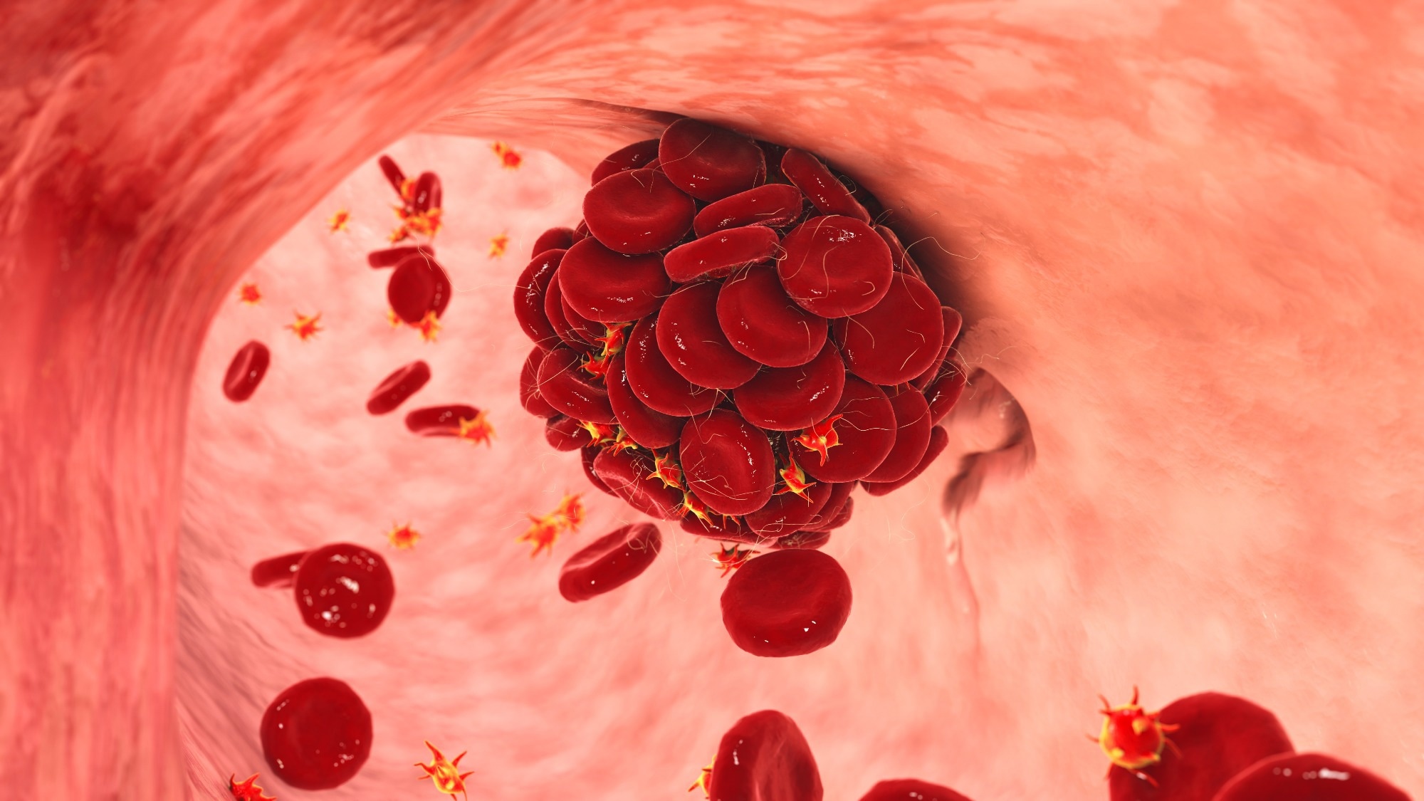
    Before we delve into the environmental factors, it is important to have a basic understanding of rheumatoid arthritis. RA is a chronic inflammatory disease that mainly affects the joints, but can also affect other organs. It is characterized by the immune system mistakenly attacking the body’s own tissues, especially the synovium, which lines the joints.

    Environmental triggers of rheumatoid arthritis

    Although genetics contribute to a person’s susceptibility to RA, environmental factors can trigger the disease in individuals with a genetic predisposition. These triggers can be external factors that interact with the body’s immune system and potentially lead to the development of RA. Several environmental factors have been identified and studied in association with rheumatoid arthritis.

    Genetic predisposition and environmental factors

    Individuals with certain genetic variations are more likely to develop rheumatoid arthritis. However, genetic predisposition alone is not sufficient to cause the disease. Environmental factors play a crucial role in the onset of RA in genetically susceptible individuals. Factors such as infections, smoking, diet, air pollution, stress, hormonal changes, weather conditions and exposure to chemicals are mentioned as possible triggers.

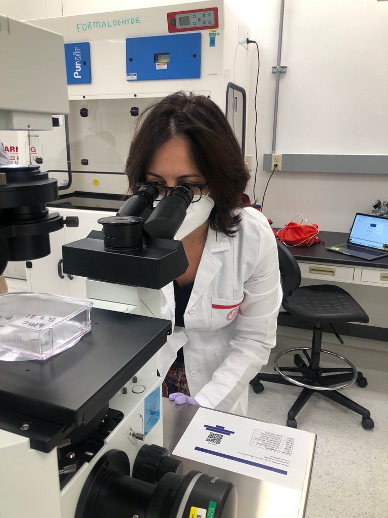
    The role of infections in causing rheumatoid arthritis

    Infections, especially those caused by certain bacteria and viruses, have been linked to the development of rheumatoid arthritis. It is believed that these infections can trigger an abnormal immune response, leading to the production of antibodies that attack the body’s own tissues. This process can eventually cause the onset of RA in susceptible individuals.

    The impact of smoking on rheumatoid arthritis

    Smoking has long been recognized as a major environmental cause of rheumatoid arthritis. Research has shown that smokers have a higher risk of developing RA than non-smokers. The chemicals in tobacco smoke can activate immune cells and promote inflammation, contributing to the development and progression of the disease.

    Diet and rheumatoid arthritis

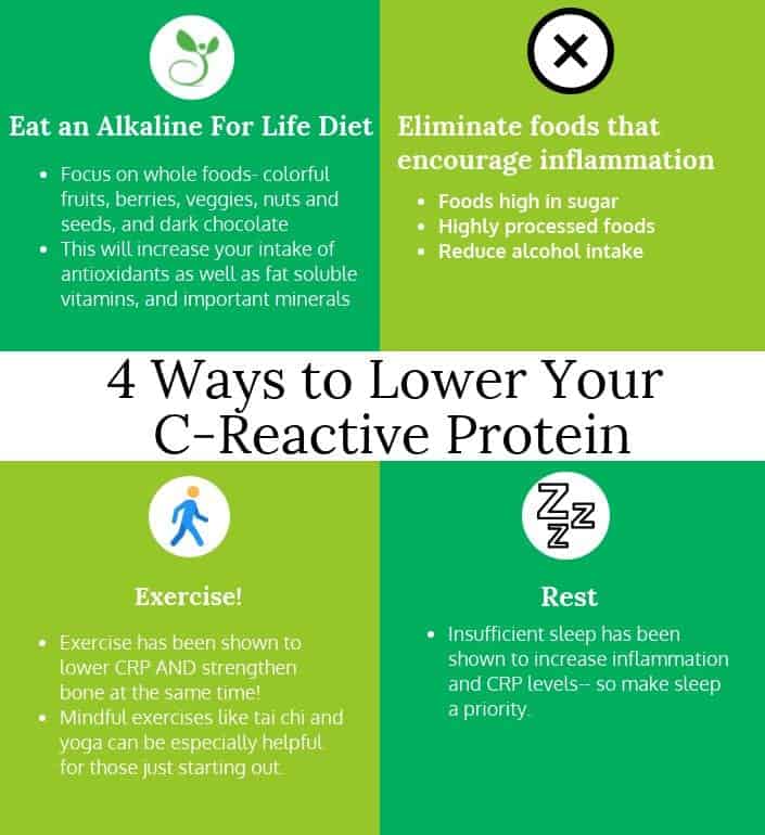
    Although the relationship between diet and rheumatoid arthritis is complex and not yet fully understood, certain dietary factors have been linked to the risk and severity of RA. For example, diets rich in omega-3 fatty acids, found in oily fish and flaxseed, have shown potential anti-inflammatory effects and may help reduce symptoms of RA. On the other hand, diets high in saturated fats and processed foods can promote inflammation and worsen the condition.

    The influence of air pollution

    Air pollution, especially particulate matter (PM2.5) and diesel exhaust, has been identified as a potential environmental trigger for rheumatoid arthritis. Inhalation of these pollutants can lead to oxidative stress, inflammation and immune system dysfunction. Long-term exposure to air pollution has been linked to an increased risk of developing RA and worsening symptoms in individuals already diagnosed with the condition.

    Stress and rheumatoid arthritis

    Chronic stress has been implicated as a trigger for rheumatoid arthritis. Stress can compromise the immune system and promote inflammation, potentially worsening RA symptoms. Although stress alone does not cause the disease, it can contribute to its onset and influence its progression.

    Hormonal factors

    Hormonal changes, especially in women, have been linked to an increased risk of rheumatoid arthritis. The fluctuation of hormones, such as estrogen, during the reproductive phase and menopause can affect the immune response and contribute to the development or worsening of RA symptoms.

    Weather and rheumatoid arthritis

    Many people with rheumatoid arthritis report that changes in weather conditions, especially cold and damp weather, can worsen their symptoms. Others say humidity increases swelling and pain in their joints. Although the exact mechanisms behind this association are not yet fully understood, it is thought that changes in temperature and barometric pressure may influence joint inflammation and pain perception in some individuals with RA.

    Chemical exposure and rheumatoid arthritis

    Exposure to certain chemicals, such as solvents, pesticides and heavy metals, has been linked to an increased risk of developing rheumatoid arthritis. These chemicals can disrupt the immune system and promote inflammation, potentially triggering the onset of RA or worsening its symptoms.

    Preventive measures and lifestyle changes

    While it may not be possible to completely prevent rheumatoid arthritis, certain preventative measures and lifestyle changes can help reduce the risk and control symptoms. These include maintaining a healthy diet, avoiding smoking and exposure to secondhand smoke, managing stress levels, staying physically active, protecting oneself from infections and minimizing exposure to environmental pollutants and chemicals.

    Conclusion

    In conclusion, rheumatoid arthritis is a complex disease influenced by both genetic and environmental factors. Environmental factors of rheumatoid arthritis, such as infections, smoking, diet, air pollution, stress, hormonal changes, weather conditions, and chemical exposure may contribute to the onset and progression of RA in genetically predisposed individuals. Understanding these triggers and taking preventative measures can play an important role in controlling the disease and improving the quality of life for people with rheumatoid arthritis.

    Frequently Asked Questions

    1. Can rheumatoid arthritis be completely prevented?

    Rheumatoid arthritis cannot be completely prevented, but certain lifestyle changes can help reduce the risk and manage symptoms effectively.

    2. Are all infections associated with rheumatoid arthritis?

    Although certain infections have been linked to the development of rheumatoid arthritis, not all infections have been linked to the disease.

    3. Is there a specific diet for rheumatoid arthritis?

    There is no one-size-fits-all diet for rheumatoid arthritis. However, a balanced and healthy diet that is rich in nutrients and low in processed foods can support overall health and possibly alleviate symptoms.

    4. How does air pollution affect rheumatoid arthritis?

    Air pollution, especially particulate matter and diesel exhaust, can promote inflammation and oxidative stress, potentially worsening symptoms and increasing the risk of developing rheumatoid arthritis.

    5. Can stress alone cause rheumatoid arthritis?

    Stress alone may not cause rheumatoid arthritis, but it can contribute to its onset and influence its progression by affecting the immune system and promoting inflammation.

    Source link

  • Lemony Sardine Salad – Food for Healthy Bones

    Lemony Sardine Salad – Food for Healthy Bones

    One of my favorite recipes Sardine salad for your bones with a refreshing lemon flavor.

    Oily fish is a superfood and sardines are the unsung heroes of this group. They are rich in selenium and calcium and are an excellent source of omega-3 fatty acids. They are high in EPA and DHA, essential fatty acids that perform a variety of tasks in your body, including stopping inflammation at the cellular level, breaking down fat cells and maintaining healthy hormonal balance.

    My kitchen partner created a recipe (found below). As a result, my Skelly felt nourished by eating her lemony sardine salad for my bones. If you’re wondering who Skelly is, she’s with me in the featured photos on my website. Maybe you’ve seen us dancing or cooking. She is also a metaphor for the skeleton in all of us and a symbol for the inner strength you feel when you nourish your bones properly.

    Remember that the sardines must have bones for the calcium.

    Ortiz Sardines

    The brand I enjoy is Ortiz, which comes in a glass bottle of olive oil. You can buy them on Amazon.com. I drain the olive oil from the jar before making the recipe. I prefer to use my own organic olive oil.

    I hope you and your bones enjoy our Lemony Sardine Salad.

    Delicious Sardines in Organic Olive Oil from Spain
    Sardine salad with high calcium and omega 3 content

    Lemony Sardine Salad

    Oily fish is a superfood and sardines are the unsung heroes of this group. They are rich in selenium and calcium and are an excellent source of omega-3 fatty acids. They are high in EPA and DHA, essential fatty acids that perform a variety of tasks in your body, including stopping inflammation at the cellular level, breaking down fat cells and maintaining healthy hormonal balance.

    Find more bone-loving recipes on my Comprehension Bone Health Program (CBHP) waiting list here!

    View all recipes here.

    Preparation time 15 minutes

    Cooking time 0 minutes

    Total time 20 minutes

    Class Starter, lunch, salad, snack

    kitchen Mediterranean

    Portions 2

    Calories 235 kcal

    • 1 Tablespoon olive oil extra virgin
    • 1 teaspoon lemon peel
    • 1/4 cup lemon juice
    • 1 teaspoon Dijon mustard
    • 1 Tablespoon capers
    • 1 Tablespoon parsley – dried 2 tbsp if fresh
    • 1 Tablespoon Tarragon – dried 2 tablespoons, if fresh
    • 2 stems celery cut in pieces
    • 4.9 oz sardines
    • 1 dash salty samples
    • 1 dash pepper samples
    • cut the celery into pieces

    • chop the herbs finely if they are fresh

    • Mix oil, lemon zest and juice, mustard, capers, parsley, tarragon and celery in a bowl.

    • Carefully fold in the sardines and season with salt and pepper.

    • Serve on lettuce, greens or with crackers or on a sandwich

    These nutrition facts apply to 1 serving of your recipe.
    Total servings for this recipe: 2


    The nutritional information is calculated automatically and may vary based on the ingredients and products used.

    Calories: 235kcalCarbohydrates: 6GEgg white: 19GFat: 16GSaturated fat: 2GPolyunsaturated fat: 5GMonounsaturated fat: 8GCholesterol: 99mgSodium: 410mgPotassium: 551mgFiber: 2GSugar: 1GVitamin A: 431IUVitamin C: 18mgVitamin D: 3µgVitamin K: 32µgCalcium: 339mgIron: 4mgMagnesium: 52mgPhosphorus: 371mgSelenium: 38µgZinc: 1mg

    Keyword Gluten free, lemon, osteopenia, osteoporosis, sardines


    Want more bone-loving recipes?

    Below is the link to purchase mine downloadable guide:

    Dr. Approved: 7-Day Meal Plan – Simple Cooking for Bone Health

    With 27 bone-loving recipes, shopping lists, nutrition plans, storage tips, weekly tips (why soak), nutrition panel for each recipe, 5 cooking videos for inspiration and much more.

    My Fully Clickable (Table of Contents) download guide helps you navigate recipes and all the information in this guide.

    The best $47 dollars you can spend on your bones.

    Click the button below for more information and to make your purchase.


    From my bones to yours,

    Irma Jennings INHC,

    Your holistic bot coach

    30 Essential Foods for Bone Health

    Source link

  • Blood flow restriction therapy can speed your recovery

    Blood flow restriction therapy can speed your recovery

    Whether you are an athlete looking to improve performance or recovering from an injury or surgery, Blood Flow Restriction Therapy (BFR) can be a game-changer for your life. BFR has been one of the major breakthroughs in the physical therapy industry, allowing physical therapists to alleviate atrophy and muscle weakness and achieve strength gains while minimizing joint stress. With BFR, physiotherapists can improve patient outcomes and unlock new possibilities.

    What is blood flow restriction therapy (BFR)?

    Blood flow restriction therapy, also called occlusion training, uses a device such as a cuff or wrap and places it around an arm or leg to stop partial blood flow to the affected area. Blood flow is restricted by restricting arterial flow and venous return. Restricting blood flow creates higher levels of metabolic stress. Metabolic stress and mechanical strain are associated with increased muscle growth. Exercises can be performed with low-load resistance training and achieve the same (if not better results) as traditional strength training.

    Benefits of blood flow restriction therapy

    1. Increases muscle growth and strength

    One of the biggest challenges after an injury or surgery is regaining muscle mass and strength. Women have a greater challenge than men in recovering muscle function after injury or surgery. Healthy men naturally have high levels of muscle-building testosterone and growth hormone to restore muscle mass, while women have less of both. After surgery, losing muscle size lasts about three days without use. The muscle shuts down due to the stress of a surgical process or injury. Myostatin, the hormone that muscle cells produce, inhibits muscle rebuilding and promotes scar tissue to limit growth.

    Active young athlete receiving BFR treatment.

    2. Speeds up recovery

    Occlusion training allows patients to achieve their goals in fewer sessions because it can produce similar results to traditional high-load training with lighter weights.

    3. Restores muscle function

    Rehabilitation with BFR can help minimize muscle and strength loss during immobilization or injury. In addition, it can aid in postoperative recovery and the recovery of muscle function.

    4. Improves cardiovascular health

    Blood flow restriction training can affect your cardiovascular system similar to traditional aerobic exercise. It can potentially improve important measures of cardiovascular health, such as VO2 max (a measure of aerobic fitness) and endothelial function (the health of your blood vessels).

    PT assists patient with BFR treatment.

    Common conditions BFR Physiotherapy treats

    1. Osteoarthritis (OA): According to studies in the Strength and Conditioning Journal, BFR may help improve OA muscle function and aid in pain management, leading to better overall function and quality of life. Overall, BFR can improve joint health.
    2. Sports injuries: BFR treats muscle strains, ligament sprains or tendonitis
    3. Postoperative conditions: BFR treats ACL, fractures, Achilles tendon, rotator cuff repairs, joint replacements, etc.
    4. Chronic conditions: BFR treats common cardiovascular diseases such as COPD and high blood pressure, which affect more than millions of people.
    5. Muscle weakness: BFR addresses atrophy due to various causes, such as disuse, immobilization or neurological disorders.
    6. Edema (inflammation and swelling): BFR stimulates fluid mobilization and improves circulation in the affected area. This may especially benefit patients with acute or chronic swelling conditions, such as postoperative or post-traumatic cases.

    BFR assessment in physiotherapy

    Patients are assessed for the appropriate level of bladder flow restriction. Physiotherapists can use a specialized Doppler, which determines when complete occlusion of blood flow has been achieved. Additionally, a personalized treatment plan and exercise program are designed to ensure the most effective results.

    Is blood flow restriction therapy safe?

    Many patients are initially concerned about the safety of BFR. Blow Flow Restriction Therapy is safe for post-surgical patients, athletes, seniors and anyone who wants to accelerate effective strengthening results. Therefore, we recommend the use of blood flow restrictions under the guidance of a physiotherapist, as the exact occlusion pressure will be assessed in each patient.

    The patient receives BFR treatment.

    Risks and side effects

    At Foothills we are often asked, “Does BFR cause blood clots?” BFR should not impede overall circulation or increase the risk of blood clots. Many case studies have concluded that BFR can release anticoagulant hormones when the cuff is released.

    Blood flow restriction therapy is not recommended for patients with uncontrolled high blood pressure. However, if you have uncontrolled high blood pressure or a cardiovascular condition, it is critical to consult a physical therapist or your doctor before considering BFR training. Additionally, they can assess your specific medical situation, evaluate potential risks, and provide personalized advice on whether BFR is right for you.

    It is always important to prioritize your health and consult a physiotherapist who specializes in BFR for personalized guidance. If you’re curious if BFR is right for you, schedule a FREE assessment near a Foothills location! We’ve enjoyed helping people from all walks of life return to the sports and activities they love. We look forward to introducing you to the BFR training world and witnessing its incredible impact on your fitness and rehabilitation goals.

    Source link