Category: Knee fractures

  • knee pain in athletes

    knee pain in athletes

    Knee pain is a common issue for athletes at all levels. It can affect their performance and even stop them from playing sports1. Young athletes, especially girls, often face anterior knee pain, also known as patellofemoral pain syndrome1. This pain is not usually from a physical problem. Instead, it’s often due to overusing muscles or not stretching and strengthening enough1.

    Key Takeaways

    • Knee pain is a common problem for athletes, impacting their sports performance and ability to play.
    • Young athletes, especially girls, often experience anterior knee pain from overusing muscles or not training properly.
    • Symptoms include dull pain during activity, sounds when moving the knee, and pain at night or during certain activities.
    • Treatments include low-impact exercises, strengthening, ice therapy, and sometimes medication or physical therapy.
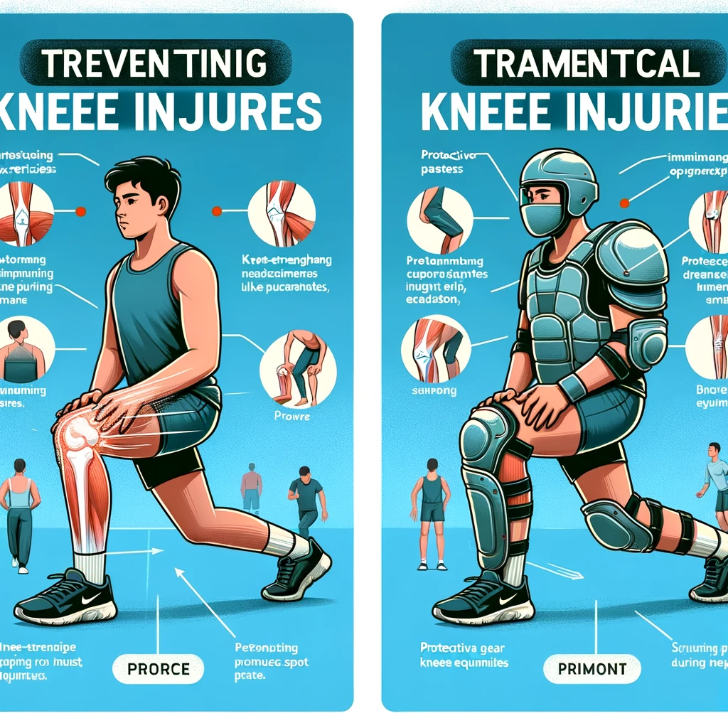
    • Preventing knee pain means warming up properly, staying at a healthy weight, wearing the right shoes, and slowly increasing exercise intensity.

    Understanding Patellofemoral Pain Syndrome

    Patellofemoral pain syndrome (PFPS), also known as “runner’s knee,” is a common injury. It affects the knee joint2. This condition causes pain in the front of the knee, near the kneecap. It’s especially common in athletes who do a lot of knee bending and straightening, like running, cycling, and skiing.

    What is Patellofemoral Pain Syndrome?

    Patellofemoral pain syndrome is a disorder that affects the muscles around the knee2. It happens when these muscles are weak or out of balance. This can make the kneecap move wrongly, causing friction and irritation in the joint.

    Causes of Patellofemoral Pain Syndrome

    The main reasons for patellofemoral pain syndrome are:

    • Muscle weakness, especially in the quadriceps and hip muscles2
    • Misalignment or instability of the kneecap2
    • Overuse, from doing too much training or suddenly increasing activity2
    • Trauma, like a fall or injury2
    • Tight muscles, in the quadriceps and hamstrings2

    These issues can lead to patellofemoral pain syndrome. This causes discomfort, less mobility, and could lead to more serious problems if not treated2.

    Key Findings Study
    Patellofemoral pain syndrome often doesn’t fully heal with just conservative treatment. Rathleff MS et al., 20122
    Adolescent basketball players show different signs of anterior knee pain based on gender. Foss KD et al., 20142
    Studies show structural issues on MRI in people with patellofemoral pain. van der Heijden RA et al., 20162

    Understanding patellofemoral pain syndrome helps athletes and healthcare workers find ways to prevent, manage, and treat it23.

    Knee Pain in Athletes: Symptoms and Diagnosis

    Knee pain is a big issue for athletes who push their bodies hard. The main signs include pain, swelling, and trouble moving the knee4. Athletes might hear a popping sound, feel unstable, or weak4.

    Doctors check for tenderness, alignment problems, and muscle imbalances during a physical exam4. They might use X-rays or MRI scans to see what’s going on inside4.

    About 30% of teens get knee pain that doctors often check out4. Girls are 2–10 times more likely to get it than boys4.

    1. Things like an odd Q angle, flat feet, tight Achilles, and muscle imbalances can cause knee pain4.
    2. Many knee injuries in young athletes come from too much stress on the muscles and bones4.
    3. Intrinsic causes of knee pain include various conditions like anterior knee pain syndrome and Osgood-Schlatter disease4.

    Patellofemoral pain syndrome makes going up or down stairs, sitting a long time, and squatting hard4. It might take two years to fully get better4.

    “Patellofemoral pain syndrome is a big reason for knee pain in young athletes5. It’s common in sports that involve running, jumping, or squatting5.”

    Doing activities like squatting can put over 1,000 pounds of pressure on the kneecap5. Flat feet and other issues can make the kneecap track wrongly, causing pain5.

    Risk Factors for Patellofemoral Pain Syndrome Other Knee Conditions
    • Flatfeet
    • Intoeing
    • Knock-knees
    • Tight hamstring
    • Weak thigh and hip muscles
    • Osgood-Schlatter disease
    • Patellar tendonitis
    • Iliotibial band friction syndrome

    Athletes with patellofemoral pain syndrome often play sports like basketball and volleyball5. The pain gets worse with running, jumping, and squatting5.

    knee pain symptoms

    To fix patellofemoral pain syndrome, find and fix the root cause with help from a doctor or physical therapist5. Treatment includes resting, icing, taking anti-inflammatory drugs, and doing other exercises5.

    Patellofemoral pain syndrome, also known as runner’s knee, causes pain in front of the knee6. It can come from kneecap misalignment, too much training, injury, or muscle weakness6.

    Runner’s knee symptoms include pain when moving, after sitting a long time, and sounds from the kneecap6. Doctors use a health history, physical exam, and might do X-rays to diagnose it6.

    Treatment for runner’s knee depends on how bad it is and what symptoms you have6. It might include resting, exercises, cold therapy, using a knee brace, and taking medication6. To prevent it, stay at a healthy weight, warm up, and wear good shoes6.

    Important things to know about PFPS: it causes pain and noises around the knee, can come from overuse or structural problems, and treatment includes rest, exercises, cold therapy, and medication6.

    When visiting a healthcare provider, prepare by asking questions, bring someone with you, take notes, and understand what they tell you about your condition and treatment6.

    Conclusion

    Knee pain is a big issue for athletes, caused by things like patellofemoral pain syndrome, overuse, and injuries7. Knowing why kids and teens often get chronic knee pain7 and what makes young athletes more likely to get knee injuries7 helps us find better ways to prevent and treat it.

    Using the right exercises7 and injury management can help athletes get over knee pain and lower the chance of it happening again7. Also, things like strength training, proper warm-ups, and the right gear can help prevent knee pain from starting7.

    Patellofemoral pain syndrome, or anterior knee pain, is really common in teen athletes, affecting up to 39% of those in sports like basketball and tennis8. Over half of these teens still had pain after 2 years8. By understanding this, we can help young athletes stay healthy and perform well in sports.

    FAQ

    What is knee pain in athletes?

    Knee pain is a common issue for athletes. It affects their performance and ability to play sports. It can come from patellofemoral pain syndrome, overuse, or traumatic injuries.

    What is patellofemoral pain syndrome (PFP syndrome)?

    Patellofemoral pain syndrome, or “runner’s knee,” is pain in the front of the knee near the kneecap. It’s a common injury for athletes who do a lot of knee bending and straightening.

    What causes patellofemoral pain syndrome?

    Causes include muscle weakness, especially in the quadriceps and hip muscles. Misalignment of the kneecap is another factor. Overuse from too much training or suddenly doing more can also cause it. Trauma, like a fall, can lead to it too.

    What are the symptoms of knee pain in athletes?

    Symptoms include pain, swelling, and less movement. Athletes might hear a popping or cracking sound in their knee. They may also feel unstable or weak.

    How is knee pain in athletes diagnosed?

    Healthcare providers do a detailed physical check to find tenderness, alignment problems, and muscle imbalances. They might use X-rays or MRI scans to see what’s really going on.

    Source Links

    1. Adolescent Anterior Knee Pain – OrthoInfo – AAOS – https://orthoinfo.aaos.org/en/diseases–conditions/adolescent-anterior-knee-pain/
    2. Patellofemoral pain in athletes – https://www.ncbi.nlm.nih.gov/pmc/articles/PMC5476763/
    3. Patellofemoral pain in athletes: clinical perspectives – https://www.ncbi.nlm.nih.gov/pmc/articles/PMC5640415/
    4. Evaluation and management of knee pain in young athletes: overuse injuries of the knee – https://www.ncbi.nlm.nih.gov/pmc/articles/PMC5532199/
    5. Knee Pain and Patellofemoral Pain Syndrome – https://www.healthychildren.org/English/health-issues/injuries-emergencies/sports-injuries/Pages/Knee-Pain-and-Patellofemoral-Pain-Syndrome.aspx
    6. Patellofemoral Pain Syndrome (Runner’s Knee) – https://www.hopkinsmedicine.org/health/conditions-and-diseases/patellofemoral-pain-syndrome-runners-knee
    7. Knee pain in young sports players aged 6–15 years: a cross-sectional study in Japan – https://www.ncbi.nlm.nih.gov/pmc/articles/PMC9906902/
    8. Nearly 40% of adolescent athletes report anterior knee pain regardless of maturation status, age, sex or sport played – https://www.sciencedirect.com/science/article/abs/pii/S1466853X21001097
  • knee cap injury

    knee cap injury

    Kneecap injuries, also known as patellar injuries, are common, especially among athletes and active individuals. These injuries can happen due to various reasons such as falls, accidents, or sports activities. The kneecap, or patella, plays a crucial role in the knee joint, helping with leg movement and providing protection. Understanding the causes, types, symptoms, and treatment options for kneecap injuries can help in managing and preventing them effectively.

    Common Causes of Kneecap Injuries

    knee cap injury

    Kneecap injuries can happen for many reasons. Here are some of the most common causes:

    Trauma and Accidents

    A direct hit to the knee can cause serious damage. Falls on hard surfaces like concrete, car accidents where the knee hits the dashboard, and even gunshot wounds can lead to kneecap injuries. The patella, located at the front of the knee, is especially vulnerable during these events.

    Sports-Related Injuries

    Playing sports can also put your kneecap at risk. Activities where the knee might get hit by a ball, bat, or stick are common culprits. Sudden movements or sharp impacts can also cause injuries, especially if the quadriceps muscle pulls on the kneecap too hard.

    Anatomical Issues

    Sometimes, the way your body is built can make you more likely to hurt your kneecap. Problems within the knee joint itself can lead to injuries over time. This can include issues like misalignment or weak muscles around the knee.

    Kneecap injuries are often the result of a combination of factors, making it important to understand the various causes to prevent them effectively.

    Types of Kneecap Injuries

    Patellar Fractures

    Patellar fractures occur when the kneecap bone breaks. This can happen due to a direct blow or a fall onto the knee. These fractures can be very painful and may limit your ability to straighten your knee. Treatment often involves immobilization with a cast or brace, and in severe cases, surgery may be required.

    Dislocations

    A dislocated kneecap happens when the patella is forced out of its normal position in the groove at the end of the femur. This can damage nearby soft tissues and cause significant pain and swelling. Immediate medical attention is usually necessary to realign the kneecap, followed by physical therapy to strengthen the surrounding muscles.

    Tendon Tears

    Tendon tears involve the ripping of the tendons that connect the kneecap to the muscles of the thigh and shin. These injuries can result from sudden, forceful movements or overuse. Symptoms include pain, swelling, and difficulty moving the knee. Treatment options range from rest and physical therapy to surgical repair, depending on the severity of the tear.

    Kneecap injuries can vary widely in severity, but early diagnosis and appropriate treatment are crucial for effective recovery.

    Symptoms of Kneecap Injuries

    kneecap injury illustration

    Pain and Swelling

    Pain is the most common symptom of a kneecap injury. It can be sharp or dull and often worsens with movement. Swelling usually follows, making the knee appear red and puffy.

    Locking and Instability

    The knee might lock up, making it hard to straighten. It can also feel unstable, as if it might give way, which is often due to patellofemoral instability.

    Difficulty in Movement

    Injuries can make bending or straightening the knee difficult. In severe cases, you might not be able to put any weight on the leg at all.

    Diagnosis Methods for Kneecap Injuries

    Physical Examination

    A physical examination is often the first step in diagnosing a kneecap injury. During this exam, the doctor will check for signs of pain, swelling, and instability. They may also perform manual tests, such as trying to extend your knee against gravity, to assess the damage to the kneecap or tendons. The straight leg raise test can reveal issues with the extensor mechanism, which includes the quadriceps tendon, patella, and patellar tendon.

    Imaging Tests

    Imaging tests are crucial for a more detailed look at the injury. Common imaging tests include X-rays, MRI scans, and CT scans. These tests help to identify fractures, dislocations, and other structural problems. An X-ray is often the first imaging test done to check for broken bones. If more detail is needed, an MRI or CT scan may be used to get a clearer picture of the soft tissues and bones.

    Medical History Review

    Reviewing the patient’s medical history is another important step. The doctor will ask about any previous knee injuries, surgeries, or conditions that might affect the current injury. They will also inquire about the circumstances leading to the injury, such as a fall or accident, and any symptoms experienced at the time, like hearing a pop or feeling the knee give way.

    Quick and accurate diagnosis is key to effective treatment and recovery. If you suspect a kneecap injury, seek medical attention promptly.

    Treatment Options for Kneecap Injuries

    Home Remedies and Rest

    For minor kneecap injuries, the RICE method is often recommended. This includes rest, ice, compression, and elevation. Over-the-counter anti-inflammatory medications like ibuprofen can help reduce pain and swelling. It’s important to avoid putting weight on the injured knee to allow it to heal properly.

    Medical Interventions

    More serious injuries may require medical interventions. These can include wearing a cast or brace to keep the leg stable while the bone heals. Pain medications, including opioids for severe pain, may be prescribed initially, followed by non-opioid options. Physical therapy is often recommended to help regain strength and mobility.

    Surgical Procedures

    In cases where the injury is severe or doesn’t respond to other treatments, surgery may be necessary. Surgical options can include repairing torn tendons or ligaments, or fixing fractures with screws and plates. Recovery from surgery will typically involve a period of immobilization followed by physical therapy to restore function.

    Preventing Kneecap Injuries

    knee cap injury illustration

    Protective Gear

    Wearing the right protective gear can make a big difference. Knee pads and braces can help shield your kneecaps during activities that put them at risk. Always choose gear that fits well and is appropriate for your specific sport or activity.

    Strengthening Exercises

    Doing exercises to strengthen your thigh muscles, like quadriceps and hamstrings, can help keep your knees stable. Strong muscles support your kneecaps better, reducing the chance of injury. Include both strength training and flexibility exercises in your routine.

    Safe Practices in Sports

    Practicing safe techniques in sports is crucial. Warm up before you start, and gradually increase the intensity of your workouts. Avoid sudden, intense movements that can strain your knees. If something causes knee pain, it’s best to stop and rest.

    Taking simple steps like wearing the right shoes and maintaining a healthy weight can also help protect your knees. These actions lower the stress on your kneecaps and keep them healthy.

    Recovery and Rehabilitation

    Physical Therapy

    Physical therapy is essential for regaining movement and strength in the knee. Rehabilitation focuses on improving range of motion, building up muscle strength, and decreasing knee stiffness. Recovery can take several months, depending on the severity of the injury.

    Pain Management

    Managing pain is crucial during the recovery process. If your pain is severe, your doctor may suggest a prescription-strength medication for a few days. Over-the-counter pain relievers can also be effective.

    Long-term Care

    Long-term care involves regular check-ups and possibly ongoing physical therapy. This ensures that the knee continues to heal properly and helps prevent future injuries. Staying active and following your doctor’s advice are key components of long-term care.

    Whether your treatment is surgical or nonsurgical, rehabilitation will play a vital role in getting you back to your daily activities.

    Conclusion

    Kneecap injuries are common, especially among active individuals and athletes. They can range from minor bruises to severe fractures and dislocations. It’s important to recognize the symptoms early and seek appropriate medical attention. While some injuries can be managed with rest and home care, others may require more intensive treatments like braces or even surgery. Understanding the causes and types of kneecap injuries can help in preventing them and ensuring a quicker recovery if they do occur. Always consult with a healthcare provider for a proper diagnosis and treatment plan.

    Key Takeaways

    • Kneecap injuries often result from trauma, sports activities, or anatomical problems.
    • Common types of kneecap injuries include fractures, dislocations, and tendon tears.
    • Symptoms of kneecap injuries usually involve pain, swelling, and difficulty in movement.
    • Diagnosis typically involves a physical exam and imaging tests like x-rays.
    • Treatment can range from home remedies and rest to medical interventions and surgery.

    Frequently Asked Questions

    What are common causes of kneecap injuries?

    Kneecap injuries can happen due to falls, sports activities, or car accidents. Anything that hits your knee hard can cause an injury.

    What types of kneecap injuries are there?

    There are several types of kneecap injuries, including fractures, dislocations, and tendon tears.

    How do I know if I have a kneecap injury?

    Common signs include pain, swelling, difficulty moving your knee, and feeling like your knee might give out.

    How are kneecap injuries diagnosed?

    Doctors usually do a physical exam and may use imaging tests like x-rays to see the injury clearly.

    What are the treatment options for kneecap injuries?

    Treatment can range from rest and home care for minor injuries to medical interventions like braces or surgery for more serious cases.

    How can I prevent kneecap injuries?

    Wearing protective gear, doing exercises to strengthen your knees, and following safe practices in sports can help prevent injuries.

  • Performing Exercises After an ACL Injury or Surgery: Tips and Advice.

    Performing Exercises After an ACL Injury or Surgery: Tips and Advice.

    Performing Exercises After an ACL Injury or Surgery: A Comprehensive Guide

    Anterior cruciate ligament (ACL) injuries are among the most common and potentially devastating knee injuries, particularly in athletes and active individuals. Whether caused by a sudden twist, an awkward landing, or a direct impact, an ACL injury can sideline you for months. However, with the right approach to rehabilitation and a well-structured exercise plan, you can regain strength, mobility, and confidence in your knee. This comprehensive guide offers in-depth information, tips, and advice for performing exercises after an ACL injury or surgery.

    17033499 2886 4722 a6dd d7527948ec3a 1

    Understanding ACL Injuries and Surgery

    What is the ACL?

    The anterior cruciate ligament (ACL) is one of four major ligaments in the knee joint. It runs diagonally through the center of the knee, connecting the femur (thigh bone) to the tibia (shin bone). The ACL plays a crucial role in stabilizing the knee during dynamic movements such as:

    • Cutting or changing direction quickly
    • Pivoting
    • Jumping and landing
    • Decelerating rapidly

    The ACL prevents the tibia from sliding forward in relation to the femur and provides rotational stability to the knee. This stability is essential for activities that involve quick changes in direction, such as soccer, basketball, skiing, and football.

    Mechanisms of ACL Injury

    ACL injuries typically occur through one of the following mechanisms:

    1. Non-contact injuries: These account for about 70% of ACL injuries. They often occur when an athlete changes direction rapidly, stops suddenly, or lands from a jump incorrectly.
    2. Contact injuries: These occur when there’s a direct blow to the knee, often from the side, which can happen in contact sports like football or rugby.
    3. Hyperextension: When the knee is forced beyond its normal range of motion, it can cause the ACL to tear.

    Types of ACL Injuries

    ACL injuries are typically classified into three grades:

    • Grade 1: A mild sprain where the ligament is stretched but not torn.
    • Grade 2: A partial tear of the ACL (relatively rare).
    • Grade 3: A complete tear of the ACL, often referred to as a “rupture.”

    ACL Surgery Options

    For many active individuals and athletes, especially those with complete ACL tears, surgery is often recommended. The most common surgical treatment is ACL reconstruction, where the damaged ligament is replaced with a graft. There are two main types of grafts:

    1. Autograft: Tissue taken from the patient’s own body. Common autograft sources include:
      • Patellar tendon
      • Hamstring tendon
      • Quadriceps tendon
    2. Allograft: Tissue taken from a donor, usually a cadaver.

    Each graft type has its pros and cons:

    Graft TypeAdvantagesDisadvantages
    Patellar Tendon AutograftStrong initial fixation, faster healing to bonePotential for anterior knee pain, risk of patellar fracture
    Hamstring Tendon AutograftLess harvest site morbidity, smaller incisionsSlower healing to bone, potential for hamstring weakness
    Quadriceps Tendon AutograftGood size and strength, less harvest site morbidity than patellar tendonLess commonly used, potential for quadriceps weakness
    AllograftNo harvest site morbidity, shorter surgery timeSlower incorporation, slightly higher failure rate in young, active patients

    The choice of graft depends on various factors, including the surgeon’s preference, the patient’s age, activity level, and specific anatomical considerations.

    ACL Injury Statistics

    To understand the scope of ACL injuries, consider these statistics:

    StatisticValue
    Annual ACL injuries in the US~200,000
    Percentage of ACL injuries requiring surgery50-60%
    Average recovery time to return to sports6-9 months
    Risk of re-injury within 2 years15-25%
    Percentage of female athletes compared to male athletes suffering ACL injuries2-8 times higher
    Percentage of ACL injuries that occur in non-contact situations~70%

    These statistics highlight the importance of proper rehabilitation and the need for a comprehensive approach to recovery and prevention.

    f0e06c3a 1e03 4adc 8104 bdb61cd7c0bc

    The Importance of Rehabilitation

    Rehabilitation following an ACL injury or surgery is crucial for several reasons:

    1. Restoring Strength and Stability

    ACL injuries and subsequent surgery can lead to significant muscle weakness, particularly in the quadriceps and hamstrings. A structured exercise program helps rebuild strength in these key muscle groups, which is essential for knee stability. Strong muscles around the knee joint can help:

    • Absorb shock and reduce stress on the knee
    • Improve joint alignment
    • Enhance overall leg strength and power

    2. Regaining Range of Motion

    After surgery, scar tissue formation and swelling can limit knee mobility. Flexibility exercises ensure that the knee can move through its full range of motion without pain or stiffness. This is crucial for:

    • Normal walking gait
    • Climbing stairs
    • Performing sports-specific movements

    3. Reducing Swelling and Pain

    Early movement and specific exercises can help reduce post-surgical swelling and manage pain by:

    • Promoting circulation and lymphatic drainage
    • Preventing adhesions
    • Stimulating the release of endorphins, the body’s natural pain relievers

    4. Improving Proprioception

    Proprioception is the body’s ability to sense the position and movement of its parts. ACL injuries can disrupt this sense in the knee. Proprioceptive exercises enhance:

    • Balance and coordination
    • Neuromuscular control
    • Reaction time

    These improvements are crucial for preventing future injuries and regaining confidence in the knee’s stability.

    5. Psychological Recovery

    Rehabilitation is not just about physical recovery. It plays a significant role in psychological healing by:

    • Building confidence in the injured knee
    • Providing a sense of progress and accomplishment
    • Preparing the athlete mentally for return to sport

    6. Preventing Re-injury

    A comprehensive rehabilitation program addresses factors that may have contributed to the initial injury, such as:

    • Muscle imbalances
    • Poor movement patterns
    • Inadequate core strength

    By correcting these issues, the risk of re-injury can be significantly reduced.

    Show Image

    Detailed Guide to Performing Exercises After an ACL Injury or Surgery

    Pre-Exercise Considerations

    Before beginning any exercise program, it’s crucial to:

    1. Consult with your surgeon and physical therapist: They will provide clearance and guidelines specific to your case.
    2. Understand your restrictions: Know what movements or activities to avoid at each stage of recovery.
    3. Set up a safe exercise environment: Ensure you have a non-slip surface and any necessary equipment (e.g., resistance bands, stability ball).
    4. Warm up properly: Start with gentle movements to increase blood flow to the area.

    Phase 1: Early Rehabilitation (Weeks 1-4)

    The focus in this phase is on protecting the graft, reducing swelling, and gradually restoring range of motion.

    Key Exercises:

    1. Heel Slides:
      • Lie on your back with legs straight.
      • Slowly slide your heel towards your buttocks, bending your knee.
      • Hold for 5 seconds, then slowly straighten.
      • Perform 3 sets of 10 repetitions.
    2. Quad Sets:
      • Sit with your leg straight and supported.
      • Tighten your thigh muscle, pressing the back of your knee down.
      • Hold for 5 seconds, then relax.
      • Perform 3 sets of 10 repetitions.
    3. Ankle Pumps:
      • Lie on your back or sit with your leg elevated.
      • Point your toes away from you, then flex them back towards you.
      • Perform 3 sets of 20 repetitions.
    4. Passive Knee Extension:
      • Sit with your leg supported, a small rolled towel under your heel.
      • Relax and allow gravity to straighten your knee.
      • Hold for 5 minutes, gradually increasing duration as tolerated.
    5. Straight Leg Raises:
      • Lie on your back with your uninjured leg bent.
      • Tighten your thigh muscle and lift your straight leg about 6 inches.
      • Hold for 3 seconds, then slowly lower.
      • Perform 3 sets of 10 repetitions.

    Phase 2: Intermediate Rehabilitation (Weeks 4-8)

    This phase focuses on progressive strengthening and introducing weight-bearing exercises.

    Key Exercises:

    1. Mini Squats:
      • Stand with feet shoulder-width apart, holding onto a stable surface.
      • Slowly bend your knees to about 30 degrees.
      • Hold for 3 seconds, then slowly straighten.
      • Perform 3 sets of 10 repetitions.
    2. Step-Ups:
      • Stand in front of a low step (4-6 inches high).
      • Step up with your surgical leg, bringing your other foot onto the step.
      • Step back down, leading with the non-surgical leg.
      • Perform 3 sets of 10 repetitions on each leg.
    3. Hamstring Curls:
      • Lie on your stomach.
      • Slowly bend your knee, bringing your heel towards your buttocks.
      • Hold for 3 seconds, then slowly lower.
      • Perform 3 sets of 10 repetitions.
    4. Stationary Cycling:
      • Start with no resistance, focusing on smooth motion.
      • Gradually increase duration and resistance as tolerated.
      • Aim for 10-20 minutes per session.
    5. Calf Raises:
      • Stand holding onto a stable surface.
      • Rise up onto your toes, then slowly lower.
      • Perform 3 sets of 15 repetitions.

    Phase 3: Advanced Rehabilitation (Weeks 8-12)

    This phase introduces more dynamic exercises and focuses on improving balance and proprioception.

    Key Exercises:

    1. Lunges:
      • Stand with feet hip-width apart.
      • Take a large step forward, lowering your hips until both knees are bent at 90 degrees.
      • Push back to the starting position.
      • Perform 3 sets of 10 repetitions on each leg.
    2. Single-Leg Balance:
      • Stand on your surgical leg.
      • Maintain balance for 30 seconds.
      • Progress to closing your eyes or standing on an unstable surface.
      • Perform 3 sets.
    3. Leg Press:
      • Start with light weight and gradually increase.
      • Press the weight away until your knees are almost straight, then slowly return.
      • Perform 3 sets of 12 repetitions.
    4. Lateral Step-Downs:
      • Stand on a 4-6 inch step with your surgical leg.
      • Slowly lower your other foot to touch the ground, then raise back up.
      • Perform 3 sets of 10 repetitions on each leg.
    5. Swimming or Water Jogging:
      • Start with 15-20 minutes and gradually increase duration.
      • Focus on proper form and avoid breaststroke kick initially.

    Phase 4: Return to Activity (Weeks 12 and Beyond)

    This phase prepares you for a return to sports or high-level activities.

    Key Exercises:

    1. Plyometric Drills:
      • Box Jumps: Start with a low box and progress in height.
      • Lateral Hops: Hop side to side over a line or low obstacle.
      • Progress from double-leg to single-leg exercises.
    2. Agility Drills:
      • Shuttle Runs: Start with straight-line runs and progress to change of direction.
      • Ladder Drills: Focus on quick, precise foot movements.
    3. Sport-Specific Exercises:
      • Gradually introduce movements specific to your sport.
      • Start at low intensity and progress as tolerated.
    4. Strength Training:
      • Continue with progressive resistance training for legs and core.
      • Include exercises like squats, deadlifts, and leg press.
    5. Cardiovascular Conditioning:
      • Progress to running on various surfaces and inclines.
      • Include interval training to improve endurance.
    8e9b0504 8de8 46ab 86df 22ef30487aee

    Nutrition and Recovery

    Proper nutrition plays a crucial role in recovery from ACL surgery and rehabilitation. A well-balanced diet can help reduce inflammation, support tissue repair, and provide the energy needed for rehabilitation exercises.

    Key Nutritional Considerations:

    1. Protein Intake:
      • Essential for muscle repair and rebuilding.
      • Aim for 1.6-2.0 grams of protein per kilogram of body weight daily.
      • Include sources like lean meats, fish, eggs, dairy, and plant-based proteins.
    2. Anti-Inflammatory Foods:
      • Help reduce inflammation and support healing.
      • Include omega-3 rich foods like salmon, flaxseeds, and walnuts.
      • Consume a variety of colorful fruits and vegetables for antioxidants.
    3. Complex Carbohydrates:
      • Provide energy for rehabilitation exercises.
      • Choose whole grains, fruits, and vegetables.
    4. Hydration:
      • Crucial for overall health and recovery.
      • Aim for 8-10 glasses of water per day, more if exercising intensely.

    Recommended Daily Nutrient Intake for ACL Recovery

    NutrientRecommended Daily IntakeKey Sources
    Protein1.6-2.0 g/kg body weightLean meats, fish, eggs, dairy, legumes
    Omega-3 Fatty Acids1-2 gFatty fish, flaxseeds, chia seeds, walnuts
    Vitamin C75-90 mgCitrus fruits, berries, bell peppers, broccoli
    Vitamin D600-800 IUSunlight exposure, fatty fish, fortified foods
    Calcium1000-1200 mgDairy products, leafy greens, fortified foods
    Iron8-18 mgRed meat, spinach, lentils, fortified cereals

    Supplements

    While a balanced diet should be the primary source of nutrients, some supplements may be beneficial during ACL recovery:

    1. Collagen: May support joint health and ligament healing.
    2. Vitamin D and Calcium: Essential for bone health, especially if mobility is limited.
    3. Omega-3 Supplements: Can help reduce inflammation if dietary intake is insufficient.
    4. Protein Powder: Can help meet increased protein needs, especially for athletes.

    Always consult with your healthcare provider before starting any new supplement regimen.

    Pain Management and Rest

    Managing pain and getting adequate rest are crucial components of ACL rehabilitation:

    Pain Management Strategies:

    1. RICE Method: Rest, Ice, Compression, and Elevation, particularly in the early stages of recovery.
    2. Proper Form: Ensure correct technique during exercises to avoid unnecessary strain.
    3. Gradual Progression: Increase intensity and duration of exercises slowly to prevent overexertion.
    4. Pain Medication: Use as directed by your healthcare provider, typically for short-term relief.

    Importance of Rest:

    1. Sleep: Aim for 7-9 hours of quality sleep per night to support tissue repair.
    2. Recovery Days: Include rest days in your exercise routine to prevent overtraining.
    3. Listen to Your Body: If you experience increased pain or swelling, take a break and consult your physical therapist.

    Psychological Support and Mental Health

    Recovering from an ACL injury can be mentally challenging. It

    Psychological Support and Mental Health

    Recovering from an ACL injury can be mentally challenging. It’s not just a physical journey, but an emotional one as well. Understanding and addressing the psychological aspects of recovery is crucial for a successful return to activity.

    Common Psychological Challenges:

    1. Fear of Re-injury: Many patients develop a fear of returning to the activity that caused their injury. This fear can be paralyzing and hinder progress.
    2. Loss of Identity: For athletes, being sidelined can lead to a loss of identity and purpose.
    3. Depression and Anxiety: The long recovery process and uncertainty about the future can trigger symptoms of depression and anxiety.
    4. Frustration with the Recovery Process: The lengthy rehabilitation can be frustrating, especially when progress seems slow.
    5. Loss of Confidence: Doubts about physical capabilities can erode self-confidence.

    Strategies for Mental Health Support:

    1. Set Realistic Goals:
      • Break down your recovery into small, achievable goals.
      • Celebrate minor milestones to maintain motivation.
    2. Visualization Techniques:
      • Mentally rehearse successful performance of exercises and sports movements.
      • Visualize yourself returning to your sport or activity successfully.
    3. Mindfulness and Relaxation:
      • Practice mindfulness meditation to stay present and manage anxiety.
      • Use deep breathing exercises to reduce stress.
    4. Seek Professional Support:
      • Consider working with a sports psychologist or counselor.
      • Join support groups for individuals recovering from similar injuries.
    5. Stay Connected:
      • Maintain contact with teammates and coaches.
      • Engage in team activities in non-physical roles when possible.
    6. Education:
      • Learn about your injury and the recovery process to feel more in control.
      • Understand that setbacks are a normal part of the recovery journey.
    7. Focus on Overall Health:
      • Use this time to improve other aspects of your health and fitness.
      • Explore new hobbies or skills to maintain a sense of progress and accomplishment.
    b5bfdf2a 91b7 48ff a2b1 ac5993a328ee 1

    Returning to Sport or Activity

    Returning to sport or high-level activity after an ACL injury is a critical phase that requires careful planning and execution. It’s important to remember that being cleared to return to sport doesn’t necessarily mean you’re ready for full competition.

    Criteria for Return to Sport:

    1. Full Range of Motion: The affected knee should have the same range of motion as the unaffected knee.
    2. Adequate Strength: Typically, the strength of the affected leg should be at least 90% of the unaffected leg.
    3. Neuromuscular Control: Demonstrated ability to perform sport-specific movements with proper form and control.
    4. Psychological Readiness: Confidence in the knee and readiness to return to full activity.
    5. Passing Functional Tests: Successfully completing a series of physical tests that mimic sport demands.

    Functional Tests for Return to Sport:

    TestDescriptionPassing Criteria
    Single Leg Hop TestHop for distance on each legAffected leg ≥ 90% of unaffected leg
    Triple Hop TestThree consecutive hops for distanceAffected leg ≥ 90% of unaffected leg
    Crossover Hop TestThree hops crossing over a lineAffected leg ≥ 90% of unaffected leg
    6-meter Timed HopHop on one leg for 6 metersAffected leg ≤ 10% slower than unaffected leg
    Y-Balance TestSingle-leg reach test in 3 directions< 4 cm side-to-side difference

    Gradual Return to Sport Protocol:

    1. Phase 1: Controlled Practice
      • Participate in non-contact drills
      • Focus on sport-specific movements in a controlled environment
    2. Phase 2: Team Practice
      • Introduce contact drills gradually
      • Participate in scrimmages with teammates
    3. Phase 3: Simulated Competition
      • Full-speed practice sessions
      • Scrimmages against other teams
    4. Phase 4: Limited Game Play
      • Return to competition with limited playing time
      • Gradually increase duration of play
    5. Phase 5: Full Return
      • Unrestricted participation in competition

    It’s crucial to progress through these phases under the guidance of your healthcare team and to listen to your body throughout the process.

    Long-Term Considerations and Injury Prevention

    After recovering from an ACL injury, it’s important to focus on long-term knee health and preventing future injuries.

    Ongoing Maintenance:

    1. Continue Strength Training: Maintain strength in the quadriceps, hamstrings, and core muscles.
    2. Regular Flexibility Work: Keep up with stretching routines to maintain good range of motion.
    3. Balance and Proprioception Exercises: Incorporate these into your regular workout routine.
    4. Proper Warm-up: Always perform a thorough warm-up before physical activity.

    Injury Prevention Strategies:

    1. Proper Technique: Focus on correct landing and cutting techniques in your sport.
    2. Neuromuscular Training: Participate in programs designed to improve movement patterns and reduce injury risk.
    3. Cross-Training: Engage in a variety of activities to promote overall fitness and reduce overuse injuries.
    4. Listen to Your Body: Pay attention to signs of fatigue or pain and adjust your activity accordingly.
    5. Regular Check-ups: Schedule periodic assessments with a sports medicine professional to monitor your knee health.

    Long-Term Outlook:

    While an ACL injury can be a significant setback, many individuals return to their pre-injury level of activity or sport. However, it’s important to be aware of the long-term implications:

    • Increased Risk of Osteoarthritis: ACL injuries, even when properly treated, may increase the risk of developing osteoarthritis in the knee later in life.
    • Potential for Re-injury: There is a slightly elevated risk of re-injury or injuring the opposite knee’s ACL.
    • Ongoing Maintenance: Long-term success often requires ongoing attention to knee health and overall fitness.

    Conclusion

    Recovering from an ACL injury or surgery is a challenging but achievable journey. By following a structured rehabilitation program, paying attention to nutrition and mental health, and gradually progressing through the phases of recovery, you can optimize your chances of a successful return to activity.

    Key takeaways:

    1. Follow a structured rehabilitation program guided by healthcare professionals.
    2. Progress gradually through the phases of recovery, from gentle movements to sport-specific exercises.
    3. Pay attention to proper form and technique during exercises.
    4. Support your recovery with proper nutrition and rest.
    5. Address the psychological aspects of recovery and seek support when needed.
    6. Use functional tests to gauge readiness for return to sport.
    7. Focus on long-term knee health and injury prevention strategies.

    Remember, every individual’s recovery is unique. Stay patient, stay committed, and celebrate the small victories along the way. With dedication and the right approach, you can overcome an ACL injury and return to an active, healthy lifestyle.

    97e014b7 0e67 474f b1b1 2eef378afee0

  • Why Women Are 2-10 Times More Likely Than Men to Tear Their Anterior Cruciate Ligament – ​​ACL Strong

    Why Women Are 2-10 Times More Likely Than Men to Tear Their Anterior Cruciate Ligament – ​​ACL Strong


    Anterior cruciate ligament (ACL) injuries are a common and debilitating injury in sports, and unfortunately young women are more susceptible to a torn ACL compared to men. But Why?

    Why are we seeing more and more female athletes tearing their anterior cruciate ligament?

    What can women do to reduce their risk?

    In this article, we uncover the factors behind this gender disparity and provide valuable insights into what women can do to reduce their risk. By understanding the anatomical, hormonal, neuromuscular, and biomechanical factors at play, as well as the importance of training and prevention strategies, we can pave the way for safer athletic experiences and a lower incidence of ACL tears in women.

    Let’s take a closer look at these variables:

    Anatomy and hormonal differences

    There are anatomical and hormonal variations in women that affect the knee joint. With a wider pelvis, a greater Q angle, and hormonal fluctuations at the forefront, the delicate balance of ligament laxity is noticeably affected, making women more susceptible to the dreaded ACL tears.

    A larger Q angle, between the natural width of the hips and the narrower knee position, is thought to place more stress on the knee than a more vertical hip-to-knee-to-foot alignment. This added stress, combined with the forces already placed on the ligaments during twisting and playing sports, may increase the risk of tearing in women with a larger Q angle.

    Hormonal fluctuations, particularly related to the menstrual cycle, are also thought to contribute to susceptibility to ligament tears. Research has yet to confirm this theory, although many experts believe there is some correlation, even if it is not yet fully understood.

    Neuromuscular imbalances:

    Neuromuscular control refers to the communication between the brain (nervous system) and muscles and is responsible for actions such as responding to a stimulus or controlling body movement. Neuromuscular control deficits are more pronounced in young females compared to their male counterparts… and these imbalances can create a perfect storm for injury. Most commonly seen is a relative weakness in the hamstrings compared to the quadriceps, decreased core stability, and less controlled mechanics during landing. These imbalances place added stress on the ACL during dynamic movements, contributing to a greater likelihood of a tear.

    While the ideal ratio of quadriceps to hamstring strength is around 3:2, certain women typically exhibit a range closer to 3:1. This lack of hamstring strength hinders the muscle group’s ability to contract effectively and maintain proper knee joint alignment, a problem that is particularly prevalent in adolescent women. Implementing exercises to improve neuromuscular imbalances is crucial to reducing your risk of injury.

    In scenarios where the core, hamstrings, and hip stabilizing muscles do not have sufficient strength and stability to maintain optimal joint alignment, female athletes subject their knees to significant forces while performing cutting, pivoting, and landing maneuvers. A force on the ACL that exceeds its capacity will result in a ligament injury.

    Biomechanical differences:

    Biomechanical differences in women can affect optimal sport-specific movements such as landing mechanics. A common occurrence in women is an increased tendency for knee valgus, an inward collapse of the knee, during actions such as landing or cutting. This alignment places excessive stress on the ACL, increasing the risk of injury.

    Research also suggests that women may have relatively smaller and weaker ACLs compared to their male counterparts. This disparity in size and strength may compromise the ligament’s ability to withstand the forces encountered during dynamic movements, making it more susceptible to injury.

    Sports participation and injury risk:

    Women’s involvement in high-risk sports such as soccer, basketball, gymnastics, and skiing significantly increases the risk of ACL tears. These sports, characterized by activities

    Movements such as cutting, planting, pivoting and landing naturally increase susceptibility to injury.

    The problem is further compounded by the fact that many young women participate in these risky sports without proper training or preparation, further increasing the risks.

    Training and prevention strategies:

    Training opportunities and injury prevention strategies may not be accessible to all athletes. It is critical for female athletes to have access to specialized training programs that prioritize improving neuromuscular control, strength, and proper movement mechanics when they begin participating in sports, because even the right exercise, done incorrectly, can actually set you back more susceptible to injuries.

    Historically, men have had the advantage of getting stronger earlier, leading to a lower risk of injury in their careers. When female athletes are given the same opportunity to strengthen and train their bodies, they also experience a lower risk of injury.

    So…How do I reduce the risk?

    Understanding the factors that contribute to the increased susceptibility to ACL tears gives female athletes the opportunity to proactively implement measures to prevent these injuries. (For more insight, download the FREE report on why injury prevention programs are ESSENTIAL for female athletes playing collegiate sports.)

    By emphasizing the importance of tailored training, promoting neuromuscular balance, and implementing injury prevention strategies, women are empowered to minimize their risks and pursue their athletic ambitions with unwavering confidence.

    At ACL Strong, we’ve created a safer and more inclusive sports environment for female athletes by developing a plan that reduces the risk of injury and makes your knees bulletproof!
    Bulletproof Secrets
    Snow Course V6 V5

    Source link

  • Boost your ACL Prehab exercises with the Neubie

    Boost your ACL Prehab exercises with the Neubie

    Another study, published in the American Journal Of Sports Medicine, found that the following exercise routine helped participants perform better on the single leg hop test, improve self-reported knee function scores, improve quadriceps and hamstring strength, and even showed a positive trend for return to sports after surgery

    According to the study:

    “The program consisted of 4 exercise periods per week: 2 supervised gym sessions alternating with 2 supervised home sessions… Patients were allowed to perform 1 repetition of their maximal effort to determine starting weights in the gym. Each gym activity consisted of 3 sets of 12 repetitions and the weights were increased by 10% to 15% each week. During the last gym session, the weights were returned to the previous week’s value to prevent preoperative fatigue and to promote muscular endurance response and mass gain. Home exercises consisted of the same program as in the gym, but with the use of a TheraBand instead of weights. Patients used a DonJoy knee brace locked at 20 degrees of extension to reduce the risk of further ACL loading pre- or postoperatively during open kinetic chain (OKC) exercise. The brace also acts as a confidence factor during OKC exercises. Proprioception training was performed on a wobble cushion.”

    The exercise routine, highlighted in the appendix of the study, consists of the
    next. Although the full text of the study is not freely available, you can access the appendix here by clicking on “Supplementary Material” in the left column.

    Exercise group 1 – All exercises 3×12

    • Leg press
    • Leg curl
    • Leg extension

    Exercise group 2 – All exercises 3×12

    • Half drop
    • 45 degree squat
    • Calf raises

    Exercise group 3 – All exercises 3×12

    • Hip abduction
    • Hip adduction
    • Hip flexion

    Balancing/Proprioception – 3×30 seconds – Use a wobble cushion

    • Step 1: Stand on one leg with
      eyes open
    • Step 2: Stand on one leg with
      eyes closed
    • Step 3: Stand on one leg with
      eyes closed, head raised

    Stretching – All stretching exercises 3×30 seconds

    • Quadriceps
    • Hamstring
    • Calf


    Source link

  • Study reveals link between contact sports participation and parkinsonism in individuals with chronic traumatic encephalopathy

    Study reveals link between contact sports participation and parkinsonism in individuals with chronic traumatic encephalopathy

    The largest study of CTE to date has found a new link between contact sports participation, chronic traumatic encephalopathy (CTE) and the development of a movement disorder known as parkinsonism.

    The study of 481 deceased athletes by researchers from Boston University Chobanian and Avedisian School of Medicine and VA Boston Healthcare, published today in JAMA Neurologyit appears that most individuals with CTE developed parkinsonism, and CTE pathology appears to be the cause of the parkinsonism symptoms in most cases.

    Parkinsonism is a condition characterized by symptoms similar to Parkinson’s disease, such as tremor, abnormal slowness of movement, or abnormal stiffness of the arms or legs. It has long been associated with traumatic brain injury (TBI) and CTE in boxers. However, the specific pathologies underlying these symptoms in CTE were unknown.

    Parkinson’s disease is classically associated with the buildup of proteins called Lewy bodies in brain cells, but researchers found that 76% of individuals with CTE and Parkinsonism did not do Have Lewy body pathology.

    “We were surprised to find that most individuals with CTE and parkinsonism did not have Lewy body pathology,” noted Thor Stein, MD, PhD, associate professor of pathology and laboratory medicine at BU and VA Boston Healthcare, and one of the study’s corresponding authors. “Instead,” Stein explained, “subjects with parkinsonism were more likely to have more severe CTE-related brain cell death in a region of the brainstem important for controlling movement.”

    CTE is a degenerative brain disease whose only known cause is repeated head blows, such as those that occur in contact sports. A 2018 study by the same research team found that the duration of contact sports is associated with an increased risk of developing Lewy body disease. However, the current study is the first to describe a link between contact sports participation, brainstem pathology, and parkinsonism in CTE.

    “Increased CTE severity has been shown to be associated with longer playing time,” noted Daniel Kirsch, an MD/PhD student at BU and one of the study’s first authors. “In this study, we found that eight additional years of contact sports play was associated with a 50 percent increased risk of more severe disease in a specific area of ​​the brainstem that controls movement.”

    The study subjects donated their brains to the Understanding Neurologic Injury and Traumatic Encephalopathy (UNITE) brain bank. People with parkinsonism were compared to those without to identify the types of pathologies that might explain why some people with CTE develop these symptoms and to examine relationships with the duration of contact sports.

    This study underscores the importance of understanding the long-term effects of repeated head impacts and the need for preventive measures in contact sports to reduce the risk of neurodegenerative diseases such as CTE and parkinsonism.

    Source link

  • Video: 5 Important Exercises to Do After ACL Surgery

    Video: 5 Important Exercises to Do After ACL Surgery


    Document

    Are you recovering from ACL surgery and wondering what exercises you can do to aid in your recovery? This video will cover the early stages of recovery, explain what factors are important during this stage, and outline key exercises you can do every day to aid in your recovery.

    During the early stages of recovery after anterior cruciate ligament surgery, the main goals are to achieve full range of motion and regain neuromuscular control of the knee.[1] To achieve these goals, it is essential to manage pain, swelling, and inflammation of the knee to ensure that you can complete your rehabilitation exercises. Using ice, applying compression, elevating your foot, and doing ankle pumps are great ways to manage pain, swelling, and inflammation.

    While achieving both flexion and extension range of motion is important for activities of daily living, achieving full knee extension is emphasized for people recovering from ACL surgery.[1] Knee extension is especially important in the early stages of recovery, as numerous studies have shown that loss of full knee extension can be a predictor of postoperative complications and poor outcomes.[2][3][4][5] Therefore, exercises to increase knee range of motion are especially important during the early phase of ACL recovery.

    Achieving neuromuscular control of the knee is another important goal after ACL surgery. Neuromuscular control of the knee refers to the coordination of your brain, nerves, and muscles to create movement and stability.[6] Regaining neuromuscular control is essential to not only ensure proper movement and function, but also to prevent further injury to your knee.[6] Some ways to regain neuromuscular control of the knee include strengthening the supporting muscles around the knee and doing balance exercises.[1]

    During the early recovery phase following ACL surgery, exercises that focus on knee flexion, knee extension, knee muscle strengthening, and proprioception are recommended for the first 4 weeks following surgery. Five safe and effective exercises that you can do daily to help you recover functionally include quadriceps and hamstring co-contractions, heel glides, knee extension over a roller, straight leg raises, and chair assisted squats. Click on this link for a more detailed description of these exercises. Keep in mind that everyone recovers at their own pace, so your ability to perform these exercises may vary depending on your condition. Consult with your healthcare provider to ensure that these exercises are safe for you.

    Listen to Joey Wong, kinesiologist, explain the key exercises to focus on after ACL surgery, and learn about ACL recovery goals.

    The Curovate Physical Therapy App has detailed videos and descriptions of these 5 essential exercises, as well as many other important exercises for your recovery. Click on one of the download links below to get started on your ACL recovery today! Curovate is an evidence-based app that provides guided physical therapy plans, daily guided video exercises, ways to measure and monitor your progress, and measures your knee range of motion! Download the Curovate App by clicking on the links below.

    If you require further, tailored assistance during your ACL recovery, you can book a 1-on-1 video session with a physiotherapist on our Virtual Physiotherapy page.


    Download it on Google Play

    Blogs related to ACL surgery

    References


    Source link

  • Treatment Options for Traumatic Knee Injury: A Professional Overview

    Treatment Options for Traumatic Knee Injury: A Professional Overview

    When it comes to traumatic knee injuries, treatment options vary depending on the severity of the injury. Knee injuries can be caused by a variety of factors, including sports injuries, falls, and car accidents. Symptoms of a knee injury may include pain, swelling, stiffness, and difficulty moving the knee joint.

    v2 3hh2j

    If you suspect that you have a knee injury, it is important to seek medical attention as soon as possible. A doctor can diagnose the injury and recommend the appropriate treatment. Diagnosis may involve a physical exam, imaging tests such as X-rays or MRI scans, and possibly arthroscopy, which involves inserting a small camera into the knee joint to examine the damage.

    Treatment options for a traumatic knee injury may include rest, ice, compression, and elevation (RICE), physical therapy, medications, and surgery. The specific treatment plan will depend on the type and severity of the injury. In some cases, conservative treatment such as RICE and physical therapy may be sufficient to heal the injury. In more severe cases, surgery may be necessary to repair or replace damaged tissue.

    Assessment and Immediate Care

    When a traumatic knee injury occurs, prompt assessment and care are essential for optimal healing. In this section, we will discuss the initial evaluation, pain management, and swelling reduction.

    Initial Evaluation

    The initial evaluation of a traumatic knee injury includes a thorough physical exam and imaging studies. During the physical exam, we will assess the knee for tenderness, swelling, and range of motion. We may also perform special tests to evaluate the ligaments and menisci of the knee. X-rays may be obtained to evaluate for fractures or dislocations, while an MRI may be necessary to assess for soft tissue injuries.

    f7b8c365 3679 4413 9947 5966b4487944 1

    Pain Management

    Pain management is an important aspect of the immediate care of a traumatic knee injury. We recommend the use of non-steroidal anti-inflammatory drugs (NSAIDs) for pain relief. In addition, we may prescribe stronger pain medications if necessary. We also recommend the use of ice to reduce pain and inflammation. Applying ice to the knee for 20 minutes every 2-3 hours can help to alleviate pain.

    Swelling Reduction

    Swelling is a common symptom of a traumatic knee injury and can be reduced with the use of rest, ice, compression, and elevation (RICE). We recommend that patients rest the affected knee and avoid weight-bearing activities until the swelling has subsided. Applying ice to the knee can help to reduce swelling, while compression with an elastic bandage can help to prevent further swelling. Elevating the knee above the level of the heart can also help to reduce swelling.

    In conclusion, prompt assessment and care are essential for the treatment of a traumatic knee injury. The initial evaluation should include a thorough physical exam and imaging studies. Pain management can be achieved with the use of NSAIDs and ice, while swelling can be reduced with RICE.

    Rehabilitation and Treatment Options

    When it comes to treating a traumatic knee injury, there are several rehabilitation and treatment options available. These options can be divided into two categories: non-surgical therapies and surgical interventions.

    Non-Surgical Therapies

    For less severe knee injuries, non-surgical therapies such as physical therapy and exercise can be effective in promoting healing and restoring function. Physical therapy can help to strengthen the muscles around the knee, improve range of motion, and reduce pain and swelling. Exercises such as straight leg raises, hamstring curls, and calf raises can also be beneficial in promoting strength and function.

    In addition to physical therapy and exercise, medication such as nonsteroidal anti-inflammatory drugs (NSAIDs) can be used to reduce pain and inflammation. Bracing and compression can also be used to provide support and reduce swelling.

    Surgical Interventions

    For more severe knee injuries, surgical intervention may be necessary. The type of surgery required will depend on the nature and extent of the injury. Common surgical interventions for knee injuries include arthroscopic surgery, meniscus repair, ACL reconstruction, and ligament reconstruction.

    Arthroscopic surgery is a minimally invasive procedure that uses small incisions and a camera to visualize and treat the injured area. Meniscus repair involves repairing or removing the damaged meniscus. ACL reconstruction involves replacing the torn ACL with a graft. Ligament reconstruction involves repairing or replacing the damaged ligament.

    Recovery time for surgical interventions can vary depending on the type and extent of the surgery. Rehabilitation is an important part of the recovery process and typically involves physical therapy to restore strength and function to the knee.

    In conclusion, there are several rehabilitation and treatment options available for traumatic knee injuries. Non-surgical therapies such as physical therapy and exercise can be effective for less severe injuries, while surgical interventions may be necessary for more severe injuries. It is important to work closely with a healthcare professional, such as an orthopedic surgeon, to determine the best course of treatment for your specific injury. Clinical trials may also be available for those interested in exploring new treatment options.

  • Preventing Traumatic Knee Injuries: Tips from a Professional

    Preventing Traumatic Knee Injuries: Tips from a Professional

    Knee injuries are common and can be caused by various factors such as sports, accidents, and repetitive motion. Traumatic knee injuries can be devastating and can lead to long-term complications. Therefore, it is essential to take preventive measures to avoid such injuries. In this article, we will discuss some effective ways to prevent traumatic knee injuries.

    v2 3hh3x znuda

    One of the most effective ways to prevent knee injuries is by strengthening the muscles around the knee. Strong muscles can help support the knee joint and reduce the risk of injury. Exercises such as lunges, squats, and leg presses can help strengthen the muscles around the knee. Additionally, stretching can help improve flexibility and reduce the risk of muscle strains.

    Another way to prevent knee injuries is by wearing protective gear during sports or activities that involve jumping, running, or sudden changes in direction. Knee pads, braces, and proper footwear can help absorb shock and reduce the risk of injury. It is also essential to warm up before engaging in physical activities to prepare the muscles and joints for the stress they will endure.

    775e9c8b 8af4 4bb2 8cc5 d0517fcd25b5 1

    In summary, taking preventive measures can help reduce the risk of traumatic knee injuries. By strengthening the muscles around the knee, wearing protective gear, and warming up before physical activities, we can protect our knees and avoid long-term complications.

    Understanding Knee Injuries

    When it comes to knee injuries, prevention is key. Knee injuries can be caused by various factors, including trauma, overuse, and degenerative conditions. Understanding the anatomy of the knee, common causes and types of knee injuries, as well as their symptoms and diagnosis can help you take steps to prevent knee injuries.

    Anatomy of the Knee

    The knee is a complex joint that connects the thigh bone (femur) to the shin bone (tibia). The knee joint also includes the kneecap (patella) and other structures such as ligaments, tendons, cartilage, and meniscus. The ligaments provide stability to the knee, while the tendons connect the muscles to the bones. The cartilage and meniscus act as shock absorbers and help to distribute weight evenly across the joint.

    Common Causes and Types of Knee Injuries

    Knee injuries can be caused by a variety of factors, including trauma, overuse, and degenerative conditions such as osteoarthritis and rheumatoid arthritis. Common types of knee injuries include fractures, dislocations, ligament tears, meniscus tears, and torn cartilage.

    Fractures occur when there is a break in the bone, while dislocations occur when the bones in the joint are forced out of position. Ligament tears can occur when the ligaments that connect the bones in the knee are stretched or torn, while meniscus tears can occur when the cartilage in the knee is torn. Torn cartilage can also occur as a result of overuse or degenerative conditions.

    Symptoms and Diagnosis

    Symptoms of knee injuries can include pain, swelling, stiffness, redness, warmth, and fever. Chronic pain can also be a symptom of knee injuries. Diagnosis of knee injuries typically involves a physical examination, imaging tests such as X-rays and MRI scans, and sometimes arthroscopy, a minimally invasive surgical procedure.

    In conclusion, knee injuries can be caused by various factors, and understanding the anatomy of the knee, common causes and types of knee injuries, as well as their symptoms and diagnosis can help you take steps to prevent knee injuries.

    Prevention and Management Strategies

    Exercise and Physical Activity

    Engaging in regular physical activity and exercise can help prevent traumatic knee injuries. It is important to incorporate exercises that focus on strengthening the muscles around the knee joint, such as the quadriceps, hamstrings, and calf muscles. These exercises can include lunges, squats, leg presses, and step-ups.

    Stretching is also important in preventing knee injuries. It is recommended to stretch before and after exercise to improve flexibility and reduce the risk of injury. Proper technique and form during exercise is crucial in preventing knee injuries. Coaches and trainers can provide guidance on proper technique and form during exercise.

    Protective Gear and Techniques

    Wearing proper protective gear, such as knee braces, can help prevent knee injuries during sports and physical activity. Knee braces can help provide support and stability to the knee joint, reducing the risk of injury.

    Techniques such as jumping and landing properly can also help prevent knee injuries. It is important to land softly and use proper technique when jumping, especially during sports such as basketball or volleyball.

    Treatment and Recovery

    If a knee injury does occur, it is important to seek medical attention from a doctor or physical therapist. Treatment may include rest, compression, elevation, and ice to reduce swelling and pain. Anti-inflammatory drugs such as ibuprofen may also be prescribed to reduce pain and inflammation.

    In some cases, surgery may be necessary to repair a torn ligament or tendon. Physical therapy may also be recommended to help with recovery and prevent future injuries.

    Overall, taking steps to prevent knee injuries through exercise, proper technique, and protective gear can help reduce the risk of injury and promote long-term knee health.

  • Right Way For Managing Knee Pain: by Professional

    Right Way For Managing Knee Pain: by Professional

    Our knees are fundamental to our mobility and daily activities. Yet, knee pain is a widespread issue affecting millions worldwide. This comprehensive guide will delve into the causes, symptoms, and treatments for knee pain, providing you with the knowledge to better understand and manage this common condition.

    Introduction

    Knee pain is a pervasive problem that doesn’t discriminate by age or lifestyle. Whether you’re an athlete pushing your limits, an office worker with a sedentary job, or somewhere in between, knee pain can strike and significantly impact your quality of life. Understanding the complexities of knee pain is crucial for effective management and treatment.

    According to recent studies, approximately 25% of adults suffer from frequent knee pain. This staggering statistic underscores the importance of knee health education and proactive care.

    [Image: Anatomical diagram of a knee] Alt text: Detailed anatomical diagram showing the structure of a human knee joint

    Anatomy of the Knee

    To truly understand knee pain, we must first explore the intricate anatomy of the knee joint.

    Basic Structure

    The knee is one of the largest and most complex joints in the human body. It consists of:

    • Bones: The femur (thighbone), tibia (shinbone), and patella (kneecap)
    • Cartilage: Including the meniscus, which provides cushioning between bones
    • Ligaments: Connect bones and provide stability (e.g., ACL, PCL, MCL, LCL)
    • Tendons: Connect muscles to bones (e.g., patellar tendon)
    • Bursae: Small fluid-filled sacs that reduce friction between tissues

    Function of Each Component

    • Bones: Provide the structural framework and enable movement
    • Cartilage: Acts as a shock absorber, reducing friction between bones
    • Ligaments: Stabilize the joint and guide motion
    • Tendons: Allow muscles to move the joint
    • Bursae: Reduce friction and provide smooth movement

    The knee’s design allows for a range of motions, including flexion, extension, and a small degree of rotation. This complexity, while providing great functionality, also makes the knee susceptible to various types of injuries and conditions.

    [Image: Labeled diagram of knee anatomy] Alt text: Labeled anatomical diagram of a knee showing bones, cartilage, ligaments, and tendons

    Common Causes of Knee Pain

    Knee pain can result from a wide array of factors, ranging from acute injuries to chronic conditions. Understanding these causes is crucial for proper diagnosis and treatment.

    Injuries

    • Sprains and strains: Overstretching or tearing of ligaments or muscles
    • Meniscus tears: Damage to the cartilage that cushions the knee joint
    • Ligament tears: Such as ACL (Anterior Cruciate Ligament) or MCL (Medial Collateral Ligament) injuries
    • Fractures: Breaks in the kneecap, lower femur, or upper tibia

    Arthritis

    • Osteoarthritis: The most common form, caused by wear and tear of cartilage
    • Rheumatoid arthritis: An autoimmune condition that causes joint inflammation
    • Post-traumatic arthritis: Develops after an injury to the knee

    Overuse Conditions

    • Tendinitis: Inflammation of tendons, often due to repetitive activities
    • Bursitis: Inflammation of the bursae, frequently caused by prolonged kneeling
    • Patellofemoral pain syndrome: Pain around the kneecap, common in runners

    Other Causes

    • Gout: Build-up of uric acid crystals in the joint
    • Infections: Bacterial or viral infections in the joint or surrounding tissues
    • Osgood-Schlatter disease: Common in adolescents, causing pain below the kneecap
    • Iliotibial band syndrome: Common in runners, causing outer knee pain

    [Image: Infographic showing percentage breakdown of common knee pain causes] Alt text: Pie chart illustrating the percentage distribution of common causes of knee pain

    Statistics on Knee Pain Causes

    I have created a detailed table showing the prevalence of different causes of knee pain, including the age group most affected, common symptoms, and potential treatments. If you need any further modifications or additions, please let me know!

    These statistics highlight the significant role that degenerative conditions like osteoarthritis play in knee pain, while also showing the substantial impact of acute injuries.

    Symptoms Associated with Knee Pain

    Knee pain can manifest in various ways, and understanding these symptoms can help in early identification and treatment.

    Types of Pain

    • Sharp pain: Often associated with injuries or meniscus tears
    • Dull ache: Common in chronic conditions like arthritis
    • Burning pain: May indicate nerve issues or inflammation
    • Throbbing pain: Could be related to fluid buildup or inflammation

    Other Symptoms

    • Swelling: Indicates inflammation or injury
    • Stiffness: Common in arthritis or after periods of inactivity
    • Instability: Feeling like the knee might give way, often due to ligament issues
    • Locking: The knee gets stuck in one position, often due to a meniscus tear
    • Popping or crunching noises: May indicate cartilage wear or loose bodies in the joint

    Warning Signs

    • Severe pain or swelling
    • Inability to bear weight
    • Visible deformity
    • Fever accompanying knee pain
    • Redness and warmth around the joint

    It’s important to note that symptoms can vary greatly depending on the underlying cause. For instance, osteoarthritis pain often worsens with activity, while rheumatoid arthritis may cause more pain in the morning.

    [Image: Photo series showing visual symptoms like swelling] Alt text: Series of photos showing different visual symptoms of knee problems, including swelling and redness

    Diagnosis of Knee Pain

    Accurate diagnosis is crucial for effective treatment of knee pain. Healthcare providers use a combination of methods to determine the underlying cause.

    Physical Examination

    A thorough physical exam is often the first step in diagnosis. This may include:

    • Assessment of range of motion
    • Palpation for areas of tenderness
    • Stability tests to check ligament integrity
    • Gait analysis to observe walking patterns

    Imaging Tests

    Various imaging techniques can provide detailed views of the knee structures:

    • X-rays: Show bone structure and alignment, useful for diagnosing fractures or arthritis
    • MRI (Magnetic Resonance Imaging): Provides detailed images of soft tissues, excellent for diagnosing ligament or meniscus injuries
    • CT (Computed Tomography) scan: Offers cross-sectional images of the knee, useful for complex fractures
    • Ultrasound: Can show real-time images of tendons and ligaments in motion

    Other Diagnostic Procedures

    • Blood tests: To check for infections or inflammatory conditions
    • Arthroscopy: A minimally invasive surgical procedure to view inside the joint
    • Synovial fluid analysis: Examination of fluid from the joint to diagnose infection or gout

    The choice of diagnostic tools depends on the suspected condition and the information needed to make an accurate diagnosis.

    [Image: Doctor examining a patient’s knee] Alt text: Healthcare professional performing a physical examination of a patient’s knee.

    Treatment Options

    Treatment for knee pain varies widely depending on the underlying cause, severity of symptoms, and individual patient factors. Here’s an overview of common treatment approaches:

    Non-surgical Treatments

    1. Rest and activity modification
      • Avoiding activities that exacerbate pain
      • Using assistive devices like canes or crutches when necessary
    2. Physical therapy and exercises
      • Strengthening exercises for muscles supporting the knee
      • Flexibility exercises to improve range of motion
      • Balance and proprioception training
    3. Medications
      • Non-steroidal anti-inflammatory drugs (NSAIDs) for pain and inflammation
      • Acetaminophen for pain relief
      • Topical medications like creams or patches
      • Corticosteroid injections for severe inflammation
    4. Braces and supports
      • Knee sleeves for compression and support
      • Functional braces to support unstable knees
      • Unloader braces for arthritis
    5. Alternative therapies
      • Acupuncture
      • Massage therapy
      • Cold and heat therapy

    Surgical Treatments

    When non-surgical treatments fail to provide relief, surgical options may be considered:

    1. Arthroscopy
      • Minimally invasive procedure to repair damaged cartilage or ligaments
      • Remove loose bodies in the joint
    2. Knee replacement
      • Partial knee replacement: Replaces only the damaged part of the knee
      • Total knee replacement: Replaces the entire knee joint
    3. Other surgical procedures
      • Osteotomy: Realignment of the knee to relieve pressure
      • Cartilage grafting: To repair damaged cartilage
      • Synovectomy: Removal of inflamed synovial tissue

    The choice of treatment depends on various factors, including the patient’s age, activity level, overall health, and the specific condition causing the knee pain.

    [Image: Collage of various treatment methods] Alt text: Collage showing different knee pain treatment methods including medication, physical therapy, and surgery .

    Home Remedies and Self-Care

    Many knee pain issues can be effectively managed at home, especially in the early stages or for mild cases.

    RICE Method

    The RICE method is a well-established approach for managing acute knee injuries:

    • Rest: Avoid activities that aggravate pain
    • Ice: Apply cold packs for 15-20 minutes every 2-3 hours to reduce swelling
    • Compression: Use an elastic bandage to minimize swelling
    • Elevation: Raise the leg above heart level to reduce blood flow and swelling

    Gentle Exercises and Stretches

    • Low-impact activities like swimming or cycling to maintain fitness without stressing the knee
    • Stretching exercises to improve flexibility of muscles around the knee
    • Strengthening exercises for quadriceps, hamstrings, and calf muscles

    Proper Footwear and Posture

    • Wear shoes that provide good support and cushioning
    • Use orthotic inserts if recommended by a healthcare provider
    • Maintain good posture to reduce stress on knees

    <Weight Management

    Maintaining a healthy weight is crucial for knee health. Every pound of weight lost reduces the load on the knee by about 4 pounds during walking.

    Dietary Considerations

    • Consume anti-inflammatory foods like fish, nuts, and leafy greens
    • Stay hydrated to maintain joint lubrication
    • Consider supplements like glucosamine and chondroitin (consult with a doctor first)
    36332d02 750b 4cc1 adce de97b2ded648 1

    Prevention Strategies

    Preventing knee pain is often easier and more effective than treating it. Here are some strategies to maintain healthy knees:

    Maintain a Healthy Weight

    Excess weight puts additional stress on your knees. Losing weight if you’re overweight or maintaining a healthy weight can significantly reduce your risk of knee pain and osteoarthritis.

    Exercise Regularly

    • Engage in low-impact exercises like swimming, cycling, or elliptical training
    • Incorporate strength training to build muscles that support the knee
    • Practice flexibility exercises to maintain good range of motion

    Use Proper Technique

    • Always warm up before and cool down after exercise
    • Learn and use proper form during physical activities
    • Gradually increase the intensity and duration of your workouts

    Protect Your Knees

    • Wear appropriate protective gear during sports or high-risk activities
    • Use knee pads when kneeling for prolonged periods

    Listen to Your Body

    • Don’t ignore pain or discomfort in your knees
    • Take breaks and rest when needed
    • Modify activities that cause knee pain

    Maintain Good Overall Health

    • Stay hydrated to keep joints lubricated
    • Eat a balanced diet rich in nutrients that support joint health
    • Get enough sleep to allow your body to recover and repair

    [Image: Person demonstrating proper exercise form] Alt text: Individual demonstrating proper form for knee-strengthening exercises

    When to See a Doctor

    While many cases of knee pain can be managed at home, there are situations where professional medical attention is necessary. Seek medical care if you experience:

    • Severe pain or swelling
    • Inability to bear weight on the affected leg
    • Visible deformity of the knee or leg
    • Signs of infection (fever, redness, warmth around the knee)
    • Pain that persists for several days despite home care
    • Instability or giving way of the knee
    • Locking of the knee joint
    • Numbness or tingling around the knee

    Remember, early intervention can often prevent more serious problems from developing.

    [Image: Infographic of warning signs] Alt text: Infographic listing key warning signs that indicate when to seek medical attention for knee pain

    Living with Chronic Knee Pain

    For some individuals, knee pain becomes a chronic condition. Learning to live with ongoing knee issues requires a multifaceted approach:

    Lifestyle Adjustments

    • Modify your home or workplace to reduce stress on your knees
    • Use assistive devices when necessary (canes, walkers, raised toilet seats)
    • Adapt your activities to accommodate your knee condition

    Pain Management Techniques

    • Learn relaxation techniques like deep breathing or meditation
    • Consider cognitive behavioral therapy to manage pain perception
    • Explore alternative therapies like acupuncture or tai chi

    Emotional and Psychological Considerations

    • Recognize the emotional impact of chronic pain
    • Seek support from friends, family, or support groups
    • Consider counseling if you’re struggling with depression or anxiety related to your knee pain

    Stay Active

    • Work with a physical therapist to develop a safe exercise routine
    • Focus on low-impact activities that don’t exacerbate your knee pain
    • Maintain social activities and hobbies as much as possible

    Ongoing Medical Care

    • Attend regular check-ups with your healthcare provider
    • Be proactive about discussing new treatments or management strategies
    • Stay informed about advancements in knee pain treatment

    Living with chronic knee pain can be challenging, but with the right approach, many people can maintain a good quality of life and continue to engage in meaningful activities.

    [Image: Person using assistive devices] Alt text: Individual using a cane or walker to assist with mobility due to knee pain

    Conclusion

    Knee pain is a complex issue that affects millions of people worldwide. By understanding the anatomy of the knee, recognizing common causes and symptoms of knee pain, and familiarizing yourself with various treatment and prevention strategies, you can take proactive steps to maintain healthy knees and manage pain effectively when it occurs.

    Remember that each person’s experience with knee pain is unique, and what works for one individual may not work for another. It’s essential to work closely with healthcare professionals to develop a personalized approach to knee health and pain management.

    By implementing preventive measures, seeking timely medical attention when needed, and following appropriate treatment plans, you can optimize your knee health and maintain an active, fulfilling lifestyle. Your knees play a crucial role in your mobility and independence – taking care of them is an investment in your overall quality of life.