Blog

  • What are some common knee injuries that may require surgery?

    What are some common knee injuries that may require surgery?

    Knee injuries are disruptive at best, but can lead to pain and damage in the long term if you don’t get the right treatment. Knowing when to have knee surgery can save you from chronic pain and complications. Learn about the most common types of knee injuries that require surgery to repair.

    A knee injury occurs when part of the knee is broken down due to overuse or trauma. These injuries are common in people of all ages, especially athletes. The reason these injuries are so common is that our knees are one of the most used joints in our entire body, making them susceptible to damage and blunt force during physical activity.

    Knee injuries that may require surgery

    Among the knee injuries that often require surgery, these are the most common:

    ACL tears

    ACL tears are very common in athletes, but they can happen to anyone during a sudden twist. This injury occurs when the anterior cruciate ligament (ACL) is torn. This is one of the ligaments that keep your knee stable and is often torn due to rapid changes of direction during sporting events.

    Fractions

    Fractures in the kneecap, medically referred to as the patella, are often caused by trauma. This can be the result of a fall or a direct impact on the bone.

    Torn meniscus

    The meniscus is the rubbery cartilage that sits between the tibia and the femur. It acts as a shock absorber for shock, so if it is torn it can lead to painful instability in the knee.

    Patellar tendonitis

    The patellar tendon is the tendon at the front of the knee and connects that shin bone to the quadriceps. Patellar tendonitis is common in skiers, runners and athletes due to the repetitive motions and stress of their sport.

    Bone and Joint Specialists Orthopedic and Spine Center features highly qualified physicians with extensive experience and skills in the field of bones and joints. The team here is ready to treat your knee injury with precision and care. If you are interested in working together to find a solution for your knee injury, call us at 219-795-3360 or schedule a consultation online.

    Source link

  • Which graft is better: patellar tendon or hamstring tendon for an ACL

    Which graft is better: patellar tendon or hamstring tendon for an ACL

    The two most common areas where a surgeon can take a graft to replace your ACL are the patellar tendon and the hamstring tendon. If this is new information to you, please refer to our previous blog on where the most common areas for an ACL graft are. Also keep in mind that the patellar tendon and hamstring tendon are the most common but not the only sources for an ACL graft.

    As you read the differences below, it is important to remember that your surgeon will choose the graft site for your ACL surgery. In some cases, due to a previous injury or surgery, one or more of the graft sites may not be feasible. The good news is that our ACL rehabilitation app, Curovate, has a protocol for hamstring tendon, patellar tendon, quadriceps tendon, allograft, and non-surgical ACL rehabilitation.

    Overall, the patellar tendon is the more stable ACL graft and has a lower re-injury rate compared to the hamstring tendon graft. Read the advantages and disadvantages of each graft site in the table below.

    Hamstring Graft Surgery (HT) Patellar tendon surgery, also called bone-patellar-bone surgery (BTB).
    1. The surgery takes longer and the tourniquette, a device used during surgery to restrict blood flow in the leg, stays on longer2 1. The knee is stiffer after surgery2
    2. The knee moves more freely forward and backward when tested with a KT-1000 Arthrometer machine2a device that measures the forward and backward movement of your lower leg compared to your thigh. 2. Increased knee pain when kneeling on the ground3
    3. During a period of 15 years after surgery, more reinjury was reported.3 3. Over a period of 15 years after surgery, increased stiffness and difficulty straightening the knee were reported3.
    4. The need to perform another surgery to address problems with the first surgery is called revision surgery. The revision rate for hamstring operations increases by 0.65% within 1 year and by 4.45% within 5 years4. 4. The 1-year revision rate is 0.16% and the 5-year revision rate is 3.03% for patellar tendon graft surgery4.
    5. Hamstring muscle strength reduced by 15%1. Muscle strength is the maximum force you can exert in the shortest possible time.
    6. Muscle strength decreases by 11% when stretching the knee2. Muscle strength is the amount of force a muscle can produce during maximum effort.
    7. There is increased instability in the knee when force is applied to it2. Instability is unwanted movement in a joint.

    Conclusion

    Based on the research reviewed in this blog, the patellar tendon has a lower re-injury rate and is more stable at 1 and 5 years after ACL surgery. However, the location of the graft should be discussed with your surgeon and ultimately the surgeon will decide the most suitable area for the ACL graft.

    If you have had ACL surgery and want clear daily, weekly and monthly guidance for your knee extension and daily exercises for your recovery, try our Cuorvate app. Curovate offers video-guided daily exercises, weekly range of motion goals and exercises, progress tracking, the ability to measure your knee and hip range of motion, and in-app chat with a physical therapist.

    If you need more tailored help during your ACL recovery, check out our Virtual Physiotherapy page to book your 1-on-1 video session with a physiotherapist.

    acl knee physical therapy 1080x1080 2
    Download it on Google Play

    Other recommended blogs

    References

    1. Ageberg, E., Roos, HP, Silbernagel, KG, Thomeé, R., Roos, EM, akademin, S., Gothenburg University. (2009). Knee extension and flexion muscle strength after anterior cruciate ligament reconstruction with patellar tendon graft or hamstring tendon graft: a cross-sectional comparison 3 years after surgery. Knee Surgery, Sports Traumatology, Arthroscopy, 17(2), 162-169.
    2. Beynnon, B.D., Johnson, R.J., Fleming, B.C., Kannus, P., Kaplan, M., Samani, J., and Per Renstrom. (2002). Anterior cruciate ligament replacement: comparison of bone-patellar tendon-bone grafts with two-strand hamstring grafts A prospective, randomized study. The Journal of Bone & Joint Surgery, 84(9), 1503-1513.
    3. Leys, T., Salmon, L., Waller, A., Linklater, J., and Pinczewski, L. (2012). Clinical outcomes and risk factors for re-injury 15 years after anterior cruciate ligament reconstruction: a prospective study of hamstring and patellar tendon grafts. The American Journal of Sports Medicine, 40(3), 595-605.
    4. Rahr-Wagner, L., Thillemann, T.M., Pedersen, A.B., and Lind, M. (2014). Comparison of hamstring tendon and patellar tendon grafts in anterior cruciate ligament reconstruction in a nationwide population-based cohort study: results from the Danish Knee Ligament Reconstruction Register. The American Journal of Sports Medicine, 42(2), 278-284.

    Source link

  • 2024-2025 Orthopedic Research Fellowship @ Kettering Health

    Kettering Health is pleased to announce a partnership with Orthopedic Associates of SW Ohio and extend an invitation to ambitious individuals looking to improve their residency application and further develop their foundation in clinical research. Fourth-year medical students and graduate medical students (DO/MD applicants only) with an interest in orthopedic surgery are encouraged to apply for a funded clinical research fellowship in Dayton, Ohio.

    Position: Orthopedic researcher

    Duration: 12 months (July 2024 – July 2025)

    Place: Kettering Health Campuses, Dayton, OH

    Salary: 1099 Contract with Biweekly Allowance ($800/week or $1600/biweekly or $3200/month @ $38,400/year)

    The goal of the Orthopedic Surgery Residency Program at Kettering Health is to develop competent, caring, versatile, practice-ready orthopedic surgeons. We focus on providing high-quality evidence-based treatment; the orthopedic assistants are involved in the coordination and delivery of that care during the educational experience. Residents are instructed by a host of dedicated general and fellowship-trained orthopedic clinical faculty, many of whom practice with Orthopedic Associates of SW Ohio.

    The Orthopedic Research Fellow will work closely with clinical faculty and orthopedic residents as an Orthopedic Research Fellow. In all aspects of the orthopedic specialty, research fellows will be pioneers in the coordination and conduct of clinical orthopedic research examining a broad scope of general and subspecialty orthopedic surgery. The research fellow will have the opportunity to work with clinical faculty whose interests are similar to their own, as well as a wide range of sub-specialty orthopedic surgeons. Previous experience in academic or clinical research, particularly with previous peer-reviewed publications, is preferred but not required to apply. Consider submitting writing samples if this applies to your experience with your application. In addition to the experience gained in the field of clinical orthopedic research, the orthopedic researcher can expect to enhance their residency application with robust exposure to clinical orthopedics and orthopedic surgery, further develop research skills that will be important in their medical career, networks with orthopedic faculty and residents and may be able to demonstrate their own technical skills. The experience will be invaluable as they pursue a career in orthopedic surgery.

    Responsibilities of research staff:

    – Collaborate with industry to establish cadaver workshops for orthopedic implants and research

    – Ambassador of Virtual Reality Training with Orthopedic Resident and Guidance with PrecisionOS

    – Collaborate with orthopedic residents and faculty by organizing quarterly research meetings and overseeing all department clinical research projects.

    – Create and evaluate clinical research questions, coordinate relevant literature searches and plan clinical trials

    – Preparation of manuscripts, posters and oral presentations related to orthopedic clinical research

    – Attend all relevant conferences, including the preparation of research abstracts, posters and presentations (registration and costs covered for activities related to research presentations at academic and clinical meetings across the country)

    – Coordinating and managing orthopedic medicine interns during the summer period

    – Educate patients and enroll them in research studies (may include calling and coordinating follow-up with patients).

    – Some examinations require participation in a clinic and physical examination, as well as the use of specific equipment.

    – Collaborate with the hospital clinical research team, EMR clarity team and IRB to ensure appropriate research oversight/integrity and patient safety.

    – Collect, organize and analyze clinical data

    – Maintain and develop relationships with industry partners to support research efforts

    – Working closely with Dr. Brent Bamberger, orthopedic surgery program director and clinical orthopedic faculty.

    Registration procedure:

    – Interested applicants should apply to the Orthopedic Surgery Residency Program Coordinator, Machele Maus (This email address is being protected from spambots. You need JavaScript enabled to view it.), with the following:

    – Declaration of interest (maximum one paragraph, 250 words or less)

    – CV (including USMLE/COMLEX 1 and 2 scores)

    – Read subject line: Orthopedic Research Fellowship Application – [Full Name]

    – Writing sample

    – Two letters of recommendation

    Talk to our 2022-2023 Research Fellow: Madison Messmer: This email address is being protected from spambots. You need JavaScript enabled to view it.

    https://secure.ketteringhealth.org/grandviewmeded/residency/orthopedic-surgery.cfm

    Questions about the position or the application process can be directed via email to Machele Maus or Madison Messmer.

    Source link

  • Save Our Bones Bulletin: Newly Discovered Genetic Pathway for Bone Formation;  3D scans can predict the risk of fractures;  Evolutionary biology and bone regeneration

    Save Our Bones Bulletin: Newly Discovered Genetic Pathway for Bone Formation; 3D scans can predict the risk of fractures; Evolutionary biology and bone regeneration

    New research enriches our understanding of bone function and provides insight into maintaining its quality and strength. This bulletin delves into three new studies that point the way to the future of bone health.

    First we’ll look at a report on the genetics of bone loss. Scientists have identified a protein that plays an important role in regulating the genes that control bone regeneration.

    Next, you’ll learn about a group of Finnish scientists who are using 3D modeling to revolutionize the way we assess bone health and predict fracture risk.

    Finally, we look at a report from the PhyloBone project. The project’s leaders are using techniques from evolutionary biology to identify and study proteins in the bone matrix that may have the power to regulate bone regeneration.

    Study focuses on genes for another new osteoporosis drug

    A new study published in The FASEB Journal linked the expression of a certain gene to reduced bone loss in postmenopausal women.

    Researchers have identified a gene that regulates the high mobility group AT-hook 1 (Hmga1) protein. This protein regulates the expression of genes that convert bone marrow-derived mesenchymal stem cells (BMSCs) into cells that build new bone.

    Relevant excerpt

    “Tests on rats showed an increase in Hmga1 expression during bone formation, but a decrease when the rats underwent ovariectomy, which simulated the conditions of menopause. Introducing more Hmga1 to these rats led to a remarkable recovery in bone resorption.

    Yihe Hu, PhD, from Zhejiang University in China, the lead author, noted: “Our study showed that Hmga1 prevents bone loss by promoting the osteogenic differentiation of BMSCs in osteoporosis rats, suggesting that Hmga1 could be an important therapeutic target for osteoporosis. are.”1

    Unfortunately, when Dr. Yihe Hu calls “therapeutic target,” he is referring to a potential new osteoporosis drug that would be developed, marketed and sold by Big Pharma. Nevertheless, new information about the genetic pathways that lead to bone formation is welcome, as it could also prove to have non-pharmaceutical applications.

    Short content

    A new study identified a genetic pathway that increases bone formation. Researchers discovered that a protein called Hmga1 regulates the expression of genes that instruct bone marrow-derived mesenchymal stem cells (BMSCs) to become bone-building cells. Increasing Hmga1 in rats with induced bone loss led to restoration of bone loss.

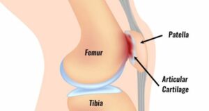
    3D modeling can provide more accurate assessments of bone health

    Researchers in Sweden have developed a method to create a 3D model of a patient’s bones based on information from 2D X-ray images.

    The report, published in the Journal of Bone and Mineral Research, concluded that they were able to identify thousands of patients at risk of fractures who would be overlooked by current assessment methods.

    Relevant excerpt

    Lorenzo Grassi, associate professor of biomedical engineering at Lund University, was responsible for evaluating the method. He explains that the new method uses 2D X-ray images of bone density measurements to produce 3D models of the femur.

    “The shift from 2D to 3D is performed using a computer-simulated template that describes how bone geometry and density varies in the population.”

    The 3D model of the femur can be used to simulate various situations and scenarios that could have an influence, for example in the event of a fall. Information that makes it easier to estimate the risk of fractures.”2

    Grassi developed 3D simulations using data from 400 study participants who had previously undergone X-ray-based bone mineral density assessments. When he compared the accuracy of each method in predicting which patients would break their hip over the next decade, the 3D simulation provided a more accurate prediction.

    This new method could provide patients with a more comprehensive understanding of their bone health compared to DXA scans. However, because the modeling uses the information from the 2D X-ray, it would complement rather than replace DXA technology.

    Short content

    Researchers in Sweden developed a method for creating a 3D model of a patient’s bone from 2D DXA scans. Their 3D model proved to be more effective than DXA scans in predicting fractures.

    Evolutionary biology provides new data on bone regeneration

    Scientists in Finland have discovered hundreds of non-collagenous proteins in the bone matrix that may play a role in bone formation and regeneration.

    Their research, conducted as part of the PhyloBone project, uses the principles of evolutionary biology to identify molecular mechanisms that maintain bone health.

    Relevant excerpt

    “Since the bone matrix, which constitutes the majority of bone mass, plays both structural and regulatory roles, non-collagenous organic components play a key function in bone regulation. For example, it is known that few non-collagenous proteins, such as osteopontin, play an important role in bone formation. However, the bone matrix consists of hundreds of proteins that are still poorly understood and that may play an important regulatory role in bone regeneration and osteoporosis.

    “Our project has identified 255 proteins in 30 species of vertebrates. The aim of the project is to serve as a valuable resource for further research in bone regeneration, osteoporosis and related areas,” says Dr. Puigbò, co-principal investigator of the PhyloBone project.”3

    The project will continue to conduct studies to determine the regulatory role of bone proteins. They have shared their data on these understudied proteins with the scientific community, with the aim of encouraging further research and new discoveries in the field of bone matrix proteins.

    Hopefully, this research will lead to new knowledge on how bone regeneration can be supported and enhanced through behavioral and lifestyle changes. However, it may also stimulate the development of new drugs against osteoporosis.

    Short content

    Finnish scientists have identified hundreds of proteins in the bone matrix that may play a role in bone formation and regeneration. They have made their data on these underdeveloped proteins available to the scientific community, which will hopefully lead to more research and new discoveries.

    What this means for you

    New research offers new insights and information. We can apply this knowledge in our pursuit of stronger, healthier bones.

    The connection between the latest scientific research and an informed, drug-free approach to bone health is at the heart of the Osteoporosis Reversal Program. The Save Institute uses scientific research published in mainstream journals to ensure its drug-free approach is the safest and most effective method for reversing and preventing osteoporosis.

    As science advances, it increasingly emphasizes the importance of diet, exercise and lifestyle as the key tools for maintaining healthy bones and ensuring a robust, long and independent life.

    References

    1 https://www.revyuh.com/news/lifestyle/health-and-fitness/how-to-avoid-the-risk-of-osteoporosis-study-suggests-a-new-way-to-fight-brittle- bone disease/

    2 https://www.news-medical.net/news/20230913/New-method-could-improve-prediction-of-osteoporotic-fracture-risk.aspx

    3 https://medicalxpress.com/news/2023-08-bone-regeneration-osteoporosis-evolution.html


    Source link

  • Management of complex regional pain syndrome (CRPS)

    Management of complex regional pain syndrome (CRPS)


    By Russell Seemler, South Chandler Clinic

    Living with Complex Regional Pain Syndrome (CRPS) can be a challenging and disabling experience. However, physiotherapy can tackle CRPS. Physiotherapy is an essential part of the overall treatment plan for CRPS and is usually tailored to the specific needs of the individual and the severity of their condition.

    What is complex regional pain syndrome?

    Complex regional pain syndrome (CRPS) is considered by some to be the most painful medical condition in the world. CRPS is considered an injury or malfunction of the peripheral and central nervous systems, manifesting as extreme pain and other physiological symptoms rolled into one. or more limbs of the body. These symptoms can last for months to years and, if not treated, can become permanent.

    Diagram of complex pain syndrome.

    The causes and triggers of complex regional pain syndrome

    The central nervous system consists of the brain and spinal cord. The peripheral nervous system consists of all other nerves in the body, minus the brain and spinal cord. The peripheral nerves deliver signals to and from the brain and spinal cord to different parts of the body. An injury to one of the peripheral nerves is believed to be the initial onset of CRPS. An injury such as a broken bone, a strain or sprain, a burn, a cut, surgery or something as minor as a needle stick can also be the first onset of CRPS.

    In some cases, infections or blood vessel problems can also trigger the cascade to CRPI. Once an initial injury occurs, signals from the peripheral nerves to the brain tell a person that he or she has been injured. The brain then sends signals to different parts of the body telling them to start releasing chemicals that will trigger many physiological responses in the body necessary to initiate the healing process. One of the first things a body does to protect and heal itself is send out chemicals and nerve signals that cause swelling, increased blood flow to the injured area, and pain. These are normal parts of the healing process.

    Signals from the brain

    With CRPS, a major problem is that the signals from the brain that cause pain, swelling, and increased blood flow never go away. Even after the injury has completely healed. The brain then tricks the body into believing it is still injured, even though it is not. As this continues, the signals can become stronger and stronger to the point where constant, extreme pain lasts for months to years and even permanently.

    There is no clear reason why someone who breaks their arm, for example, gets CRPS, while many others who break their arm do not. However, they have discovered that CRPS is more common in women than in men and that the risk is greatest around the age of 40. However, it can occur in either sex at any age.

    Treatment of complex regional pain syndrome.

    The symptoms of complex regional pain syndrome

    Signs and symptoms of CRPS can vary slightly from person to person depending on what stage of the condition they are in. Some patients may have all the symptoms mentioned or just some. Also, symptoms may change over time depending on how long ago CRPS started.

    • Extreme pain – burning, tingling and crushing/squeezing pain
    • Changes in skin color – the skin may become more pink, red, purple or blue compared to the other limb
    • Changes in hair or nail growth in the extremities – brittle nails and increased or decreased hair growth, depending on the person
    • Skin changes – the skin becomes shiny and thin
    • Sweating changes – excessive sweating or less sweating compared to the other limb
    • Skin temperature – although people feel like their leg is burning, when they touch the limb it is very cold.
    • Increased joint stiffness and reduced movement in the joint
    • Increased muscle tightness and spasms in the limbs
    • Osteoporosis – shown on x-ray
    • Allodynia – severe sensitivity to normal skin touch, as if being touched after a sunburn

    As the condition progresses and lasts longer, people may also experience:

    • Atrophy of muscles in the limbs due to reduced use
    • Short-term memory problems
    • Difficulty coming up with words while speaking
    • Irritability
    • Depression
    • Increased swelling in the extremities
    • Sensitivity to noise and vibration

    Research has also shown that people in pain have difficulty distinguishing between the injured right and left legs when looking at a photo of the same body part. Many with CRPS have the same problem, as they cannot determine if it is a right or left hand or leg when they look at a photo.

    Treatment for complex regional pain syndrome

    There are many forms of treatment for CRPS, although treatment will be individual for each person depending on their signs and symptoms. What works for one CRPS patient may not work for another, and a combination of treatments may also be necessary.

    • Physiotherapy

    • Your physical therapist will work with you to develop a treatment plan to help you manage your condition. Because the symptoms of CRPS vary, the approach to care will also vary. CRPS treatment is based on how regular and severe your symptoms are. More importantly, physical therapy is focused on helping you achieve your personal goals.
    • Medicines

      • Bisphosphonates – to prevent bone reabsorption, help with osteoporosis
      • Corticosteroids – to prevent inflammation and swelling
      • Botox – for muscle spasms and tightness
      • Drugs for nerve pain – Gabapentin, Amitriptyline, etc.
      • Over-the-counter medications for pain/inflammation – ibuprofen, aspirin, naproxen
      • Topical creams for pain – usually lidocaine
    • Procedural**

      • Sympathetic nerve blocks – they have been shown to help in the short term, but long term benefits are usually not seen.n
      • Spinal cord stimulator – a surgical procedure in which a stimulator is placed directly into the spinal column to control the signals traveling from the brain to the body using electrical stimulation.
      • Surgical sympathectomy – A procedure in which sympathetic nerves that travel from the brain to the affected limb are surgically severed to prevent pain signals from reaching the limb. This is a controversial operation because some doctors think it causes CRP.S
    • Psychotherapy

      • Many people with CRPS develop anxiety, depression and even PTSD. Seeing a psychiatrist or psychologist for help is a highly encouraged treatment strategy.
    • Eating pattern

      • Maintaining a well-balanced diet has been shown to reduce symptoms of CRPS
    • Sleep

      • Maintaining a consistent sleep-wake cycle has been shown to reduce symptoms of CRPS. Because the body regulates hormones such as cortisol, it can worsen pain and sympathetic nerve symptoms.

    *These medications are still in the research phase and are considered experimental
    **It is recommended to try non-procedural treatments before procedural treatments due to reduced risks and potential complications.

    Therapist helping patient with complex pain syndrome.

    How physiotherapy can improve complex regional pain syndrome

    Physical therapy is one of the best treatments for CRPS because of its ability to preserve limb function. Physical therapy will focus primarily on maintaining limb function by using stretches and exercises to keep the muscles loose and strong and the joint moving. In addition, PT can also help with pain relief and edema/swelling. There are many types of physical therapy treatments that have been shown to be helpful.

    • Desensitization therapy – a treatment aimed at reducing allodynia

      • This treatment involves lightly stroking various types of textured objects over the affected or unaffected limb to retrain the pain receptors in the area. The purpose is to tell the pain receptors that light touch should not cause pain in a limb. And to stop sending pain signals to the brain.
    • Graded Motor Imagery (GMI) – a treatment that focuses on retraining the distinction between the right and left limbs and restoring function while reducing pain to the limb

      • This treatment style uses images of right and left arms or legs in different positions.
      • In phase one, the patient only needs to determine whether a limb is a right or left limb.b
      • For phase two, the patient only needs to remember to move the same-sided limb into the correct position; no movement takes place. This turns on the premotor cortex areas of the brain.
      • During phase three, the patient moves the limb on the same side to the position shown in the image.
      • The goal is for all phases to be performed pain-free. And that the next phase will only start if the previous phase is performed pain-free and with 100% accuracy
    • Mirror Box Therapy – This treatment is used to restore function and reduce pain in the affected limb

      • The affected limb is placed in a box that is not visible to the patient
      • A mirror attached to the box is aimed at the unaffected limb. This causes the patient to see a mirror image of the unaffected limb.
      • The mirror image visually tricks the brain into believing it is the affected limb.
      • The patient then performs exercises, stretches, desensitization therapy, graded motor imagery, etc. with the unaffected limb. However, they look in the mirror all the time and trick their brain into believing that the affected limb is performing all movements pain-free.
    • Edema massage/edema taping

      • This treatment focuses on controlling swelling and edema in the area
      • Various massage and taping techniques promote swelling and edema to leave an area. This usually reduces pain, restores normal blood flow, and increases mobility and strength in an area

    CRPS is a serious and extreme pain condition that can be disabling. Physical therapy treatments are the best strategies to maintain function and relieve pain.

    If you think you have CRPS based on the information above, contact your doctor immediately. The sooner a diagnosis is made, the better your chances of going into remission for CRPS. Patients with CRPS should adhere to their physical therapy program. Care and communication with their care team will ensure that the treatment plan is optimized for their specific needs and goals.

    CRPS can be challenging to treat, but with a comprehensive approach that includes physical therapy, individuals can experience improved function and less pain. If you are ready to see a physical therapist, make an appointment with us.

    Source link

  • Game-Changers for Bone Health and Aging: The Power of Vitamin E, Tocotrienols, and Geranylgeraniol with Dr.  Barrie Tan + BoneCoach™ – BoneCoach™

    Game-Changers for Bone Health and Aging: The Power of Vitamin E, Tocotrienols, and Geranylgeraniol with Dr. Barrie Tan + BoneCoach™ – BoneCoach™

    Ready to hidden power of age-old nutrients proven that it benefits your health and your bones?

    Do you want to learn why people with osteopenia, osteoporosis and… those affected by bisphosphonates may benefit of what is in the annatto plant?

    Interested in breakthrough compounds that many consider “aging game changers” for heart, liver, bones, blood sugar, oxidative stress and much more?

    Search no further!

    I had the privilege of working with Dr. Barrie Tan, a leading scientist and pioneer specializing in vitamin E research. Dr. Tan shared the revolutionary science of tocotrienols and geranylgeraniol and their crucial role in this intervention and prevention of osteoporosis.

    Episode timeline

    0:00 – Episode begins

    2:24 – Meet our guest, Dr. Barrie Tan

    4:03 – Dr. Tan’s discovery of the annatto plant and his motivation for studying it
    7:48 – An introduction to tocotrienols, geranylgeraniol and their differences

    9:48 – Understanding vitamin E and the meaning of tocopherol

    19:01 – The function of geranylgeraniol

    20:53 – Benefits of tocotrienols for osteoporosis and bone health

    30:31 – The reason behind Dr.’s specific research Barrie Tan

    32:17 – The role of geranylgeraniol in MK4 synthesis and its importance for bone health

    39:19 – Benefits of MK4: Kidney stones, calcification of the arteries and dementia

    41:34 – Discussion on preclinical and animal studies on geranylgeraniol, its effects on bone turnover, stiffness and its benefits for people taking bisphosphonates

    47:52 – Overview of 20 clinical studies on tocotrienols and their various benefits

    57:02 – Summary of key studies and results specifically focused on tocotrienols

    1:00:17 – Dr. Tan elaborates on recent studies, insights about statins and other crucial findings

    1:06:35 – Guidance on where to find the exact dosages and amounts of the nutrients and ingredients discussed

    Sources mentioned

    **Show notes @ https://bonecoach.com/drbarrietan-vitamine

    Below you will find resources from Dr. Barrie Tan!

    >> CLICK HERE TO SAVE 10% ON ANNATROL BONE SUPPORT

    >> Click here to use the code “BONECOACH” for your copy of Dr.’s Vitamin E book. Barrie Tan!

    What can you do to support your bone health and this podcast?

    1. Press the “Subscribe” button on your respective podcast player (i.e. Apple, Google, Spotify, Stitcher, iHeart Radio and TuneIn). Never miss an episode that can help improve your bone health.

    2. Leave a review. The more positive ratings and reviews and the more subscribers we have, the more people can find us and get the answers to the questions they need. Thank you! 🙂

    3. Tell a friend about The Bone Coach Podcast or share via text, email or social. Do you know of a Facebook group where people can benefit from this information? Feel free to hit any of the share buttons below.

    About Dr.Barrie Tan:

    Dr. Primarily a scientist with a PhD in chemistry/biochemistry, Barrie Tan was previously an assistant professor at the University of Massachusetts Amherst (Chemistry, Food Sciences and Nutrition). For the past 35 years, Barrie has immersed himself in the world of vitamin E and is considered one of the world’s foremost experts, credited with commercializing tocotrienol in three major natural sources: palm, rice and annatto. His first discoveries involved palm and rice, but due to their high tocopherol content they also lacked potency. Barrie continued his research and was soon rewarded when he found the best in class tocotrienol in the annatto plant! Barrie’s relationship with Annatto spans more than twenty years and multiple clinical trials in chronic diseases. He was rewarded again when he discovered geranylgeraniol, an endogenous nutrient important for promoting healthy aging.

    Medical disclaimer

    The information shared above is for informational purposes only and is not intended as medical or nutritional therapy advice; it does not diagnose, treat or cure any disease or condition; it should not be used as a substitute or substitute for medical advice from physicians and trained medical professionals. If you are under the care of a healthcare professional or are currently taking prescription medications, you should discuss any changes in your diet and lifestyle or possible use of nutritional supplements with your doctor. You should not stop prescribed medications without first consulting your doctor.

    Source link

  • What is that pain under my kneecap?  Understanding Patellofemoral Pain Syndrome – ACL Strong

    What is that pain under my kneecap? Understanding Patellofemoral Pain Syndrome – ACL Strong


    Patellofemoral pain syndrome

    That dull, aching, throbbing, sometimes sharp pain around the knee or kneecap that comes and goes based on activity may be a condition known as Patellofemoral Pain Syndrome. Sometimes associated with cracking and popping, patellofemoral pain can range from not so bad to disabling discomfort causing people of all ages to avoid activities such as walking, jogging or participating in a sport.

    What causes patellofemoral pain?

    With patellofemoral pain syndrome, discomfort in or around the front of the knee is often due to irritation of the cartilage that lines the underside of the kneecap in the joint. Stress on the knee can cause the cartilage to become inflamed, which can ultimately lead to thinning and fraying of the tissue over time. In some cases, athletes begin to notice symptoms after years of competition and training, even without immediate injury. In other cases, people only report this pain days, weeks, or months after an increase or change in activity. If the causes of patellofemoral pain syndrome are not treated, they can eventually lead to tissue degeneration in the knee, such as osteoarthritis.

    patellofemoral pain800 800x426 1 800x426 1

    The patella, or kneecap, is a floating bone connected to the femur and shin bone by tendons and ligaments. The bottom, or back part, of the patella is covered with a layer of cartilage, which helps it slide smoothly over the thigh bone when you bend and straighten your knee. There should be sufficient joint space between the patella and the underlying femur, although this joint space sometimes narrows, increasing the chance of contact or friction between the bony surfaces.

    A variety of factors including anatomy, soft tissue mobility, biomechanics (movement quality), and physical activity may contribute to a higher risk of patellofemoral syndrome. For example…

    • The anatomical resting position of the patella may be slightly higher than normal, also called ‘patella alta’, which can make the patella more sensitive to gradual wear.
    • A patella that is hypermobile or has too much mobility can bump and rub against the underlying bone, irritating the cartilage and causing pain. Conversely, a patella that is hypomobile or limited in mobility may become painful due to too much force being placed on a particular part of the patella.
    • In terms of movement, the patella moves over the femur, but the femur also moves under the patella. The biomechanics of the entire leg must be addressed to understand what is happening at the knee. The hip and foot play an important role in what the knee experiences. For example, someone with overpronated feet or weakness in the hip muscles may experience knee pain due to altered mechanics and resulting pressure on the cartilage lining of the patella. Abnormal biomechanics can be corrected through exercises prescribed by a physical therapist.
    • Physical activity also contributes to patellofemoral pain. The intensity or load of the activity can place more force on the knees than the body can safely tolerate. Activity modification may be necessary to control symptoms. For example, exercises such as running can be avoided for a while to calm symptoms such as swelling and pain.

    If the contributing factors are not addressed, the friction and friction between the surfaces can worsen, leading to thinning and fraying of the cartilage, wear and tear of the joint, and ultimately bone-on-bone osteoarthritis.

    What is the best treatment for patellofemoral pain syndrome?

    The recommended treatment for patellofemoral pain generally focuses on strengthening the muscles of the leg, from the hip and trunk to the foot, along with maintaining or increasing flexibility to reduce stress on the patella. Normalizing strength, flexibility and biomechanics through the limbs will help reduce pressure on specific areas of the cartilage that have become irritated and painful, allowing activity and exercise to be comfortable again.

    In addition to strengthening, ice and anti-inflammatory medications, such as ibuprofen, are often recommended to reduce swelling and relieve pain during the acute phase. Reducing painful activities such as climbing stairs can be helpful in recovering from patellofemoral pain syndrome. Although you limit painful exercises, you can still be active by incorporating alternative, non-painful exercises that will strengthen the knee around the knee and reduce irritation to the cartilage and other structures that support the patella. For example, a runner with patellofemoral pain can substitute swimming a few times a week to stay active without aggravating the knee.

    Have I waited too long? Is it too late for me?

    No matter how long you have been suffering from knee pain, it is never too late to build strength. Focusing on correcting your mechanics during exercise and exercise can help reduce your pain over time. Strengthening the muscles around the knees allows you to move with better control and precision, which also improves performance. Increasing body control provides more stability in the patellofemoral joint, which can take pressure off the patella and reduce the risk of further cartilage breakdown.

    Can it be cured?

    Patellofemoral pain syndrome is one of the most common causes of knee pain and one of the most common conditions resolved with physical therapy. A supervised exercise program to address the underlying causes is the best treatment strategy to relieve patellofemoral pain.

    Our courses at ACL Strong are designed and programmed by physiotherapists to give you complete confidence that the exercises you perform are both safe and effective.

    Many of our members experience an unexpected benefit when they enroll in an ACL Strong course… their patellofemoral pain improved because they trained smarter and reduced the strain on their knees.

    “I can’t believe I’m skiing without pain for the first time in years!”

    “The knee pain I used to have while playing football is no longer there!”

    “One of the biggest benefits we have seen in our athletes through ACL Strong is that it helps them resolve old injuries that have been bothering them.”

    When you take part in the ACL Strong Snow Course or Classic Course, you will learn how to care for your knees in the long term, so you can be as active or competitive as you want, and for as long as you want.

    👉 Start strengthening and training smarter today!

    Source link

  • “If you’re trying to get pregnant, you should actually have sex”

    “If you’re trying to get pregnant, you should actually have sex”


    Screen Shot 2021 03 14 at 2.19.43 PM

    When I first started thinking about getting pregnant after being diagnosed with rheumatoid arthritis, I was eager to hear from other women who had succeeded in this goal. At the time there wasn’t much social media, but I did find a great book called Arthritis, Pregnancy and the Path to Parenthood. It contained quotes and advice from real women, and I found it invaluable.

    But I also discovered a problem: the book started by talking about the possibility of changing some of your medications before getting pregnant, and then skipped straight to being pregnant. But wait! If I stop taking my meds, won’t I flare up? And if I’m in pain, how can I ever get pregnant? Is there a chapter missing from this book about trying to get pregnant while living with arthritis?

    That’s the question I asked Iris Zink, a rheumatology nurse who recently wrote a book with Jenny Thorn Palter about intimacy and chronic illness. (The book is called “Sex – Interrupted” and you can read my review of it here!) Their book recommends many alternatives to intercourse – which I think is generally good advice for maintaining intimacy in a relationship while dealing with a chronic illness! But what if you want to start a family? If you’re trying to get pregnant, you actually have to have sex!

    Iris and I decided to have a discussion about a topic we haven’t seen anyone else talk about: the challenges many women face when actually trying to conceive while living with a chronic illness. I share my personal experiences, and Iris shares her expert advice this video!

    Source link

  • 6 tips for weeding your mental garden

    6 tips for weeding your mental garden

    I love gardening, tilling the soil, planting little seeds and watching them grow. A few weeks ago, while picking crabgrass from the kale, I found myself thinking about my clients and their concerns about bone health. I was thrilled to think about the extent to which mental and emotional stress robs us of both happiness and bone health. With this thought in mind, a “Garden life” metaphor came to mind. The metaphor was simple and applies to backyards as well as mental landscapes. The metaphor is: “Nourish the desired and remove the unwanted.”

    Two weeks later, after attending the revival of Acharya Shunya Roar like a goddess workshop, I was inspired to write this one 6 tips for weeding your mental garden. They are simple “Acharya Shunya-inspired” self-awareness exercises that you and I can use to create inner peace and a mind that works for us, not against us. (In case you haven’t heard of her work, Acharya Shunya is a leading spiritual leader of our time, a best-selling author, a Vedic scholar, and the first female custodian of a 2,000-year-old Vedic wisdom lineage. I had the great fortune to recently interviewed her at the Omega Institute. At the end of this blog, you can learn more about her work and how to enjoy this wise video interview.) Susan Garden 4

    This is how the story goes and here are your 6 powerful tips for creating the bone strength and destiny you desire:

    Every spring, six of my friends and I plant a beautiful vegetable garden in the backyard. While none of us are expert gardeners, we enjoy watching the bloom of life and thinking of ways to nourish this new life with fertilizer, water, sunshine and our love. We also keep an eye on weeds as unwanted visitors can easily take over the entire garden if not kept in line. The more we care for our garden, the more beautiful and fruitful it becomes.

    The garden-life metaphor

    What I realized was that we plant “seeds” in our minds with every thought, belief, desire, and intention. These ‘seeds’ can be life-sustaining positive thoughts that carry an energy of appreciation, happiness, contentment or even love. Or the thought may carry an energy of negativity, blame, anger, worry and even fear. In our mindscape we fertilize these ‘thought seeds’ by the attention we pay to them. Whether we are aware of it or not, what we focus on becomes more powerful in our daily lives and in our mental garden.

    Just as we can produce a richer and more beautiful garden if we limit the weeds, we can with awareness control the unwanted “mental weeds” that take up space and spread their vibration throughout our entire mindscape.

    And what exactly are ‘mental weeds’?

    “Mental weeds” are repetitive thoughts and beliefs that invade and disrupt the natural, calm, peaceful, and happy feelings in our minds. These weeds are disruptive, negative, voluntary mental pop-ups that include fear, self-doubt, anger, worry and anxiety. The mental and emotional negativity of this “thought weed” has been well documented as a contributing factor to many chronic degenerative diseases, including osteoporosis (as described in the Better Bot Solutions Course and my blog, How bot responds to danger).

    This mind-body connection is well established. The Bible says that, for example “A cheerful heart is good medicine, but a crushed spirit dries up the spirit bones.(Proverbs 17:22) Even more striking is the 5,000 year old Ayurvedic wisdom that Acharya shared with me. She said that in our conversation another word for ‘health’ in the ancient Vedic texts was ‘happiness’. The crowning achievement of all this came at the end of our interview, when Acharya Shunya applauded me for discovering that worry and fear damage the bone. She talked about the fact that ancient Vedic texts mention this Bone is the strongest material in the body, the strongest, but the only thing that can erode it is negativity. Thinking about those pearls of wisdom fueled my motivation to increase the level of my mental/emotional gardening!

    6 tips for weeding your mental garden

    Inspired by the Vedic scholar Acharya Shunya

    1. Take inventory of your thoughts.
    • Experiment with focusing on one thought and recording the “feel” of the thought. Was the thought associated with a feeling of well-being or even contentment? Or was it critical of others or yourself? Was it humiliating to yourself or to someone else? Was it a good feeling?
    • Ask yourself, “Is this a thought or belief that I want to continue and influence in my life every day?” Or can I just let this article pass?
    • Be aware and remind yourself that the thoughts you repeat over and over become the strongest weeds.
    1. Do some mental housekeeping.
    • Remember: you are not your thoughts. You are the witness of your mental wanderings.
    • With practice you can create a little distance between yourself and your thoughts.
    • You can become a witness to your thoughts. You can realize yourself as the consciousness behind your thoughts.
    • As a witness, you then consciously choose which thoughts you want to strengthen and which thoughts you want to let through.
    • Once again, remember that what you focus your attention on will become stronger in your life.
    • Remove the thoughts and associated feelings that you would rather not have in your life.
    • Moreover, you don’t have to hold on to every thought; you have enough left.
    • In fact, you have about 90,000 thoughts a day, most of which passed through your mindscape yesterday.
    • As spiritual teacher Eckhart Tolle suggests, it’s a good habit not to take your thoughts so seriously.
    1. Meditate on the wisdom of this ancient Vedic observation that Acharya taught me:
    • Every action leads to habit.
    • Every habit builds character.
    • And character leads to fate.
    • As she suggests, don’t give space in your beautiful mind to a single thought without purpose.
    1. Make your mind your servant, not your master.
    • When your mind is in turmoil or overshadowed by worries, stop whatever you are doing.
    • Take a few deep breaths and shift your attention to what it feels like to breathe in and out.
    • Creating even a small space between you and your worrying thought calms the mind.
    • Then ask yourself, “Can I acknowledge this thought and the associated feeling and then let it go?”
    • Or should I do a reality check to verify whether what I think is actually true?
    • Or is it time to gather my sovereign power and address the situation?
    1. Be kind and gentle to yourself and others.
    • Remember that it is the nature of the mind to move quickly, just as it is the nature of the monkey to jump from branch to branch.
    • You can’t always control your monkey mind.
    • However, your sovereign self can witness the thoughts, and you realize that you are not your mind.
    • You can create a gap between your thoughts and emotions and your deepest self.
    • As you repeatedly rest in this gap, gently sow the seeds of your desires and imagine the mental landscape you desire.
    1. Check your mental/emotional tone every morning when you get up.
    • Where did your thoughts wander during the night, what are your feelings?
    • When I wake up cheerful and happy, I am grateful to the great goddess for a refreshing sleep.
    • If I wake up less cheerful, I immediately call on one of my inspiring spiritual teachers, consciously setting a higher vibration.
    • You might start by asking yourself: What vibrational tone do I want for today?
    • Consciously determine the tone you want to spend the day.
    • And then ask: what can I do to move my energy field in that direction?
    • There are countless activities that are cutting edge, and the choice is yours. Options include meditation, prayer, reading or listening to uplifting speakers, wisdom teachings or scriptures, taking a sunrise walk, hitting the yoga mat, music, chanting. The tone options are endless. Enjoy it and see what works best for you.

    In closing, let me say that this is easier than you might think. As Acharya reminded me, it takes some time to change the constitution/organization of the physical body; you don’t change the bone fragility score overnight. But the constitution/organization of the mind can change in a flash. If you change your mind, you immediately change your life!

    So let’s all get started! Do the work, start designing a mind that serves your bones and your greatness! I’ll be there next to you.

    Join my upcoming video interview with Acharya Shunya on YouTube on October 5 by clicking Notify Me on my YouTube channel

    Acharya Shunya and Dr. Susan Brown

    Acharya Shunya’s website: awakensself.com

    The books of Acharya Shunya:

    • Ayurveda lifestyle wisdom: A complete recipe to optimize your health, prevent diseases and live with vitality and joy, Sounds true, 2017.
    • Sovereign self: Claim your inner joy and freedom with the empowering wisdom of the Vedas, Upanishads and Bhagavad Gita, Sounds true, 2020.
    • Shout like a goddess: Every woman’s guide to becoming unapologetically powerful, prosperous and peaceful, Sounds true, 2022.

    Dr. Susan BrownI am Dr. Susan E. Brown. I am a clinical nutritionist, medical anthropologist, writer and motivational coach speaker. Learn my proven 6-step natural approach to bone health in my online courses.

    Source link

  • How Sleep Improves Bone Health – Bone Talk

    How Sleep Improves Bone Health – Bone Talk

    shutterstock 124050577 2

    You’ve probably heard someone say that children seem to grow bigger overnight evidence gathered from extensive research suggests that this is probably true. Many people have a busy schedule that prevents them from sleeping well. However, you should not forget that good sleep is good for your mental state And physical health. This is how sleep affects your bone health.

    The Healing Power of Zzz’s: Sleep and Bone Remodeling

    Your body constantly renews, repairs and grows its bones. There is a direct correlation between the amount of sleep you get and the health of your bones. Researchers have made the link individuals who receive shorter sleep duration have lower bone mineral density and a higher risk of osteoporosis. The study, conducted in postmenopausal women, found that women who get five or fewer hours of sleep per night have lower bone mineral density in the spine, neck, hip and even across the body.

    Several healthy processes are affected by the amount of sleep you get, and one of these processes is bone remodeling. Your body’s special bone cells, osteocytes, manage bone remodeling. These cells cause various actions in the body, such as helping your bones maintain optimal mineral levels and healing damaged areas. For example, the cells will activate other cells known as osteoclasts, causing them to remove minerals from your bones if your calcium levels get too low. The cells also give rise to osteoblasts, which help them rebuild and repair your bones if you suffer several fractures. The bone remodeling processes are likely to be less effective for individuals who do not get enough rest.

    Level up: Improve your sleep hygiene for better bone health

    The general rule of thumb is that individuals who sleep and rest longer tend to have healthier bones than those who don’t. Bone growth and repair are facilitated by a good night’s sleep, as the rest gives your body enough time to repair and reshape itself.

    Signs of poor sleep hygiene include having trouble sleeping, experiencing daytime sleepiness, and experiencing sleep disturbances. These are the most telling signals; However, another concern to consider is persistently poor sleep quality. Over time, poor sleep hygiene can cause these problems to persist and potentially worsen other health problems.

    Creating one healthy sleep routine is important for both your physical and mental health. It improves your productivity and quality. Good sleep hygiene is vital for children and adults; however, it is even more important for individuals likely to be affected by bone-related conditions.

    Good sleep habits are good for your health because they create consistency and positive reinforcement for all aspects of life. Good sleep hygiene can be the result of adapting your environment to your needs and establishing the right routines.

    Pillow Talk: tips for bone-strengthening sleep

    There are many of them steps you can take to improve your sleep experience. You need to optimize your sleep schedule, daily routines, and bedtime routines to help you get better sleep quality. Eat rightget enough fysical activityand create a pleasant environment in which you can relax and fall asleep easily. Here are a few more tips to help you improve your sleep routine:

    • Prioritize your sleep: You need to prioritize sleep if you want healthy bones, body and mind. Calculate your ideal sleep duration by taking into account the time you wake up and make this a regular part of your daily routine.

    • Set a fixed alarm time: Keeping a consistent wake-up time, regardless of the day of the week, can help you regulate your sleep patterns and improve sleep quality.

    • Maintain a regular sleep schedule: Consistency helps synchronize your body’s internal clock.

    • Create a sleep-conducive environment: Keep your bedroom dark, quiet and cool. The ideal temperature is about 65-68 degrees.

    • Limit screen time before bed: Exposure to blue light from screens can disrupt melatonin production.

    • Make gradual adjustments: Try to adjust your sleep pattern gradually. Make adjustments of half an hour or an hour each day until you adjust to your schedule.

    • Don’t take many naps: Avoid taking many naps during the day as this can affect your sleep patterns. Keep naps short and limit them to the early afternoon.

    • Prioritize nutrition: Avoid heavy meals and caffeine right before bed, opting for sleep-supporting snacks instead.

    • Add physical activity: Regular exercise can improve sleep quality, but avoid vigorous exercise before bed.

    • Deal with stress: Techniques such as meditation and deep breathing can alleviate sleep-disrupting stressors.

    Incorporating these practical tips into your daily life can make a significant difference in both the quality of your sleep and the health of your bones. By taking proactive steps to improve your sleep habits, you’re investing in a healthier, more resilient future for your bones. So go ahead, prioritize sleep and let your body do its nightly magic for stronger, healthier bones. Good night!

    Source link