Tag: Knee Anatomy

  • Effective Knee Injury Exercises for Rehabilitation

    Effective Knee Injury Exercises for Rehabilitation

    Knee injuries can be debilitating, but with the right rehabilitation exercises, individuals can regain strength and mobility. This article explores effective exercises for knee injury rehabilitation and provides insights into implementing a comprehensive rehabilitation program to support recovery and long-term wellness.

    Key Takeaways

    • Proper exercise techniques are crucial for knee injury rehabilitation.
    • Range of motion exercises help improve flexibility and reduce stiffness in the knee joint.
    • Strengthening exercises are essential for rebuilding muscle strength and stability around the knee.
    • Balance and stability exercises aid in improving coordination and preventing future injuries.
    • A comprehensive rehabilitation program should include physical therapy, nutritional considerations, and psychological support for holistic recovery.

    Understanding Knee Injuries and Rehabilitation

    thumbnail

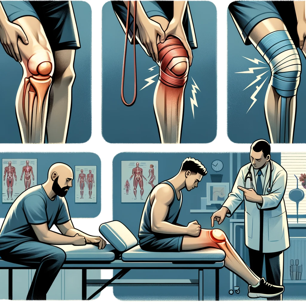
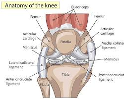
    Common Types of Knee Injuries

    Knee injuries are a common occurrence, and they can significantly impact our daily lives. Understanding the specific type of knee injury is crucial for effective rehabilitation. It is important to note that each injury requires a tailored approach to rehabilitation, taking into account the severity and nature of the injury. For example, a ligament tear may require a different rehabilitation program compared to a meniscus injury. Understanding the nuances of each injury type is essential for successful recovery.

    Principles of Knee Rehabilitation

    In our approach to knee rehabilitation, we prioritize a thorough understanding of the injury’s nature and the individual’s specific needs. It’s crucial to tailor the rehabilitation program to address these unique factors, ensuring the most effective recovery process.

    Rehabilitation should be a progressive journey, starting with the reduction of pain and swelling, followed by the restoration of joint mobility. Once these initial goals are achieved, we focus on rebuilding strength and coordination. Here’s a simple list of principles we adhere to:

    • Gradual progression of exercise intensity
    • Regular monitoring of pain and discomfort levels
    • Incorporation of functional exercises that mimic daily activities

    Remember, patience and consistency are key. Rushing the process can lead to setbacks, so it’s important to advance only when ready.

    We also emphasize the importance of education in rehabilitation. Understanding the purpose behind each exercise and how they contribute to recovery can significantly enhance motivation and adherence to the program.

    Importance of Proper Exercise Techniques

    Proper exercise techniques are crucial for effective knee injury rehabilitation. It is essential to perform each exercise with precision and attention to detail. This ensures that the targeted muscles and ligaments are engaged optimally, promoting a more efficient recovery process. Additionally, maintaining proper form during exercises helps prevent further injury and promotes overall joint health. It is important to note that exercises should be tailored to individual needs and capabilities, taking into account the specific nature of the injury and the stage of rehabilitation. This personalized approach ensures that the exercises are both challenging and safe, maximizing the benefits of the rehabilitation program.

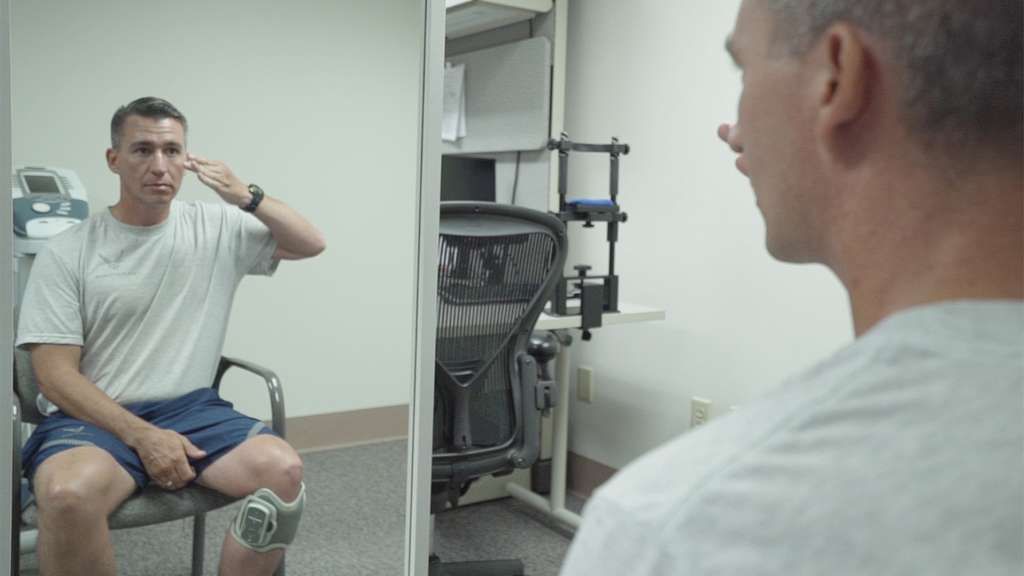
    Effective Exercises for Knee Injury Rehabilitation

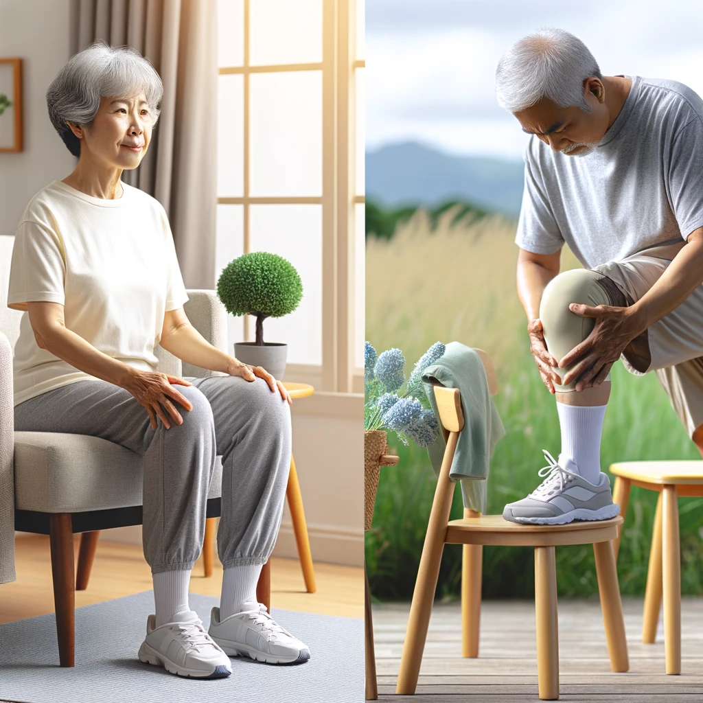
    Range of Motion Exercises

    After completing the range of motion exercises, we move on to strengthening exercises to build muscle strength around the knee joint. These exercises are crucial for improving stability and preventing future injuries. It’s important to gradually increase the intensity and resistance of the exercises to promote muscle growth and endurance. Additionally, incorporating a variety of exercises that target different muscle groups is essential for a well-rounded rehabilitation program. We recommend consulting with a physical therapist to customize a strengthening regimen that suits individual needs and limitations.

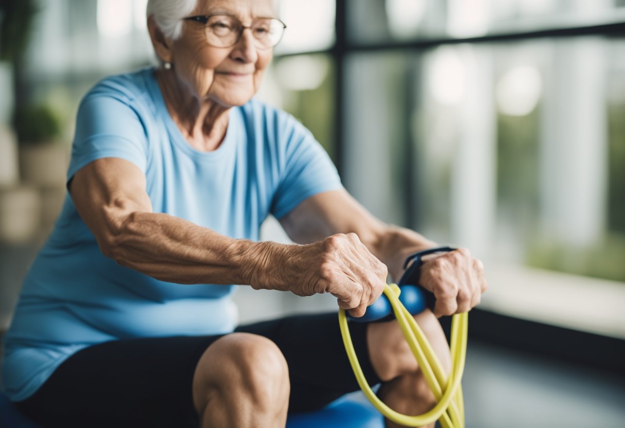
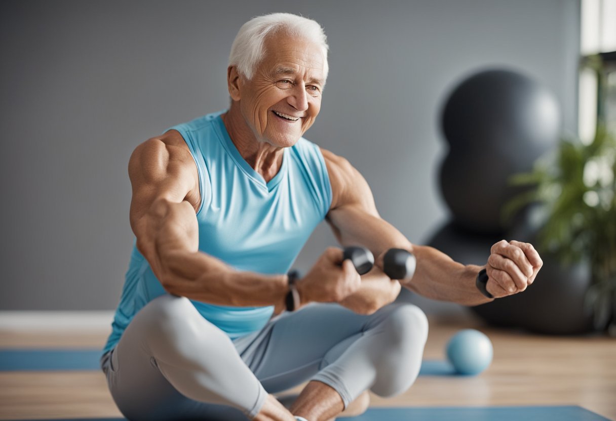
    Strengthening Exercises

    In our rehabilitation journey, we recognize the pivotal role of strengthening exercises. These exercises aim to rebuild muscle power around the knee, which is crucial for both stability and movement. We start with isometric exercises, where the muscle length doesn’t change during contraction, and gradually progress to more dynamic movements.

    Quadriceps sets and straight-leg raises form the foundation of our strengthening routine. It’s essential to maintain proper form to avoid undue stress on the knee. As strength builds, we incorporate exercises like step-ups and mini-squats, always mindful of the knee’s alignment and pain thresholds.

    Remember: Consistency is key in strengthening exercises, but never at the expense of correct technique.

    We also emphasize the importance of a tailored exercise regimen. Here’s a simple progression outline we follow:

    1. Isometric exercises
    2. Straight-leg raises
    3. Half-squats
    4. Leg presses
    5. Gradual introduction of resistance

    Each step is taken with careful consideration of the individual’s pain levels and overall recovery progress. If a client experiences a setback, such as an ACL setback, we reassess and adjust the program, ensuring they can overcome the challenge and emerge stronger.

    Balance and Stability Exercises

    After focusing on balance and stability exercises, we must transition towards implementing a comprehensive rehabilitation program. This holistic approach ensures that all aspects of recovery are addressed, including the physical, nutritional, and psychological components.

    Physical therapy plays a crucial role in guiding patients through the recovery process. It’s essential to work with a qualified therapist who can tailor exercises to individual needs and progress. A therapist’s expertise can help prevent re-injury and ensure that rehabilitation goals are met efficiently.

    Nutritional considerations are also vital. A balanced diet rich in anti-inflammatory foods can aid in reducing swelling and promoting healing. Incorporating omega-3 fatty acids, antioxidants, and adequate protein is crucial for tissue repair and overall health.

    Lastly, psychological support cannot be overlooked. The journey to recovery is not just physical; it’s also an emotional challenge. Providing encouragement and mental health resources can significantly impact a patient’s motivation and resilience. Remember:

    “A positive mindset is a key to successful rehabilitation.”

    By integrating these elements into a rehabilitation program, we can offer patients the best chance for a full and timely recovery.

    Implementing a Comprehensive Rehabilitation Program

    Role of Physical Therapy

    In our comprehensive rehabilitation program, we emphasize the integration of various therapeutic modalities to address the multifaceted nature of knee injuries. Our approach combines physical therapy, nutritional considerations, and psychological support to ensure a holistic recovery experience. Additionally, we prioritize individualized care to tailor the rehabilitation program to the specific needs of each patient. This personalized approach allows us to address weaknesses, promote flexibility, enhance stability, and improve mobility, ultimately reducing limitations and promoting recovery.

    Nutritional Considerations

    Nutrition plays a crucial role in the rehabilitation process. It is essential to focus on consuming a balanced diet that includes a variety of nutrients such as proteins, carbohydrates, and vitamins. Additionally, maintaining proper hydration is vital for overall recovery. A well-structured meal plan can help in achieving nutritional goals and supporting the body’s healing process. Consider incorporating a mix of lean proteins, whole grains, and antioxidant-rich fruits and vegetables into your diet to promote optimal recovery. It’s important to consult with a registered dietitian or nutritionist to personalize your meal plan according to your specific needs and dietary preferences. Furthermore, it’s beneficial to monitor your progress and make adjustments to your diet as needed to ensure that you are meeting your nutritional requirements for effective rehabilitation.

    Psychological Support for Recovery

    Psychological support is crucial for the successful recovery of individuals with knee injuries. It plays a significant role in addressing the emotional and mental aspects of rehabilitation, promoting a positive mindset and overall well-being. Encouragement and motivation from family, friends, and healthcare professionals can greatly impact the recovery process. Additionally, establishing a support network and engaging in open communication can help individuals cope with the challenges of rehabilitation and foster a sense of community and belonging. Emotional resilience and a positive outlook are key factors in achieving successful rehabilitation outcomes.

    Conclusion

    In conclusion, the effectiveness of knee injury exercises for rehabilitation cannot be overstated. By following a structured exercise program, individuals can significantly improve their knee function and reduce the risk of future injuries. It is imperative to consult with a qualified healthcare professional to develop a tailored exercise plan that addresses specific needs and limitations. With dedication and consistency, individuals can regain strength, mobility, and confidence in their knee health.

    Frequently Asked Questions

    What are the most common types of knee injuries?

    The most common types of knee injuries include ACL tears, meniscus tears, patellar tendonitis, and knee sprains.

    What are the principles of knee rehabilitation?

    The principles of knee rehabilitation include reducing pain and inflammation, improving range of motion, strengthening the muscles around the knee, and gradually returning to normal activities.

    Why is proper exercise technique important for knee injury rehabilitation?

    Proper exercise technique is important for knee injury rehabilitation to prevent further damage, promote healing, and ensure effective recovery.

    What are range of motion exercises for knee injury rehabilitation?

    Range of motion exercises for knee injury rehabilitation focus on improving flexibility and mobility in the knee joint through gentle movements and stretches.

    What are strengthening exercises for knee injury rehabilitation?

    Strengthening exercises for knee injury rehabilitation target the muscles around the knee, including the quadriceps, hamstrings, and calf muscles, to improve stability and support.

    How do balance and stability exercises benefit knee injury rehabilitation?

    Balance and stability exercises benefit knee injury rehabilitation by improving proprioception, coordination, and overall stability, reducing the risk of future injuries.

  • Knee Pain Exercises for Seniors: Effective Ways to Relieve Discomfort

    Knee Pain Exercises for Seniors: Effective Ways to Relieve Discomfort

    Knee pain is a common condition that affects many seniors. It can be caused by a variety of factors, including aging, arthritis, and injury. Knee pain can be debilitating, making it difficult for seniors to perform everyday tasks and enjoy their favorite activities. Fortunately, there are exercises that can help alleviate knee pain and improve mobility.

    v2 3n7uu hlzjc

    At our clinic, we recommend a variety of exercises to help seniors manage knee pain. These exercises are designed to strengthen the muscles around the knee joint, improve flexibility, and reduce inflammation. They can be done at home or with the guidance of a physical therapist. It’s important to note that exercise is just one part of a comprehensive treatment plan for knee pain. Seniors should also work with their healthcare provider to address any underlying conditions that may be contributing to their knee pain.

    As we age, it’s common to experience aches and pains in various parts of the body. However, knee pain can be particularly challenging, as it can limit mobility and impact overall quality of life. By incorporating targeted exercises into their daily routine, seniors can take an active role in managing their knee pain and improving their health and wellbeing.

    Understanding Knee Pain in Seniors

    As we age, our body undergoes changes that can lead to various health conditions, including knee pain. Knee pain can be caused by a variety of factors, including injuries, overuse, inflammation, and osteoarthritis. In this section, we will discuss the causes and risk factors of knee pain in seniors, as well as when to consult a doctor.

    Causes and Risk Factors

    Knee pain can be caused by a variety of factors, including injuries, overuse, inflammation, and osteoarthritis. Injuries such as meniscus tears, ligament sprains, and fractures can cause knee pain. Overuse injuries, such as tendinitis and bursitis, can also lead to knee pain. Inflammation caused by conditions such as rheumatoid arthritis can also cause knee pain.

    Osteoarthritis is a common cause of knee pain in seniors. It is a degenerative joint disease that occurs when the cartilage in the knee joint wears away over time. This can lead to pain, stiffness, and swelling in the knee joint.

    Aging is also a risk factor for knee pain. As we age, our bones and joints become weaker and more prone to injury and wear and tear. This can lead to conditions such as osteoarthritis and other joint problems.

    When to Consult a Doctor

    If you are experiencing knee pain, it is important to consult a doctor. A doctor can help you determine the cause of your knee pain and recommend appropriate treatment options. Physical therapy, rehabilitation, and medication are common treatment options for knee pain. In some cases, surgery may be necessary to repair or replace damaged knee joints.

    An orthopedic surgeon or physician can provide medical advice and treatment options for knee pain. A physical therapist can also help with rehabilitation and physical therapy exercises to help improve knee function and reduce pain.

    In conclusion, knee pain can be caused by a variety of factors, including injuries, overuse, inflammation, and osteoarthritis. If you are experiencing knee pain, it is important to consult a doctor to determine the cause of your pain and recommend appropriate treatment options.

    Exercise Program for Knee Pain Relief

    If you are experiencing knee pain, incorporating a regular exercise program can help reduce pain, improve flexibility, and increase strength. In this section, we will discuss low-impact exercises, strengthening and flexibility exercises, and exercise safety and techniques.

    Low-Impact Exercises

    Low-impact exercises are a great way to stay active while minimizing the stress on your knees. Some examples of low-impact exercises include cycling, swimming, water aerobics, and walking. These exercises can help improve cardiovascular health and increase range of motion without putting too much pressure on your knees.

    90e423f3 b6e6 487f 82c6 63a762872275

    Strengthening and Flexibility Exercises

    Strengthening and flexibility exercises can help improve the muscles around your knee joint, providing more support and reducing pain. Some examples of strengthening exercises include squats, lunges, leg raises, and calf raises. For flexibility, try quadriceps, hamstring, and calf stretches. Additionally, exercises like clamshells, leg extensions, and hamstring curls can help target specific muscles like the quadriceps, hamstrings, and hip muscles.

    Exercise Safety and Techniques

    It’s important to warm up before any exercise program and to use proper technique to prevent injury. Stretching exercises, foam rolling, and myofascial release can help prepare your muscles for exercise. Resistance bands can also be used to add resistance to exercises like squats and lunges.

    When starting an exercise program, start slowly and gradually increase the intensity and duration of your workouts. Listen to your body and stop if you experience any pain or discomfort. Always wear proper footwear and use proper technique to prevent injury.

    In summary, incorporating low-impact exercises, strengthening and flexibility exercises, and proper exercise safety and techniques can help reduce knee pain and improve overall knee health.

    Frequently Asked Questions

    v2 3n7vd lzcdk

    What are effective seated knee strengthening exercises for older adults?

    Seated knee strengthening exercises are beneficial for seniors with knee pain who may not be able to stand for long periods. Some effective seated knee strengthening exercises include:

    • Seated leg extension: Lift one leg at a time and extend it straight out in front of you, hold for a few seconds, and lower it back down. Repeat with the other leg.
    • Seated knee lift: Sit on a chair and lift one knee up towards your chest, hold for a few seconds, and lower it back down. Repeat with the other leg.

    Which exercises should be avoided when experiencing knee pain?

    High-impact exercises such as running and jumping should be avoided when experiencing knee pain. Exercises that put excessive strain on the knees, such as lunges and deep squats, should also be avoided. Instead, low-impact exercises such as walking, swimming, and cycling are recommended.

    How can knee osteoarthritis be managed with exercises for the elderly?

    Knee osteoarthritis can be managed with exercises that focus on strengthening the muscles around the knee joint. Some effective exercises for knee osteoarthritis include:

    • Straight leg raises: Lie on your back and lift one leg straight up towards the ceiling, hold for a few seconds, and lower it back down. Repeat with the other leg.
    • Wall squats: Stand with your back against a wall and lower yourself down into a squatting position, hold for a few seconds, and stand back up.

    Can you recommend knee strengthening exercises suitable for seniors to do at home?

    Yes, there are many knee strengthening exercises that seniors can do at home. Some effective knee strengthening exercises for seniors include:

    • Step-ups: Step up onto a low step with one foot, then step back down. Repeat with the other foot.
    • Mini-squats: Stand with your feet shoulder-width apart and squat down slightly, hold for a few seconds, and stand back up.

    What types of leg exercises are safe for seniors with knee issues?

    Low-impact leg exercises such as walking, swimming, and cycling are safe for seniors with knee issues. Strength training exercises that focus on the muscles around the knee joint, such as leg curls and leg presses, can also be beneficial.

    What are the best practices for walking with knee pain?

    When walking with knee pain, it’s important to wear comfortable, supportive shoes and to walk on flat, even surfaces. Start with short walks and gradually increase the distance over time. Use a walking aid, such as a cane or walker, if necessary. If the pain persists, consult a doctor or physical therapist for further guidance.

  • Knee Pain After ACL Surgery

    Knee Pain After ACL Surgery

    Managing Knee Pain After ACL Surgery

    Anterior cruciate ligament (ACL) reconstruction is a common surgical procedure for individuals who have suffered a torn ACL. While outcomes have improved dramatically, knee pain and re-injury continue to be concerns during the recovery process. Recent advances in rehabilitation protocols and biomechanical research are shedding light on optimal recovery strategies.

    9566c6af 8e44 4b28 9740 7d191e646e96

    Understanding Knee Pain After ACL Surgery

    Understanding Knee Pain after ACL Surgery

    Knee pain following ACL reconstruction arises due to multiple interrelated biomechanical and physiological factors. The initial trauma of surgery triggers an inflammatory response which can lead to fluid buildup, swelling, and stiffness, resulting in pain with movement. This acute inflammatory phase usually lasts 6-8 weeks. Concurrently, muscular imbalances around the knee joint develop, most notably quadriceps inhibition and weakness. The quadriceps muscles play a crucial role in stabilizing the knee joint. When weakened and inhibited after surgery, abnormal forces are transmitted through the joint, exacerbating pain and dysfunction. Finally, biomechanical alterations including asymmetric gait patterns and improper landing mechanics cause further pain. A careful evaluation of all these contributing trigger points through a full physical therapy examination is key to pinpointing the optimal treatment approach.

    Causes of Knee Pain

    Knee pain following ACL reconstruction arises due to multiple interrelated biomechanical and physiological factors. The initial trauma of surgery triggers an inflammatory response which can lead to fluid buildup, swelling, and stiffness, resulting in pain with movement.

    Inflammation

    This acute inflammatory phase usually lasts 6-8 weeks.

    A 2022 study found that altered walking biomechanics were present even 2 years after surgery, especially in females (Smith et al). These persistent abnormalities highlight the need for rehabilitation focused on normalizing movement patterns.

    1: Common Causes of Knee Pain after ACL Surgery

    CauseContributing Factors
    Inflammation– Surgical trauma<br>- Swelling<br>- Fluid buildup
    Muscle imbalances– Quadriceps inhibition<br>- Hamstring dominance<br>- Calcium deposits
    Biomechanical abnormalities– Gait asymmetries<br>- Improper landing mechanics<br>- Overpronation

    Managing Knee Pain

    Effective knee pain management requires a multifaceted rehabilitation program tailored to the individual’s specific deficits and dysfunctions. The priority initially is controlling inflammation through ice, compression, medication, and activity modification. Once pain and swelling have been reined in, restoring full knee mobility is essential for reducing stiffness and dysfunction. Gentle manual therapy techniques and range of motion exercises facilitate this process. As mobility improves, muscular imbalances must be addressed through targeted stretching and progressive strength training, focused especially on reactivating the quadriceps. Finally, movement pattern retraining helps ingrain proper mechanics. Areas to emphasize include squatting, lunging, walking gait, running, jumping and landing. The goal is to eliminate any asymmetries or compensations that could lead to abnormal forces through the joint. Throughout this process, communication with the care team is key to calibrate the program to avoid flare-ups while still stimulating progress.

    • Anti-inflammatory medications
    • Ice, compression, elevation
    • Restoring range of motion
    • Eliminating muscle imbalances
    • Normalizing movement patterns

    2: Rehabilitation Phases and Focus Areas

    PhaseTimingGoals
    Inflammation & Early ROM0-2 weeks– Manage swelling/pain<br>- Regain mobility
    Intermediate ROM & Early Strengthening3-6 weeks– Improve quad strength<br>- Single leg stability
    Advanced Strengthening & Neuromuscular Control6-12 weeks– Running mechanics<br>- Multi-plane agility
    Return to Sport4+ months– Sport-specific training<br>- Confidence in knee function

    Muscular Imbalances

    Concurrently, muscular imbalances around the knee joint develop, most notably quadriceps inhibition and weakness. The quadriceps muscles play a crucial role in stabilizing the knee joint. When weakened and inhibited after surgery, abnormal forces are transmitted through the joint, exacerbating pain and dysfunction.

    Biomechanical Changes

    Finally, biomechanical alterations including asymmetric gait patterns and improper landing mechanics cause further pain. A careful evaluation of all these contributing trigger points through a full physical therapy examination is key to pinpointing the optimal treatment approach.

    Managing Knee Pain

    Rehabilitation Program

    Effective knee pain management requires a multifaceted rehabilitation program tailored to the individual’s specific deficits and dysfunctions.

    Early Phase

    The priority initially is controlling inflammation through ice, compression, medication, and activity modification. Once pain and swelling have been reined in, restoring full knee mobility is essential for reducing stiffness and dysfunction. Gentle manual therapy techniques and range of motion exercises facilitate this process.

    Later Phase

    As mobility improves, muscular imbalances must be addressed through targeted stretching and progressive strength training, focused especially on reactivating the quadriceps.

    Movement Retraining

    Finally, movement pattern retraining helps ingrain proper mechanics. Areas to emphasize include squatting, lunging, walking gait, running, jumping and landing. The goal is to eliminate any asymmetries or compensations that could lead to abnormal forces through the joint. Throughout this process, communication with the care team is key to calibrate the program to avoid flare-ups while still stimulating progress.

    Optimizing Long-Term Outcomes

    Preventing Re-Injury

    Due to the high risk of retear after ACL reconstruction, prevention of re-injury is paramount during recovery. Gradually building up strength and then progressing activity in a controlled manner based on specific objective criteria allows tissues to heal while regaining function.

    Maintaining Stability

    Furthermore, consistently practicing ideal movement patterns and positioning trains the neuromuscular system to maintain knee stability. For example, focusing on knee alignment over toes during squats and engaging core muscles protects the ACL graft.

    Return to Sports

    As the graft incorporation stretches beyond 6 months, impact activities can be cautiously integrated. However, pivoting sports may need to wait 9 months or more. Maintaining quadriceps and core strength and allowing the full return of power and agility prior to discharge from therapy ensures the knee can withstand demands of sport. Sticking to activity guidelines, along with smart training periodization can optimize recovery even years after ACL surgery.

    Conclusion

    Knee pain and dysfunction after ACL surgery remain ongoing challenges. However, recent research into biomechanics and rehabilitation is improving long-term outcomes. An individualized, progressive recovery program focused on resolving muscle imbalances and normalizing movement is essential to optimizing recovery.

  • Knee Ligaments

    Knee Ligaments

    Comprehensive Guide to Knee Ligaments: Anatomy, Function, and Injuries

    Knee ligament injuries can be painful and debilitating, affecting the quality of life of many individuals. Understanding knee ligament anatomy and associated conditions can help prevent injuries and ensure proper treatment. In this comprehensive guide, we will explore knee ligaments and their role in knee stability, common knee ligament injuries, and treatment and management strategies.

    v2 3299b pnn1f

    The knee joint is one of the most complex joints in the human body, consisting of bones, cartilage, tendons, and ligaments. Knee ligaments play a crucial role in providing stability and support to the joint. The four main knee ligaments include the anterior cruciate ligament (ACL), posterior cruciate ligament (PCL), medial collateral ligament (MCL), and lateral collateral ligament (LCL). These ligaments work together to control knee movement and prevent excessive rotation and translation.

    Knee ligament injuries are common among athletes and can occur due to sudden twisting or impact to the knee joint. These injuries can cause pain, swelling, and limited mobility, which can lead to disability if left untreated. Early diagnosis and proper treatment can help prevent long-term complications and improve outcomes.

    Understanding Knee Ligaments and Associated Conditions

    At the core of knee joint stability are the ligaments that connect the bones of the knee together. The knee joint is comprised of four major ligaments that connect the femur to the tibia and fibula: the anterior cruciate ligament (ACL), the posterior cruciate ligament (PCL), the medial collateral ligament (MCL), and the lateral collateral ligament (LCL).

    Anatomy of Knee Ligaments

    The ACL and PCL are located inside the knee joint, while the MCL and LCL are located on the outside of the joint. These ligaments are made up of collagen and elastic fibers that provide strength and stability to the knee joint. The ACL is responsible for preventing the tibia from moving too far forward, while the PCL prevents the tibia from moving too far backward. The MCL and LCL work together to prevent the knee from moving side to side.

    Common Injuries and Disorders

    Ligament injuries are a common cause of knee pain and can range from mild sprains to complete tears. ACL tears are one of the most common knee injuries and often occur in athletes who participate in high-impact sports. PCL injuries are less common and typically occur as a result of a direct blow to the front of the knee. MCL and LCL injuries are usually caused by a direct blow to the outer or inner knee, respectively.

    Osteoarthritis is another common disorder that affects the knee joint. It is a degenerative joint disease that is characterized by the breakdown of cartilage in the knee joint. Rheumatoid arthritis is an autoimmune disorder that can also affect the knee joint.

    Epidemiology and Risk Factors

    Knee ligament injuries are more common in athletes who participate in high-impact sports such as football, basketball, and soccer. Overuse injuries are also common in athletes who participate in running and jumping sports. Advancing age, sedentary lifestyle, smoking, and low income are all risk factors for knee osteoarthritis.

    Pain and Mental Health Considerations

    Knee pain can have a significant impact on an individual’s mental health and quality of life. Chronic pain syndromes, such as chronic musculoskeletal pain, can lead to anxiety, depression, and sleep disorders. Local pain, neuropathic pain, somatic pain, and visceral pain are all types of pain that can be associated with knee injuries and disorders. Insomnia and other sleep disorders are common in individuals with chronic pain and can further exacerbate the impact on quality of life.

    The Impact on Quality of Life

    Knee pain and associated conditions can have a significant impact on an individual’s quality of life. Disability, social interactions, and overall physical and mental health can all be affected by knee pain and related disorders.

    Diagnostic Tools and Techniques

    Diagnostic tools and techniques for knee injuries and disorders include X-rays, MRI scans, and physical examinations. Patient assessments are also important in determining the extent of the injury or disorder and developing an appropriate treatment plan.

    In conclusion, understanding knee ligaments and associated conditions is essential for maintaining knee joint health and preventing injury. By understanding the anatomy of knee ligaments, common injuries and disorders, epidemiology and risk factors, pain and mental health considerations, the impact on quality of life, and diagnostic tools and techniques, individuals can take steps to prevent knee injuries and manage knee pain and related disorders.

    Treatment and Management Strategies

    When it comes to knee ligament injuries, treatment and management strategies vary depending on the severity and type of injury. In this section, we will discuss the various treatment options for knee ligament injuries. We will divide the treatment options into five subsections: non-surgical interventions, surgical procedures, pharmacological approaches, interventional strategies, and prognosis and outcome measures.

    Non-Surgical Interventions

    Non-surgical interventions are often the first line of treatment for knee ligament injuries. These interventions include rest, ice, compression, and elevation (RICE), physical therapy, and neuromuscular training. Physical therapy and neuromuscular training are particularly effective for patients with moderate-to-severe chronic pain or brief episodes of knee pain. These interventions can help improve lower extremity kinematics, reduce patellofemoral pain syndrome, and prevent sarcopenia.

    Patient education is an essential component of non-surgical interventions. Patients need to understand the importance of proper posture, body mechanics, and preventive strategies. Patients should also be taught non-pharmacological pain control techniques like meditation, deep breathing exercises, and relaxation techniques.

    Surgical Procedures

    Surgical procedures are typically reserved for patients with severe knee ligament injuries that do not respond to non-surgical interventions. The most common surgical procedures include knee arthroplasty, total knee arthroplasty, hip arthroplasty, and total hip arthroplasty. Preoperative planning and surgical technique are critical to ensure successful outcomes.

    Pharmacological Approaches

    Pharmacological approaches are often used in conjunction with non-surgical and surgical interventions. The most common pharmacological therapies include corticosteroids, mesenchymal stem cells, nonsteroidal anti-inflammatory drugs (NSAIDs), and opioids. Pharmacological therapies can help reduce pain and inflammation, improve functional outcomes, and enhance the patient’s overall quality of life.

    Interventional Strategies

    Interventional strategies are minimally invasive procedures that can help manage knee ligament injuries. These strategies include interventional pain therapy, multimodal analgesia, and multidisciplinary intervention. Interventional strategies can help improve functional level, reduce pain, and enhance functional outcomes.

    Prognosis and Outcome Measures

    Prognosis and outcome measures are essential to assess the effectiveness of treatment and management strategies for knee ligament injuries. Outcome measures include functional outcomes, patient satisfaction, and quality of life. Prognosis is influenced by age and sex, pediatric population, manual labor, urban and rural populations, and rheumatologic problems.

    In conclusion, knee ligament injuries require a multidisciplinary approach to treatment and management. Non-surgical interventions, surgical procedures, pharmacological approaches, interventional strategies, and prognosis and outcome measures are all important components of a comprehensive treatment plan. With the right treatment and management strategies, patients can achieve improved outcomes and a better quality of life.

  • Knee Injuries

    Knee Injuries

    Treating and Recovering from Knee Injuries

    Introduction:
    Knee injuries are an extremely common problem that can happen to athletes, weekend warriors, and even sedentary individuals. The knee joint is complex and vulnerable to various forms of trauma and overuse. Knee problems can develop at any age and affect day-to-day activities. This comprehensive guide covers the most prevalent knee injuries, their typical symptoms and diagnosis, all the treatment options available, as well as the rehabilitation process. Read on to learn how to identify knee issues, seek proper medical care, and safely return to activities after sustaining a knee injury.

    Most Common Knee Injuries:

    a0fa2479 45f9 4bec 8c78 5d910548489b

    There are several injuries that frequently occur around the knee joint. These include sprains, strains, tears in ligaments or tendons, fractures, dislocations, and inflammatory conditions.

    Knee sprains and strains involve overstretching or partial tears of the ligaments and tendons. The most commonly affected ligaments are the ACL, MCL, and LCL. Knee strains can happen to muscles like the quadriceps or hamstrings. These injuries cause pain, swelling, and instability in the knee.

    More serious knee injuries involve complete tears of key ligaments like the anterior cruciate ligament (ACL) or posterior cruciate ligament (PCL). Tears can also occur in shock-absorbing structures like the menisci cartilage or patellar tendon. Knee dislocations are when the knee cap slips out of position. Fractures around the knee may occur in the patella, tibia, or femur.

    Inflammatory conditions like tendinitis, bursitis, and arthritis can develop over time with overuse or anatomical issues. Patellar tendinitis (jumper’s knee) involves irritation of the tendon below the kneecap. Bursitis affects the small fluid-filled sacs around the knee joint. Arthritis causes progressive cartilage damage and bone spur formation.  

    Causes and Risk Factors:

    Knee injuries can result from sudden trauma or impact to the joint, like a sports tackle or fall. Twisting the knee forcefully can also damage ligaments. Prolonged overuse through repetitive motions, improper training, or imbalance in strength and flexibility increases injury risk. Some anatomical factors that predispose people include being bow-legged or knock-kneed, having flat feet, or a previous knee injury.

    Signs and Symptoms:

    – Pain – This may be focal or diffuse depending on the injury. Severe initial pain suggests serious trauma. Chronic, progressive pain indicates an overuse injury or degenerative arthritis.  

    – Swelling – Fluid accumulation indicates inflammation or bleeding inside the joint. Localized swelling affects specific structures.

    – Instability – A feeling that the knee will buckle or give out. Significant instability points to ligament tears.

    – Locking or catching – Impaired mobility suggesting loose cartilage or tears is blocking joint motion.

    – Redness and warmth – The knee appears flushed and inflamed.

    – Limited range of motion – The knee lacks full extension or flexion due to pain, swelling, or structural damage.

    – Popping or crunching sounds – Results from cartilage and bone rubbing abnormally.

    Diagnosis and Testing:

    A physician gathers a medical history and performs a physical exam assessing knee range of motion, strength, alignment, swelling, and sites of pain. Imaging tests help evaluate soft tissue and bone structures. X-rays show fractures and arthritis. MRI scans visualize ligaments, tendons, and cartilage in detail. CT scans also assess bony anatomy. Joint fluid analysis under a microscope can identify inflammatory conditions. Assessing walking and alignment helps identify biomechanical issues.

    Treatment Options:

    – RICE – Rest, ice packs, compression, and elevating the knee reduces swelling and pain.

    – Medications – Anti-inflammatories and targeted injections provide pain relief and reduce inflammation.

    Physical therapy – Exercises strengthen muscles, improve range of motion, and promote proprioception.

    – Assistive devices – Braces, sleeves, crutches, or canes help immobilize and offload the knee.

    – Surgery – Repairing torn ligaments or resurfacing damaged cartilage can restore function.

    – Alternative medicine – Treatments like acupuncture, massage, cryotherapy, or prolotherapy may augment conventional care.

    Rehabilitation and Recovery:

    The initial rehab goals are to control pain and swelling while regaining passive knee mobility. Later, the focus shifts to building muscle strength, endurance, and stability. Sport-specific drills prepare the patient for returning to full activity. Recovery timelines vary based on the injury. Most knee issues require at least 4-6 weeks of rehab. Severe ligament tears may need 3-9 months. Patience during rehab is key to prevent re-injury.

    Outlook and Prevention:

    With appropriate treatment, most knee injuries can fully recover within 3-12 months depending on severity. However, some may lead to lasting issues like osteoarthritis later in life. Preventive measures include building muscular strength, proprioception, and flexibility through training. Use proper technique when exercising. Wear protective knee pads if necessary. Taping or bracing can also stabilize vulnerable knees during activities.  

    Conclusion:

    Knee injuries are diverse, encompassing everything from minor sprains to severe fractures and ligament tears. Prompt diagnosis and treatment tailored to the specific injury leads to an optimal recovery. A well-structured rehabilitation program further helps restore knee mobility and strength. While knees are susceptible to injury, focusing on prevention and prompt care when problems arise allows most people to return to an active lifestyle.

  • Knee Anatomy and Functions And the Key Components of Your Knee

    Knee Anatomy and Functions And the Key Components of Your Knee

    As a physical therapist, I have seen many patients with knee injuries and conditions. Understanding the anatomy and function of the knee joint is essential to prevent injuries, manage pain, and recover from surgery. In this article, I will explain the basic structure of the knee, the role of ligaments and tendons, and the muscles responsible for movement.

    v2 2i6y0

    The knee joint is the largest joint in the body and connects the thigh bone (femur) to the shin bone (tibia). The kneecap (patella) is a small bone that sits in front of the knee joint and protects the joint. The knee joint is a hinge joint that allows for flexion and extension of the leg. It also has some rotational movement, which is important for activities such as walking and running.

    The knee joint is supported by several ligaments and tendons. Ligaments are tough bands of tissue that connect bones to bones, while tendons connect muscles to bones. The ligaments and tendons around the knee joint work together to provide stability and support to the joint. In the next section, I will discuss the role of these structures in more detail.

    Key Takeaways

    • The knee joint is the largest joint in the body and allows for flexion, extension, and some rotational movement.
    • The knee joint is supported by ligaments and tendons that provide stability and support to the joint.
    • Understanding the basic structure and function of the knee joint is essential for preventing injuries, managing pain, and recovering from surgery.

    Basic Structure of the Knee

    As the largest joint in the body, the knee is a complex structure that allows us to walk, run, jump, and perform other physical activities. It is a synovial joint, meaning it contains a fluid-filled capsule that lubricates the joint and reduces friction during movement.

    Bones and Joints

    The knee joint is formed by the articulation of three bones: the femur, tibia, and patella. The femur, or thigh bone, is the longest bone in the body and forms the upper part of the knee joint. The tibia, or shin bone, is the larger of the two bones in the lower leg and forms the lower part of the knee joint. The fibula is the smaller bone in the lower leg and is not directly involved in the knee joint.

    The knee joint is actually two joints in one: the tibiofemoral joint and the patellofemoral joint. The tibiofemoral joint is the main joint between the femur and tibia, while the patellofemoral joint is the joint between the patella and the femur.

    Patella

    The patella, or kneecap, is a sesamoid bone that sits in front of the knee joint and helps to protect the knee and improve the leverage of the quadriceps muscle. The patella is unique in that it is not directly attached to any other bone in the body. Instead, it is connected to the quadriceps tendon and the patellar ligament.

    In summary, the knee joint is a complex structure that is formed by the articulation of three bones: the femur, tibia, and patella. The knee joint is actually two joints in one: the tibiofemoral joint and the patellofemoral joint. The patella, or kneecap, is a sesamoid bone that sits in front of the knee joint and helps to protect the knee and improve the leverage of the quadriceps muscle.

    Knee Ligaments and Tendons

    The knee joint is stabilized and supported by a network of ligaments and tendons. These structures work together to provide strength and stability to the knee joint, allowing us to perform various activities such as walking, running, and jumping.

    Collateral Ligaments

    The collateral ligaments are located on the sides of the knee joint. The medial collateral ligament (MCL) is located on the inner side of the knee, while the lateral collateral ligament (LCL) is found on the outer side. These ligaments help to prevent excessive side-to-side movement of the knee joint.

    Cruciate Ligaments

    The cruciate ligaments are located inside the knee joint and cross each other to form an “X” shape. The anterior cruciate ligament (ACL) is located in the front of the knee, while the posterior cruciate ligament (PCL) is located at the back. These ligaments help to prevent excessive forward and backward movement of the knee joint.

    Tendons

    The patellar tendon and quadriceps tendon are two important tendons that are located in the knee joint. The patellar tendon connects the patella (kneecap) to the tibia (shinbone), while the quadriceps tendon connects the quadriceps muscle to the patella. These tendons help to provide stability to the knee joint and allow us to perform various movements such as jumping and climbing stairs.

    In summary, the knee joint is stabilized and supported by a network of ligaments and tendons. The collateral ligaments help to prevent excessive side-to-side movement of the knee joint, while the cruciate ligaments help to prevent excessive forward and backward movement. The patellar and quadriceps tendons provide stability to the knee joint and allow us to perform various movements.

    Muscles and Movement of the Knee

    v2 2i6yj oazrc

    The knee joint is a hinge joint that allows for flexion and extension of the lower leg. The movement of the knee is controlled by a complex system of muscles, tendons, and ligaments that work together to stabilize and move the joint.

    9d23c210 985b 4333 a7fa ab54f1b02ac7

    Anatomical illustration of the human knee joint, showing all the components along with their names. This includes the bones, ligaments, cartilage, and tendons, with clear labels for each part such as the femur, tibia, fibula, patella, meniscus, and various ligaments and tendons.

    Quadriceps Muscles

    The quadriceps muscles are a group of four muscles located on the front of the thigh that work together to extend the knee joint. These muscles include the rectus femoris, vastus lateralis, vastus intermedius, and vastus medialis. The rectus femoris also works to flex the hip joint.

    Hamstring Muscles

    The hamstring muscles are a group of three muscles located on the back of the thigh that work together to flex the knee joint. These muscles include the biceps femoris, semitendinosus, and semimembranosus. The gracilis muscle also works to flex the knee joint.

    Movements of the Knee

    The knee joint allows for a variety of movements, including flexion, extension, lateral rotation, and medial rotation. Flexion is the movement that brings the heel towards the buttocks, while extension is the movement that straightens the leg. Lateral rotation is the movement that turns the lower leg outward, while medial rotation is the movement that turns the lower leg inward.

    The popliteus muscle is a small muscle located at the back of the knee joint that works to unlock the knee joint during flexion. The flexors and extensors of the knee joint work together to stabilize the joint during movement.

    The tibiofemoral joint is the main joint of the knee, while the patellofemoral joint is the joint between the kneecap and the femur. The articularis genus muscle is a small muscle located at the front of the knee joint that works to pull the synovial membrane of the joint upward during extension.

    The gastrocnemius and plantaris muscles are located at the back of the knee joint and work to plantarflex the ankle joint. Instability of the knee joint can lead to pain and difficulty with activities such as running and walking.

    Knee Injuries and Treatment

    v2 2i6yr 6vmiv

    Common Knee Injuries

    The knee is a complex joint that is susceptible to a variety of injuries. Some common knee injuries include:

    • Anterior Cruciate Ligament (ACL) Tear: This is a common injury that occurs when the ACL is stretched or torn. It can happen during sports that involve sudden stops or changes in direction, such as basketball or soccer. Symptoms include pain, swelling, and instability in the knee.
    • Meniscus Tear: The meniscus is a piece of cartilage that helps cushion the knee joint. A tear can occur when the knee is twisted or bent forcefully. Symptoms include pain, swelling, and difficulty moving the knee.
    • Patellar Tendinitis: This is an injury to the tendon that connects the kneecap to the shinbone. It is often caused by overuse and can result in pain and swelling in the front of the knee.

    how to prevent knee injuries and arthritis

    SectionDescription
    1.Maintain a healthy weight to reduce stress on the knee joint.
    2.Wear appropriate shoes for the activity you are doing.
    3.Warm up before exercising to increase blood flow to the muscles.
    4.Use proper technique when exercising to avoid unnecessary stress on the knee joint.
    5.Incorporate exercises that strengthen the muscles around the knee joint.
    6.Avoid activities that put excessive stress on the knee joint, such as jumping or running on hard surfaces.
    7.Take breaks during activities to rest and stretch the knee joint.
    8.Use knee pads or braces for added support during high-impact activities.
    9.Stay hydrated to help keep the joints lubricated.
    10.Consult with a healthcare professional before starting a new exercise program.

    Treatment and Rehabilitation

    Treatment for knee injuries varies depending on the severity and type of injury. Some common treatments include:

    • Rest: Resting the knee and avoiding activities that aggravate the injury can help reduce pain and swelling.
    • Ice: Applying ice to the knee can help reduce pain and swelling. Ice should be applied for 15-20 minutes at a time, several times a day.
    • Physical therapy: Physical therapy can help strengthen the muscles around the knee joint and improve flexibility and range of motion.
    • Medication: Over-the-counter pain relievers, such as ibuprofen or acetaminophen, can help reduce pain and swelling.

    In some cases, surgery may be necessary to repair a knee injury. Rehabilitation after surgery may include physical therapy and exercises to help regain strength and range of motion.

    Overall, it is important to take steps to prevent knee injuries and to seek treatment promptly if an injury does occur. With proper care and treatment, many knee injuries can be successfully treated, allowing individuals to return to their normal activities.

    Frequently Asked Questions

    v2 2i6yz bljme

    What is the knee joint called?

    The knee joint is a synovial joint that connects three bones: the femur, tibia, and patella. It is a complex hinge joint composed of two articulations: the tibiofemoral joint and patellofemoral joint.

    Is the knee a hinge joint?

    Yes, the knee is a hinge joint. It allows for flexion and extension, as well as a small degree of medial and lateral rotation.

    What are the 3 most commonly injured knee structures?

    The three most commonly injured knee structures are the anterior cruciate ligament (ACL), medial collateral ligament (MCL), and meniscus.

    What are the major anatomical features of the knee?

    The major anatomical features of the knee include bones (femur, tibia, and patella), cartilage, ligaments, tendons, and muscles.

    What is the functional anatomy of the knee?

    The knee joint is responsible for weight-bearing and movement, allowing for flexion and extension, as well as a small degree of medial and lateral rotation. The major muscles involved in knee movement include the quadriceps, hamstrings, and calf muscles.

    What are the symptoms of a torn ligament in your knee?

    Symptoms of a torn ligament in your knee include pain, swelling, instability, and difficulty bearing weight. Depending on the severity of the tear, surgery may be necessary to repair the ligament.

  • A Comprehensive Guide to Knee Anatomy, Morphology, Function, and Common Issues

    The knee is arguably the most important and complex joint in the human body. It bears our weight, allows movement and flexibility, and absorbs tremendous impact forces. Understanding knee anatomy and function is crucial for keeping your knees healthy and recovering from injury.

    This in-depth knee guide covers everything you need to know about knee anatomy, morphology, function, and common problems. Read on to learn how your knees work so you can keep them in top shape!

    Knee Joint Anatomy

    The knee joint connects three bones: the femur (thighbone), tibia (shinbone), and patella (kneecap). These bone structures provide the foundation of the knee.

    knee anatomy

    Knee joint components:

    • Femur
    • Tibia
    • Patella
    • Joint capsule
    • Cartilage
    • Synovial membrane
    • Menisci
    • Ligaments
    • Tendons
    • Muscles
    • Nerves and blood vessels

    The ends of the femur and tibia are covered in articular cartilage, a smooth substance that protects the bones and allows them to glide smoothly against each other.

    The whole knee joint is surrounded by a joint capsule lined with synovial membrane. This produces synovial fluid that lubricates the joint and reduces friction.

    There are two menisci between the femur and tibia – the medial meniscus and lateral meniscus. These C-shaped discs of cartilage act as cushions or shock absorbers in the knee.

    Ligaments connect bones and provide stability to the knee:

    • Anterior cruciate ligament (ACL)
    • Posterior cruciate ligament (PCL)
    • Medial collateral ligament (MCL)
    • Lateral collateral ligament (LCL)

    Tendons connect muscles to bones. The quadriceps and patellar tendons are key structures that straighten the knee.

    Powerful muscles like the quadriceps and hamstrings control knee movement. Smaller muscles provide additional support.

    Nerves carry messages between the knee and brain to facilitate movement. The knee joint also has a rich blood supply to provide nutrients.

    download knee anatomy

    Knee Morphology

    Knee morphology refers to the shape and form of the knee joint structures. Here are some key morphological features:

    • The femur has two rounded condyles that sit on the flat tibial plateau. This shape allows the knee to flex, extend, and rotate.
    • The patella is a triangular sesamoid bone embedded within the quadriceps tendon. It protects the knee joint and increases quadriceps leverage.
    • Menisci are crescent-moon shaped discs between the femur and tibia. This distributes body weight and provides congruency.
    • Collateral ligaments run vertically on the medial and lateral knee to resist side-to-side motion.
    • Cruciate ligaments cross each other inside the joint to enable rotation while limiting front-to-back translation.
    • Muscles like the quadriceps have large attachment sites for strong contraction leverage.
    • Articular cartilage is smooth and dome-shaped over bony surfaces to facilitate gliding.
    • The synovial membrane lines the joint capsule and folds into crevices for lubrication access.

    Understanding the shape and alignment of knee structures is critical when diagnosing injuries or dysfunction.

    Knee Joint Function and Biomechanics

    The complex anatomy of the knee allows for specialized motions and weight bearing functions.

    Main Knee Functions

    • Flexion and extension for walking, running, and jumping
    • Slight internal and external rotation for foot positioning
    • Weight bearing as the body’s central support joint
    • Shock absorption to reduce impact loading

    Knee Flexion and Extension

    The femur and tibia rotate against each other to produce knee flexion and extension. This hinge-like motion ranges from 0° when straight to over 140° during deep flexion.

    Key structures involved in knee flexion:

    • Hamstrings – Flex the knee by pulling the tibia posteriorly
    • Gastrocnemius – Flexes knee through its connection with the hamstrings
    • Popliteus – Rotates femur internally during flexion

    Key structures for knee extension:

    • Quadriceps – Straighten the knee by pulling the patella and tibia anteriorly
    • Soleus – Assists knee extension via synergy with the gastrocnemius

    Knee Rotation

    The knee joint can rotate slightly inward and outward when flexed to position the foot and lower leg. This is mediated by:

    • Popliteus – Internally rotates the femur
    • Iliotibial band – Facilitates external femoral rotation

    Rotational stability is provided by the cruciate ligaments and collateral ligaments.

    Shock Absorption and Impact Reduction

    The knee joint structures work together to reduce damaging impact forces:

    • Menisci – Act as shock absorbers between the femur and tibia
    • Cartilage – Compresses to cushion bone articulation
    • Ligaments – Provide dynamic restraint to overloaded joints
    • Muscles – Eccentrically contract to dissipate energy

    Proper knee biomechanics are essential for absorbing up to 6 times your body weight during activities like jogging!

    Common Knee Problems and Injuries

    Due to its complexity and heavy use, the knee joint is susceptible to various injuries and pathologies. Some of the most common knee problems include:

    Sprains and Strains

    • Medial collateral ligament (MCL) sprain – Overstretching or tearing of the MCL from impact or twisting. Causes inner knee pain and laxity.
    • Lateral collateral ligament (LCL) sprain – Injury to the LCL from hyperextension or direct trauma. Results in outer knee pain.
    • Patellar tendon strain – Overload and inflammation where the tendon attaches to the tibia. Causes anterior knee pain.
    • Quadriceps/hamstring strain – Muscle overuse leading to partial tears and weakness. Hampers knee stability.

    Meniscal Injuries

    • Meniscus tear – Occurs when abruptly twisting or rotating the knee. Torn menisci cause pain, catching, locking, and swelling.
    • Discoid meniscus – An abnormal thickened meniscus prone to tearing. More common medially.
    • Degenerative meniscal tear – Fraying and deterioration seen with aging. Associated with osteoarthritis.

    Ligament Tears

    • ACL tear – Sudden change in direction tears the ACL. Instability, pain, and swelling follow.
    • PCL tear – Hyperextension injury that strains or tears the PCL. Posterior knee pain and laxity result.

    Patellofemoral Problems

    • Patellar dislocation – The patella dislocates laterally from the femoral trochlear. Causes intense pain.
    • Patellofemoral pain syndrome – Anterior knee pain from improper patellar tracking. Aggravated by climbing stairs.
    • Patellar tendinitis (jumper’s knee) – Overuse injury of the patellar tendon. Leads to inflammation and localized pain.

    Arthritis

    • Osteoarthritis – Degenerative joint disease causes knee cartilage to thin and wear down. Results in stiffness, swelling, and activity limitations.
    • Rheumatoid arthritis – Autoimmune condition that inflames the knee joint linings. Manifests as pain, warmth, and joint destruction over time.

    Osgood-Schlatter Disease

    • Tibial tubercle apophysitis – Inflammation where the patellar tendon inserts into the tibial tubercle. Common in adolescents during growth spurts.

    Treatment Options for Knee Injuries

    Treating knee injuries and conditions aims to reduce pain, resolve any instability, prevent re-injury, and restore function. Treatment options may include:

    • RICE – Rest, ice, compression, and elevation for acute knee injuries.
    • Medications – NSAIDs, analgesics, corticosteroid injections to relieve pain and inflammation.
    • Bracing – Provides external knee support and stability during ligament injuries.
    • Physical therapy – Stretches, strengthening, neuromuscular retraining, manual therapy, and modalities to facilitate recovery.
    • Surgery – Necessary for severe ligament/meniscus tears, joint repairs, or realignment procedures.
    • Alternative medicine – Acupuncture, massage, cryotherapy, prolotherapy injections are complementary options.

    The best treatment approach depends on the specific knee condition as well as patient factors like age, activity level, and expectations.

    Knee Injury Prevention Tips

    You can help avoid many common knee injuries and keep your knees healthy with these proactive prevention measures:

    • Maintain appropriate strength, flexibility, and endurance through exercise. Emphasize hamstrings, quads, glutes, calves, hip abductors.
    • Use proper biomechanics and technique when exercising, running, jumping, or performing manual labor. Avoid locking knees fully straight.
    • Warm up adequately before activities and sports with dynamic stretches. Cool down and stretch afterwards as well.
    • Wear supportive footwear designed for your sport or workplace conditions. Replace shoes regularly.
    • Tape or brace knees as needed for extra support during activity. Especially helpful following prior injury.
    • Lose excess weight to reduce stress on the knees during weight bearing.
    • Listen to your body pain signals. Rest and recover from activities that aggravate the knees.
    • Ensure any knee injuries are fully rehabilitated before returning to sports or strenuous work tasks.

    Key Takeaways

    • The knee joint is formed by the articulation of the femur, tibia, patella, and associated soft tissue structures.
    • Normal knee function involves flexion/extension, slight rotation, shock absorption, and weight bearing ability.
    • Key knee structures include bones, articular cartilage, menisci, ligaments, tendons, muscles, and nerves/blood vessels.
    • Proper knee morphology like rounded condyles and angled ligaments facilitates biomechanics.
    • Sprains, strains, arthritis, tendinitis, and ligament tears are common knee problems.
    • Rehab, medications, bracing, surgery, and preventive measures can help manage knee issues.

    Protecting your knees through strength, flexibility, proper mechanics, and smart training is crucial for longevity. Understanding fundamental knee anatomy and biomechanics provides great insight into keeping this vital joint healthy!

    Frequently Asked Questions About the Knee Joint

    Here are answers to some frequently asked questions about knee anatomy, function, injuries, and care:

    What are the four bones of the knee?

    The four bones that form the knee joint are the femur, tibia, fibula, and patella. The femur is the thigh bone, the tibia is the shin bone, and the patella is the kneecap. The fibula runs parallel to the tibia but does not directly articulate with the knee.

    What does the ACL do?

    The anterior cruciate ligament (ACL) runs diagonally through the middle of the knee to prevent the tibia from sliding forward in relation to the femur. ACL tears are unfortunately very common knee injuries, especially in sports that involve pivoting.

    Why does my knee make a cracking or popping sound sometimes?

    Occasional cracking or popping sounds are fairly normal in the knee and generally nothing to worry about. This can occur from tendons sliding over bony bumps or gases releasing from the fluid-filled joint spaces during movement.

    What causes knee buckling?

    Knee buckling or instability has multiple potential causes, including ligament tears, meniscus tears, knee arthritis, knee cap tracking problems, muscle weakness, and nerve injuries. Having a sudden giving out of the knee should be evaluated by a medical professional.

    What are the symptoms of a meniscus tear?

    Common symptoms of a torn meniscus include knee pain, swelling, catching or locking during activity, and a feeling that the knee may give out. Tenderness along the joint line and limited range of motion are also possible.

    How long does it take to recover from knee surgery?

    The recovery time after knee surgery depends on the specific procedure performed. Minor arthroscopic surgery may require only 2-4 weeks for return to normal activities, while major reconstructive surgery can take 4-9 months for full recovery. Following all post-op protocols can optimize results.

    What are the best knee exercises?

    Some of the best knee exercises to strengthen the structures around the joint include: quad sets, straight leg raises, heel slides, knee extensions, hamstring curls, squats, lunges, and calf raises. Low-impact exercises like swimming and cycling are also great knee-friendly options.

    What causes knee osteoarthritis?

    Knee osteoarthritis develops from wear and tear to the joint over time. Contributing factors include aging, obesity, joint injury, repetitive impact, muscle weakness, and misalignment. Genetics can also play a role in the degeneration of the knee cartilage, synovium, and underlying bone.

    How can I tell if I sprained my MCL?

    Symptoms of a grade 1 or 2 MCL knee sprain include pain on the inner knee, swelling, and tenderness over the MCL. There is usually no joint instability. A complete MCL tear can cause significant inner knee instability with valgus force. Imaging or physical exam by a doctor can confirm an MCL sprain.

    I hope this comprehensive guide gives you a great overview of knee anatomy, biomechanics, injuries, treatment, and prevention tips to keep your knees in good working order for life! Let me know if you have any other knee-related questions.