As we continue to navigate the aftermath of the COVID-19 pandemic, a growing concern has emerged among patients and healthcare providers alike: Can COVID-19 trigger persistent knee pain? This question is particularly relevant for individuals who have experienced a seemingly successful recovery from the virus, only to be left with lingering musculoskeletal issues.
Recent observations suggest that some patients are experiencing knee pain as a post-COVID symptom, sparking interest in understanding the potential link between SARS-CoV-2 infection and long-term musculoskeletal manifestations.
We will explore the emerging phenomenon of knee pain following COVID recovery, examining the prevalence among long COVID sufferers and the implications for patients and healthcare providers.
Key Takeaways
- Understanding the link between COVID-19 and knee pain
- The prevalence of knee pain among long COVID sufferers
- Potential causes of musculoskeletal issues post-COVID
- Implications for patients and healthcare providers
- Overview of the latest research on post-COVID musculoskeletal manifestations
Understanding the Link Between COVID-19 and Joint Pain
The phenomenon of joint pain post-COVID-19 recovery has sparked intense investigation into the virus’s impact on the musculoskeletal system. As research unfolds, it becomes increasingly clear that the relationship between COVID-19 and joint pain is complex and multifaceted. We are beginning to understand that the virus’s influence extends beyond the respiratory system, affecting various bodily systems, including the musculoskeletal system.
How COVID-19 Affects the Musculoskeletal System
COVID-19’s impact on the musculoskeletal system is a significant area of study. The virus enters human cells via the angiotensin-converting enzyme 2 (ACE2) receptor, which is expressed in skeletal muscle and synovial tissue cells. This interaction can lead to various musculoskeletal symptoms in post-COVID-19 patients.
Prevalence of Joint Pain in Post-COVID Patients
Studies have shown that the prevalence of arthralgia (joint pain) in patients after COVID-19 infection varies widely, ranging from 2% to 65% within a timeframe of 4 weeks to 12 months post-infection. This wide range is indicative of the diverse impact of COVID-19 on different populations.
| Study | Prevalence of Arthralgia | Timeframe Post-Infection |
|---|---|---|
| Study A | 2% | 4 weeks |
| Study B | 65% | 12 months |
As we continue to study the effects of COVID-19 on the musculoskeletal system, it becomes clear that joint pain is a significant concern for many patients recovering from the infection.
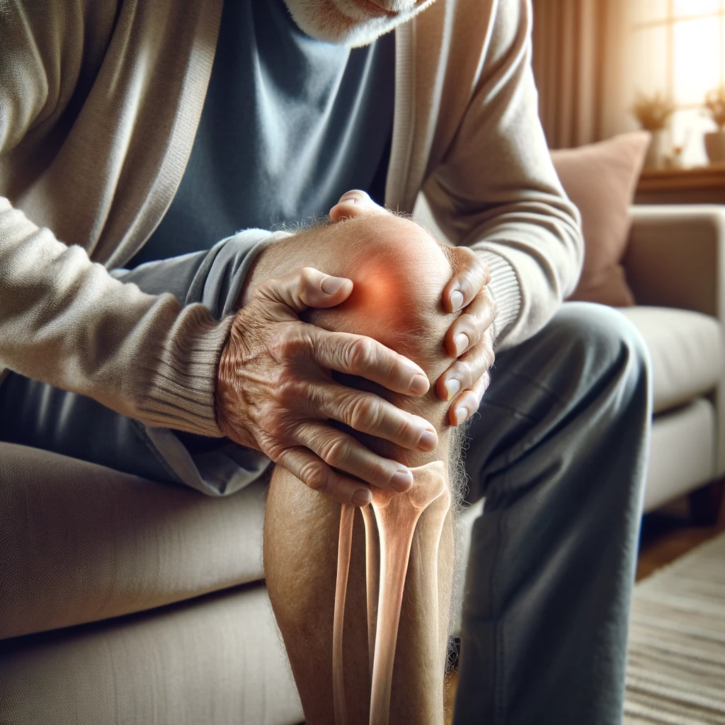
Knee Pain After COVID Recovery: A Common Symptom
Knee pain following COVID-19 recovery is more than just a minor complaint; it’s a symptom that warrants attention and understanding. As we delve into the specifics of post-COVID knee pain, it becomes clear that this issue affects a significant number of patients.
The knee joint, being a complex and weight-bearing part of our musculoskeletal system, is particularly susceptible to various forms of stress and inflammation. Understanding why knee pain emerges as a common symptom after COVID-19 recovery is crucial for both patients and healthcare providers.
Why the Knee Joints Are Particularly Vulnerable
The knee joints are especially vulnerable due to their anatomical and physiological characteristics. As weight-bearing joints, they endure constant mechanical stress, making them more prone to inflammation and pain.
Several factors contribute to the vulnerability of knee joints:
- The complex structure of the knee, involving bones, ligaments, and tendons, provides multiple sites for potential inflammation.
- Regular mechanical stress from daily activities can exacerbate any underlying inflammatory condition.
- Previous injuries or conditions, such as osteoarthritis, can make the knee more susceptible to post-COVID pain.
Distinguishing Post-COVID Knee Pain from Other Conditions
Distinguishing post-COVID knee pain from other common knee conditions is essential for appropriate treatment. The characteristics of post-COVID knee pain can provide clues about its origin.
| Condition | Typical Characteristics | Response to Rest and Activity |
|---|---|---|
| Post-COVID Knee Pain | Often bilateral, inflammatory in nature | May improve with rest, but can persist |
| Osteoarthritis | Degenerative, often unilateral | Worsens with activity, improves with rest |
| Meniscus Tears | Acute onset, mechanical symptoms like locking | Worsens with activity, especially twisting |
By understanding these differences, patients and healthcare providers can better identify the cause of knee pain and develop an appropriate treatment plan.
The Science Behind Post-COVID Joint Inflammation
Understanding the science behind post-COVID joint inflammation is crucial for addressing the lingering effects of the virus on the musculoskeletal system. The immune system’s response to SARS-CoV-2 can sometimes lead to prolonged inflammatory states, affecting joints even after the virus has been cleared.
Inflammatory Responses and Cytokine Activity
Viral infections, including COVID-19, are known to trigger inflammatory responses in the body. Cytokines such as IL-1, IL-6, and TNF-alpha play a crucial role in this process, contributing to the development of joint pain and inflammation. The table below summarizes the key cytokines involved and their effects.
| Cytokine | Role in Inflammation |
|---|---|
| IL-1 | Promotes inflammation and joint damage |
| IL-6 | Contributes to systemic inflammation and pain |
| TNF-alpha | Involved in systemic inflammation and joint destruction |
The Role of ACE2 Receptors in Joint Tissues
The interaction between SARS-CoV-2 and ACE2 receptors in joint tissues is an area of ongoing research. It’s believed that this interaction may lead to direct viral damage or immune-mediated inflammation, contributing to the development of post-COVID joint pain.
By understanding the mechanisms behind post-COVID joint inflammation, we can better address the needs of patients experiencing persistent joint pain after COVID-19 recovery.
Reactive Arthritis Following COVID-19 Infection
Reactive arthritis, an autoimmune condition typically associated with bacterial infections, has also been observed in patients after COVID-19 infection. This development has sparked interest in understanding the link between viral infections like SARS-CoV-2 and autoimmune joint inflammation.
What Is Reactive Arthritis?
Reactive arthritis is characterized by joint inflammation that occurs as a reaction to an infection elsewhere in the body, often developing days to weeks after the initial infection. It’s an autoimmune response where the body’s immune system mistakenly attacks the joints, leading to pain, swelling, and stiffness. This condition is known to be triggered by certain bacterial infections, but recent evidence suggests that viral infections, including COVID-19, may also play a role.
How SARS-CoV-2 Triggers Reactive Arthritis
The exact mechanisms by which SARS-CoV-2 triggers reactive arthritis are still under investigation. However, researchers propose that molecular mimicry and altered immune regulation may be key factors. Molecular mimicry occurs when the immune system confuses the body’s own tissues with the virus due to similarities in their molecular structures. Altered immune regulation refers to the disruption of the body’s normal immune response, leading to an inappropriate attack on the joints.

Timeline and Development of Symptoms
Studies have shown that reactive arthritis can develop anywhere from 1 to 4 weeks after SARS-CoV-2 infection, with an average onset of approximately 22 days. This timeline suggests that patients recovering from COVID-19 should be aware of the potential for developing joint pain and inflammation in the weeks following their recovery.
By understanding reactive arthritis and its potential link to COVID-19, healthcare providers can better diagnose and manage this condition, offering relief to patients experiencing post-COVID joint pain.
Common Symptoms of Post-COVID Knee Pain
The aftermath of COVID-19 infection can lead to knee pain, characterized by a range of distinct symptoms. As we explore these symptoms, it becomes clear that post-COVID knee pain is not just about the pain itself, but a complex interplay of various physical manifestations and pain patterns.
Physical Manifestations
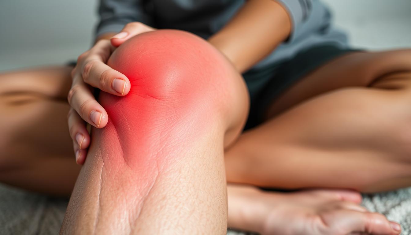
Post-COVID knee pain is often accompanied by several physical symptoms. These can include swelling, redness, warmth, and a reduced range of motion in the affected knee. Some individuals may also experience soft tissue sensitivity or redness around the knee area.
The physical symptoms associated with post-COVID knee pain can be summarized in the following table:
| Symptom | Description |
|---|---|
| Swelling | Inflammation around the knee joint |
| Redness | Visible redness or discoloration |
| Warmth | Increased temperature around the knee |
| Reduced Range of Motion | Decreased mobility or stiffness |
Pain Patterns and Progression
The pain associated with post-COVID knee pain can follow specific patterns. For instance, some individuals may experience morning stiffness or pain that worsens after periods of inactivity. The pain can also be accompanied by muscle weakness or pain in the surrounding muscles.
The progression of post-COVID knee pain varies among individuals. While some may experience improvement within weeks, others may develop more persistent joint pain lasting months. Understanding these patterns is crucial for managing the condition effectively.
Risk Factors for Developing Knee Pain After COVID
Understanding the risk factors for knee pain after COVID-19 is crucial for prevention and management. Several elements can contribute to an individual’s likelihood of experiencing knee pain post-recovery. We will explore these factors to better comprehend their implications.
Pre-existing Joint Conditions
Individuals with pre-existing joint conditions such as osteoarthritis or previous knee injuries are more susceptible to developing knee pain after COVID-19. These conditions compromise the joint’s integrity, making it more vulnerable to post-viral inflammation. A patient’s medical history plays a significant role in determining their risk profile.
Severity of COVID-19 Infection
The severity of the COVID-19 infection is another critical factor. Studies suggest that people who experienced more severe cases of COVID-19 are at a higher risk of developing subsequent joint problems, including knee pain. This correlation highlights the potential long-term effects of severe SARS-CoV-2 infection on the musculoskeletal system, observed in various cases.
Genetic Predispositions
Genetic factors, such as the presence of the HLA-B27 gene, can predispose certain individuals to post-viral joint inflammation. This gene is associated with various forms of inflammatory arthritis, suggesting a genetic component to the risk of developing knee pain after COVID-19.
| Risk Factor | Description |
|---|---|
| Pre-existing Joint Conditions | Increases vulnerability to post-COVID knee pain |
| Severity of COVID-19 Infection | More severe cases correlate with higher risk |
| Genetic Predispositions | Genes like HLA-B27 may increase risk |
Diagnosing Post-COVID Knee Pain
Healthcare providers use a multifaceted diagnostic strategy to identify the underlying causes of knee pain in patients who have recovered from COVID-19. This approach is crucial for developing an effective treatment plan.
Medical Evaluation and History
A thorough medical evaluation is essential for diagnosing post-COVID knee pain. This involves taking a detailed medical history, including the severity of the COVID-19 infection, the timeline of symptoms, and any pre-existing health conditions. Understanding the patient’s overall health status helps healthcare providers identify potential contributing factors.
Imaging and Laboratory Tests
imaging techniques such as X-rays, ultrasound, or MRI may be employed to assess joint structures and look for signs of inflammation. Laboratory tests, including inflammatory markers (ESR, CRP) and autoantibody tests, are also critical in determining the cause of knee pain in these patients.
Ruling Out Other Causes
Differential diagnosis is vital to rule out other potential causes of knee pain. Many study findings emphasize that accurate diagnosis is crucial for proper treatment of this condition. By considering alternative explanations for the patient’s symptoms, healthcare providers can develop a more targeted treatment plan.
Treatment Options for Post-COVID Knee Pain
The treatment of knee pain following COVID-19 recovery involves a range of strategies. Effective management of post-COVID knee pain requires a comprehensive approach that may include medication, physical therapy, and lifestyle adjustments.
Medications for Pain and Inflammation
For many patients, the first line of treatment for post-COVID knee pain involves medications aimed at reducing pain and inflammation. Nonsteroidal anti-inflammatory drugs (NSAIDs) are commonly recommended. Research has shown that NSAIDs such as ibuprofen and naproxen are effective in managing knee pain. In some cases, prescription NSAIDs like celecoxib or indomethacin may be prescribed for more severe pain.
- Over-the-counter NSAIDs for initial pain management
- Prescription NSAIDs for more severe cases
- Potential use of corticosteroid injections for inflammation
Physical Therapy Approaches
Physical therapy plays a crucial role in the treatment of post-COVID knee pain. Specific exercises and techniques can help reduce inflammation, improve joint mobility, and strengthen the muscles supporting the knee. A physical therapist can tailor a program to the individual’s needs and abilities.
When to Consider Advanced Treatments
In cases where initial treatments do not provide adequate relief, more advanced treatments may be considered. This can include disease-modifying antirheumatic drugs (DMARDs) for persistent inflammation or corticosteroid injections to directly address joint inflammation. A healthcare provider can help determine the best course of action based on the individual’s condition and response to initial treatments.
The Brain’s Role in Post-COVID Pain Persistence
Emerging research suggests that the brain plays a significant role in the continuation of knee pain even after the initial COVID-19 infection has passed. Recent breakthroughs in understanding neuroplasticity, the brain’s ability to adapt and change, have provided insights into how COVID-19 may lead to persistent pain.
Neuroplasticity and Pain Pathways
Neuroplasticity allows our body to learn new skills, but it can also enable our immune system to perpetuate certain responses that may not be warranted after the infection has resolved. The brain develops a “pain memory” that can continue to produce pain sensations in the body even after the original inflammatory trigger has been resolved.
This concept is crucial in understanding how COVID-19 can lead to chronic pain conditions. The brain’s pain processing pathways can be altered, resulting in persistent pain.
Brain Retraining Techniques
Fortunately, neuroplasticity also allows for the possibility of “relearning” or “retraining” the brain. Techniques such as mindfulness practices, cognitive behavioral therapy, and specific pain reprocessing therapies can help reset pain pathways. By addressing the neurological component of post-COVID knee pain, patients who do not respond to conventional anti-inflammatory treatments may find relief.
These brain retraining techniques offer a promising approach to managing post-COVID knee pain by targeting the brain’s response and its effects on the body, helping to change the way pain is perceived and processed.
Self-Management Strategies for Recovery
The journey to recovery from post-COVID knee pain involves implementing practical self-management techniques that support both physical and mental well-being. As we explore these strategies, it’s essential to understand the importance of balance and tailored approaches in managing knee pain and associated fatigue.
Appropriate Rest and Movement Balance
One of the challenges in improving joint pain is finding the right balance between rest and movement. While it’s uncomfortable to move affected joints, deliberate motion is often necessary to mobilize the area and increase circulation, thereby speeding up recovery. Maintaining a level of non-draining physical activity is crucial. Practices such as yoga, tai chi, and aquatic exercises are beneficial as they provide movement without excessive joint stress.
Stress Reduction Techniques
Stress can exacerbate pain and inflammation. Techniques such as mindfulness meditation, deep breathing exercises, and progressive muscle relaxation can help manage pain by reducing the body’s inflammatory response. These practices not only alleviate stress but also contribute to overall well-being and support the recovery process.
Dietary Considerations for Inflammation
Diet plays a significant role in managing inflammation. Incorporating anti-inflammatory foods and supplements into one’s diet can support joint health and overall recovery from post-COVID fatigue and pain. A well-balanced diet is a critical component of a comprehensive recovery plan.
| Self-Management Strategy | Benefits |
|---|---|
| Gentle Movement Practices | Improves circulation, reduces stiffness |
| Stress Reduction Techniques | Reduces inflammatory response, alleviates pain |
| Anti-Inflammatory Diet | Supports joint health, aids in recovery |
By incorporating these self-management strategies into daily life, individuals can significantly enhance their recovery from post-COVID knee pain and associated fatigue, ultimately improving their overall health and well-being.
Long-term Outlook for Post-COVID Knee Pain
As research continues to emerge, we’re gaining a clearer picture of what to expect regarding post-COVID knee pain over time. The available evidence suggests that many cases of knee pain following COVID-19 infection resolve within a few months with appropriate treatment.
Recovery Timeline Expectations
Studies have shown promising results in terms of recovery time. For instance, one study found that symptoms resolved within a few days with treatment in all 54 participants. Most people treated with medications in various case studies seem to make a full recovery, indicating a positive long-term outlook for many patients.
The recovery timeline can vary based on several factors, including the severity of initial symptoms, the promptness and effectiveness of treatment, and individual health factors. Generally, evidence suggests that many cases resolve within weeks to months.
When to Be Concerned About Persistent Pain
While many individuals experience resolution of knee pain within a few months, some may develop persistent or chronic symptoms as part of long COVID. It’s essential to be aware of when knee pain might be a sign of a more chronic condition. If knee pain persists for more than 3-6 months, it may be indicative of an underlying chronic inflammatory condition, warranting further medical evaluation.
Understanding the relationship between post-COVID knee pain and other long COVID symptoms is also crucial. As research continues to unfold, we’re learning more about how these symptoms can evolve over time and what this means for overall recovery expectations.
Moving Forward: Life After Post-COVID Knee Pain
The journey to recovery from post-COVID knee pain involves understanding the condition, adopting appropriate care strategies, and making necessary lifestyle adjustments. As we move forward, it’s crucial to maintain joint health through regular exercise, good nutrition, and proper body mechanics to prevent the recurrence of pain.
For many, the experience of post-COVID arthritis or joint pain may necessitate certain lifestyle modifications. We will explore how to implement these changes in a positive, sustainable way, ensuring that they contribute to overall well-being and resilience against future challenges.
Addressing the psychological aspects of long COVID recovery is also vital. We will discuss strategies for coping with uncertainty and maintaining a positive outlook, which are crucial for navigating the aftermath of COVID-19.
Finally, we will touch on the ongoing research into post-COVID conditions and how patients can stay informed about new developments in care approaches and treatment options for long-term COVID effects, supporting their continued journey towards full recovery.
FAQ
What is the link between COVID-19 and joint pain?
We know that COVID-19 can affect the musculoskeletal system, leading to joint pain and inflammation in some patients. The exact mechanisms are still being studied, but it’s believed that the virus triggers an immune response that can cause inflammation in the joints.
Why are knee joints particularly vulnerable to post-COVID pain?
Our knee joints bear a significant amount of our body weight and are subject to various stresses, making them more susceptible to pain and inflammation, especially in the context of a systemic condition like COVID-19.
How long does post-COVID knee pain typically last?
The duration of post-COVID knee pain can vary significantly from person to person. In some cases, it may resolve on its own within a few weeks, while in others, it can persist for months, requiring ongoing management and treatment.
Can pre-existing joint conditions increase the risk of developing post-COVID knee pain?
Yes, we have found that individuals with pre-existing joint conditions, such as arthritis, may be more likely to experience knee pain after COVID-19 infection due to their compromised joint health.
What are the common symptoms of post-COVID knee pain?
Patients often report pain, stiffness, and swelling in the knee joint, which can be accompanied by reduced mobility and, in some cases, warmth or redness around the affected area.
How is post-COVID knee pain diagnosed?
Diagnosing post-COVID knee pain involves a comprehensive medical evaluation, including a detailed history of the patient’s COVID-19 infection and any subsequent symptoms, as well as imaging and laboratory tests to rule out other causes of knee pain.
What treatment options are available for post-COVID knee pain?
Treatment may include medications to manage pain and inflammation, physical therapy to improve joint mobility and strength, and in some cases, more advanced treatments such as corticosteroid injections or surgery.
Can lifestyle changes help manage post-COVID knee pain?
Yes, maintaining a healthy weight, engaging in appropriate exercise, using stress reduction techniques, and making dietary changes to reduce inflammation can all be beneficial in managing post-COVID knee pain.