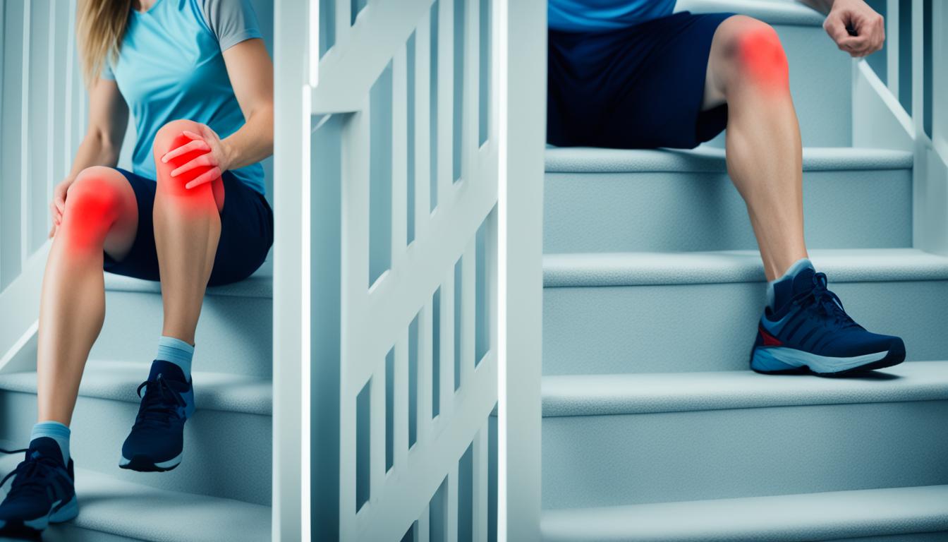
Are you experiencing knee pain when going up stairs? You’re not alone. Many individuals struggle with discomfort and limitations while climbing stairs due to various reasons such as muscle weakness, wear and tear, knee injuries, and inflammation.
Fortunately, there are effective exercises that can help alleviate knee pain, strengthen the surrounding muscles, improve flexibility, and enhance your overall mobility when ascending stairs.
In this article, we will explore different exercises and strategies to combat knee pain while climbing stairs. By incorporating these exercises into your routine, you can promote knee health, reduce discomfort, and regain your freedom to navigate stairs with confidence.
Key Takeaways:
- Exercises can help strengthen knee muscles and improve flexibility.
- Knee pain on stairs can have various causes, including muscle weakness and inflammation.
- Strategies such as handrail usage and leading with the correct leg can reduce knee pain on stairs.
- Warming up the knee and strengthening hip muscles play a crucial role in managing knee pain.
- Gradual progression and building tissue resilience are essential for long-term knee health.
Understanding Knee Pain on Stairs
Knee pain on stairs can be a debilitating issue, affecting daily activities such as climbing stairs or even walking. To effectively manage and reduce knee pain, it is crucial to understand the underlying causes. There are several factors that can contribute to knee pain on stairs, including:
- Muscle Weakness: Weakness in the muscles surrounding the knee can lead to increased stress and pressure on the joint, resulting in pain while climbing stairs.
- Wear and Tear: Over time, the knee joint can experience wear and tear, causing discomfort when bearing weight on stairs.
- Knee Injuries: Previous knee injuries, such as ligament tears or meniscus damage, can cause ongoing pain and difficulty on stairs.
- Kneecap Damage: The kneecap, or patella, can be susceptible to damage or misalignment, leading to knee pain on stairs.
- Altered Biomechanics: Poor alignment or altered movement patterns can place excessive strain on the knee joint, resulting in pain on stairs.
- Inflammation: Inflammatory conditions, such as arthritis or bursitis, can cause knee joint irritation and pain while climbing stairs.
By identifying the specific cause of knee pain on stairs, healthcare professionals can provide targeted treatment plans to alleviate discomfort and improve function. This may involve a combination of exercise therapy, pain management techniques, and lifestyle modifications.

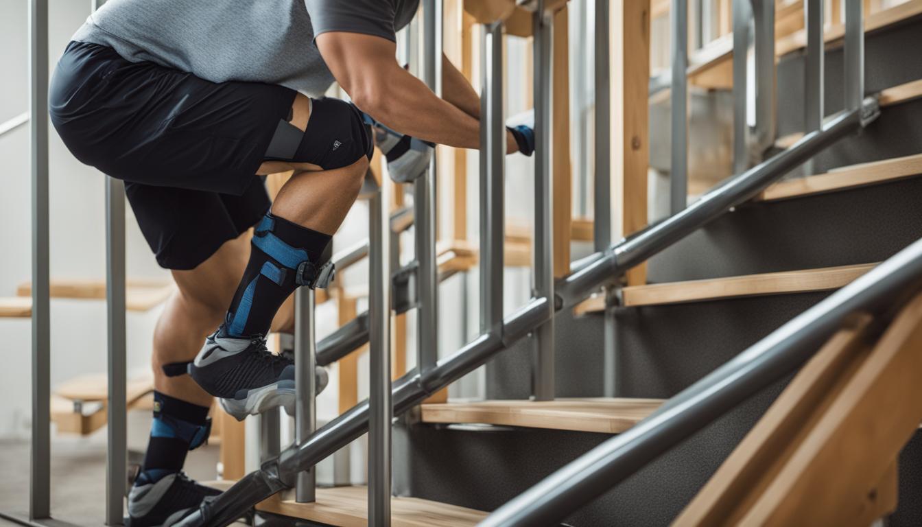
Tips for Reducing Knee Pain on Stairs
If you experience knee pain while climbing stairs, there are effective strategies that can help minimize discomfort and provide relief. By following these tips, you can reduce knee pain on stairs and make your daily activities more manageable.
- Take one step at a time: It’s important to take your time and avoid rushing when climbing stairs. By going slowly and placing one foot at a time on each step, you can decrease the strain on your knee joints.
- Lead with the correct leg: When climbing stairs, leading with the leg that has less pain or better functionality can help distribute the load more evenly and reduce stress on the affected knee.
- Utilize hand rail for support: Using the hand rail while climbing stairs provides extra stability and support, allowing you to shift some of the weight from your knees to your upper body. This can significantly reduce the pressure on your knee joints.
- Consider using crutches or sticks: If your knee pain is severe or you have difficulty bearing weight on your knee joints, using crutches or sticks can help alleviate the strain. These assistive devices provide additional support and stability, allowing you to climb stairs more comfortably.
By incorporating these strategies into your daily routine, you can effectively reduce knee pain on stairs and improve your mobility. Remember to consult with your healthcare professional or physical therapist for personalized guidance and recommendations.

Note: Always consult with a healthcare professional before starting any new exercise or treatment plan for knee pain relief.
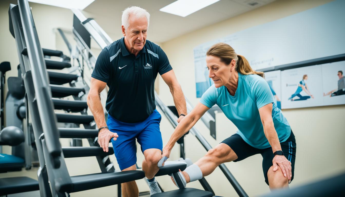
Warm Up and Strengthen Your Knee
Before climbing stairs, it is important to warm up your knee to reduce stiffness and improve lubrication in the joint. A proper warm-up routine can help prepare your knee for the physical demands of stair climbing, allowing for greater comfort and flexibility. Start with gentle exercises that target the knee joint and surrounding muscles.
Here are some warm-up exercises to help with knee flexibility and strength:
- Knee Bending and Straightening: Perform slow and controlled knee bends, focusing on the full range of motion. This exercise helps to increase blood flow to the knee joint and promote flexibility.
- Quadriceps Stretch: Stand near a wall or use a chair for support. Grab your ankle and gently pull your heel towards your buttocks. Hold the stretch for 20-30 seconds and repeat on both legs. This stretch targets the quadriceps muscles, which are important for knee stability and strength.
- Hamstring Stretch: Sit on the edge of a chair with one leg extended in front of you. Lean forward, reaching towards your toes while keeping your back straight. Hold the stretch for 20-30 seconds and switch legs. This stretch targets the hamstring muscles, which play a role in knee alignment and function.
In addition to warming up, it is essential to strengthen the muscles that support the knee to improve stability and reduce pain while climbing stairs. Focus on exercises that target the quadriceps, hamstrings, and hip muscles.
“By strengthening the muscles around your knee, you can help alleviate pain and reduce the risk of further injury.”
Here are some knee-strengthening exercises to incorporate into your routine:
| Exercise | Description |
|---|---|
| Squats | Stand with your feet shoulder-width apart. Bend your knees and lower your buttocks towards the ground as if sitting on an imaginary chair. Keep your back straight and your knees aligned with your toes. Return to the starting position and repeat. |
| Lunges | Step forward with one leg, bending both knees to a 90-degree angle. Make sure your front knee stays aligned with your ankle and doesn’t extend past your toes. Push down through your front heel to return to the starting position. Alternate legs and repeat. |
| Step-ups | Find a stable step or platform. Step onto the platform with one foot, lifting your body up and bringing your opposite knee towards your chest. Step down and repeat on the opposite side. |
By incorporating warm-up exercises and targeted knee-strengthening exercises into your routine, you can improve knee flexibility, strength, and muscle endurance. These exercises help to support the knee joint, reduce discomfort, and enhance your ability to navigate stairs with ease.
The Role of Hip Muscle Strength
Weakness in the hip muscles can contribute to knee pain while climbing stairs. When the hip muscles are weak, more stress is placed on the knee joints, leading to pain and discomfort. To alleviate knee pain and promote proper alignment and stability during stair climbing, it is crucial to strengthen the hip muscles through targeted exercises.
Causes of Knee Pain
Knee pain can have various causes, including muscle weakness. Hip muscle weakness, in particular, can negatively impact the knee joint and result in pain and discomfort during activities such as climbing stairs. By addressing the underlying weakness in the hip muscles, we can reduce stress on the knee joints and alleviate knee pain.
Hip Strategy in Stair Climbing
Proper hip strategy plays a vital role in maintaining knee health while climbing stairs. When the hip muscles are strong and functional, they assist in stabilizing the knee joints and distributing the load more evenly. This reduces the strain placed on the knees, minimizing the risk of pain and injury.
Weak hip muscles can disrupt the hip strategy in stair climbing, increasing the reliance on the knee joints to bear the load. This can lead to overuse of the knee muscles and exacerbate knee pain.
Hip Muscle Exercises
To strengthen the hip muscles and improve the hip strategy during stair climbing, incorporating targeted exercises into your fitness routine is essential. Here are a few effective hip muscle exercises:
- Squats: A compound exercise that targets the hip muscles, quadriceps, and glutes.
- Lunges: Helps strengthen the hip muscles while improving balance and stability.
- Step-ups: Mimics the movement of climbing stairs and engages the hip muscles.
By consistently performing these exercises, you can enhance hip muscle strength and reduce the strain on your knees during stair climbing.
| Exercise | Description |
|---|---|
| Squats | A compound exercise that targets the hip muscles, quadriceps, and glutes. Stand with feet shoulder-width apart, lower into a squat position by bending the knees, and then return to the starting position. |
| Lunges | Step forward with one leg, lowering your body until both knees are bent at a 90-degree angle. Push back up using the front leg and repeat on the other side. |
| Step-ups | Place one foot on a step or platform, push through the heel of the elevated foot, and step up, bringing the other foot onto the platform. Step back down and repeat on the other side. |
Gradual Progression and Tissue Resilience
Building tissue resilience in the knee is essential for resolving knee pain and improving overall function. Tissue resilience refers to the ability of the tissues, such as cartilage, ligaments, tendons, and bone, to withstand stress and recover from injury or strain. By gradually progressing the intensity and frequency of knee strengthening exercises, we can promote the healing and remodeling of these tissues, ultimately reducing knee pain while going up stairs.
To achieve tissue resilience, it is crucial to gradually increase the challenge placed on the knee joint through exercise progression. Starting with gentle exercises, such as knee bends and straightening, allows the tissues to adapt and become stronger over time. As our knee strengthens and pain levels decrease, we can gradually introduce more advanced exercises that target specific muscle groups, such as the quadriceps and hamstrings.
Exercise progression should be individualized and guided by pain levels. It is important to listen to our body and not push beyond our limits, as this can worsen knee pain and lead to further injury. A gradual and systematic approach to exercise progression ensures that we build strength and resilience while minimizing the risk of aggravating knee pain.
In addition to exercise progression, it is crucial to incorporate a variety of knee strengthening exercises into our routine. This helps to target different muscle groups, improve overall knee stability, and enhance the resilience of the surrounding tissues. Some effective knee strengthening exercises include:
- Leg presses
- Step-ups
- Lunges
- Wall squats
- Clamshells
Performing these exercises regularly, under proper guidance, can help boost tissue resilience and alleviate knee pain when going up stairs. It is essential to speak with a healthcare professional or a qualified exercise specialist to determine the appropriate exercises and progression plan for your specific needs.
Quote:
“By gradually progressing our knee strengthening exercises, we can enhance tissue resilience, promote healing, and ultimately reduce knee pain while performing daily activities, such as climbing stairs.”
Remember, tissue resilience is a gradual process that takes time and consistent effort. With dedication and a well-structured exercise program, we can achieve significant improvements in knee pain resolution and overall knee health.
Conclusion
Knee pain when going up stairs can be effectively managed and relieved through a range of strategies, including targeted exercises, warm-up routines, and strength-building techniques. By addressing the underlying causes of knee pain, such as muscle weakness and biomechanical issues, we can significantly reduce discomfort and improve our ability to climb stairs without experiencing pain.
Regular exercise plays a crucial role in knee pain management. Performing specific knee pain relief exercises, such as quadriceps strengthening exercises and flexibility-enhancing movements, helps to strengthen the surrounding muscles and improve joint stability. Additionally, engaging in warm-up routines before stair climbing activities can reduce stiffness and improve lubrication in the knee joint, making the process more comfortable and less painful.
Prevention is equally important in maintaining knee health and reducing the risk of future pain. By incorporating regular exercise into our daily routine, we can promote tissue resilience and long-term knee health. Gradual progression in exercise intensity and frequency, guided by individual pain levels, allows for the healing and remodeling of tissues, including cartilage, ligaments, tendons, and bones.
In conclusion, by adopting a holistic approach that combines targeted exercises, warm-up routines, and gradual progression, we can effectively manage knee pain when going up stairs. Prioritizing knee pain management, engaging in knee pain relief exercises, and implementing preventive measures will enable us to enjoy pain-free stair climbing and maintain optimal knee health for the long term.
FAQ
What are some exercises for knee pain when going up stairs?
There are several exercises that can help strengthen the knee muscles, improve flexibility, and reduce knee pain while climbing stairs. Some examples include knee bending and straightening exercises, squats, and step-ups.
What are the causes of knee pain on stairs?
Knee pain on stairs can be caused by muscle weakness, wear and tear on the knee bones and cartilage, knee injuries, kneecap damage, altered biomechanics, and inflammation.
How can I reduce knee pain on stairs?
To reduce knee pain on stairs, you can take one step at a time, lead with the correct leg, use the hand rail for support, and consider using a crutch or stick. These strategies can help minimize stress and pressure on the knee joints.
How can I warm up and strengthen my knee?
Before climbing stairs, it is important to warm up your knee to reduce stiffness and improve lubrication in the joint. Simple knee bending and straightening exercises can help prepare the knee joint and alleviate discomfort. Additionally, strengthening the muscles that support the knee, such as the quadriceps and hip muscles, can improve knee stability and reduce pain while going up stairs.
How does hip muscle strength affect knee pain on stairs?
Weak hip muscles can contribute to knee pain while climbing stairs. Strengthening the hip muscles through exercises such as squats and step-ups can alleviate knee pain and promote proper alignment and stability during stair climbing.
How does gradual progression and tissue resilience help with knee pain management?
Gradually progressing the intensity and frequency of knee strengthening exercises can promote the healing and remodeling of tissues such as cartilage, ligaments, tendons, and bone. This can help build tissue resilience, reduce knee pain, and improve overall knee function.
How can I manage and alleviate knee pain when going up stairs?
Knee pain when going up stairs can be managed and alleviated through a combination of exercises, warm-up routines, and strengthening techniques. By addressing the underlying causes of knee pain, including muscle weakness and biomechanical issues, you can reduce discomfort and improve your ability to climb stairs without pain.