Healing Process for Knee Ligament Damage: A Professional Guide
As healthcare professionals, we understand the importance of knee ligaments in maintaining stability and mobility in the knee joint. Unfortunately, ligament injuries are common, especially in individuals who engage in physical activity. A ligament injury can be debilitating and may limit an individual’s ability to perform daily activities. In this article, we will discuss the three-step healing process for knee ligament damage.

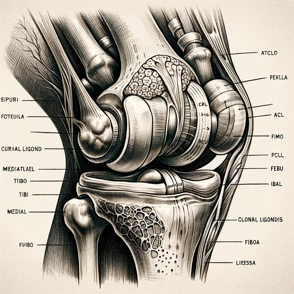
Understanding Ligament Damage and Its Implications is the first step in the healing process. Knee ligaments are strong, fibrous tissues that connect the bones in the knee joint. When a ligament is damaged, it can result in pain, swelling, and instability in the knee joint. The severity of the injury can range from mild to severe, depending on the degree of damage to the ligament. It is crucial to diagnose and treat a ligament injury promptly to prevent further damage and complications.
Treatment and Rehabilitation of Ligament Damage is the second step in the healing process. The treatment plan for a ligament injury depends on the severity of the injury. Mild injuries may require rest, ice, compression, and elevation (RICE) therapy, while severe injuries may require surgery. Rehabilitation is an essential aspect of the healing process, and it involves exercises and physical therapy to restore strength, flexibility, and mobility in the knee joint.
Key Takeaways
- Understanding the implications of ligament damage is crucial in the healing process.
- Treatment plans for knee ligament damage vary based on the severity of the injury.
- Rehabilitation is a vital aspect of the healing process and can help restore strength, flexibility, and mobility in the knee joint.
Understanding Ligament Damage and Its Implications
When it comes to knee injuries, ligament damage is one of the most common. Ligaments are the tough, fibrous bands of tissue that connect bones to each other and provide stability and strength to the joint. However, when these ligaments are damaged, it can lead to pain, tenderness, swelling, inflammation, stiffness, and a limited range of motion.
Types of Ligament Injuries
There are several types of ligament injuries that can occur in the knee, including sprains, ruptures, and strains. The most common type of knee ligament injury is an anterior cruciate ligament (ACL) tear, which can occur during sports or other physical activities that involve sudden stops or changes in direction.
Signs and Symptoms
The signs and symptoms of a knee ligament injury can vary depending on the severity of the injury. Common symptoms include pain, tenderness, swelling, inflammation, stiffness, and a limited range of motion. In more severe cases, the knee may feel unstable or give out when weight is placed on it.
Diagnosis and Evaluation
If you suspect that you have a knee ligament injury, it is important to seek medical attention from a specialist or physical therapist. They will perform a thorough evaluation to determine the severity of the injury and the best course of treatment. This evaluation may include X-rays or an MRI to assess the extent of the damage.
Overall, understanding the implications of ligament damage is critical to properly diagnose and treat knee injuries. If you experience any symptoms or have any questions, it is important to seek medical attention as soon as possible.
Treatment and Rehabilitation of Ligament Damage
At times, knee ligament damage can be a painful experience that may require treatment and rehabilitation. We have put together a comprehensive guide to help you understand the treatment process and how to rehabilitate your knee ligament damage.
Initial Treatment and Pain Management
The initial treatment for knee ligament damage involves rest, ice, compression, and elevation (RICE). RICE helps reduce swelling, pain, and inflammation around the affected area. We recommend using nonsteroidal anti-inflammatory drugs (NSAIDs) such as ibuprofen or naproxen to manage pain and inflammation.
different types of knee injuries, their typical healing times, and recovery notes:
| Injury | Healing Time | Recovery Notes |
|---|---|---|
| Bone bruise | 2-4 weeks | Rest, ice, compression. Gradual return to activity over 1-2 months. |
| Meniscus tear – minor | 4-6 weeks | Often heals with conservative treatment. |
| Meniscus tear – moderate/severe | 12+ weeks | Often requires surgery. Multi-month recovery common. |
| MCL tear – grade 1/2 | 3-6 weeks | Healing progresses well if braced. |
| MCL tear – grade 3 | 8-12 weeks | Often requires surgical repair/reconstruction followed by extensive rehab. |
| LCL sprain | 4-8 weeks | Bracing to stabilize knee throughout healing. |
| ACL tear – partial | Up to 8 weeks | May heal with rehab/bracing or require surgical reconstruction. |
| ACL tear – complete | 30+ weeks | Requires reconstructive surgery with graft. Months of rehab. |
| Patellar dislocation | 6 weeks | Knee immobilized in early recovery. Later rehab focuses on realignment. |
| Patellar fracture | 6-8 weeks | Cast/brace to immobilize. Surgical fixation may be necessary. |
| Arthritis flare up | 1-4 weeks | Resting the joint along with anti-inflammatory meds helps recovery. |
Recovery times are general estimates and can vary significantly depending on injury severity. Re-injury risks can persist even months after the acute healing phase
Surgical Intervention and Post-Surgery Care
In some cases, surgical intervention may be necessary to repair the damaged ligament. Surgery is typically followed by a period of rest, ice, compression, and elevation. Physical therapy and rehabilitation exercises may also be recommended to help regain range of motion and strength.
Rehabilitation and Therapy
Physical therapy and rehabilitation exercises are an essential part of the recovery process. Rehabilitation exercises help to strengthen the muscles around the knee and improve range of motion. We recommend working with a physical therapist to develop a personalized rehabilitation plan.
Advanced Treatments and Techniques
Advanced treatments and techniques such as tissue engineering, mesenchymal stem cells, and platelet-rich plasma may be used to promote healing and regeneration of the damaged ligament. We recommend discussing these options with your doctor to determine if they are appropriate for your specific condition.
Prevention and Future Care
Prevention and future care are essential to avoid re-injury and maintain healthy knee ligaments. We recommend engaging in regular physical activity and exercise to improve strength and flexibility. We also recommend wearing appropriate protective gear during sports activities to minimize the risk of injury.
In conclusion, knee ligament damage can be a painful experience, but with proper treatment and rehabilitation, you can recover and regain your strength and mobility. Remember to always consult with your doctor and physical therapist to develop a personalized treatment plan that meets your specific needs.