Tag: Orthopedic health

  • Knee Pain Painting on a Ladder

    Knee Pain Painting on a Ladder

    Imagine you’re in the middle of painting, but knee pain stops you. Many face this when they have a meniscus tear. Climbing ladders then becomes a big challenge, affecting your project and mobility.

    Dealing with ladder climbing and a meniscus tear is tough. We aim to give you expert advice to stay safe. Our tips help you avoid more injuries and keep working on your projects.

    Key Takeaways

    • Safely assess your knee condition before climbing a ladder.
    • Understand the proper techniques for ladder climbing with a meniscus injury.
    • Learn how to prevent further knee damage while on a ladder.
    • Discover exercises to strengthen your knee before and after ladder climbing.
    • Explore alternative solutions for painting projects that minimize ladder use.

    Understanding Knee Pain: Common Causes

    Knee pain comes from many sources. It’s key to know these causes to avoid more harm. Activities like ladder climbing put extra stress on our knees. This can make existing problems worse or cause new ones.

    Types of Knee Pain

    Knee pain falls into two main groups: acute injuries and chronic conditions. Acute injuries happen suddenly, like from a fall. Chronic conditions develop over time, often from repeated strain or wear.

    Some common knee pain types include meniscus tears, ligament sprains, and tendonitis. Knowing the exact type of pain helps find the right treatment and ways to prevent it.

    Why Ladder Work Can Exacerbate Pain

    Climbing ladders puts a lot of strain on the knees, especially when bending or standing on uneven ground. This strain can make knee problems worse or cause new ones like patellofemoral pain syndrome. The constant stress from ladder work can lead to knee inflammation and pain.

    To reduce the risk of making knee pain worse, it’s important to understand how ladder work affects the knee. Knowing this helps us take steps to lessen the strain and protect our knees.

    Safety Measures When Using a Ladder

    Ladder Safety Measures

    To stay safe while using a ladder, we need to take certain steps. Ladder climbing can be dangerous, especially for those with knee problems. By picking the right ladder and setting it up right, we can lower the chance of accidents.

    Choosing the Right Ladder

    Picking the right ladder is key. Think about how high you need to go and how much weight the ladder can hold. For painting, a ladder with a strong base and non-slip feet is best. Look for ladders with comfy rungs and think about the material. Aluminum ladders are light, while fiberglass ladders are more stable.

    Ladder TypeMaterialWeight Capacity
    Step LadderAluminum300 lbs
    Extension LadderFiberglass250 lbs

    Proper Ladder Setup

    After picking the right ladder, setting it up correctly is crucial for safety. Make sure the ladder is on a solid, level ground. Having someone hold the ladder can also help. The ladder’s angle is important too; it should be at a 75-degree angle or one foot away from the wall for every four feet of ladder height.

    By following these tips, we can make ladder climbing safer. Remember, safety steps are not just suggestions; they’re essential to avoid serious injuries.

    Preparing for Your Painting Project

    knee injury prevention

    Getting ready for painting is more than just picking up supplies. It also means getting your body ready to avoid knee injury.

    Stretching Exercises Before Climbing

    Do some stretching exercises before you start climbing. This helps your knees a lot. Try:

    • Leg swings to loosen up your leg muscles
    • Knee bends to prepare your knees for the climb
    • Calf raises to strengthen your calf muscles, which support your knees

    Essential Gear to Minimize Knee Strain

    Wearing the right gear can also help your knees. Use:

    1. Knee pads for extra cushioning and support
    2. Supportive footwear to stabilize your knees
    3. Comfortable clothing that allows for a full range of motion

    With these steps, you can lower the chance of knee pain and injury while painting.

    Painting Techniques to Reduce Knee Strain

    There are ways to lessen knee strain while painting on a ladder. Painting can put a lot of pressure on our knees, especially if we have conditions like meniscus tears. Using the right techniques can help avoid making knee pain worse.

    Opt for a Brush Instead of a Roller

    Choosing a brush over a roller can help reduce knee strain. Brushes allow for more precise strokes and need less force, which is easier on the knees. Plus, they help you paint more accurately, so you might not have to climb the ladder as much.

    Take Breaks to Relieve Pressure

    It’s important to take breaks to ease knee strain. Stepping down from the ladder often can help take the pressure off your knees. These breaks are a chance to stretch and prevent knee pain.

    By using these techniques, we can make painting easier on our knees. This is especially helpful for those with knee pain or meniscus tear symptoms.

    When to Seek Medical Advice

    Knee pain can really get in the way of doing things. There are times when you need to see a doctor. If you’re feeling pain in your knee while on a ladder, it’s important to know when to get help.

    Signs Your Knee Pain Needs Attention

    There are signs that mean you should see a doctor. These include:

    • Persistent pain that doesn’t improve with rest
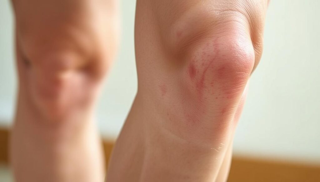
    • Swelling or redness around the knee
    • Instability or feeling like the knee is going to give way
    • Warmth or tenderness to the touch
    • Loss of mobility or difficulty straightening the knee

    If you notice any of these, it’s best to talk to a healthcare professional. Ignoring these signs can lead to further complications.

    Recommended Specialists for Knee Pain

    Choosing the right specialist is crucial for knee pain. Here’s a table of specialists you might consider:

    SpecialistDescription
    Orthopedic SurgeonSpecializes in surgical treatments for musculoskeletal disorders, including knee pain. They can perform knee surgery if necessary.
    RheumatologistExpert in diagnosing and treating rheumatic diseases that can cause knee pain.
    Physical TherapistCan help improve knee mobility and strength through targeted exercises.

    An orthopedic surgeon is often the first choice for serious knee pain or when surgery is needed. They can give a detailed diagnosis and suggest the best treatment.

    Home Remedies for Knee Pain Relief

    Home remedies are a simple way to handle knee pain and boost your quality of life. We often seek easy, effective solutions to ease discomfort before seeking medical help. Two popular methods are ice and heat therapy, and over-the-counter pain relief.

    Ice and Heat Therapy

    Ice and heat therapy are easy yet effective for knee pain. Ice therapy reduces swelling and numbs pain, great for sudden injuries or pain spikes. Heat therapy, on the other hand, relaxes muscles and boosts blood flow, helping with long-term pain and stiffness.

    To use ice, wrap an ice pack in a towel and apply it for 15-20 minutes, several times a day. For heat, try a warm bath, heating pad, or a warm cloth on your knee. It’s key to alternate between ice and heat based on your pain type.

    “The application of ice or heat can significantly alter the pain perception and is a crucial component of a comprehensive pain management plan.”

    – Expert in Pain Management

    Over-the-Counter Pain Relief Options

    Over-the-counter (OTC) pain meds are also effective for knee pain. Common ones are acetaminophen and ibuprofen. Acetaminophen mainly relieves pain, while ibuprofen also fights inflammation.

    • Always follow the recommended dosage on the label.
    • Consult with a healthcare provider if you’re unsure about interactions with other medications.
    • Be aware of potential side effects and contraindications.

    By adding these home remedies to your daily routine, you can better manage knee pain. This lets you enjoy activities like painting on a ladder more comfortably.

    Prolonging Knee Health During Painting

    To enjoy painting without harming our knees, we need to focus on knee health. Painting on a ladder can be tough on our knees, especially for those with a torn meniscus. By keeping our knees strong and moving regularly, we can protect them.

    Tips for Maintaining Strong Knees

    Keeping our knees strong involves exercises and careful movements. Physical therapy helps strengthen knee muscles, offering better support and injury prevention. Exercises like squats, lunges, and leg presses are helpful. Also, using the right ladder techniques and taking breaks can ease knee strain.

    ExerciseBenefitFrequency
    SquatsStrengthens quadriceps and hamstrings3 sets of 10 reps, 3 times a week
    LungesImproves balance and strengthens knee muscles3 sets of 10 reps (per leg), 3 times a week
    Leg PressesTargets quadriceps, hamstrings, and glutes3 sets of 12 reps, 3 times a week

    Importance of Regular Movement

    Regular movement is key for healthy knees. Sitting still for too long can make knees stiff and painful, especially for those with injuries. Adding gentle exercises to our day keeps knees flexible and lowers injury risk. It’s also important to listen to our bodies and rest when needed.

    By using these tips, we can keep our knees healthy while painting. Regular physical therapy, exercises, and careful movements are all important for a safe painting experience.

    Equipment to Assist While Painting on a Ladder

    To have a safe and fun painting time, the right tools are key. Using the right gear when painting on a ladder helps avoid knee pain and injuries.

    Knee Pads and Supportive Gear

    Knee pads are a simple yet effective way to protect your knees while working on a ladder. They offer cushioning and support, easing the pressure on your knees. Supportive gear, like knee braces, adds extra stability and protection. Pick knee pads or supportive gear based on your work and needed support.

    Look for knee pads that are durable, comfortable, and adjustable. It’s also important to pick knee pads that fit your task. For example, if you’re working on a rough surface, you’ll need sturdier knee pads.

    Type of Knee PadFeaturesBest For
    Hard Cap Knee PadsDurable, protectiveRough surfaces, heavy-duty work
    Soft Cap Knee PadsComfortable, flexibleGeneral painting, smooth surfaces
    Gel Knee PadsCushioned, shock-absorbingLong periods of kneeling, comfort

    Choosing the Right Footwear

    Along with knee pads, choosing the right footwear is crucial for stability and to prevent slipping on the ladder. Find shoes with good grip and a snug fit to keep your feet steady on the ladder.

    • Non-slip soles
    • Comfortable fit
    • Ankle support

    By using knee pads with the right shoes, you can greatly lower the risk of knee injury. This makes painting more comfortable and enjoyable.

    Final Thoughts: Enjoying Your Painting Project

    As we finish talking about managing knee pain while painting on a ladder, remember it’s not just about the end. It’s also about how we feel during it. By taking care of our knees, we can enjoy painting more without worrying about injuries.

    It’s important to love the journey of creating. We can do this by following the tips we’ve shared. From picking the right ladder to stretching, every step matters.

    Practical Adjustments for Future Projects

    For future painting, we can reduce knee pain. Use knee pads and supportive gear. Also, pick shoes that help you stay stable on the ladder.

    Maintaining Knee Health

    Keeping our knees healthy is key to enjoying painting and other activities. Regular exercise and movement help make our knees stronger. This makes it easier to handle ladder work.

    FAQ

    What are the symptoms of a meniscus tear?

    Symptoms include knee pain, swelling, and stiffness. Sometimes, we hear a popping sound when it happens.

    How can I prevent further injury while climbing a ladder with a meniscus tear?

    To avoid more harm, choose the right ladder and set it up right. Wear knee pads and supportive shoes.

    What are the treatment options for a torn meniscus?

    Treatments include physical therapy, pain meds, and sometimes surgery. An orthopedic surgeon can help decide the best treatment.

    Can I continue with my painting projects if I have a meniscus tear?

    Yes, you can keep painting. Just be careful to avoid making the injury worse. Use proper techniques, take breaks, and wear supportive gear.

    When should I seek medical advice for knee pain?

    Get medical help if your knee pain, swelling, or stiffness doesn’t go away. An orthopedic surgeon can find the cause and suggest treatment.

    What are some home remedies for knee pain relief?

    Try ice and heat, over-the-counter pain meds, stretching, and keeping a healthy weight. These can help ease knee pain.

    How can I maintain strong knees while painting on a ladder?

    Keep your knees strong by doing physical therapy, wearing supportive gear, and taking breaks. This helps relieve knee pressure.

    What type of equipment can assist me while painting on a ladder?

    Use knee pads, supportive gear, and the right shoes. They help protect and support your knees while painting.

  • First signs of cartilage wear in knees

    First signs of cartilage wear in knees

    Have you ever brushed off knee discomfort as “just getting older”? What if those twinges during stairs or stiffness after sitting could reveal early joint changes? We’re here to help you spot subtle shifts in your knee health before they escalate.

    Cartilage acts as your knees’ natural shock absorber. When this cushion wears down, even routine activities can trigger discomfort. The Cleveland Clinic confirms: early intervention slows osteoarthritis progression by up to 50% in some cases.

    Common red flags include:

    • Morning stiffness lasting over 30 minutes
    • Popping/grinding sensations during movement
    • Swelling recurring after exercise

    Our guide explores both conservative strategies and advanced treatments. Whether you’re considering physical therapy or consulting a knee specialist, timely action preserves mobility. Let’s decode your body’s signals together.

    Key Takeaways

    • Early cartilage changes often show as stiffness, not constant pain
    • Osteoarthritis develops gradually over 5-10 years in most cases
    • Morning symptoms that improve with movement warrant attention
    • Non-surgical options effectively manage 80% of early-stage cases
    • Specialized imaging often detects wear before X-rays show damage

    Understanding Cartilage and Knee Joint Anatomy

    Your knees are engineering marvels—three bones working with precision through every step and bend. The femur, tibia, and patella form a dynamic partnership, connected by ligaments that act like biological seatbelts. Between them lies the unsung hero: cartilage.

    Anatomy of the Knee Joint

    Four key players keep your knee functional:

    • Bones: Thighbone (femur) meets shinbone (tibia), capped by the kneecap (patella)
    • Ligaments: ACL and PCL control rotation, while MCL/LCL prevent sideways slips
    • Cartilage: Two types—slippery articular coating and shock-absorbing meniscus pads

    Role of Cartilage in Joint Health

    Cartilage isn’t just padding—it’s active tissue reducing bone friction by 20x during movement. Johns Hopkins research confirms:

    “Healthy cartilage absorbs up to 3x body weight during walking.”

    Weight management matters. Every pound lost reduces knee stress by 4 pounds during daily activities. High-impact sports accelerate wear, while swimming preserves this vital tissue.

    Subtle differences in knee alignment—like being knock-kneed or bowlegged—change pressure points. These variations explain why some people develop cartilage issues earlier than others, even with similar lifestyles.

    Recognizing Early Symptoms and Indicators

    Knee discomfort often whispers before it screams. Early-stage joint changes frequently appear as fleeting sensations rather than constant pain. We’ve observed patients who dismissed initial stiffness as “normal aging,” only to face accelerated arthritis progression later.

    early knee arthritis symptoms

    Pain, Swelling, and Stiffness

    Three warning signs dominate clinical reports:

    • Persistent ache lasting 48+ hours after activity
    • Visible puffiness without recent injuries
    • Morning rigidity needing 15+ minutes to ease

    Research from Hospital for Special Surgery reveals:

    “65% of early arthritis cases present with intermittent symptoms patients initially self-treat.”

    This pattern allows damage to advance silently. Swelling that recurs after exercise often signals tissue irritation, while clicking sounds may indicate uneven cartilage surfaces.

    Signs You Shouldn’t Ignore

    Two red flags demand immediate attention:

    1. Pain waking you at night
    2. Locking sensations during movement

    These symptoms suggest mechanical issues requiring professional evaluation. Patients with prior injury history or genetic arthritis risks should act faster—delayed care increases surgical likelihood by 40%.

    We recommend tracking symptom frequency. If stiffness occurs 3+ times weekly or limits daily tasks, schedule a knee specialist consultation. Early intervention preserves natural joint function better than late-stage treatments.

    First signs of cartilage wear in knees

    Early joint changes often reveal themselves through patterns rather than dramatic events. We’ve seen countless cases where subtle sensations during routine motions became critical clues for proactive care.

    Patterns in Daily Movement

    Patients often describe a “new normal” in their body awareness:

    • Basketball players feeling joint instability after layups
    • Yoga practitioners noticing uneven pressure during lunges
    • Walkers sensing gravel-like textures when climbing hills

    A construction worker shared with us: “My knee would click like an old door hinge every time I carried tools upstairs.” These narratives highlight how cartilage damage often announces itself through functional changes rather than constant knee pain.

    Sports-related injuries frequently accelerate wear. Weekend warriors might dismiss a minor twist during tennis, only to develop persistent swelling weeks later. Research shows 1 in 3 recreational athletes underreport early wear tear symptoms, risking further deterioration.

    Key triggers emerge in clinical reports:

    1. Discomfort peaking 12-24 hours after activity
    2. Intermittent locking sensations during rotation
    3. Heat radiating from joint spaces

    Monitoring these patterns helps intercept problems before they escalate. As one physical therapist noted: “The knees keep score—they’ll tell you when the load exceeds their capacity.”

    Diagnosis Through Imaging and Medical Evaluation

    Unlocking knee mysteries starts with smart detective work. Doctors combine patient stories with advanced tools to map joint health. This two-part approach reveals hidden issues invisible to casual observation.

    Medical History and Physical Examination

    Your doctor becomes a biological historian during evaluations. They’ll ask:

    • When stiffness typically occurs
    • Specific movements triggering discomfort
    • History of sports injuries or accidents

    Physical tests assess range of motion and stability. A rheumatologist we work with notes: “How someone climbs onto an exam table often tells me more than their X-rays.”

    The Importance of X-Rays and MRI Scans

    Imaging acts like a truth serum for knee joints. X-rays show bone alignment and spacing, while MRIs expose soft tissue details. Consider these differences:

    • X-rays detect bone spurs in 15 minutes
    • MRI scans reveal 90% of early cartilage changes

    Johns Hopkins research found MRI accuracy exceeds 85% for diagnosing early arthritis. These tools help doctors separate temporary inflammation from permanent damage. One patient’s scan recently showed cartilage thinning that standard exams missed—allowing targeted treatment before bone-on-bone contact developed.

    Accurate imaging guides personalized care plans. It prevents unnecessary procedures by distinguishing between arthritis flare-ups and mechanical injuries. Early detection through these methods preserves natural joint function better than delayed interventions.

    Exploring Non-Surgical Treatments

    Effective solutions exist before considering surgery. Many patients achieve lasting relief through targeted conservative approaches that address both symptoms and root causes.

    RICE and Pain Management Strategies

    The RICE method remains foundational for acute flare-ups:

    • Rest: 48-hour activity modification protects damaged cartilage
    • Ice: 15-minute cold therapy sessions reduce swelling
    • Compression: Knee sleeves improve blood flow during recovery
    • Elevation: Reduces fluid accumulation by 30% in clinical studies

    NSAIDs like ibuprofen temporarily ease pain but work best when combined with activity adjustments. We recommend limiting medication use to 10 days unless supervised by a physician.

    Benefits of Physical Therapy and Injections

    Customized exercise programs yield impressive results:

    • Quad-strengthening routines improve joint stability by 40%
    • Low-impact cycling maintains mobility without cartilage stress

    For persistent cases, injections offer targeted relief. Corticosteroids reduce inflammation within 72 hours, while hyaluronic acid supplements lubricate knee joints. Research shows 60% of patients delay surgery for 5+ years using these treatments.

    Early intervention proves critical. A recent Johns Hopkins study found:

    “Patients starting non-surgical care within 6 months of symptoms preserved 25% more cartilage thickness over two years.”

    Regular monitoring ensures treatment plans evolve with your joint needs. Combining multiple approaches often yields better long-term outcomes than single solutions.

    Understanding Surgical Options for Knee Cartilage Damage

    Modern medicine offers precise solutions when knee preservation becomes critical. Surgeons now tailor approaches using advanced imaging and minimally invasive techniques. Decisions hinge on damage severity, patient age, and activity goals.

    Arthroscopic Procedures and Meniscal Repair

    Keyhole surgery addresses isolated damage effectively. Common interventions include:

    • Meniscal repair: Preserves natural cushioning using bioabsorbable anchors
    • Partial meniscectomy: Removes torn fragments causing mechanical symptoms

    Research shows 75% of arthroscopic patients resume light activities within 6 weeks. A recent study noted: “MRI-guided planning improves surgical accuracy by 30% compared to traditional methods.”

    When Knee Replacement Becomes Necessary

    Advanced degeneration often requires joint resurfacing. Orthopedic specialists consider replacement when:

    1. Bone erosion appears on X-rays
    2. Daily pain persists despite 6+ months of conservative care

    Total knee cartilage surgery replaces damaged surfaces with metal/plastic components. Recovery typically spans 3-6 months, with most patients reporting 90% pain reduction.

    Risks versus benefits vary significantly:

    • Arthroscopy: Low complication rates (under 2%) but possible retears
    • Replacement: Lasts 15-20 years but requires activity modifications

    Early surgical consultation prevents irreversible joint damage. As one surgeon explains: “Timing matters more than technique—we aim to intervene when repair remains feasible.”

    Conclusion

    Your knees’ long-term health depends on recognizing subtle changes before they escalate. Early intervention transforms outcomes—studies show patients addressing joint issues within six months maintain 30% better mobility than those delaying care. We’ve outlined how stiffness patterns and activity-related swelling often precede severe arthritis.

    Accurate diagnosis combines physical exams with advanced imaging. MRI scans detect cartilage damage years before X-rays reveal bone changes. Non-surgical approaches like targeted exercises and injections successfully manage 70% of early-stage cases when implemented promptly.

    When conservative methods fall short, modern procedures offer precision solutions. Partial meniscus repairs and minimally invasive techniques help active individuals regain function without major surgery. Remember: persistent knee symptoms warrant professional evaluation—delaying assessment risks irreversible tissue damage.

    We empower patients through education because informed decisions preserve independence. Track changes in your knee function, prioritize weight management, and partner with trusted specialists. Your mobility journey starts with acknowledging those first whispers of change—we’re here to help you respond effectively.

    FAQ

    What does knee cartilage damage feel like?

    Early cartilage wear often causes dull aches, stiffness after rest, or sharp pain during activities like climbing stairs. Swelling may come and go, and some people hear grinding or popping sounds when moving the joint.

    Can cartilage repair itself without surgery?

    Cartilage has limited blood supply, so it rarely heals fully on its own. However, non-surgical treatments like physical therapy, hyaluronic acid injections, or platelet-rich plasma (PRP) therapy can reduce symptoms and improve joint function.

    How do doctors confirm cartilage loss?

    We use MRI scans to visualize soft tissue damage and X-rays to assess bone alignment. During exams, we check for tenderness, range of motion, and perform specific tests like the McMurray test for meniscus injuries.

    Are weight management strategies effective for knee health?

    Yes—every pound lost reduces 4 pounds of pressure on knees. Combining low-impact exercises like swimming with anti-inflammatory diets helps slow cartilage breakdown and eases osteoarthritis symptoms.

    What surgical options exist for severe cartilage damage?

    For advanced cases, we consider arthroscopic debridement, microfracture surgery, or osteochondral grafting. Total knee replacement becomes necessary when bone rubs against bone, causing chronic pain and mobility loss.

    Do corticosteroid injections weaken joints over time?

    While effective for short-term inflammation control, frequent steroid injections may accelerate tissue degeneration. We typically limit them to 3-4 per year and combine them with strengthening exercises for better outcomes.

    Can young athletes recover from meniscus tears?

    Yes—with prompt treatment. Arthroscopic meniscal repair preserves tissue better than removal. Recovery includes 6-12 weeks of rehab focusing on quadriceps strengthening and avoiding pivoting motions during healing.

    Does weather really affect knee pain from cartilage loss?

    Barometric pressure changes can expand joint fluids and tissues, increasing discomfort. Many patients report flare-ups before storms. Using warm compresses and staying active indoors helps manage weather-related symptoms.

  • Early Knee Osteoarthritis & Subtle Symptoms

    Early Knee Osteoarthritis & Subtle Symptoms

    Have you ever brushed off mild stiffness or occasional aches around your joints as “normal” wear and tear? Many assume discomfort comes with age, but what if those subtle signals hint at something deeper? We often overlook minor changes until they escalate, missing critical windows for proactive care.

    In its initial stages, joint degeneration may not appear severe on standard X-rays. Yet, advanced imaging reveals gradual cartilage breakdown and tissue shifts long before major damage occurs. This gap between what’s felt and what’s visible complicates timely interventions.

    Recognizing these quiet warnings matters. Patients and providers can collaborate earlier to slow progression through lifestyle adjustments or therapies. Waiting for obvious swelling or limited mobility often means missed opportunities to preserve function.

    Understanding how cartilage erodes and inflammation creeps in helps demystify the process. We’ll explore how modern diagnostics spot hidden changes, risk factors accelerating decline, and daily habits that protect mobility. Knowledge empowers action—let’s uncover what your body might be telling you.

    Key Takeaways

    • Minor joint stiffness or discomfort may indicate early degeneration, not just aging.
    • Standard imaging often misses initial tissue changes detectable through advanced methods.
    • Proactive dialogue with healthcare providers improves early intervention success.
    • Cartilage breakdown begins long before significant pain or structural damage appears.
    • Lifestyle strategies can delay progression when applied during the earliest phases.

    Understanding Osteoarthritis and Its Early Phases

    Joint discomfort isn’t always just a sign of getting older. Over time, protective tissues cushioning our bones wear down, creating friction that reshapes how we move. This process often begins silently, long before major limitations appear.

    Overview of Osteoarthritis

    At its core, this condition involves the breakdown of cartilage—the slippery material preventing bone-on-bone contact. Unlike injuries causing sudden pain, degeneration happens gradually. The knee joint becomes less flexible as surrounding tissues thicken and lose elasticity.

    Standard X-rays frequently miss these initial changes. Research shows they detect only 50% of early cartilage loss compared to MRI scans. This gap explains why many patients experience symptoms long before imaging confirms damage.

    Progression From Early to Advanced Disease

    Initial tissue alterations set off a chain reaction. Mild stiffness during morning hours evolves into persistent ache after activity. Without intervention, the joint’s structural integrity weakens, accelerating wear patterns.

    StageCartilage ConditionVisible Changes
    Early PhaseSurface frayingMild swelling
    Advanced PhaseFull-thickness lossBone spurs

    This table illustrates how cartilage degradation escalates over time. Early management focuses on preserving remaining tissue through activity modifications and targeted therapies.

    Identifying Early Knee Osteoarthritis & Subtle Symptoms

    Does morning stiffness linger longer than usual after sitting? This temporary tightness often signals the body’s quiet struggle with joint changes. Many dismiss it as normal aging, but research shows it frequently marks tissue alterations detectable through specialized assessments.

    Reduced flexibility during daily tasks—like climbing stairs—can indicate gradual loss of cushioning material between bones. Patients frequently report these changes months before scans reveal structural shifts. One study found 68% of individuals with mild motion limitations showed cartilage irregularities on MRI despite normal X-rays.

    IndicatorInitial PhaseDelayed Response
    Stiffness DurationUnder 30 minutesOver 1 hour
    Motion Range5-10% reduction20%+ loss

    Timely treatment strategies become crucial here. Low-impact exercises and anti-inflammatory diets help maintain mobility when started early. Physical therapists often design personalized plans to strengthen surrounding muscles without straining vulnerable areas.

    Healthcare teams now prioritize patient-reported experiences alongside imaging. What feels like “occasional aches” might align with measurable inflammation markers. Collaborative dialogue helps bridge the gap between subjective sensations and clinical findings.

    Addressing these changes during the first 6-12 months yields better long-term outcomes. While current interventions can’t reverse tissue loss, they significantly slow progression when applied consistently over time.

    Recognizing Subtle Symptoms and Early Signs

    How often do we dismiss fleeting discomfort after a walk as mere fatigue? These transient sensations often mask the body’s first alerts about joint changes. Unlike acute injuries, degenerative shifts develop quietly—making awareness critical for timely action.

    Initial Pain and Stiffness Patterns

    Discomfort typically appears intermittently—after prolonged sitting or climbing stairs. Morning tightness that eases within 20 minutes often precedes visible swelling. Patients report:

    • Dull aches improving with light movement
    • Temporary stiffness after periods of inactivity
    • Mild warmth around joints post-activity

    One study found 42% of individuals with these patterns showed cartilage irregularities on MRI. Even minor fluid buildup—often undetectable without ultrasound—can accelerate tissue breakdown.

    Changes in Range of Motion and Joint Function

    Reduced flexibility manifests subtly. Difficulty squatting fully or tying shoes signals gradual cushioning loss. Consider this comparison:

    Normal FunctionEarly Decline
    160° knee bend140-150° range
    Pain-free stair climbingPost-activity soreness

    Activity avoidance often begins unconsciously. Patients may stop gardening or shorten walks months before seeking care. Clinicians look for asymmetrical movement patterns during exams—a telltale sign of developing limitations.

    Microscopic tissue damage triggers cascading effects. Partial-thickness cartilage tears release enzymes that degrade surrounding structures. Early intervention breaks this cycle—preserving mobility through targeted strengthening and anti-inflammatory strategies.

    Risk Factors Contributing to Knee Osteoarthritis

    What makes some joints wear out faster than others? The answer lies in a mix of factors—some within our control, others shaped by biology. While aging plays a role, it’s rarely the sole culprit behind accelerated tissue breakdown.

    risk factors for joint degeneration

    Age, Gender, and Genetic Influences

    Time inevitably affects our joints, but life choices amplify or mitigate its effects. Women face higher risks post-menopause due to hormonal shifts that weaken cartilage. Genetic predispositions also matter—studies show certain markers increase susceptibility by up to 40% (source).

    The Impact of Obesity and Joint Injury

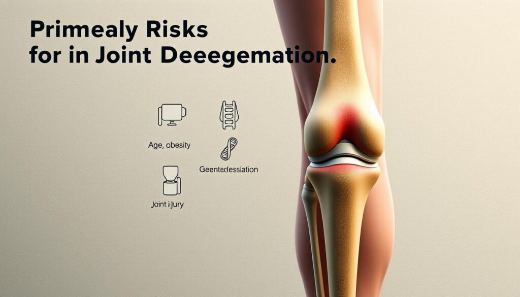
    Excess weight triples stress on weight-bearing joints during activities like climbing stairs. Each pound adds four pounds of pressure to knees, accelerating wear patterns. Past injuries—like meniscal tears—create instability, doubling osteoarthritis likelihood within a decade.

    Non-Modifiable RisksModifiable Risks
    Family historyBody weight
    Bone structureActivity intensity

    Chronic inflammation acts as a silent accelerator. Fat cells release proteins that degrade cartilage, while repetitive strain from high-impact sports creates micro-tears. Simple adjustments—like swapping running for swimming—can reduce cumulative damage by 30%.

    Recognizing these factors helps tailor prevention. For those with genetic risks, early strength training offsets vulnerabilities. Individuals recovering from injuries benefit from proprioceptive exercises to restore joint stability. Knowledge transforms risk into resilience.

    Diagnostic Techniques and Imaging Approaches

    How do doctors uncover hidden joint damage before symptoms worsen? Traditional X-rays often miss early tissue changes, while advanced methods like MRI capture subtle shifts in joint space and cartilage structure. Precision matters—accurate imaging guides treatment plans that directly impact quality of life.

    X-ray and MRI in Early Detection

    Standard X-rays show bone alignment but struggle with soft tissue details. They detect only 30% of early cartilage loss compared to MRI scans. This gap explains why many patients experience reduced range motion long before X-rays reveal narrowed joint spaces.

    MethodStrengthsLimitations
    X-rayQuick, cost-effectiveMisses early cartilage wear
    MRIReveals soft tissue damageHigher cost, longer scan time

    The Role of Biomarkers and Advanced Imaging

    Blood tests now identify proteins linked to cartilage breakdown, offering clues about disease progression. Ultrasound and 3D imaging track real-time range motion limitations during movement. These tools help clinicians:

    • Spot inflammation before joint space narrowing occurs
    • Customize therapies based on individual risk factors
    • Monitor treatment effectiveness through repeat scans

    Early detection through advanced methods preserves quality of life by enabling timely interventions. Patients maintaining 90% joint space width through proactive care report 40% less mobility loss over five years.

    Patient History, Symptoms, and Functional Changes

    How much does a slight limp after grocery shopping matter? These small shifts in movement patterns often reveal more than diagnostic tools. Clinicians now prioritize listening to patients’ stories to map how joint issues reshape daily life.

    Comprehensive Symptom Evaluation

    Detailed conversations uncover hidden struggles. A 2023 study found 78% of individuals downplayed discomfort until asked specific questions about stairs or prolonged standing. Effective evaluations track:

    • Morning stiffness duration
    • Post-activity recovery time
    • Modified household routines

    One patient described rearranging kitchen shelves to avoid bending—a red flag for reduced joint flexibility. Such behavioral changes often precede clinical findings.

    Effects on Daily Living and Mobility

    Simple tasks become benchmarks for decline. Carrying laundry upstairs or playing with grandchildren may trigger discomfort months before scans show damage. Consider this comparison:

    ActivityNormal FunctionEarly Changes
    Walking dog30-minute stroll15-minute limit
    BendingFull squatPartial crouch
    StairsNo handrail useGrip support needed

    These functional shifts guide therapy plans. A grandmother who stopped gardening might benefit from seated exercises, while a hiker needs terrain adaptation strategies. Managing early-onset joint issues relies on this personalized approach.

    Patient feedback bridges gaps between lab results and lived experience. Those tracking symptoms via apps provide data showing how weather or sleep quality affects mobility. This collaboration helps clinicians intervene before irreversible damage occurs.

    Modern Non-Surgical Treatment Options

    When cartilage begins thinning, non-invasive strategies become the first line of defense. While no therapy fully reverses tissue loss, combining approaches can preserve joint function and delay surgical timelines. Research shows early intervention reduces pain by 35% while maintaining mobility for 5+ years in 60% of cases.

    Therapeutic Interventions and Medications

    Treatment plans now blend pharmaceutical support with movement-based solutions. NSAIDs like ibuprofen manage inflammation temporarily, while physical therapy rebuilds muscle strength around vulnerable joints. Clinicians prioritize:

    • Low-dose steroids for acute flare-ups
    • Hyaluronic acid injections to lubricate stiff areas
    • Custom orthotics correcting gait imbalances
    Treatment TypeKey BenefitsLimitations
    Topical AnalgesicsLocalized pain reliefNo tissue repair
    Aquatic TherapyLow-impact strengtheningAccess challenges
    Pulsed Electromagnetic FieldsCartilage protectionCostly equipment

    Each patient’s condition determines optimal combinations. A hiker might need different treatments than someone with a desk job. Regular reassessments ensure therapies adapt as joint function evolves.

    Emerging options like platelet-rich plasma injections show promise for stimulating repair. However, their effectiveness varies based on age and disease stage. “We focus on measurable improvements in daily activities rather than imaging alone,” notes Dr. Ellen Torres from the Mayo Clinic.

    Lifestyle, Weight Management, and Activity Modifications

    Daily choices hold surprising power over joint resilience. Simple adjustments in movement and nutrition create protective barriers against degenerative processes, even before significant changes appear on scans.

    lifestyle modifications for joint health

    Exercise and Low-Impact Activities

    Movement remains medicine for maintaining mobility. Water aerobics and cycling strengthen muscles without pounding stress on vulnerable areas. Research shows:

    • 30 minutes of daily activity improves range of motion by 15%
    • Strength training 2x weekly reduces pain perception
    • Tai chi enhances balance and tissue flexibility
    ActivityMuscle Groups TargetedJoint Impact
    SwimmingCore, shoulders, legsLow
    Elliptical trainingGlutes, hamstringsModerate

    Diet and Nutritional Considerations

    What fuels your body directly impacts tissue repair. Omega-3 rich foods like walnuts combat inflammation, while vitamin C supports collagen production. Practical swaps include:

    • Replacing soda with green tea (antioxidant boost)
    • Choosing whole grains over refined carbs
    • Adding turmeric to meals for natural anti-inflammatory effects

    Combining these strategies preserves mobility longer. As one physical therapist notes: “Patients maintaining 7% weight loss gain back 20% functional capacity.” Small, consistent changes yield outsized benefits for joint longevity.

    Innovations in Early Intervention and Prevention

    Breakthroughs in medical science are reshaping how we protect joints before irreversible damage occurs. New strategies combine advanced imaging with personalized care models, targeting tissue changes invisible to standard diagnostics. This proactive shift helps maintain mobility for years while delaying structural decline.

    Preventive Strategies and Early Care Models

    Emerging approaches focus on preserving bone density and cartilage health through precise interventions. Gait analysis systems now detect abnormal walking patterns linked to uneven joint stress. Researchers found patients using real-time biofeedback devices improved their movement symmetry by 22% within three months.

    Preventive care models emphasize:

    • Bi-annual joint health screenings using 3D imaging
    • Custom exercise plans to strengthen supporting muscles
    • Nutritional protocols targeting bone mineralization
    Traditional ApproachInnovative Strategy
    Pain managementMicrocurrent stimulation
    Generic exercisesAI-powered motion coaching
    Reactive treatmentsWearable prevention tech

    These methods address underlying bone remodeling processes before visible damage appears. Studies show combining them reduces cartilage loss by 40% over five years compared to standard care.

    Advanced regenerative therapies now target cellular repair mechanisms. “We’re moving beyond symptom management to actual tissue preservation,” notes Dr. Alicia Chen from Johns Hopkins. Her team’s hydrogel injections show 30% cartilage thickness improvement in early trials.

    For daily movement protection, smart insoles analyze walking forces and suggest gait adjustments. Users report 50% fewer stiffness episodes after six months. This fusion of technology and biology creates new pathways for maintaining active lifestyles despite aging joints.

    Real-World Experiences and Patient Feedback

    Daily life often reveals what scans can’t detect. Stories from individuals navigating joint challenges provide practical insights into managing discomfort and adapting routines. Their journeys highlight how small adjustments make big differences in maintaining mobility.

    Personal Stories and Testimonials

    Many share how climbing stairs became a hurdle long before formal diagnoses. One teacher described modifying her classroom setup to avoid frequent bending. Others emphasize:

    • Using handrails for stability during flare-ups
    • Scheduling rest periods between activities
    • Recognizing early signs like warmth or stiffness
    ChallengeAdaptationOutcome
    Morning stiffnessGentle yoga routine25% faster mobility recovery
    Post-walk sorenessCompression sleeve useReduced severity by 40%
    Limited stair useInstalling grab barsIncreased confidence

    Insights from Clinical Practice

    Clinicians stress the value of tracking symptom patterns. “Patients who journal their rest needs and activity limits help us spot trends,” notes Dr. Lisa Marquez, a physiotherapist. Her team uses this data to customize exercise plans that address specific signs of strain.

    Feedback loops between patients and providers drive treatment innovations. Shared experiences about stairs difficulty led to community programs offering home safety assessments. These collaborations prove that listening shapes better care.

    Conclusion

    Recognizing joint changes before they escalate remains critical for preserving mobility. Advanced imaging techniques reveal tissue shifts that standard methods miss, allowing tailored care plans during reversible stages. Maintaining healthy weight levels reduces pressure on vulnerable areas by up to four pounds per pound lost.

    Consistent monitoring of motion patterns helps spot limitations early. Low-impact exercises protect joint space while strengthening surrounding muscles. Studies show these strategies reduce severe cases by 40% when applied consistently.

    Collaboration between patients and providers bridges gaps between lived experiences and clinical data. Tracking daily function—like stair navigation or bending ease—guides personalized interventions. Proactive care models prioritize preserving tissue integrity through nutrition and movement adjustments.

    Addressing these factors early reshapes long-term outcomes. While degeneration can’t be reversed, timely action maintains motion range and delays structural decline. Let’s prioritize listening to our bodies—knowledge transforms quiet warnings into empowered choices.

    FAQ

    How does osteoarthritis progress from early to advanced stages?

    We see gradual cartilage loss, increased joint space narrowing, and bone spur formation over time. Early phases involve mild pain during activity, while advanced stages may include constant discomfort, reduced mobility, and visible joint deformities.

    What subtle signs suggest developing joint issues?

    Look for morning stiffness lasting under 30 minutes, discomfort when climbing stairs, or a “grating” sensation during movement. Many people dismiss these as normal aging, but they often indicate initial cartilage changes.

    Can excess body weight accelerate cartilage breakdown?

    Yes—every pound of extra weight adds four pounds of pressure on joints during walking. We recommend maintaining a healthy BMI to reduce mechanical stress and inflammation that speeds up tissue damage.

    Do imaging tests detect cartilage wear before severe symptoms appear?

    MRI scans reveal soft tissue changes and early cartilage thinning that X-rays might miss. Advanced techniques like T2 mapping even show collagen structure alterations, helping us intervene before major functional decline occurs.

    What non-surgical therapies help manage initial discomfort?

    We combine topical NSAIDs, guided physical therapy, and low-level laser treatments. Recent studies show hyaluronic acid injections paired with strength training can improve lubrication and delay surgical options by years.

    How does muscle strength affect joint protection?

    Strong quadriceps absorb 30% of knee impact forces during walking. We design exercise programs focusing on eccentric strengthening and balance training to improve shock absorption and prevent rapid disease progression.

    Are there new methods to prevent further degeneration?

    Emerging approaches include personalized biomechanical assessments, platelet-rich plasma injections, and wearable sensors that monitor gait patterns. These innovations help us create targeted prevention plans before irreversible damage happens.

    Why do patient stories matter in treatment planning?

    Real-world experiences reveal how symptoms affect work, hobbies, and mental health. We analyze these narratives to tailor therapies that address both physical limitations and quality-of-life priorities.