Are you or someone you know experiencing knee pain due to patellar subluxation or dislocation? If so, you’re not alone. Patellar instability is a condition that affects many individuals, often resulting from injury, ligamentous laxity, or an increased Q angle of the knee.
The good news is that treatment is often nonoperative, with bracing being a highly effective option for first-time dislocation without bony avulsion or presence of articular loose bodies. In this article, we’ll explore the importance of using a knee brace for patellar instability and provide recommendations for the best knee braces available.
Key Takeaways
- Understanding patellar instability and its causes
- The role of bracing in treating patellar instability
- Factors to consider when choosing a knee brace
- Top recommendations for patellar instability braces
- The benefits of using a knee brace for patellar instability
Understanding Patellar Instability
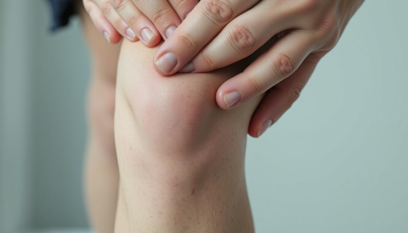
The patella, or kneecap, plays a vital role in knee function, and its instability can lead to significant discomfort and impairment. Patellar instability occurs when the patella slips out of its normal position, often causing pain and discomfort.
What Is Patellar Instability?
Patellar instability is characterized by the abnormal movement of the patella, which can lead to it slipping out of place. This condition can cause significant pain and discomfort, especially during activities that involve knee movement.
Key aspects of patellar instability include:
- Abnormal patellar movement
- Pain and discomfort
- Instability during knee activities
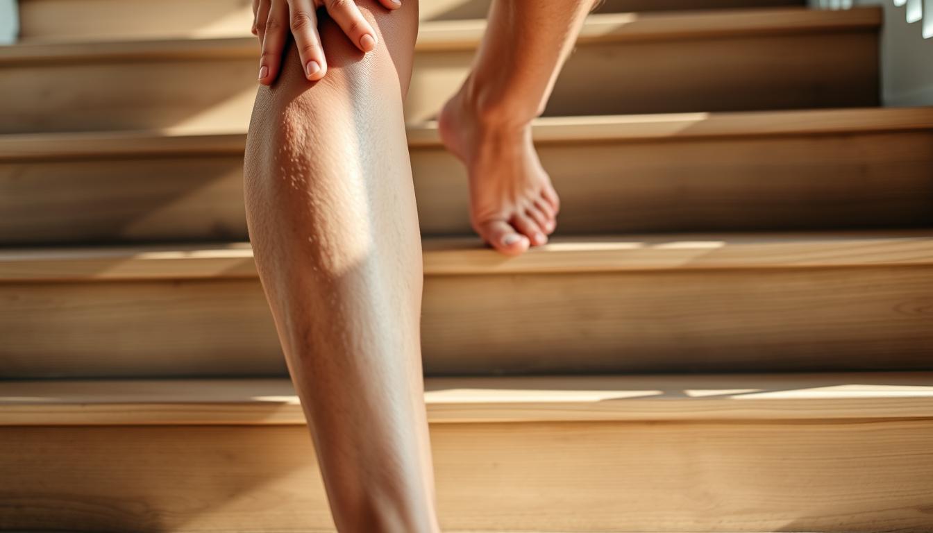
Symptoms to Watch For
Identifying the symptoms of patellar instability is crucial for early diagnosis and treatment. Common symptoms include:
- Complaints of instability
- Anterior knee pain
- A painful “pop” or “clunk” felt with patellar dislocation
Recognizing these symptoms can help individuals seek medical attention early, potentially preventing further complications.
Causes and Risk Factors
Several factors contribute to patellar instability, including:
| Cause/Risk Factor | Description |
|---|---|
| Ligamentous Laxity | Looseness in the ligaments that support the patella |
| Previous Patellar Instability Events | History of patellar dislocation or subluxation |
| Anatomical Factors | Conditions like patella alta and trochlear dysplasia |
Understanding these causes and risk factors is essential for choosing the right patellar brace and managing the condition effectively.
As noted by medical professionals, “Understanding the underlying causes of patellar instability is crucial for effective management and treatment.” This emphasizes the importance of a comprehensive approach to addressing patellar instability.
“The choice of patellar brace depends on the severity of the instability and the individual’s specific needs.”
Benefits of Using a Knee Brace
For individuals dealing with patellar instability, incorporating a knee brace into their daily routine can be a game-changer. Knee braces are designed to provide support and stability to the knee, which is crucial for managing patellar instability.
Enhanced Stability and Support
A knee brace offers enhanced stability by keeping the patella in its correct position, thereby reducing the risk of it slipping out of place. This is particularly beneficial during physical activities that may exacerbate the condition. The brace provides additional support to the knee structure, helping to alleviate stress on the surrounding tissues.

Pain Relief and Recovery
The use of a knee brace can significantly aid in pain relief by minimizing the movement of the patella and reducing irritation to the surrounding soft tissues. Furthermore, by providing support and stability, knee braces facilitate a smoother recovery process, enabling individuals to return to their normal activities more quickly.
Prevention of Further Injury
One of the key benefits of wearing a knee brace is the prevention of further injury. By stabilizing the knee and patella, the brace reduces the risk of additional trauma or strain, which is especially important during sports or activities that involve running, jumping, or quick changes of direction.
Features such as variable compression systems and breathable materials enhance the comfort and effectiveness of knee braces. These features allow for a customized fit and help in managing the condition more effectively.
Types of Patellar Instability Braces
Patellar instability braces come in various types, each designed to address specific needs. The choice of brace depends on the severity of the instability, the patient’s activity level, and personal preferences.
Patellar Stabilizing Braces
Patellar stabilizing braces are designed to provide direct support to the patella, helping to keep it in its correct position. These braces are particularly useful for individuals with recurrent patellar subluxation or dislocation. They often feature a patellar buttress or a horseshoe-shaped pad that applies gentle pressure to guide the patella into place.
Key benefits of patellar stabilizing braces include enhanced stability and support during physical activities. For runners, a best patella stabilizer for running can be crucial in preventing injuries and ensuring a smooth recovery.
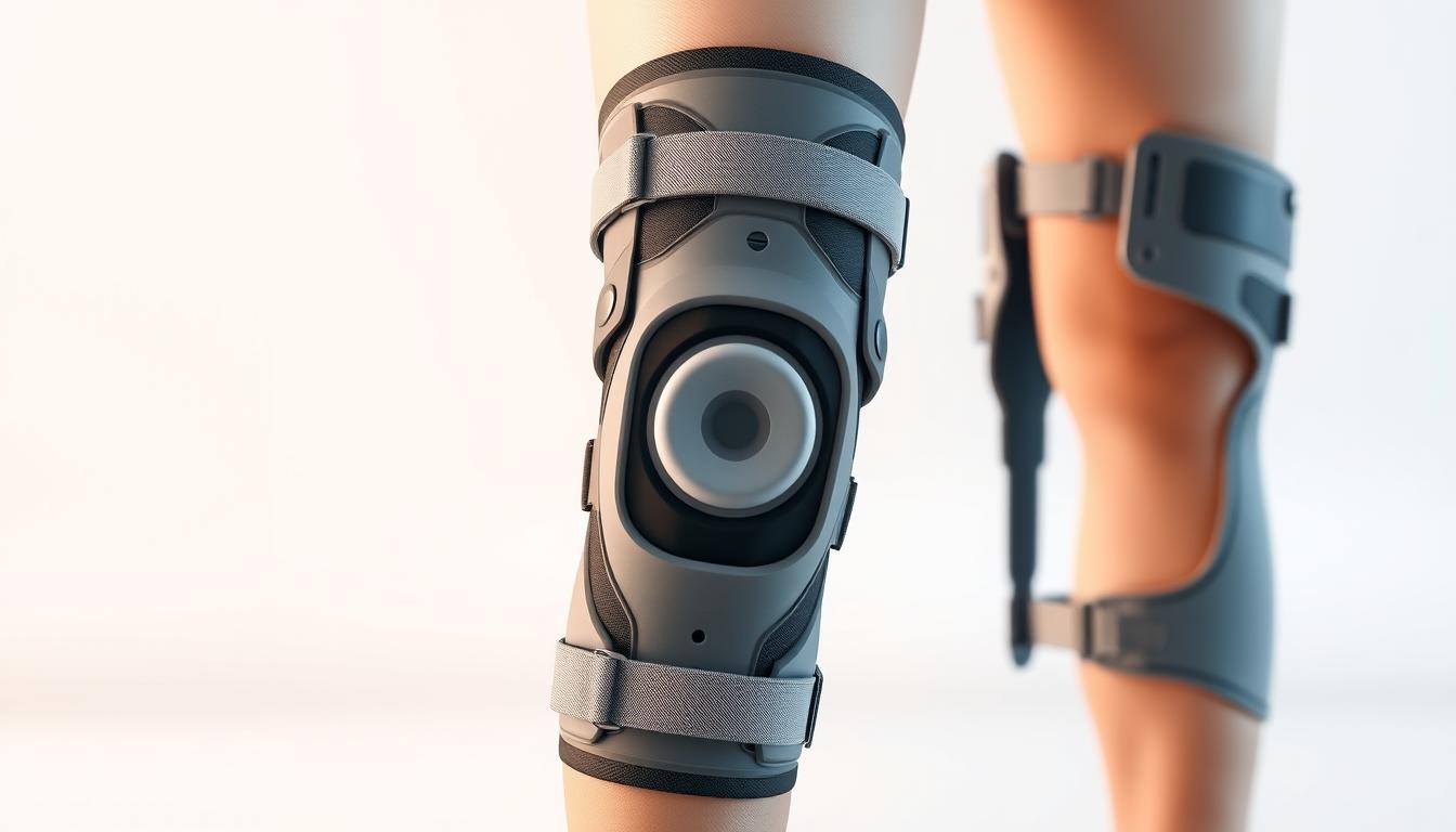
Hinged Knee Braces
Hinged knee braces offer additional support by stabilizing the knee joint, thereby indirectly supporting the patella. These braces feature hinges that allow for natural knee movement while preventing excessive motion that could exacerbate patellar instability.
The primary advantage of hinged knee braces is their ability to control knee movement, providing stability and protection. They are suitable for individuals with more severe knee instability or those who require additional support during high-impact activities.
Elastic Knee Sleeves
Elastic knee sleeves provide a more conservative approach to managing patellar instability. They offer compression and warmth to the knee area, which can help in reducing pain and swelling. While they do not provide the same level of stability as other braces, they are beneficial for mild cases of patellar instability or as a preventive measure.
For those seeking a simple, non-invasive solution, elastic knee sleeves can be a good option. They are also top-rated patellar stabilizer brace alternatives for individuals looking for comfort and flexibility.
In conclusion, the type of patellar instability brace that is most suitable depends on the individual’s specific condition, activity level, and personal comfort. Understanding the characteristics and benefits of each type can help in making an informed decision.
Key Features to Look for in a Brace
When searching for the ideal knee brace for patellar instability, several key features must be considered to ensure optimal support and comfort. The right brace can make a significant difference in managing patellar tendonitis and enhancing overall knee stability.
Adjustability and Fit
A crucial aspect of any orthopedic brace for patellar instability is its adjustability and fit. A well-fitting brace ensures that the patella is properly aligned and supported, reducing the risk of further irritation or injury. Look for braces with adjustable straps or sleeves that can be customized to fit your knee snugly.

Breathability and Comfort
The breathability and comfort of a knee brace are vital for long-term wearability. Braces made from moisture-wicking materials can help keep the skin dry and reduce irritation. For individuals with sensitive skin, hypoallergenic materials are recommended to minimize the risk of allergic reactions.
For more information on managing knee pain with the right brace, you can visit https://kneehurt.com/best-knee-brace-for-arthritis-top-picks-for-pain-relief/ to explore top picks for knee braces that can provide relief.
Durability and Material Quality
The durability of a knee brace is directly related to the quality of its materials and construction. High-quality materials not only extend the lifespan of the brace but also ensure consistent support and comfort. When choosing a brace, consider the quality of the fabric, the robustness of the stitching, and the overall build.
By focusing on these key features—adjustability and fit, breathability and comfort, and durability and material quality—you can find a knee brace that effectively supports your knee health and enhances your overall quality of life.
Recommended Patellar Instability Braces
Patellar instability can be effectively managed with the appropriate brace, and there are several top recommendations available. These braces are designed to provide the necessary support and stability to alleviate discomfort and prevent further injury.
McDavid Knee Brace with Stays
The McDavid Knee Brace with Stays is a popular choice among athletes and individuals with patellar instability. It offers excellent support and stability, thanks to its stays that help keep the patella in place. This brace is also known for its comfort and breathability, making it suitable for extended wear.
Shock Doctor Ultra Knee Support
Another highly recommended option is the Shock Doctor Ultra Knee Support. This brace is designed to provide maximum support and protection for the knee. It features a unique design that helps to absorb and disperse impact, reducing the risk of injury. Users have praised its comfort and effectiveness in managing patellar instability.
DonJoy Performance Bionic Knee Brace
The DonJoy Performance Bionic Knee Brace is also highly regarded for its performance and comfort. It is designed to provide dynamic support and stability, making it an excellent choice for individuals with active lifestyles. The brace is engineered to move with the user, offering a full range of motion while protecting the knee.
For more options and to explore these recommendations in detail, you can visit BraceAbility, which offers a range of knee braces for patellar subluxation.
As
“The right knee brace can significantly improve the quality of life for individuals with patellar instability by providing the necessary support and confidence to engage in daily activities.”
Choosing the best brace involves considering factors such as support level, comfort, and activity level to ensure the selected brace meets the individual’s specific needs.
How to Choose the Right Brace
The process of finding the perfect patellar brace involves several key considerations. With various options available, it’s crucial to make an informed decision that caters to your specific needs.
Consult with a Healthcare Professional
Before selecting a brace, it’s highly recommended to consult with a healthcare professional. They can provide valuable insights into the severity of your patellar instability and recommend the most suitable type of brace. A professional consultation can help in identifying the correct size and type of brace that would offer the necessary support.
“A healthcare professional can guide you through the process, ensuring that you choose a brace that aligns with your condition and lifestyle.” –
Consider Activity Level and Usage
Your activity level and intended usage play a significant role in choosing the right brace. For instance, if you’re highly active or participate in sports, you may require a more robust and durable brace. On the other hand, if you’re looking for a brace for everyday use, comfort and breathability might be your top priorities.
| Activity Level | Brace Type | Key Features |
|---|---|---|
| Highly Active | Robust Hinged Knee Brace | Durable, High Support |
| Moderately Active | Patellar Stabilizing Brace | Balanced Support, Comfort |
| Low Activity | Elastic Knee Sleeve | Comfortable, Breathable |
Evaluate Personal Preferences
Personal comfort and preferences are equally important when choosing a brace. Consider factors such as the material, adjustability, and overall design. A brace that is comfortable and suits your personal style is more likely to be worn consistently, thereby enhancing its effectiveness.

By considering these factors and consulting with a healthcare professional, you can make an informed decision and choose a brace that meets your needs, providing the necessary support and comfort.
Tips for Proper Brace Use
Proper use of a patellar instability brace is crucial for maximizing its effectiveness and preventing potential issues. When used correctly, a patellar stabilizing brace can provide the necessary support and stability to help manage patellar instability effectively.
Correct Application and Adjustment
To ensure your brace functions as intended, it’s vital to apply and adjust it correctly. Start by reading the manufacturer’s instructions carefully. Most top patellar stabilizing braces come with guidelines that are specific to their design. If you’re unsure, consult with a healthcare professional who can provide personalized advice.
Adjusting the brace to fit comfortably is key. A snug fit is essential, but it should not be too tight as to cause discomfort or restrict movement unnecessarily. Regularly check the fit, especially if you’re using the brace during different activities or over time as your condition improves.
Regular Maintenance and Cleaning
Maintaining your patellar support brace is crucial for its longevity and effectiveness. Regular cleaning is a must, as dirt and sweat can accumulate and affect the brace’s material and functionality. Follow the manufacturer’s cleaning instructions, and avoid using harsh chemicals that could damage the brace.
For more information on managing patella instability, you can visit this resource that provides additional insights and recommendations.
Listening to Your Body
It’s essential to be attentive to your body’s response when wearing a patellar instability brace. If you experience persistent pain, discomfort, or any unusual sensations, it may be a sign that the brace needs adjustment or that there’s an underlying issue that needs medical attention.
Regularly assessing your comfort and the brace’s effectiveness can help you make informed decisions about your treatment plan. Don’t hesitate to seek professional advice if you’re unsure about any aspect of your brace use.
Alternatives to Bracing
Beyond bracing, several alternatives can help manage patellar instability, focusing on therapy, exercise, and lifestyle adjustments. These alternatives can be used in conjunction with or instead of bracing, depending on the individual’s condition and preferences.
Physical Therapy Options
Physical therapy is a viable alternative or complement to bracing for managing patellar instability. A physical therapist can create a personalized exercise program to strengthen the muscles around the knee, improve flexibility, and enhance knee stability. Techniques may include manual therapy, stretching, and strengthening exercises tailored to the individual’s needs and condition.
Strengthening Exercises for the Knee
Strengthening the muscles around the knee is crucial for improving patellar stability. Exercises such as squats, lunges, and leg press can help strengthen the quadriceps, hamstrings, and other supporting muscles. It’s essential to start with low-intensity exercises and gradually increase the intensity to avoid putting excessive strain on the knee.
Examples of Strengthening Exercises:
- Straight leg raises
- Wall squats
- Step-ups
Activity Modifications
Making adjustments to daily activities and sports can also help manage patellar instability. This might involve avoiding activities that aggravate the condition, such as deep knee bending or high-impact sports, and incorporating low-impact alternatives like cycling or swimming. Wearing appropriate footwear and using orthotics if necessary can also help.
By exploring these alternatives to bracing, individuals with patellar instability can find a management strategy that suits their lifestyle and needs, potentially reducing reliance on bracing or enhancing its effectiveness when used in combination.
When to Seek Medical Advice
Recognizing the signs that indicate a need for medical advice can make a substantial difference in treating patellar instability. It’s crucial to monitor your condition and seek professional help when necessary.
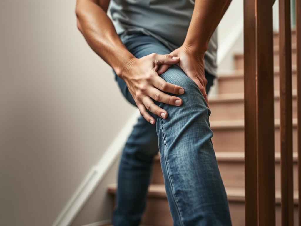
Persistent Pain and Swelling
If you experience persistent pain and swelling in your knee, it’s a clear indication that you should consult a healthcare professional. This could be a sign of an underlying issue that requires further evaluation or a different treatment approach, potentially involving a top-rated patellar stabilizer brace.
As noted by orthopedic specialists, “Persistent pain is a signal that the knee is not recovering as expected, and further assessment is needed to determine the best course of action.”
“Chronic knee pain can significantly impact one’s quality of life, making it essential to seek medical advice to address the root cause of the issue.”
Changes in Knee Function
Any noticeable changes in knee function, such as reduced mobility or instability, should prompt a visit to a medical professional. This could indicate a progression of the condition or a complication that needs to be addressed.
| Signs of Knee Function Changes | Possible Implications |
|---|---|
| Reduced mobility | Potential worsening of patellar instability |
| Increased instability | Higher risk of further injury or complications |
Signs of Complications
Signs of complications, such as increased pain, significant swelling, or difficulty walking, necessitate immediate medical attention. Using an orthopedic brace for patellar instability might be recommended as part of the treatment plan.
In conclusion, being aware of the signs that warrant medical advice is crucial for effective management of patellar instability. If you’re experiencing any of these symptoms, don’t hesitate to seek professional help.
Conclusion: Finding the Best Solution
Finding the best solution for patellar instability involves a combination of the right brace, appropriate treatment, and lifestyle adjustments. By understanding the condition and available options, individuals can take the next steps towards recovery and maintain an active lifestyle with confidence.
Effective Management Strategies
To manage patellar instability effectively, it’s crucial to choose a patellar brace that suits your specific needs. Consider factors such as adjustability, breathability, and durability when selecting a brace. Consulting with a healthcare professional can also provide valuable insights into the most suitable options.
Maintaining an Active Lifestyle
With the right patellar instability brace recommendations, individuals can confidently engage in their preferred activities without exacerbating the condition. By incorporating strengthening exercises and making necessary lifestyle adjustments, individuals can reduce the risk of further injury and maintain overall knee health.
By following these guidelines and staying informed, individuals can navigate the process of managing patellar instability with ease, ensuring a path towards recovery and continued activity.
FAQ
What is patellar instability, and how is it treated?
Patellar instability occurs when the patella (kneecap) slips out of its normal position, often causing pain and discomfort. Treatment options include using a patellar instability brace, physical therapy, and in some cases, surgery. A brace can provide stability and support to the knee, helping to alleviate symptoms.
How do I choose the best knee brace for patellar instability?
To choose the best knee brace, consider factors such as adjustability, comfort, and durability. It’s also essential to consult with a healthcare professional to determine the most suitable type of brace for your specific needs. Look for braces with features like patellar stabilizing straps or hinged designs that provide additional support.
What are the benefits of using a patellar stabilizing brace?
A patellar stabilizing brace can provide several benefits, including enhanced stability and support, pain relief, and prevention of further injury. By keeping the patella in its correct position, a brace can help alleviate symptoms and improve overall knee function.
Are there different types of patellar instability braces available?
Yes, there are various types of patellar instability braces, including patellar stabilizing braces, hinged knee braces, and elastic knee sleeves. Each type has its unique characteristics, benefits, and uses, and the most suitable one for you will depend on your specific needs and preferences.
Can I use a knee brace for patellar tendonitis?
Yes, a knee brace can be used to help manage patellar tendonitis. A brace can provide support and stability to the knee, reducing strain on the patellar tendon and alleviating pain. Look for braces with features like compression and cushioning that can help reduce inflammation.
How do I properly use a patellar instability brace?
To properly use a patellar instability brace, ensure correct application and adjustment, follow the manufacturer’s guidelines for maintenance and cleaning, and be mindful of your body’s comfort and any signs of discomfort or pain. It’s also essential to consult with a healthcare professional for personalized advice.
Are there alternatives to using a brace for managing patellar instability?
Yes, alternatives to bracing include physical therapy, strengthening exercises for the knee, and activity modifications. These approaches can help alleviate symptoms, improve knee stability, and reduce the risk of further injury. Consult with a healthcare professional to determine the best course of treatment for your specific needs.
When should I seek medical advice for patellar instability?
You should seek medical advice if you experience persistent pain and swelling, changes in knee function, or signs of complications such as infection or nerve damage. A healthcare professional can assess your condition and provide personalized guidance on the best treatment options.
Can I continue to exercise and stay active with patellar instability?
Yes, with proper management and treatment, it’s possible to continue exercising and staying active with patellar instability. Consult with a healthcare professional to determine the most suitable activities and exercises for your condition, and consider using a patellar instability brace to provide additional support and stability.
 for treating patellofemoral pain syndrome")


 activation")