Tag: Physical Therapy Management for Various Symptoms and Diagnostics

  • Osteoporosis Treatment + 6 Natural Therapies: A Comprehensive Guide

    Osteoporosis Treatment + 6 Natural Therapies: A Comprehensive Guide

    Introduction

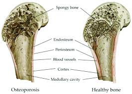
    Osteoporosis, often called the “silent disease,” is a condition characterized by decreased bone density and deterioration of bone tissue, leading to increased fragility and risk of fractures. The condition affects approximately 1 in 3 women and 1 in 5 men over the age of 50, making it a significant public health concern worldwide. The impact of osteoporosis extends beyond bone health – hip fractures, in particular, are associated with a 3-4 times greater risk of dying within 12 months compared to the general population of the same age.

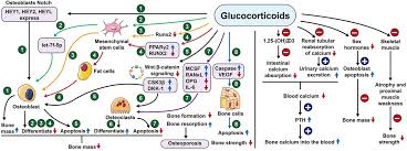
    Throughout our lives, our bones undergo a continuous process of remodeling, with old bone being removed (resorption) and new bone being formed. In healthy individuals, this process maintains a balance. However, in osteoporosis, bone resorption outpaces bone formation, resulting in a net loss of bone mass and structural deterioration.

    The good news is that osteoporosis is both preventable and treatable. While conventional medical treatments play a crucial role in managing the disease, especially for those at high fracture risk, natural approaches can complement these treatments and provide additional benefits for bone health. This article explores the full spectrum of osteoporosis management, from medical interventions to evidence-based natural therapies that can help strengthen bones and reduce fracture risk.

    By understanding both conventional treatments and natural approaches, individuals can work with their healthcare providers to develop a comprehensive strategy tailored to their specific needs and risk factors.

    download 2025 04 29T200945.128

    Understanding Osteoporosis

    To effectively address osteoporosis, it’s important to understand the disease process and the factors that contribute to its development. At its core, osteoporosis occurs when the body loses too much bone, makes too little bone, or both. This results in weakened bones that can break from minor falls or, in serious cases, even from simple actions like sneezing or bumping into furniture.

    Several risk factors contribute to the development of osteoporosis:

    • Age and Gender: Risk increases with age, with women at significantly higher risk than men. The rapid bone loss that occurs in the 5-7 years following menopause makes women particularly vulnerable.
    • Genetic Factors: Family history of osteoporosis increases risk, as does being of Caucasian or Asian descent.
    • Hormonal Changes: The decline in estrogen during menopause in women and testosterone in men accelerates bone loss. Other hormonal disorders, such as hyperthyroidism or hyperparathyroidism, can also affect bone health.
    • Body Size: Small-framed individuals and those with low body weight have less bone mass to draw from as they age.
    • Lifestyle Factors: Inadequate calcium and vitamin D intake, sedentary lifestyle, smoking, and excessive alcohol consumption all contribute to bone loss.
    • Medications: Long-term use of certain medications, including corticosteroids, anticonvulsants, and some cancer treatments, can adversely affect bone health.

    Osteoporosis is typically categorized as either primary or secondary. Primary osteoporosis is related to aging and hormonal changes, while secondary osteoporosis results from specific medical conditions or medications that affect bone metabolism.

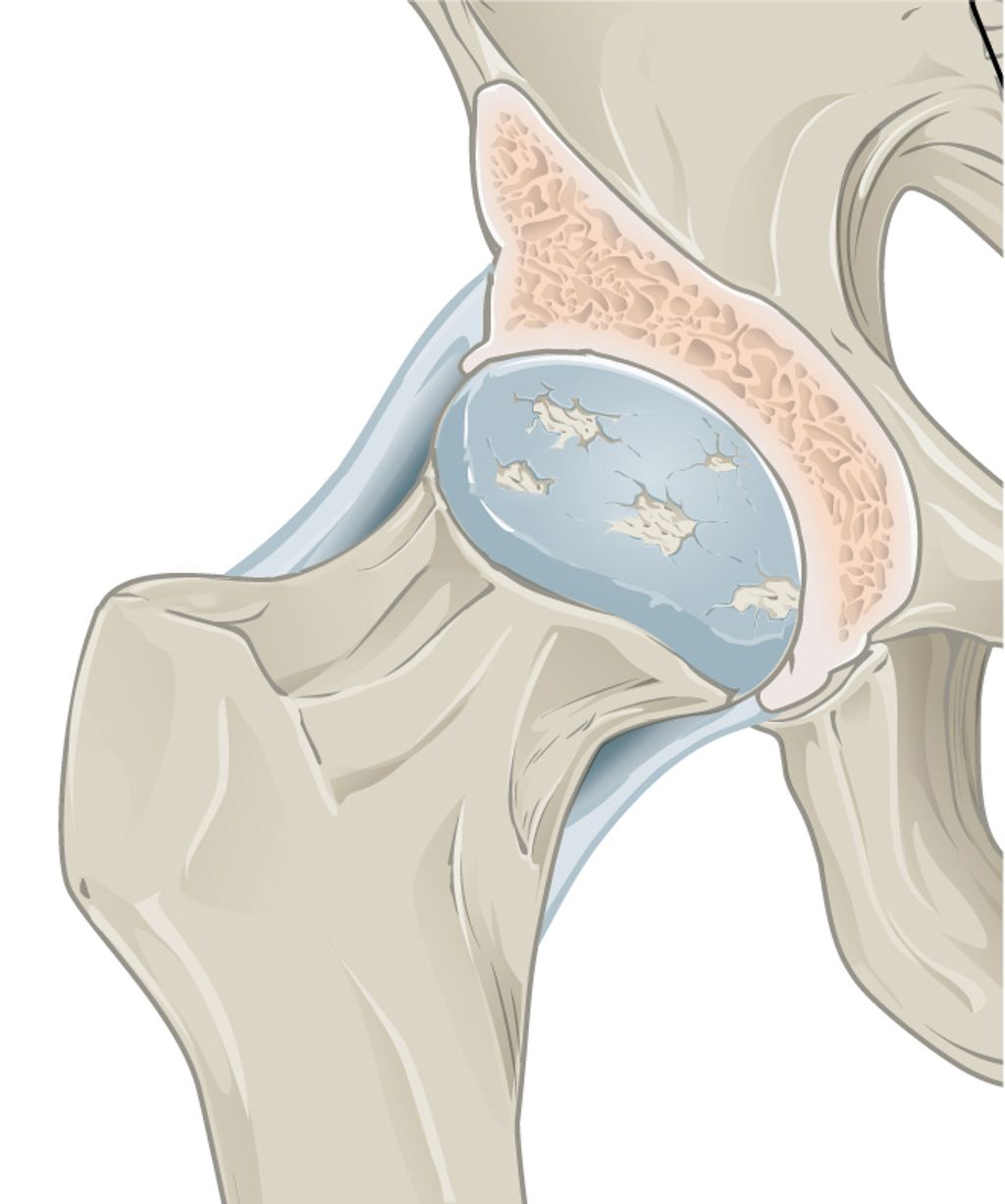
    Diagnosis typically involves dual-energy X-ray absorptiometry (DEXA scan), which measures bone mineral density (BMD). The results are presented as a T-score, comparing an individual’s bone density to that of a healthy 30-year-old of the same sex. A T-score of -1.0 or above is considered normal, while scores between -1.0 and -2.5 indicate osteopenia (low bone mass), and scores below -2.5 indicate osteoporosis.

    The Fracture Risk Assessment Tool (FRAX) may also be used to predict the 10-year probability of a major osteoporotic fracture based on individual risk factors, with or without BMD measurements.

    One of the challenges of osteoporosis is that it progresses silently, often without symptoms until a fracture occurs. This makes screening and preventive measures crucial, especially for those with known risk factors.

    Conventional Medical Treatments

    Modern medicine offers several effective treatments for osteoporosis, designed to reduce fracture risk by slowing bone loss, increasing bone formation, or both. Treatment recommendations typically consider factors such as age, sex, fracture history, bone density measurements, and overall fracture risk.

    First-Line Medications

    Bisphosphonates remain the most commonly prescribed first-line treatment for osteoporosis. These medications slow bone resorption by inhibiting the activity of osteoclasts, the cells responsible for breaking down bone. The American College of Physicians (ACP) recommends bisphosphonates as the initial pharmacologic treatment for reducing fracture risk in postmenopausal women with osteoporosis.

    Common bisphosphonates include:

    • Alendronate (Fosamax) – taken weekly or monthly
    • Risedronate (Actonel) – taken weekly or monthly
    • Ibandronate (Boniva) – taken monthly or as quarterly injections
    • Zoledronic acid (Reclast) – administered as a yearly intravenous infusion

    These medications have been shown to reduce the risk of vertebral fractures by 40-70% and non-vertebral fractures, including hip fractures, by 20-40%. Side effects can include gastrointestinal issues with oral formulations and flu-like symptoms with intravenous formulations. Rare but serious side effects include osteonecrosis of the jaw and atypical femur fractures, particularly with long-term use.

    Second-Line Treatments

    When bisphosphonates are not appropriate or effective, several second-line treatments are available:

    Denosumab (Prolia) is a RANK ligand inhibitor that blocks the development and activity of osteoclasts. Given as a subcutaneous injection every six months, it has been shown to reduce vertebral, non-vertebral, and hip fractures. Unlike bisphosphonates, denosumab does not accumulate in the bone, so its effects reverse quickly if treatment is stopped, potentially leading to rapid bone loss and increased fracture risk if not properly managed.

    Selective Estrogen Receptor Modulators (SERMs) such as raloxifene (Evista) mimic estrogen’s beneficial effects on bone without some of the risks associated with estrogen. They can reduce vertebral fracture risk but have not been shown to reduce non-vertebral or hip fracture risk.

    Hormone Replacement Therapy (HRT) was once widely used for osteoporosis prevention but is now primarily recommended for managing menopausal symptoms in women at high risk for osteoporosis, and only for the shortest duration possible due to potential risks of breast cancer, heart disease, and stroke.

    Advanced Treatments for Severe Cases

    For patients with severe osteoporosis or those who have experienced fractures despite other treatments, more potent options are available:

    Anabolic Therapies stimulate bone formation rather than simply slowing bone loss. These include:

    • Teriparatide and abaloparatide – synthetic forms of parathyroid hormone that stimulate bone formation when given intermittently. They are administered as daily injections for up to two years.
    • Romosozumab (Evenity) – a sclerostin inhibitor that both increases bone formation and decreases bone resorption. It is given as monthly injections for one year.

    These medications can increase bone density more substantially than antiresorptive drugs, especially at the spine. However, they are typically reserved for those at very high fracture risk due to their cost, route of administration, and limited treatment duration.

    Treatment Duration and Management

    The optimal duration of osteoporosis treatment continues to be studied. Many experts recommend reassessing after 3-5 years of bisphosphonate therapy, with consideration of a “drug holiday” for patients whose fracture risk has decreased. For high-risk patients, sequential therapy (starting with an anabolic agent followed by an antiresorptive) may provide optimal fracture protection.

    Regular monitoring of bone mineral density and, in some cases, biochemical markers of bone turnover, can help assess treatment response and guide decisions about continuing or modifying therapy.

    It’s important to remember that medication is just one component of osteoporosis management. All treatment approaches should be accompanied by adequate calcium and vitamin D intake, appropriate exercise, and measures to prevent falls.

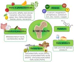
    Natural Therapy #1: Nutrition for Bone Health

    Nutrition plays a fundamental role in both preventing and managing osteoporosis. While medications can effectively slow bone loss or stimulate bone formation, they cannot compensate for nutritional deficiencies that compromise bone health. A bone-healthy diet provides the essential building blocks needed for optimal bone remodeling and maintenance.

    Calcium: The Foundation of Bone Health

    Calcium is the primary mineral found in bone, making adequate intake essential throughout life. The recommended daily intake varies by age and gender:

    • Adults aged 19-50: 1,000 mg
    • Women aged 51+: 1,200 mg
    • Men aged 51-70: 1,000 mg
    • Men aged 71+: 1,200 mg

    Dietary sources of calcium include:

    • Dairy products (milk, yogurt, cheese)
    • Fortified non-dairy alternatives (soy milk, almond milk)
    • Leafy green vegetables (kale, collard greens, bok choy)
    • Calcium-set tofu
    • Canned fish with bones (sardines, salmon)
    • Fortified cereals and juices

    While food sources are preferred, calcium supplements can help bridge dietary gaps. Two main types are available:

    • Calcium carbonate: Higher concentration of elemental calcium (40%), best absorbed with food
    • Calcium citrate: Lower concentration (21%), but better absorbed on an empty stomach and by those with reduced stomach acid

    For optimal absorption, calcium supplements should be taken in doses of 500-600 mg or less at a time, spaced throughout the day. Taking them with meals can reduce the risk of kidney stones and improve absorption, particularly for calcium carbonate.

    Vitamin D: The Essential Partner

    Vitamin D is crucial for calcium absorption and proper bone mineralization. Without adequate vitamin D, the body cannot effectively utilize calcium, regardless of intake. Current recommendations include:

    • Adults up to age 70: 600-800 IU daily
    • Adults over 70: 800-1,000 IU daily
    • Higher doses may be needed for those with vitamin D deficiency or limited sun exposure

    Vitamin D sources include:

    • Sunlight (the body produces vitamin D when skin is exposed to UVB rays)
    • Fatty fish (salmon, mackerel, tuna)
    • Fortified foods (milk, orange juice, cereals)
    • Egg yolks
    • Supplements (D3 is generally preferred over D2)

    Many healthcare providers recommend checking vitamin D levels through a blood test (25-hydroxyvitamin D) to determine if supplementation is needed. Optimal levels are generally considered to be 30-60 ng/mL.

    Beyond Calcium and Vitamin D

    While calcium and vitamin D receive the most attention, other nutrients also contribute to bone health:

    • Protein: Provides the structural matrix for bone and stimulates insulin-like growth factor I, which promotes bone formation. Aim for 0.8-1.0 g/kg of body weight daily, from both animal and plant sources.
    • Vitamin K: Important for bone protein synthesis. Found in leafy greens, broccoli, and fermented foods.
    • Magnesium: Influences crystal formation in bone and calcium metabolism. Found in nuts, seeds, whole grains, and leafy greens.
    • Potassium: Helps maintain acid-base balance, reducing calcium loss from bone. Abundant in fruits and vegetables.
    • Zinc and Manganese: Essential for bone formation enzymes. Found in whole grains, nuts, and seeds.

    Certain dietary patterns may also impact bone health. The Mediterranean diet, rich in fruits, vegetables, whole grains, fish, olive oil, and nuts, has been associated with higher bone density and lower fracture risk.

    Conversely, some dietary factors may negatively affect bone health:

    • High sodium intake increases calcium excretion
    • Excessive caffeine may interfere with calcium absorption
    • Very high protein diets can increase calcium excretion
    • Carbonated beverages, particularly colas, have been associated with lower bone density in some studies
    Figure 2: Bone-building nutrients and their food sources. A balanced diet rich in these nutrients supports optimal bone health and complements medical treatments for osteoporosis.
    Figure 2: Bone-building nutrients and their food sources. A balanced diet rich in these nutrients supports optimal bone health and complements medical treatments for osteoporosis.

    Natural Therapy #2: Exercise for Osteoporosis

    Exercise is a powerful tool for building and maintaining bone strength. Unlike medication, which primarily works to slow bone loss, appropriate physical activity can actually stimulate bone formation, improve balance and coordination (reducing fall risk), and enhance overall functional capacity.

    How Exercise Strengthens Bones

    Bones respond to mechanical loading much like muscles respond to resistance – they adapt and strengthen. This phenomenon, known as Wolff’s Law, explains why weight-bearing activities and resistance training are particularly beneficial for bone health. When muscles pull on bones during these activities, they create stress that stimulates osteoblasts (bone-building cells) to lay down new bone tissue.

    Different types of exercise affect bone health in different ways:

    Weight-Bearing Exercises

    Weight-bearing exercises force you to work against gravity while staying upright. These activities are particularly effective for strengthening the bones of the hips, legs, and lower spine. They include:

    • High-impact weight-bearing exercises: Running, jumping, high-impact aerobics, dancing, tennis, and basketball can build bone mass effectively in those without osteoporosis or previous fractures.
    • Low-impact weight-bearing exercises: Walking, elliptical training, stair climbing, and low-impact aerobics provide less bone stimulation but are safer options for those with osteoporosis, previous fractures, or other health limitations.

    A general recommendation is to engage in weight-bearing aerobic activities for 30 minutes on most days of the week.

    Resistance Training

    Resistance or strength training involves working against resistance, whether from weights, bands, water, or body weight. These exercises target specific muscle groups and the bones they attach to. Effective resistance training for bone health includes:

    • Free weights (dumbbells, barbells)
    • Weight machines
    • Resistance bands
    • Body weight exercises (push-ups, squats)

    Resistance training should be performed 2-3 times per week, targeting all major muscle groups. For bone health benefits, moderate intensity (8-12 repetitions with a weight that creates fatigue by the final repetition) is generally recommended.

    Balance and Posture Exercises

    While these exercises don’t directly build bone, they help prevent falls – a critical consideration for those with osteoporosis:

    • Tai chi
    • Yoga (with modifications for osteoporosis)
    • Posture training
    • Stability ball exercises
    • Single-leg standing

    Balance exercises should be incorporated into daily routines, even if just for a few minutes each day.

    Exercise Precautions for Osteoporosis

    For those already diagnosed with osteoporosis, exercise remains beneficial but requires certain precautions:

    • Avoid high-impact activities if you have severe osteoporosis or previous fractures
    • Avoid rapid, forceful movements that might increase fracture risk
    • Avoid extreme forward bending and twisting of the spine
    • Focus on proper form rather than amount of weight lifted
    • Start slowly and progress gradually
    • Consider working with a physical therapist to develop a safe, effective program

    Developing an Exercise Program

    An ideal exercise program for bone health includes:

    1. 30 minutes of weight-bearing aerobic activity on most days
    2. Resistance training for all major muscle groups 2-3 times weekly
    3. Balance exercises daily
    4. Posture and core strengthening exercises regularly

    For those new to exercise or with health concerns, starting with a physical therapist or qualified fitness professional experienced in working with osteoporosis is recommended. They can design a program tailored to individual needs, limitations, and goals.

    Remember that consistency is key – the bone benefits of exercise are lost when activity stops, so finding enjoyable activities that can be maintained long-term is essential for ongoing bone health.

    Natural Therapy #3: Vitamin K for Bone Health

    Vitamin K has emerged as an important nutrient for bone health that often doesn’t receive the same attention as calcium and vitamin D. Research increasingly suggests that adequate vitamin K intake is essential for optimal bone metabolism and strength.

    The Role of Vitamin K in Bone Metabolism

    Vitamin K serves as a cofactor for the enzyme that activates osteocalcin, a protein that binds calcium to the bone matrix. Without sufficient vitamin K, osteocalcin remains inactive, leading to reduced bone mineralization and potentially increased fracture risk.

    There are two main forms of vitamin K:

    • Vitamin K1 (Phylloquinone): The primary dietary form, found mainly in green leafy vegetables
    • Vitamin K2 (Menaquinones): Found in fermented foods and produced by intestinal bacteria; appears to be more effective for bone health than K1

    Studies have found that higher vitamin K intake is associated with higher bone mineral density and lower fracture risk. Low circulating levels of vitamin K have been linked to lower bone mass and increased fracture risk, particularly hip fractures.

    Dietary Sources of Vitamin K

    The best food sources of vitamin K include:

    • Vitamin K1 sources: Kale, spinach, collard greens, broccoli, Brussels sprouts, cabbage, lettuce
    • Vitamin K2 sources: Natto (fermented soybeans), cheese, egg yolks, butter from grass-fed cows, fermented foods

    The adequate intake (AI) for vitamin K is:

    • Adult women: 90 micrograms per day
    • Adult men: 120 micrograms per day

    However, optimal amounts for bone health may be higher than these basic recommendations.

    Vitamin K Supplementation

    Research on vitamin K supplementation for osteoporosis has shown mixed results. Some studies, particularly those using vitamin K2 (MK-4 form) at doses of 45mg daily, have shown reduced fracture risk in Japanese populations. Other studies using different forms or lower doses have shown more modest or inconsistent effects.

    When considering vitamin K supplementation:

    • Form matters: Vitamin K2, particularly the MK-4 and MK-7 forms, may be more beneficial for bone health than K1
    • Dosage varies: Therapeutic doses used in studies range from 45-180 micrograms for MK-7 and up to 45mg for MK-4
    • Medication interactions: Vitamin K can interfere with certain blood-thinning medications, particularly warfarin. Those taking such medications should consult their healthcare provider before supplementing

    Current Evidence and Recommendations

    While the evidence for vitamin K supplementation is promising, it’s not yet conclusive enough for most major medical organizations to recommend routine supplementation specifically for osteoporosis. Current approaches include:

    1. Ensuring adequate vitamin K intake through diet, particularly green leafy vegetables
    2. Considering supplementation under healthcare provider guidance, especially for those with low dietary intake or at high fracture risk
    3. Using vitamin K as part of a comprehensive bone health strategy that includes calcium, vitamin D, and other nutrients

    For those interested in supplementation, consulting with a healthcare provider is essential, particularly for those on medications that might interact with vitamin K.

    Natural Therapy #4: Magnesium and Bone Health

    Magnesium is an essential mineral that plays multiple roles in bone health yet is often overlooked in discussions about osteoporosis prevention and treatment. Approximately 60% of the body’s magnesium is stored in bone tissue, highlighting its importance to skeletal structure.

    Magnesium’s Role in Bone Metabolism

    Magnesium contributes to bone health through several mechanisms:

    • It influences the activity of osteoblasts and osteoclasts, the cells responsible for bone formation and resorption
    • It affects crystal formation in bone, influencing bone quality and strength
    • It regulates calcium transport and metabolism
    • It is required for the conversion of vitamin D to its active form, which is necessary for calcium absorption
    • It helps maintain appropriate calcium levels in the blood and tissues

    Research has found that magnesium deficiency is associated with reduced bone mineral density, altered bone and mineral metabolism, and increased fracture risk. One study found that 40% of women with osteoporosis or low bone density had low circulating magnesium levels.

    Dietary Sources of Magnesium

    The recommended dietary allowance (RDA) for magnesium is:

    • Adult women ages 19-30: 310 mg daily
    • Adult women ages 31+: 320 mg daily
    • Adult men ages 19-30: 400 mg daily
    • Adult men ages 31+: 420 mg daily

    Excellent food sources of magnesium include:

    • Dark leafy greens (spinach, chard)
    • Nuts and seeds (almonds, pumpkin seeds)
    • Whole grains (brown rice, quinoa)
    • Legumes (black beans, chickpeas)
    • Dark chocolate
    • Avocados
    • Bananas

    Despite its abundance in whole foods, many people don’t consume enough magnesium due to dietary patterns high in processed foods, which typically contain little magnesium. Soil depletion of minerals may also reduce the magnesium content of foods compared to historical levels.

    Magnesium Supplementation

    For those who cannot meet their magnesium needs through diet alone, supplements can be beneficial. Several forms are available, each with different properties:

    • Magnesium citrate: Well-absorbed, may have a mild laxative effect
    • Magnesium glycinate: Well-absorbed with minimal digestive side effects
    • Magnesium malate: Well-tolerated and may help with muscle pain
    • Magnesium oxide: Lower absorption rate but higher elemental magnesium content
    • Magnesium chloride: Good absorption and often available as a topical oil

    When supplementing, it’s generally recommended to start with a lower dose and gradually increase to avoid digestive discomfort. Taking magnesium supplements with food can also improve tolerance.

    Safety Considerations

    While magnesium is generally safe, excessive intake from supplements (not food) can cause diarrhea, nausea, and abdominal cramping. In people with reduced kidney function, high doses of magnesium supplements can lead to magnesium toxicity, characterized by low blood pressure, confusion, and cardiac complications.

    Those with kidney disease, heart problems, or on certain medications should consult their healthcare provider before supplementing with magnesium.

    Evidence and Recommendations

    While more research is needed on the specific effects of magnesium supplementation on fracture risk, ensuring adequate magnesium intake is a sensible strategy for supporting bone health. The greatest benefits are likely to be seen in those who are magnesium deficient.

    Current recommendations include:

    1. Prioritizing magnesium-rich whole foods in the diet
    2. Considering supplementation if dietary intake is insufficient or if deficiency is suspected
    3. Integrating magnesium into a comprehensive bone health approach alongside calcium, vitamin D, and other supportive nutrients
    Figure 3: The six natural therapies work synergistically to support bone health, complementing conventional medical treatments for osteoporosis.
    Figure 3: The six natural therapies work synergistically to support bone health, complementing conventional medical treatments for osteoporosis.

    Natural Therapy #5: Soy Isoflavones and Phytoestrogens

    The dramatic increase in osteoporosis risk that follows menopause highlights the crucial role of estrogen in maintaining bone health. As estrogen levels decline, bone resorption accelerates, often leading to significant bone loss in the first 5-7 years after menopause. This connection between estrogen and bone health has led researchers to investigate plant compounds with estrogen-like properties, known as phytoestrogens, as potential natural therapies for osteoporosis.

    Understanding Isoflavones and Phytoestrogens

    Isoflavones are a class of phytoestrogens – plant compounds that have a structure similar to human estrogen and can bind to estrogen receptors in the body, though their effects are typically much weaker than those of human estrogen. The most well-studied isoflavones include:

    • Genistein
    • Daidzein
    • Glycitein

    These compounds are found primarily in soybeans and soy products, but also in smaller amounts in other legumes such as chickpeas, lentils, and beans.

    Mechanisms of Action

    Isoflavones may support bone health through several mechanisms:

    • Binding to estrogen receptors in bone tissue, potentially slowing bone resorption
    • Inhibiting the activity of osteoclasts (cells that break down bone)
    • Supporting the activity of osteoblasts (cells that build bone)
    • Providing antioxidant effects that may protect bone cells from oxidative stress

    Research on Isoflavones and Bone Health

    Research on soy isoflavones for bone health has shown promising but mixed results:

    A meta-analysis of 19 studies in postmenopausal women found that soy isoflavone supplementation significantly increased bone mineral density by 54% and reduced bone resorption markers by 23% compared to baseline values. The most significant benefits were seen in studies lasting at least one year and using higher doses (80-90 mg of isoflavones daily).

    However, not all studies have shown positive results, and the effects may vary based on factors such as:

    • Individual metabolism of isoflavones (some people convert daidzein to equol, a more potent compound, while others do not)
    • Age and years since menopause
    • Baseline bone density
    • Dose and type of isoflavones used
    • Study duration

    Dietary Sources of Isoflavones

    The richest food sources of isoflavones include:

    • Soybeans and whole soy foods (tofu, tempeh, edamame)
    • Soy flour and soy protein
    • Soy milk and other soy beverages
    • Other legumes (chickpeas, lentils, beans)
    • Red clover (used in some supplements)

    The isoflavone content varies widely among soy foods, with whole and minimally processed soy foods generally providing higher amounts than highly processed soy ingredients.

    Supplementation Considerations

    For those considering isoflavone supplements for bone health:

    • Dosage: Studies showing benefits typically used 40-110 mg of isoflavones daily
    • Duration: Longer-term use (at least one year) appears necessary for significant effects on bone
    • Form: Supplements may contain isolated isoflavones or whole soy extracts
    • Quality: Look for standardized products from reputable manufacturers

    Safety and Concerns

    While moderate consumption of dietary soy is generally considered safe for most people, questions have been raised about the long-term safety of isolated isoflavone supplements, particularly for women with a history of hormone-sensitive conditions such as breast cancer.

    Current evidence does not indicate that moderate soy consumption increases breast cancer risk, and some studies suggest it may even be protective. However, the effects of high-dose isoflavone supplements taken for extended periods are less well understood.

    Potential side effects of isoflavone supplements may include:

    • Digestive discomfort
    • Menstrual changes in premenopausal women
    • Theoretical interactions with thyroid hormones

    Recommendations for Use

    Given the current evidence:

    1. Including whole soy foods in the diet is a reasonable approach for supporting bone health
    2. Those considering supplements should discuss them with their healthcare provider
    3. Women with a history of hormone-sensitive conditions should be particularly cautious and seek medical guidance
    4. Isoflavones should be viewed as one component of a comprehensive bone health program, not as a standalone treatment for osteoporosis

    As research continues, our understanding of the optimal use of isoflavones for bone health will likely evolve.

    Natural Therapy #6: Lifestyle Modifications

    Beyond nutrition and targeted supplements, several lifestyle modifications can significantly impact bone health and fracture risk. These changes, while sometimes overlooked, can be powerful components of a comprehensive approach to osteoporosis prevention and management.

    Smoking Cessation

    Smoking has multiple detrimental effects on bone health:

    • It reduces blood supply to bones
    • It impairs the function of osteoblasts (bone-building cells)
    • It interferes with calcium absorption
    • It alters hormonal balance, including estrogen levels
    • It may accelerate the breakdown of exogenous estrogen

    Studies have consistently shown that smokers have lower bone density and higher fracture risk compared to non-smokers. The longer one smokes, the greater the impact on bone health.

    The good news is that quitting smoking can help slow the rate of bone loss, though it may not fully reverse existing damage. Former smokers gradually see their fracture risk decrease after quitting, though it may take years to approach the risk level of people who never smoked.

    Resources for quitting smoking include nicotine replacement therapies, prescription medications, counseling programs, and support groups. Healthcare providers can help develop a personalized smoking cessation plan.

    Alcohol Moderation

    Excessive alcohol consumption negatively impacts bone health through multiple mechanisms:

    • Direct toxic effects on osteoblasts
    • Interference with vitamin D metabolism and calcium absorption
    • Disruption of hormone production and metabolism
    • Increased fall risk
    • Malnutrition associated with heavy drinking

    Moderate alcohol consumption (up to one drink daily for women and up to two drinks daily for men) has not been clearly associated with increased osteoporosis risk. However, heavy drinking significantly increases the risk of bone loss and fractures.

    For those who drink heavily, reducing alcohol consumption or abstaining completely can help preserve bone mass and reduce fracture risk. Support is available through healthcare providers, counseling, and programs like Alcoholics Anonymous.

    Fall Prevention Strategies

    For those with osteoporosis, preventing falls is crucial for avoiding fractures. Comprehensive fall prevention includes:

    Home Safety Modifications:

    • Remove tripping hazards (loose rugs, clutter)
    • Improve lighting, especially in stairways and at night
    • Install grab bars in bathrooms and on stairs
    • Use non-slip mats in bathtubs and showers
    • Consider placing frequently used items within easy reach

    Personal Safety Measures:

    • Wear properly fitting, supportive shoes with non-slip soles
    • Use assistive devices (cane, walker) if needed
    • Get up slowly from sitting or lying positions to avoid dizziness
    • Use caution when walking on wet, icy, or uneven surfaces
    • Consider wearing hip protectors if at very high fall risk

    Health Management:

    • Review medications with healthcare providers to identify those that might cause dizziness or affect balance
    • Get regular vision and hearing checks
    • Address foot problems promptly
    • Manage conditions that might affect balance, such as Parkinson’s disease or arthritis

    Stress Management

    Emerging research suggests that chronic stress may contribute to bone loss through several mechanisms:

    • Increased production of cortisol, which can directly inhibit bone formation
    • Disruption of calcium absorption and metabolism
    • Inflammation, which can accelerate bone resorption
    • Indirect effects through poor diet, reduced physical activity, and increased smoking or alcohol consumption associated with stress

    Effective stress management techniques include:

    • Mindfulness meditation
    • Progressive muscle relaxation
    • Regular physical activity
    • Adequate sleep
    • Social connection
    • Cognitive-behavioral techniques
    • Time in nature

    Incorporating stress reduction into daily routines can support overall health, including bone health.

    Weight Management

    Maintaining a healthy weight is important for bone health:

    • Being underweight (BMI < 18.5) is a significant risk factor for low bone density and fractures
    • Very high body weight increases stress on bones and risk of falls
    • Weight cycling (repeated weight loss and regain) may be detrimental to bone density

    Achieving and maintaining a healthy weight through nutritious eating and regular physical activity supports optimal bone health and reduces fracture risk.

    Integrating Conventional and Natural Approaches

    The most effective approach to osteoporosis prevention and treatment often combines conventional medical treatments with natural therapies. This integrated strategy addresses the condition from multiple angles, potentially providing more comprehensive protection against bone loss and fractures than either approach alone.

    Building a Comprehensive Treatment Plan

    An optimal osteoporosis management plan typically includes:

    1. Proper medical assessment and diagnosis, including bone density testing, fracture risk assessment, and evaluation for secondary causes of osteoporosis
    2. Appropriate medication based on individual risk factors, with higher-risk individuals typically benefiting most from pharmacologic intervention
    3. Nutritional optimization with adequate calcium, vitamin D, protein, and other bone-supporting nutrients
    4. Regular weight-bearing and resistance exercise tailored to individual fitness level and fracture risk
    5. Targeted supplementation based on individual needs, potentially including vitamin K, magnesium, and isoflavones for appropriate candidates
    6. Lifestyle modifications such as smoking cessation, alcohol moderation, and fall prevention strategies
    7. Regular monitoring of bone density, biochemical markers, and overall health status to assess progress and adjust the plan as needed

    Personalizing Your Approach

    The optimal combination of treatments varies based on individual factors:

    • Age and gender: Younger individuals may focus more on prevention through nutrition and exercise, while those at higher risk due to age or menopause may need more aggressive intervention
    • Fracture history and risk level: Those with previous fractures or very low bone density typically need medical treatment alongside natural approaches
    • Personal preferences and values: Some individuals may prefer to emphasize lifestyle and nutritional approaches, while others may be more comfortable with medication
    • Comorbidities: Other health conditions and medications may influence which treatments are most appropriate
    • Response to treatment: The approach may need adjustment based on how bone density and other markers respond over time

    Working with Healthcare Providers

    Effective osteoporosis management requires collaboration with healthcare providers, potentially including:

    • Primary care physician
    • Endocrinologist or rheumatologist
    • Registered dietitian
    • Physical therapist
    • Pharmacist

    Open communication about all therapies being used, including supplements and exercise programs, is essential to avoid potential interactions and ensure complementary rather than conflicting approaches.

    Potential Interactions to Consider

    Some natural therapies may interact with osteoporosis medications or other treatments:

    • Calcium supplements can interfere with the absorption of certain medications, including some antibiotics and thyroid medications, and should be taken at different times
    • Vitamin K supplements can interfere with warfarin and other blood-thinning medications
    • High-dose vitamin A supplements may counteract the beneficial effects of vitamin D and potentially harm bone health
    • Some herbal supplements may interact with osteoporosis medications or affect bone metabolism through unknown mechanisms

    Discussing all supplements with healthcare providers helps minimize the risk of adverse interactions.

    Monitoring Progress

    Regular assessment helps determine whether the treatment plan is working:

    • Bone density testing typically every 1-2 years while establishing treatment efficacy, then potentially less frequently
    • Blood and urine tests to assess bone turnover markers and vitamin D levels
    • Height measurements to detect potential vertebral fractures
    • Fall risk assessments
    • Review of any pain or functional limitations

    Based on these assessments, the treatment plan can be adjusted to optimize outcomes.

    Conclusion

    Osteoporosis is a complex condition that requires a multifaceted approach to prevention and treatment. While conventional medical treatments play a crucial role, especially for those at high fracture risk, natural therapies can significantly complement these approaches and provide additional benefits for bone health.

    The six natural therapies discussed – nutrition, exercise, vitamin K, magnesium, soy isoflavones, and lifestyle modifications – each address different aspects of bone health. When combined appropriately and personalized to individual needs, they create a comprehensive strategy that supports both bone quantity (density) and quality (structure).

    The most effective approach is typically one that integrates conventional and natural strategies based on individual risk factors, preferences, and needs. This may mean using medications for those at high fracture risk while simultaneously optimizing nutrition, incorporating appropriate exercise, and addressing lifestyle factors that affect bone health.

    For those at lower risk, focusing primarily on natural approaches may be appropriate, with regular monitoring to ensure bone health is maintained. The key is early intervention – whether through natural or conventional means – as preventing bone loss is easier than reversing it once significant deterioration has occurred.

    Working collaboratively with healthcare providers to develop and adjust your bone health strategy over time ensures that you receive the most appropriate combination of treatments for your specific situation. With this comprehensive approach, many individuals can maintain bone strength, reduce fracture risk, and continue to lead active, independent lives despite osteoporosis.

    References

    1. American College of Physicians. (2023). Pharmacologic Treatment of Primary Osteoporosis or Low Bone Mass to Prevent Fractures in Adults: A Living Clinical Guideline.
    2. Bone Health and Osteoporosis Foundation. (2024). Calcium/Vitamin D Requirements, Recommended Foods & Supplements.
    3. International Osteoporosis Foundation. (2024). New evidence-based guideline for the management of osteoporosis in men.
    4. National Institutes of Health. (2021). Vitamin D Fact Sheet for Health Professionals.
    5. National Osteoporosis Foundation. (2023). Clinician’s Guide to Prevention and Treatment of Osteoporosis.
    6. Palermo, A., et al. (2017). Vitamin K and osteoporosis: Myth or reality? Metabolism, 70, 57-71.
    7. Rodríguez-Olleros Rodríguez, C., & Díaz Curiel, M. (2019). Vitamin K and Bone Health. Journal of Osteoporosis.
    8. Taku, K., et al. (2010). Effect of soy isoflavone extract supplements on bone mineral density in menopausal women. Asia Pacific Journal of Clinical Nutrition.
    9. Watson, R.R., & Preedy, V.R. (2020). Bioactive Food as Dietary Interventions for the Aging Population.
    10. Weaver, C.M., et al. (2016). Calcium plus vitamin D supplementation and risk of fractures. Osteoporosis International.
  • Understanding the Anatomy of Knee Joint Bones

    Understanding the Anatomy of Knee Joint Bones

    The knee joint is a complex structure that plays a crucial role in supporting the body and facilitating movement. Understanding the anatomy of the knee joint, including its bones and surrounding structures, is essential for both medical professionals and individuals seeking to maintain optimal joint health. In this article, we will explore the structure of the knee joint, focusing on the femur and tibia bones, the patella bone, the ligaments and tendons, and the articular cartilage.

    Key Takeaways

    • The femur and tibia bones form the major weight-bearing components of the knee joint.
    • The patella bone acts as a protective shield for the knee joint and assists in the extension of the leg.
    • Ligaments and tendons provide stability and support to the knee joint, enabling various movements and preventing excessive strain.
    • Articular cartilage helps in reducing friction and providing smooth movement within the knee joint.
    • Understanding the anatomy of the knee joint is crucial for diagnosing and treating knee-related injuries and conditions.

    The Structure of the Knee Joint

    thumbnail

    The Femur and Tibia Bones

    The femur and tibia bones are the primary bones that form the knee joint. These bones play a crucial role in supporting the body’s weight and facilitating movement. The femur is the longest and strongest bone in the body, while the tibia is the second longest and provides stability to the knee joint. The interaction between these two bones is essential for the proper functioning of the knee joint.

    • The femur and tibia bones form the major weight-bearing structure of the knee joint.
    • The alignment and articulation of these bones are critical for stability and mobility.
    • Proper care and attention to these bones are essential for maintaining overall knee health.

    The Patella Bone

    Moving beyond the femur and tibia, we encounter the patella, or kneecap, which plays a crucial role in the knee joint’s function. The patella is a small, triangular bone that protects the knee joint and improves the leverage of the thigh muscles, which are essential for walking, running, and jumping.

    The patella’s posterior surface is lined with articular cartilage, which aids in smooth movement against the femur. This cartilage is vital for absorbing stress and reducing friction during knee motion.

    • The patella increases the leverage of the thigh muscles.
    • It serves as a protective shield for the knee joint.
    • Articular cartilage on the patella’s surface helps in smooth knee movements.

    Remember, the health of the patella’s articular cartilage is key to maintaining knee mobility and reducing the risk of injury.

    The Ligaments and Tendons

    After discussing the ligaments and tendons, we must emphasize the importance of proper treatment for common knee injuries. Common knee injuries requiring surgery include ACL tears, fractures in the kneecap, torn meniscus, and patellar tendonitis. Proper treatment is crucial to avoid chronic pain and complications. It is essential to consult a healthcare professional for accurate diagnosis and personalized treatment plans. Additionally, rehabilitation and physical therapy play a vital role in the recovery process. We cannot stress enough the significance of early intervention and adherence to the prescribed treatment regimen. We must prioritize the long-term health and functionality of the knee joint.

    The Articular Cartilage

    As we delve deeper into the knee joint’s anatomy, we encounter the articular cartilage, a pivotal element in facilitating smooth joint movement. This specialized structure coats the surfaces of the bones within the joint, notably the femur, tibia, and patella, providing a slick, frictionless interface that aids in the effortless bending and extending of the knee.

    The health of the articular cartilage is crucial for maintaining knee function. Over time, it can wear down or become damaged, leading to conditions such as osteoarthritis. To preserve the integrity of this cartilage, it is essential to understand its composition and the factors that affect its well-being.

    • Hyaline cartilage, the most common type found in the knee
    • Collagen fibers, providing tensile strength
    • Proteoglycans, contributing to elasticity
    • Water content, which is high in healthy cartilage

    Tip: Regular exercise and maintaining a healthy weight can help reduce the stress on knee cartilage, potentially slowing the progression of degenerative conditions.

    In our comprehensive guide to knee anatomy, we also explore the morphology and function of the knee joint, as well as common issues that can arise. Effective management of knee health includes understanding over-the-counter solutions for knee pain and adopting strategies for managing discomfort during activities such as running and walking.

    Conclusion

    In conclusion, the anatomy of the knee joint bones is a complex and intricate system that plays a crucial role in human mobility and stability. Understanding the structure and function of these bones is essential for medical professionals, researchers, and individuals seeking to maintain optimal joint health. Further exploration of the interplay between the various components of the knee joint bones holds promise for advancements in orthopedic medicine and the treatment of musculoskeletal conditions.

    Frequently Asked Questions

    What is the function of the femur and tibia bones in the knee joint?

    The femur and tibia bones form the major weight-bearing structure of the knee joint and are responsible for stability and movement.

    What is the purpose of the patella bone in the knee joint?

    The patella bone acts as a protective covering for the knee joint and provides leverage for the quadriceps muscles.

    What are ligaments and tendons in the context of the knee joint?

    Ligaments are tough bands of tissue that connect bones to each other, providing stability to the knee joint. Tendons are fibrous cords that attach muscles to bones, allowing movement of the joint.

    What is the role of articular cartilage in the knee joint?

    Articular cartilage covers the ends of the bones in the knee joint, providing a smooth and low-friction surface for movement and absorbing shock during weight-bearing activities.

    How does the knee joint support the body during various activities?

    The knee joint supports the body by distributing the weight and forces from activities such as walking, running, jumping, and standing.

    What are common injuries or conditions associated with the knee joint?

    Common knee joint injuries and conditions include ligament tears (such as ACL or MCL tears), meniscus tears, arthritis, and patellar dislocation.

  • Dealing with Hot Knees: Causes and Remedies

    Dealing with Hot Knees: Causes and Remedies

    Hot knees can be a source of discomfort and frustration, impacting daily activities and mobility. Understanding the causes and remedies for hot knees is essential for managing this issue effectively. In this article, we will delve into the anatomy of the knee, identify the causes of hot knees, assess their impact on daily activities, and explore non-invasive remedies to provide relief and improve knee health.

    Key Takeaways

    • Hot knees can result from inflammation, overuse, or underlying medical conditions.
    • Mobility and physical activities may be affected by hot knees, leading to challenges in daily life.
    • The psychological impact of hot knees should not be overlooked, as it can affect mental well-being.
    • Non-invasive remedies such as RICE therapy, physical therapy, and knee braces can help alleviate hot knee symptoms.
    • Seeking professional medical advice is crucial for understanding and addressing the root cause of hot knees.

    Understanding the Anatomy of the Knee

    thumbnail

    The Structure of the Knee Joint

    In our exploration of knee health, we must first understand the complex structure that is the knee joint. It’s a pivotal hinge that connects the femur, tibia, and patella, stabilized by ligaments and tendons. The knee joint is encapsulated in a synovial membrane, which secretes fluid for lubrication, reducing friction during movement.

    The knee’s stability is owed to four primary ligaments: the anterior cruciate ligament (ACL), posterior cruciate ligament (PCL), medial collateral ligament (MCL), and lateral collateral ligament (LCL). These ligaments work in concert to manage the knee’s range of motion and maintain its alignment.

    • Menisci, two C-shaped cartilages, act as shock absorbers between the femur and tibia.
    • The quadriceps and hamstring muscle groups are crucial for knee extension and flexion.

    Tip: Regularly engaging in exercises that strengthen the quadriceps and hamstrings can significantly enhance knee stability and prevent injuries.

    Functions of the Knee

    We often take for granted the complex role our knees play in our daily lives. The knee is not just a simple hinge; it’s a sophisticated system that allows for a range of movements including flexion, extension, and slight rotation. This enables us to perform basic actions such as walking, running, and jumping with ease.

    The knee also bears the body’s weight, acting as a vital shock absorber during activities that impact the lower body. It’s the interplay of bones, cartilage, ligaments, and tendons that work in harmony to maintain stability and mobility. Here’s a quick overview of the knee’s functions:

    • Support: Bearing the body’s weight in an upright position and during movement.
    • Flexibility: Allowing the leg to bend and straighten, facilitating motion.
    • Stability: Keeping the leg steady when standing and during dynamic activities.
    • Shock Absorption: Reducing the impact on the leg and spine during activities like walking or jumping.

    Remember, maintaining knee health is crucial for overall mobility and quality of life. Regular exercise and proper care can help preserve knee function and prevent discomfort.

    Common Knee Problems

    The Structure of the Knee Joint, Functions of the Knee, Common Knee Problems

    Identifying the Causes of Hot Knees

    thumbnail

    Inflammation and Swelling

    Inflammation and swelling are common indicators of knee distress. When the knee experiences inflammation, it becomes warm to the touch and may appear red and swollen. This can be a sign of an underlying issue that requires attention. Swelling can also restrict movement and cause discomfort, impacting daily activities. It’s important to monitor these symptoms closely and seek appropriate care when necessary.

    Overuse and Strain

    We often underestimate the stress we put on our knees through daily activities and exercise. Overuse and strain can lead to a condition commonly referred to as hot knees, where the knee feels warm to the touch and is often accompanied by pain. This is typically a result of repetitive movements or prolonged pressure that exceeds the knee joint’s capacity for stress.

    Prevention is better than cure, and this is particularly true for knee health. To avoid overuse and strain, we should adhere to proper exercise techniques and ensure we do not exceed our physical limits. Here’s a simple list of preventative measures:

    • Gradually increase the intensity of workouts
    • Incorporate rest days into exercise routines
    • Wear appropriate footwear for activities
    • Strengthen surrounding muscles to support the knee joint

    Tip: Always listen to your body’s signals. If you experience persistent knee warmth or pain, it’s crucial to rest and, if necessary, seek medical advice.

    While overuse is a common cause of hot knees, it’s important to recognize when the symptoms may indicate a more serious underlying condition. In such cases, professional medical evaluation is essential.

    Underlying Medical Conditions

    We must acknowledge that sometimes, hot knees are a manifestation of underlying medical conditions. Osteoarthritis is a prime example, characterized by the degeneration of joint cartilage and the underlying bone, most common in middle-aged and older adults. This condition can lead to a sensation of warmth in the affected joints due to increased blood flow as the body attempts to repair joint damage.

    Other medical conditions that may cause hot knees include rheumatoid arthritis, gout, and infections like septic arthritis. Each of these conditions has distinct characteristics and implications for knee health:

    • Rheumatoid arthritis: An autoimmune disorder causing chronic inflammation.
    • Gout: Caused by uric acid crystal accumulation in the joint.
    • Septic arthritis: An infection that can lead to joint damage if not treated promptly.

    Tip: Early diagnosis and management of these conditions are crucial to prevent further joint damage and alleviate the sensation of heat in the knees. It’s essential to consult a healthcare professional if you suspect your hot knees are due to an underlying medical condition.

    Assessing the Impact of Hot Knees on Daily Activities

    thumbnail

    Effect on Mobility

    Hot knees can significantly affect our daily activities, limiting our mobility and causing discomfort during movement. This can lead to a decrease in our overall physical activity levels, impacting our quality of life. It’s important to address these challenges and find effective remedies to regain our mobility and comfort.

    Challenges in Physical Activities

    We often underestimate the impact that hot knees can have on our engagement in physical activities. The discomfort and pain can significantly reduce our ability to participate in sports, exercise, and even simple tasks like walking or climbing stairs. For instance, a jogger with hot knees may find their running regimen severely compromised.

    • The intensity and duration of physical activities are often the first to be adjusted when dealing with hot knees. Here’s a brief overview of how we might modify our routines:
      • Reduce high-impact activities like running or jumping
      • Incorporate low-impact exercises such as swimming or cycling
      • Shorten workout sessions to prevent exacerbating knee pain

    Tip: Always listen to your body’s signals. If an activity causes pain in your knees, it’s crucial to stop and rest. Ignoring these warnings can lead to further injury.

    We must also consider the psychological toll that these limitations can impose. The frustration of not being able to perform at one’s best can be disheartening. It’s important to acknowledge these feelings and seek support when needed.

    Psychological Impact

    Hot knees can have a significant impact on our daily lives, affecting our mobility and ability to engage in physical activities. The psychological impact of hot knees should not be overlooked, as it can lead to feelings of frustration and limitation. It’s important to address these psychological effects in addition to the physical symptoms.

    When dealing with hot knees, it’s crucial to maintain a positive mindset and seek support from healthcare professionals. Engaging in activities that promote mental well-being, such as meditation or relaxation techniques, can also be beneficial in managing the psychological impact of hot knees.

    It’s essential to remember that addressing the psychological impact of hot knees is an integral part of comprehensive care.

    Exploring Non-Invasive Remedies for Hot Knees

    thumbnail

    RICE Therapy

    After implementing the RICE Therapy, rest is crucial for allowing the knee to heal. It’s important to avoid putting excessive weight on the affected knee and to refrain from activities that may exacerbate the condition. Additionally, gentle stretching exercises can help improve flexibility and reduce stiffness in the knee. Here’s a simple table to summarize the recommended non-invasive remedies:

    RemedyDescription
    RICE TherapyRest, Ice, Compression, Elevation
    Physical TherapyCustomized exercises to strengthen and stabilize
    Use of Knee BracesProvides support and stability for the knee joint

    Lastly, it’s advisable to seek professional advice before starting any new treatment regimen. As a general rule, it’s essential to listen to our bodies and give them the care they need to recover.

    Physical Therapy

    After completing a course of physical therapy, we can consider implementing a personalized exercise routine to maintain knee strength and flexibility. Additionally, we should focus on low-impact activities such as swimming or cycling to minimize stress on the knees. It’s important to consult with a healthcare professional to determine the most suitable exercises for our specific condition. Here are some key exercises that we can incorporate into our routine:

    1. Quadriceps strengthening exercises
    2. Hamstring stretching exercises
    3. Calf raises

    It’s essential to perform these exercises under the guidance of a qualified physical therapist to ensure proper form and technique. Remember, consistency is key in maintaining the benefits of physical therapy and exercise. As the saying goes, "Use it or lose it!"

    Use of Knee Braces

    After considering the use of knee braces, physical therapy emerges as a crucial non-invasive remedy for hot knees. This therapy focuses on strengthening the muscles around the knee joint and improving flexibility, which can alleviate discomfort and enhance mobility. Additionally, RICE therapy (Rest, Ice, Compression, Elevation) is an effective approach to reduce inflammation and provide relief. When considering non-invasive remedies, it’s important to consult a healthcare professional to determine the most suitable treatment plan for individual needs. Here’s a brief overview of the benefits of physical therapy and RICE therapy:

    Conclusion

    In conclusion, hot knees can be caused by a variety of factors, including overuse, injury, and underlying medical conditions. It is important to seek medical advice if you experience persistent hot knees, as this could be a sign of a more serious issue. Additionally, implementing rest, ice therapy, and gentle exercise may provide relief for mild cases of hot knees. By understanding the causes and implementing appropriate remedies, individuals can effectively manage and alleviate the discomfort associated with hot knees.

    Frequently Asked Questions

    What are the common causes of hot knees?

    Hot knees can be caused by inflammation, overuse, strain, or underlying medical conditions such as arthritis or gout.

    How can I relieve hot knees at home?

    You can use RICE therapy (rest, ice, compression, elevation), perform gentle exercises recommended by a physical therapist, and use knee braces for support.

    When should I seek medical attention for hot knees?

    If hot knees are accompanied by severe pain, redness, or persistent swelling, it’s important to consult a healthcare professional for proper diagnosis and treatment.

    Can hot knees affect my ability to exercise?

    Yes, hot knees can cause challenges in physical activities and may limit mobility, making it important to modify exercise routines and seek appropriate guidance.

    Are hot knees a sign of a serious medical condition?

    In some cases, hot knees can be a symptom of an underlying medical condition such as arthritis, gout, or an infection. It’s important to get a proper diagnosis to address any potential health concerns.

    What lifestyle changes can help prevent hot knees?

    Maintaining a healthy weight, incorporating low-impact exercises, wearing supportive footwear, and practicing proper body mechanics can help reduce the risk of developing hot knees.

  • Effective Knee Therapy Techniques for Recovery

    Effective Knee Therapy Techniques for Recovery

    Knee injuries can be debilitating, affecting mobility and overall quality of life. Effective knee therapy techniques are essential for recovery and rehabilitation. In this article, we will explore the anatomy and function of the knee joint, common knee injuries and their causes, diagnostic techniques for knee injuries, non-surgical knee therapy approaches, and surgical options for knee recovery. Understanding these aspects is crucial for implementing the most effective therapy techniques for knee recovery.

    Key Takeaways

    • Proper diagnosis is essential for developing an effective knee therapy plan.
    • Physical therapy plays a crucial role in knee recovery and rehabilitation.
    • Bracing and supportive devices can aid in the healing process of knee injuries.
    • Arthroscopic surgery is a minimally invasive surgical option for certain knee injuries.
    • Total knee replacement may be necessary for severe cases of knee degeneration.

    Understanding Knee Anatomy and Function

    thumbnail

    Structure of the Knee Joint

    In our exploration of knee therapy, it’s crucial to grasp the complex structure of the knee joint. This hinge-like joint is pivotal for movement, bearing the body’s weight during activities such as walking, running, and jumping. The knee comprises bones, cartilage, ligaments, and tendons that work in harmony.

    The femur (thigh bone), tibia (shin bone), and patella (kneecap) are the primary bony structures that form the knee joint. These bones are connected by a network of ligaments that provide stability, while the tendons attach muscles to the bones, facilitating movement. The menisci, two C-shaped pieces of cartilage, act as shock absorbers and ensure smooth motion.

    Prevention of injury to the knee joint begins with understanding its vulnerability and the importance of maintaining strength and flexibility. Regular exercise and proper technique can help safeguard the knee’s intricate architecture.

    Tip: Always warm up before engaging in physical activity to prepare the knee joint for the stress of exercise and reduce the risk of injury.

    Function of the Knee Joint

    We must appreciate the knee joint’s critical role in our daily activities to understand its function fully. As the largest and one of the most complex joints in the body, the knee enables a range of movements such as flexion, extension, and slight rotation. It is essential for weight-bearing activities like standing, walking, and running, and also plays a pivotal role in maintaining balance and stability.

    The knee joint works in concert with muscles, ligaments, and tendons to execute these movements smoothly. Here’s a simple breakdown of the knee’s functional capabilities:

    • Flexion: Bending the knee to move the heel towards the buttocks
    • Extension: Straightening the knee to return the leg to a standing position
    • Rotation: Twisting movement, though limited, aids in pivoting

    Tip: Regular exercise and maintaining a healthy weight can significantly reduce the strain on your knees, enhancing their function and longevity.

    Understanding these functions is vital for anyone undergoing knee therapy, as the goal is to restore these capabilities post-injury or surgery. We’ll explore how various knee therapy techniques can aid in this recovery process in the subsequent sections of our comprehensive guide.

    Common Knee Injuries and Causes

    thumbnail

    Types of Knee Injuries

    Common knee injuries can result from various causes, including sports-related activities, accidents, and degenerative conditions. These injuries can range from minor strains to severe ligament tears, affecting the stability and function of the knee joint. Understanding the specific type of injury is crucial for developing an effective treatment plan. Information on traumatic knee injuries, ACL tears, knee pain causes, and management techniques for knee pain can provide valuable insights into the nature and severity of the injury. It is important to consider both the acute symptoms and the long-term implications of the injury when determining the most suitable course of action.

    Causes of Knee Injuries

    In our exploration of knee injuries, we recognize that the causes are as varied as the activities we engage in daily. Osteoarthritis is a prevalent condition that we often encounter, characterized by the degeneration of joint cartilage and underlying bone, most common in middle-aged and older adults. It’s not just about wear and tear; injuries can also stem from acute trauma such as falls, direct blows, or sudden movements that strain the knee beyond its normal range of motion.

    Repetitive stress on the knee joint from activities like running, jumping, or cycling can lead to overuse injuries. These are particularly common among athletes but can affect anyone who is active. Factors such as improper technique, inadequate stretching, or poor conditioning can exacerbate the risk of injury. Additionally, obesity is a significant contributor, as the extra weight puts additional pressure on the knee joints, potentially accelerating the wear and tear process.

    Tip: To minimize the risk of knee injuries, it’s crucial to maintain a healthy weight, engage in regular conditioning and flexibility exercises, and use proper techniques during physical activities.

    Understanding the causes of knee injuries is imperative for prevention and effective treatment. Here’s a list of common causes:

    • Acute trauma (e.g., falls, collisions)
    • Repetitive stress from certain sports or activities
    • Improper sports techniques or equipment
    • Obesity and overweight
    • Genetic predisposition to joint conditions
    • Age-related wear and tear

    Diagnostic Techniques for Knee Injuries

    thumbnail

    Physical Examination

    After conducting the physical examination, we gather valuable data that informs our treatment plan. This includes assessing the range of motion, stability, and any signs of inflammation. We also take into account the patient’s medical history and previous injuries to gain a comprehensive understanding of the condition. Additionally, we may use imaging studies such as X-rays and MRI scans to visualize the internal structures of the knee and identify any abnormalities or damage.

    Imaging Studies

    After conducting the Imaging Studies, we analyze the results to identify any abnormalities or damage to the knee joint. This analysis helps us develop a personalized treatment plan tailored to the patient’s specific condition. In some cases, the use of MRI imaging provides detailed information about soft tissue injuries, which is crucial for accurate diagnosis and treatment planning.

    • The table below summarizes the key diagnostic techniques for knee injuries:
    Diagnostic TechniqueDescription
    Physical ExaminationAssessment of range of motion and stability of the knee joint.
    Imaging StudiesUtilization of X-rays, MRI, or CT scans to visualize the internal structures of the knee.

    It is important to note that a thorough physical examination combined with imaging studies is essential for a comprehensive understanding of the knee injury and the development of an effective treatment strategy.

    Non-Surgical Knee Therapy Approaches

    thumbnail

    Physical Therapy

    In our pursuit of effective knee recovery, we recognize physical therapy as a cornerstone of non-surgical treatment. It is a misconception that physical therapy is solely for athletes or that it is invariably painful. On the contrary, we tailor treatments to accommodate individuals of all ages and conditions, debunking common myths about its scope and nature.

    Physical therapy encompasses a range of exercises and techniques aimed at restoring knee function, improving mobility, and alleviating pain. The regimen typically progresses through stages, starting with gentle range-of-motion exercises and advancing to strength-building activities. Here is an example of a progressive physical therapy program:

    1. Initial Assessment
    2. Pain Management Techniques
    3. Range-of-Motion Exercises
    4. Strengthening Exercises
    5. Functional Training

    Tip: Consistency and adherence to the prescribed physical therapy program are crucial for optimal recovery. It is important to maintain open communication with your therapist to adjust the program as needed based on your progress and feedback.

    Bracing and Supportive Devices

    After discussing the use of bracing and supportive devices, we can explore the role of physical therapy in knee recovery. Physical therapy plays a crucial role in strengthening the muscles around the knee joint and improving flexibility and range of motion. Additionally, it focuses on reducing pain and swelling, allowing for a smoother recovery process. Customized exercise programs are tailored to each individual’s specific needs and may include a combination of stretching, strengthening, and balance exercises. These programs are designed to gradually improve knee function and promote long-term recovery. In some cases, physical therapy may also involve the use of modalities such as heat, ice, or electrical stimulation to alleviate discomfort and aid in the healing process. It is important to consult a qualified healthcare professional to determine the most suitable physical therapy approach for your condition.

    Surgical Options for Knee Recovery

    thumbnail

    Arthroscopic Surgery

    After undergoing Arthroscopic Surgery, it is important to follow a structured rehabilitation program to ensure optimal recovery. This may include a combination of physical therapy, low-impact exercises, and gradual increase in activity levels. It is crucial to adhere to the guidance of healthcare professionals and to communicate any concerns or setbacks during the recovery process.

    Additionally, maintaining a balanced diet and proper hydration can support the body’s healing process. Adequate rest and sleep are also essential for the body to recover effectively. It is important to monitor for any signs of infection or unusual pain and to report these to the healthcare team promptly.

    For a more detailed overview of the post-surgery rehabilitation program, refer to the following table:

    Rehabilitation Program ComponentsDescription
    Physical TherapyCustomized exercises and stretches
    Low-Impact ExercisesWalking, swimming, cycling
    Gradual Activity IncreaseIncremental increase in intensity

    In addition to the rehabilitation program, it is important to remember that recovery is a gradual process. Patience and perseverance are key, and it is normal to experience fluctuations in progress. It is essential to stay motivated and focused on the long-term goal of regaining full function and mobility in the knee.

    Tip: Keep a journal to track progress, challenges, and achievements throughout the recovery journey. This can provide valuable insight and motivation during the rehabilitation process.

    Total Knee Replacement

    After undergoing a Total Knee Replacement, it is important to follow a structured rehabilitation program to regain strength and mobility. This program typically involves a combination of physical therapy and exercise routines tailored to the individual’s needs. Additionally, maintaining a healthy diet and managing weight are crucial for successful recovery. It is essential to adhere to the guidance provided by healthcare professionals and to communicate any concerns or challenges encountered during the recovery process. Our team at the rehabilitation center is dedicated to supporting patients through every step of their recovery journey.

    Conclusion

    In conclusion, the effective knee therapy techniques discussed in this article provide valuable insights into the rehabilitation and recovery process for individuals with knee injuries. By emphasizing the importance of early intervention and personalized treatment plans, healthcare professionals can optimize the outcomes for their patients. It is evident that a comprehensive approach, including physical therapy, strength training, and pain management, plays a crucial role in promoting long-term recovery and restoring functionality. As advancements in medical research continue to evolve, the integration of innovative therapies and evidence-based practices will further enhance the efficacy of knee rehabilitation methods. This article serves as a foundation for further exploration and application of cutting-edge knee therapy techniques in clinical settings.

    Frequently Asked Questions

    What are the most common knee injuries?

    The most common knee injuries include ligament tears, meniscus tears, tendonitis, and patellar dislocation.

    How can physical therapy help with knee recovery?

    Physical therapy can improve strength, flexibility, and range of motion in the knee, as well as reduce pain and swelling.

    What is the recovery time for arthroscopic knee surgery?

    The recovery time for arthroscopic knee surgery varies, but most patients can expect to resume normal activities within 6-8 weeks.

    Are there any alternative treatments for knee injuries besides surgery?

    Yes, alternative treatments such as acupuncture, chiropractic care, and regenerative medicine can be effective for certain knee injuries.

    What are the risk factors for knee injuries?

    Risk factors for knee injuries include overuse, improper training techniques, previous injuries, and obesity.

    What is the success rate of total knee replacement surgery?

    Total knee replacement surgery has a high success rate, with more than 90% of patients experiencing significant improvement in pain and function.

  • Titanium Knee Replacement: A Durable Solution for Joint Health

    Titanium knee replacement surgery has revolutionized the field of joint health, offering a durable solution for individuals suffering from knee-related issues. With the evolution of knee replacement surgery, the use of titanium implants has significantly impacted the longevity and overall well-being of patients. This article explores the benefits, risks, surgical procedures, and lifestyle recommendations associated with titanium knee replacements, providing valuable insights into this innovative medical approach.

    Key Takeaways

    • Titanium knee replacements offer long-term durability and stability for joint health.
    • Potential complications and risks of titanium knee replacements should be carefully evaluated and discussed with healthcare professionals.
    • Comparative analysis reveals the advantages of titanium knee replacements over other materials, emphasizing their superior performance.
    • Preoperative preparation, surgical techniques, and postoperative rehabilitation play crucial roles in the success of titanium knee replacement surgery.
    • Following surgery, recipients of titanium knee replacements should adhere to physical activity guidelines and dietary considerations to maintain joint health and overall well-being.

    The Evolution of Knee Replacement Surgery

    thumbnail

    Historical Background of Knee Replacement

    Knee replacement surgery has a rich history dating back to the early 20th century. Our understanding of joint health and surgical techniques has evolved significantly over the years, leading to the development of advanced materials and procedures. The introduction of titanium as a primary material for knee replacements marked a significant milestone in the field of orthopedic surgery. This transition to titanium implants revolutionized the durability and longevity of knee replacements, offering enhanced stability and reduced wear and tear over time. The use of titanium has reshaped the landscape of joint health and paved the way for improved patient outcomes and quality of life.

    Advancements in Knee Replacement Materials

    As we delve into the realm of knee replacement materials, it’s crucial to acknowledge the significant strides made in this field. The journey from early materials like stainless steel and polyethylene to the modern use of titanium has been transformative. Titanium stands out for its exceptional strength-to-weight ratio and biocompatibility, making it an ideal choice for implants.

    In our pursuit of improved patient outcomes, we’ve seen a shift towards materials that better mimic the natural behavior of bone. This is where titanium’s ability to osseointegrate, or bond with bone tissue, becomes a pivotal advantage. It’s not just about durability; it’s about creating a harmonious relationship between implant and body.

    Consider the following points highlighting the evolution of materials used in knee replacements:

    • Early materials included metals like stainless steel and cobalt-chromium alloys.
    • Polyethylene was introduced for its cushioning effect but had wear issues.
    • The introduction of titanium revolutionized the field with its durability and compatibility.
    • Ongoing research focuses on surface treatments and coatings to enhance the performance of titanium implants.

    Tip: Patients should discuss with their surgeons the type of material used in their knee implants to understand the implications for longevity and lifestyle.

    Impact of Titanium Knee Replacements on Joint Health

    The use of titanium in knee replacements has revolutionized the field of joint health. Its exceptional strength and biocompatibility make it an ideal material for ensuring long-term durability and stability of the implant. Moreover, the low incidence of adverse reactions and the ability to promote bone growth around the implant contribute to improved joint health and function.


    Tip: Engaging in regular physical therapy and following a balanced exercise regimen can further enhance the benefits of titanium knee replacements, promoting flexibility and strength in the affected joint.

    Benefits and Risks of Titanium Knee Replacement

    thumbnail

    Long-Term Durability of Titanium Implants

    We recognize the significance of long-term outcomes in knee replacement surgeries, and it’s here that titanium implants truly shine. Their exceptional durability stems from titanium’s inherent properties, including its high strength-to-weight ratio and corrosion resistance. These characteristics ensure that titanium implants can withstand the rigors of daily activity while minimizing wear and tear.

    • Titanium’s biocompatibility is another key factor contributing to its longevity. This compatibility with the human body reduces the risk of rejection and facilitates the integration of the implant with bone tissue. To illustrate the durability of titanium knee implants, consider the following data:
    Years Post-SurgeryImplant Success Rate
    595%
    1093%
    1590%
    2085%

    Tip: Regular follow-up appointments are crucial to monitor the condition of the implant and to address any potential issues early on.

    When comparing titanium to other materials, it is evident that titanium implants offer a reliable solution for those seeking a long-term fix for knee pain. Their ability to maintain structural integrity over time is a testament to the material’s superiority in the realm of joint replacement.

    Potential Complications and Risks

    While we recognize the transformative impact of titanium knee replacements, it is our responsibility to acknowledge the potential complications and risks associated with the procedure. Infections, though rare, remain a concern and can occur at the site of the surgery. We also consider the possibility of implant failure, which, despite the durability of titanium, can arise from factors such as incorrect placement or the patient’s activity level.

    Prosthesis wear and tear is another risk, albeit a long-term one, as even the sturdiest materials eventually degrade. Allergic reactions to the metal, though uncommon, are also a potential risk that must be monitored. It is crucial for patients to understand these risks to make informed decisions about their health care.

    Tip: Regular follow-up appointments with your healthcare provider are essential to monitor the condition of your titanium knee replacement and to address any concerns promptly.

    To provide a clearer picture, here’s a list of potential risks associated with titanium knee replacements:

    • Infection at the surgery site
    • Implant failure
    • Prosthesis wear and tear
    • Allergic reactions to titanium
    • Blood clots or deep vein thrombosis (DVT)
    • Nerve damage around the knee area

    Each of these risks carries its own set of challenges and requires careful consideration when opting for a titanium knee replacement.

    Comparative Analysis of Titanium vs. Other Materials

    When comparing titanium knee replacements with other materials, long-term durability is a key factor to consider. Titanium implants have demonstrated exceptional longevity and resistance to wear, making them a preferred choice for many patients. In addition, titanium’s compatibility with the body’s natural bone structure promotes osseointegration, leading to improved stability and reduced risk of implant loosening over time. This is further supported by clinical studies that have shown a significantly lower rate of revision surgeries for titanium knee replacements compared to alternative materials.

    Surgical Procedure and Rehabilitation Process

    thumbnail

    Preoperative Preparation and Evaluation

    Before undergoing the titanium knee replacement surgery, we must undergo a thorough preoperative evaluation to ensure that we are in optimal health for the procedure. This evaluation will include a comprehensive medical history review, physical examination, and diagnostic tests such as X-rays and blood work. Additionally, we may need to make certain lifestyle adjustments and adhere to specific preoperative guidelines to enhance the success of the surgery.

    Furthermore, it is important to maintain open communication with the surgical team and ask any questions or express concerns we may have. This collaborative approach will help us feel more confident and informed about the upcoming procedure.

    As part of the preoperative preparation, we should also be aware of the potential risks and complications associated with the surgery. This awareness will allow us to make well-informed decisions and actively participate in the decision-making process alongside the healthcare professionals.

    In addition, here is a brief overview of the preoperative preparation and evaluation process:

    ProcessDescription
    Medical History ReviewComprehensive review of our medical history to assess overall health and potential risks.
    Physical ExaminationThorough examination to evaluate the condition of the knee joint and overall physical health.
    Diagnostic TestsX-rays, blood work, and other tests to provide detailed information for surgical planning.
    Lifestyle Adjustments and GuidelinesRecommendations for lifestyle modifications and preoperative preparations.

    Surgical Techniques for Titanium Knee Replacement

    When undergoing surgical techniques for titanium knee replacement, meticulous precision is crucial for ensuring the proper alignment and stability of the implant. Our team emphasizes the use of advanced imaging technology to guide the placement of the titanium implant, resulting in improved postoperative outcomes. Additionally, the surgical process involves a comprehensive approach to soft tissue management, which is essential for promoting efficient healing and reducing the risk of complications. Furthermore, the integration of minimally invasive techniques allows for reduced tissue trauma and faster recovery times, contributing to an overall positive surgical experience for patients.

    Postoperative Rehabilitation and Recovery

    Following the surgical implantation of a titanium knee replacement, we place a significant emphasis on the postoperative rehabilitation and recovery process. This phase is crucial for ensuring the longevity of the implant and the overall success of the surgery.

    • Rehabilitation typically begins within 24 hours after surgery, aiming to reduce recovery time and improve functional outcomes. Patients are guided through a series of exercises designed to restore movement and strengthen the knee.

    • Recovery protocols are tailored to individual needs, but generally include a combination of physical therapy, pain management, and gradual return to activities. It’s important to monitor treatment effects and compare pre- and postoperative functional scores, such as the KOOS (Knee injury and Osteoarthritis Outcome Score), to gauge progress.

    Tip: Consistent physiotherapy sessions and adherence to prescribed exercises are key to a successful recovery. Utilizing tools like the Curovate app can help track rehabilitation milestones and ensure a structured recovery plan.

    As patients navigate through the recovery journey, we continuously assess and adjust the rehabilitation program to align with their healing progress and personal goals.

    Lifestyle and Activity Recommendations After Surgery

    thumbnail

    Physical Activity Guidelines for Titanium Knee Recipients

    After undergoing titanium knee replacement surgery, regular physical activity is crucial for maintaining joint flexibility and strength. Our rehabilitation program emphasizes a gradual increase in activity levels, starting with low-impact exercises such as walking and swimming. As we progress, we incorporate strengthening exercises to improve muscle tone and support the knee joint. It’s important to consult with our healthcare team to develop a personalized exercise plan that aligns with our recovery goals and physical capabilities. Additionally, maintaining a healthy body weight through a balanced diet and regular exercise can further enhance the long-term success of the titanium knee replacement. Remember, consistency and patience are key to achieving optimal results in our recovery journey.

    Dietary Considerations for Joint Health

    Maintaining a balanced and nutrient-rich diet is crucial for supporting the long-term health of our titanium knee replacements. We emphasize the consumption of foods rich in calcium, vitamin D, and omega-3 fatty acids to promote bone strength and overall joint function. Additionally, we recommend limiting the intake of processed foods and sugary beverages, as they can contribute to inflammation and joint discomfort.

    Incorporating a variety of colorful fruits and vegetables into our diet provides essential antioxidants and anti-inflammatory compounds, which can aid in reducing oxidative stress and supporting joint health. Furthermore, staying hydrated by consuming an adequate amount of water throughout the day is essential for maintaining the elasticity of connective tissues and lubricating the joints.

    For a comprehensive overview of dietary considerations, please refer to the following table:

    NutrientFood Sources
    CalciumDairy products, leafy greens, almonds
    Vitamin DFatty fish, fortified dairy products
    Omega-3 Fatty AcidsFlaxseeds, walnuts, salmon

    It is important to consult with a healthcare professional or a registered dietitian to personalize dietary recommendations based on individual health needs and considerations.

    Impact of Titanium Knee Replacement on Daily Activities

    After undergoing titanium knee replacement surgery, we experienced a significant improvement in mobility and comfort. We were able to resume daily activities such as walking, climbing stairs, and standing for longer periods without experiencing discomfort or pain. Additionally, we found that engaging in low-impact exercises such as swimming and cycling helped to further strengthen the knee and improve overall joint health.

    Furthermore, we noticed that the titanium knee replacement allowed us to participate in activities that were previously challenging, such as gardening, light sports, and recreational activities. This enhanced our quality of life and provided a sense of normalcy in our daily routines.

    We also found it beneficial to maintain a balanced diet rich in nutrients that support joint health, such as omega-3 fatty acids, vitamin D, and antioxidants. This, combined with regular physical activity, contributed to the long-term success of the titanium knee replacement.

    It’s important to consult with your healthcare provider before engaging in any new physical activities post-surgery, and to follow the recommended guidelines for a safe and effective recovery.

    Conclusion

    In conclusion, the titanium knee replacement offers a durable and effective solution for improving joint health. With its remarkable strength and longevity, this advanced medical technology provides patients with the opportunity to regain mobility and enhance their quality of life. As research and innovation continue to drive advancements in orthopedic care, the titanium knee replacement stands as a testament to the ongoing pursuit of excellence in medical science and patient care.

    Frequently Asked Questions

    What is the typical lifespan of a titanium knee replacement?

    The typical lifespan of a titanium knee replacement is around 15-20 years, but it can vary based on individual factors and activity level.

    Are there any restrictions on physical activities after getting a titanium knee replacement?

    While low-impact activities like walking and swimming are generally encouraged, high-impact activities like running and jumping may need to be limited to protect the implant.

    What are the common complications associated with titanium knee replacements?

    Common complications include infection, blood clots, implant loosening, and allergic reactions to the implant materials. However, these risks are relatively low.

    Can a titanium knee replacement set off metal detectors at airports or security checkpoints?

    Yes, titanium knee replacements can trigger metal detectors. Patients are advised to carry a medical alert card or inform security personnel about the implant.

    How soon can I return to work after undergoing a titanium knee replacement surgery?

    The timeline for returning to work varies, but most patients can resume light work duties within 4-6 weeks after surgery, with full recovery taking several months.

    What dietary changes should I make to support the longevity of my titanium knee replacement?

    A balanced diet rich in vitamins, minerals, and protein can support the health of the implant and surrounding tissues. Maintaining a healthy weight is also important for joint health.

  • The Role of Physiotherapy in Alleviating Knee Pain

    The Role of Physiotherapy in Alleviating Knee Pain

    Physiotherapy plays a crucial role in alleviating knee pain by focusing on rehabilitation and strengthening of the knee joint. This article explores the anatomy of the knee joint, the principles of physiotherapy for knee pain, and evidence-based approaches in physiotherapy interventions. Understanding the role of physiotherapy in knee pain management is essential for both patients and healthcare professionals.

    Key Takeaways

    • Physiotherapy focuses on rehabilitation and strengthening of the knee joint.
    • Common knee injuries and conditions can be effectively managed through physiotherapy interventions.
    • Research supports the effectiveness of physiotherapy in alleviating knee pain.
    • Best practices and guidelines for physiotherapy interventions are essential for optimal patient outcomes.
    • Understanding the anatomy of the knee joint is crucial for developing targeted physiotherapy interventions.

    The Anatomy of the Knee Joint

    thumbnail

    Structure and Function of the Knee Joint

    We understand the knee joint as a complex hinge that connects the thigh bone (femur) to the shin bone (tibia). Its primary role is to allow for the bending and straightening of the leg, which is essential for basic movements such as walking, running, and jumping. The knee joint is supported by a network of ligaments, tendons, and muscles that work in harmony to maintain stability and facilitate motion.

    The menisci are two crescent-shaped cartilages that lie between the femur and tibia, acting as shock absorbers and aiding in load distribution. The knee’s stability is further enhanced by the anterior and posterior cruciate ligaments, which prevent the femur and tibia from sliding out of alignment.

    Tip: Regular low-impact exercises can help maintain knee joint flexibility and strength, potentially reducing the risk of injury.

    Understanding the intricacies of knee joint anatomy is crucial for identifying the root causes of knee pain and implementing effective physiotherapy interventions. We emphasize the importance of a personalized approach, as each individual’s knee structure and pain experience can vary significantly.

    Common Knee Injuries and Conditions

    In our exploration of knee health, we recognize a variety of injuries and conditions that commonly afflict this joint. Traumatic knee injuries such as anterior cruciate ligament (ACL) tears, meniscus tears, and fractures can severely impact an individual’s range of motion and overall knee function. These conditions often arise from sports, accidents, or age-related wear and tear.

    Osteoarthritis is another prevalent condition, characterized by the degeneration of joint cartilage and underlying bone, leading to pain and stiffness. Patellofemoral pain syndrome, also known as runner’s knee, is a condition that causes pain around the front of the knee and is often seen in athletes.

    Tip: Early intervention with appropriate knee pain management techniques can significantly improve outcomes for those suffering from knee conditions.

    To quantify the impact of these conditions, clinicians may measure the range of motion using a goniometer. This assessment helps in tailoring rehabilitation programs to the individual’s needs. Here’s a brief overview of common knee conditions:

    • ACL damage
    • Meniscus tears
    • Fractures
    • Osteoarthritis
    • Patellofemoral pain syndrome

    The Role of Physiotherapy in Knee Pain Management

    thumbnail

    Principles of Physiotherapy for Knee Pain

    In our practice, we emphasize the importance of individualized treatment plans tailored to each patient’s unique needs and circumstances. This approach allows us to address the specific biomechanical and functional deficits that contribute to knee pain. Additionally, we utilize a combination of manual therapy, therapeutic exercises, and neuromuscular re-education to optimize outcomes for our patients. Our goal is to empower individuals to actively participate in their recovery process and regain functional independence.

    • Implement a table for presenting structured, quantitative data. Ensure it’s succinct and formatted correctly in Markdown.
    • Use a bulleted or numbered list for less structured content, like steps, qualitative points, or a series of related items.

    Rehabilitation Techniques for Knee Pain

    After assessing the patient’s condition, individualized rehabilitation programs are designed to address specific needs and goals. These programs may include a combination of strengthening exercises, flexibility training, and functional movements to improve knee stability and mobility. Additionally, manual therapy techniques such as soft tissue mobilization and joint mobilization are employed to enhance tissue healing and reduce pain. Patients are educated on proper body mechanics and movement patterns to prevent further injury and promote long-term recovery. Progressive loading and weight-bearing exercises are gradually introduced to improve muscle strength and endurance. A multidisciplinary approach involving the collaboration of physiotherapists, orthopedic surgeons, and other healthcare professionals ensures comprehensive care and optimal outcomes for patients with knee pain.

    Evidence-Based Approaches in Physiotherapy for Knee Pain

    Research on the Effectiveness of Physiotherapy for Knee Pain

    In our exploration of the effectiveness of physiotherapy for knee pain, we have encountered a wealth of clinical evidence supporting its role as a cornerstone in the management of knee conditions. Physiotherapy interventions, tailored to individual needs, have been shown to significantly improve pain levels, functional mobility, and overall quality of life for patients suffering from various knee ailments.

    One of the key findings from our research is the positive impact of physiotherapy on patients with osteoarthritis of the knee. A structured physiotherapy regimen focusing on strength, stability, and mobility can enhance joint integrity and promote better joint function. This is particularly relevant for those seeking non-surgical options to manage their symptoms.

    Tip: Early intervention with physiotherapy can prevent the progression of knee pain and may reduce the need for more invasive treatments in the future.

    The following table summarizes the outcomes of several studies that have evaluated the efficacy of physiotherapy interventions for knee pain:

    StudyImprovement in PainImprovement in Function
    ASignificantModerate
    BModerateSignificant
    CMildMild

    These results underscore the importance of incorporating physiotherapy into treatment plans for knee pain. It is essential for healthcare providers to remain abreast of the latest research to ensure that patients receive the most effective care based on current evidence.

    Best Practices and Guidelines for Physiotherapy Interventions

    In our practice, we adhere to the best practices and guidelines that have been established through rigorous research and consensus among experts in the field. These guidelines serve as a beacon, guiding our interventions to ensure that each patient receives the most effective and safe treatment for their knee pain.

    Evidence-based practice is at the core of our approach, integrating clinical expertise with the best available evidence and patient values. To this end, we consistently apply the following principles:

    • Comprehensive assessment of the patient’s condition
    • Individualized treatment plans based on assessment findings
    • Application of manual therapy techniques as indicated
    • Use of therapeutic exercises tailored to the patient’s needs
    • Continuous evaluation and adjustment of the treatment plan

    Tip: Always consider the patient’s feedback and pain levels when planning and adjusting treatment interventions. This not only ensures a patient-centered approach but also enhances the effectiveness of the therapy.

    We recognize that staying updated with the latest research and adapting to new best practices is essential for the continuous improvement of patient outcomes. Therefore, we are committed to ongoing education and professional development in the field of physiotherapy.

    Conclusion

    In conclusion, physiotherapy plays a crucial role in alleviating knee pain and improving the overall quality of life for individuals suffering from this condition. Through a combination of targeted exercises, manual therapy, and patient education, physiotherapists are able to address the underlying causes of knee pain and facilitate the recovery process. The evidence supporting the effectiveness of physiotherapy in managing knee pain is compelling, and its integration into comprehensive treatment plans is essential for optimal patient outcomes. As research continues to advance in this field, the role of physiotherapy in addressing knee pain will undoubtedly remain a cornerstone of non-invasive, holistic care.

    Frequently Asked Questions

    What is the role of physiotherapy in treating knee pain?

    Physiotherapy plays a crucial role in treating knee pain by addressing the underlying causes, improving strength and flexibility, and promoting overall knee health.

    Can physiotherapy help with knee injuries such as ACL tears or meniscus tears?

    Yes, physiotherapy can be highly effective in rehabilitating knee injuries such as ACL tears and meniscus tears through targeted exercises and rehabilitation techniques.

    How long does it take to see improvement in knee pain with physiotherapy?

    The timeline for improvement in knee pain with physiotherapy varies depending on the severity of the condition, individual response to treatment, and adherence to the prescribed rehabilitation program.

    Are there specific exercises that can help alleviate knee pain?

    Yes, physiotherapists can recommend specific exercises tailored to the individual’s condition, such as strengthening exercises, range of motion exercises, and functional movements to alleviate knee pain.

    Is physiotherapy a suitable option for chronic knee pain?

    Physiotherapy can be an effective option for managing chronic knee pain by addressing the underlying factors contributing to the pain and improving overall knee function and mobility.

    What are the key principles of physiotherapy for knee pain management?

    The key principles of physiotherapy for knee pain management include pain relief, strengthening and stabilizing the knee joint, improving flexibility, and promoting functional recovery.

  • Finding the Best Knee Pain Doctor Near Me: Tips and Recommendations

    Knee pain can be debilitating and can significantly impact one’s quality of life. In this article, we will explore the causes, symptoms, and treatment options for knee pain, as well as provide tips for finding the best doctor to address this issue. Seeking professional help and understanding diagnostic procedures are crucial steps in managing knee pain effectively.

    Key Takeaways

    • Consulting a specialist is important for accurate diagnosis and personalized treatment plans.
    • Choosing the right doctor involves considering experience, expertise, and patient reviews.
    • Diagnostic tests for knee pain may include X-rays, MRI, and physical examinations.
    • Non-surgical treatments such as physical therapy and medication can be effective for managing knee pain.
    • Surgical interventions, including knee replacement surgery, may be necessary for severe cases of knee pain.

    Understanding Knee Pain: Causes and Symptoms

    Common Causes of Knee Pain

    We often encounter patients who are uncertain about the origins of their knee discomfort. The knee is a complex joint, and its pain can stem from a variety of sources. One of the most prevalent causes we see is osteoarthritis, a degenerative joint disease that worsens over time. Other common causes include ligament injuries, such as an ACL tear, meniscal tears, and tendonitis.

    Inflammation is a frequent underlying issue, whether it’s from overuse, an injury, or an underlying condition such as rheumatoid arthritis. Here’s a brief overview of these causes:

    • Osteoarthritis: Degeneration of joint cartilage and the underlying bone.
    • Ligament Injuries: Often due to sports or accidents.
    • Meniscal Tears: Can occur with aging or sudden twists.
    • Tendonitis: Inflammation of tendons, typically from repetitive movements.

    Remember, early diagnosis and treatment can significantly improve the outcome for knee pain. It’s crucial to pay attention to your body and seek professional advice if you experience persistent or severe discomfort.

    Symptoms to Look Out For

    When we experience knee pain, it’s crucial to be vigilant about the symptoms that accompany it. These symptoms can range from mild discomfort to severe pain and can indicate the underlying cause of our knee issues. We should pay close attention to the duration, frequency, and type of pain we feel. For instance, a sharp pain during movement might suggest a different issue than a dull, constant ache.

    Swelling, stiffness, and redness around the knee are also common indicators that something isn’t right. If we notice any of these symptoms, it’s important to monitor their progression. Here’s a quick list of symptoms that warrant our attention:

    • Persistent or worsening pain
    • Swelling or inflammation
    • Popping or crunching noises
    • Inability to fully straighten the knee

    Tip: If you’re experiencing a combination of these symptoms, especially if they’re affecting your ability to perform daily activities, it’s time to seek professional advice.

    Seeking Professional Help for Knee Pain

    thumbnail

    Importance of Consulting a Specialist

    When we face knee pain that persists or is severe, it’s crucial to seek the expertise of a specialist. A specialist has the training and experience to diagnose the root cause of our discomfort accurately. They can distinguish between a wide range of conditions, from temporary strains to chronic diseases like osteoarthritis.

    Timely consultation with a specialist can prevent the progression of knee problems and improve our chances of a successful recovery. Specialists can also guide us through the maze of treatment options, ensuring that we receive the most effective care for our specific condition.

    Here are a few reasons why consulting a specialist is indispensable:

    • They possess in-depth knowledge of knee anatomy and pathologies.
    • They can provide a precise diagnosis and tailored treatment plan.
    • They have access to advanced diagnostic tools and treatment technologies.

    Remember, the earlier we address knee pain with a specialist’s help, the better our prognosis can be.

    Choosing the Right Doctor

    When we face knee pain that disrupts our daily activities, finding the right doctor becomes a priority. It’s essential to choose a healthcare professional who is not only qualified but also specializes in treating knee conditions. We should look for a doctor with a proven track record of diagnosing and treating similar cases successfully.

    • Experience and specialization are key factors in selecting a doctor. Here’s a list to consider when making your choice:
      • Board certification in orthopedic surgery or sports medicine
      • Membership in professional organizations such as the American Academy of Orthopaedic Surgeons
      • Positive patient testimonials and reviews
      • Accessibility and willingness to answer questions
      • A collaborative approach to treatment planning

    Tip: Always verify the credentials and certifications of the doctor you plan to consult. It’s crucial to ensure they are recognized by reputable medical boards.

    Remember, the right doctor for someone else might not be the right doctor for you. It’s important to trust your instincts and feel comfortable with the doctor’s approach to your care. After all, effective communication and a strong patient-doctor relationship are fundamental to successful treatment outcomes.

    Diagnostic Procedures for Knee Pain

    thumbnail

    Types of Diagnostic Tests

    When we suspect knee pain, a variety of diagnostic tests are available to pinpoint the cause and extent of the injury. Imaging tests are often the first step, providing us with a clear picture of the knee’s internal structure. These include X-rays, which can detect bone fractures and degenerative joint disease; Magnetic Resonance Imaging (MRI) scans, offering detailed images of both hard and soft tissues; and Ultrasound, used to visualize soft tissue structures and their movement.

    Arthroscopy is a minimally invasive procedure that allows for a direct look inside the joint. A small camera is inserted through a tiny incision, giving us a real-time view of the knee’s condition. This can be particularly insightful for diagnosing conditions like knee arthritis, which may be hinted at in the provided snippet.

    For a comprehensive assessment, we may also conduct biomechanical tests to evaluate the knee’s function during movement. Here’s a simple list of common diagnostic tests:

    • X-ray
    • MRI scan
    • Ultrasound
    • Arthroscopy
    • Biomechanical assessment

    Remember, the choice of diagnostic test will depend on the suspected condition and the symptoms presented. It’s crucial to discuss the options with your doctor to understand the benefits and risks associated with each test.

    Interpreting Test Results

    When interpreting test results, it is crucial to consider the context of the patient’s symptoms and medical history. This helps us make informed decisions about the next steps in the treatment process.

    In some cases, a comparison table of pre- and post-treatment test results can provide valuable insights into the effectiveness of the chosen treatment plan. This table may include parameters such as pain levels, range of motion, and functional ability.

    Additionally, a numbered list of key indicators to look for in test results can aid in identifying patterns or anomalies. These indicators may include inflammation markers, joint stability, and structural integrity.

    It is important to note that test results should always be interpreted in conjunction with the patient’s clinical presentation and response to treatment. This holistic approach ensures comprehensive care and better outcomes.

    Treatment Options for Knee Pain

    thumbnail

    Non-Surgical Treatments

    Non-surgical treatments for knee pain are often the first line of defense. Physical therapy plays a crucial role in strengthening the muscles around the knee and improving flexibility. Additionally, weight management and lifestyle modifications can significantly reduce the impact of knee pain. It’s important to note that these treatments may not provide immediate relief, but they are essential for long-term management.

    • Physical Therapy: A structured exercise program designed to improve strength and flexibility.
    • Weight Management: Maintaining a healthy weight to reduce stress on the knee joint.
    • Lifestyle Modifications: Making changes to daily activities to minimize strain on the knee.

    Tip: Consistency is key when it comes to non-surgical treatments. Adhering to the prescribed exercise regimen and lifestyle adjustments is crucial for optimal results.

    Surgical Interventions

    After considering all non-surgical treatments, surgical interventions may be recommended for severe cases of knee pain. These interventions often involve procedures such as arthroscopic surgery, knee replacement, or ligament reconstruction. The decision to undergo surgery should be carefully discussed with a specialist, weighing the potential benefits and risks. In some cases, a surgical intervention may be the most effective option for long-term relief and improved mobility. It’s important to have a thorough understanding of the surgical procedure and post-operative care to make an informed decision.

    Conclusion

    In conclusion, finding the best knee pain doctor near you requires careful consideration and research. By following the tips and recommendations outlined in this article, individuals can make informed decisions and seek the most qualified medical professionals for their specific needs. It is essential to prioritize expertise, experience, and patient reviews when selecting a knee pain doctor. Remember, the right doctor can make a significant difference in managing and treating knee pain effectively.

    Frequently Asked Questions

    What are the common causes of knee pain?

    Common causes of knee pain include injuries, overuse, arthritis, and other medical conditions. It is important to consult a doctor for an accurate diagnosis.

    How do I know if I need to see a knee pain specialist?

    If you experience persistent or severe knee pain, difficulty in walking or bearing weight on the knee, or if the pain is accompanied by swelling, it is advisable to seek the expertise of a knee pain specialist.

    What types of diagnostic tests are used to identify the cause of knee pain?

    Diagnostic tests for knee pain may include X-rays, MRI scans, CT scans, ultrasound, and arthroscopy. These tests help in determining the underlying cause of the pain.

    What are the non-surgical treatment options for knee pain?

    Non-surgical treatment options for knee pain may include physical therapy, medications, injections, bracing, and lifestyle modifications. These approaches aim to alleviate pain and improve knee function.

    What are the surgical interventions for knee pain?

    Surgical interventions for knee pain may include arthroscopic surgery, partial or total knee replacement, and ligament reconstruction. These procedures are recommended for severe cases of knee pain that do not respond to non-surgical treatments.

    How long is the recovery period after knee surgery?

    The recovery period after knee surgery varies depending on the type of procedure performed. It may range from a few weeks to several months, during which physical therapy and rehabilitation are essential for optimal recovery.

  • The Process of Knee Joint Replacement Explained

    Knee joint replacement is a surgical procedure that aims to relieve pain and improve function in individuals with severe knee joint damage. This article will provide a comprehensive overview of the anatomy of the knee joint, indications for knee joint replacement, preoperative evaluation, the surgical procedure, and postoperative care. Understanding the process of knee joint replacement is essential for patients and caregivers to make informed decisions about treatment options and recovery.

    Key Takeaways

    • Knee joint replacement is indicated for conditions such as degenerative joint disease, rheumatoid arthritis, and post-traumatic arthritis.
    • Preoperative evaluation includes a thorough medical history assessment, physical examination, and imaging studies to determine the need for knee joint replacement.
    • The surgical procedure involves anesthesia and incision, resurfacing the joint, and implantation of the prosthesis to restore knee function.
    • Postoperative care includes a structured rehabilitation program, effective pain management, and monitoring for potential complications.
    • Understanding the process of knee joint replacement is crucial for patients and caregivers to make informed decisions and ensure successful recovery.

    Anatomy of the Knee Joint

    thumbnail

    Structure of the Knee Joint

    We understand the knee joint as a complex hinge that connects the thigh bone (femur) to the shin bone (tibia). This articulation allows for both stability and mobility, which are essential for standing, walking, and running. The knee joint is reinforced by ligaments and cushioned by cartilage, particularly the menisci, which act as shock absorbers.

    The primary components of the knee joint include the bones, cartilage, ligaments, and tendons. Each plays a vital role in maintaining the joint’s integrity and function:

    • Bones: The femur, tibia, and patella (kneecap) form the bony structures of the knee.
    • Cartilage: A smooth tissue that covers the ends of the bones, allowing for frictionless movement.
    • Ligaments: Bands of tough, elastic tissue that connect bones and stabilize the joint.
    • Tendons: Connect muscles to bones, facilitating movement.

    Tip: Maintaining strong muscles around the knee joint through regular exercise can help support and protect the joint, potentially delaying the need for surgical intervention.

    Function of the Knee Joint

    The function of the knee joint is crucial for mobility and stability. It allows us to perform activities such as walking, running, and climbing stairs. Additionally, the knee joint provides support for the body’s weight and facilitates smooth movement. Maintaining the integrity of the knee joint is essential for overall musculoskeletal health.

    • The knee joint is a complex structure comprising bones, ligaments, and cartilage.
    • It is responsible for flexion, extension, and rotation of the lower limb.

    It is important to prioritize the health and functionality of the knee joint through regular exercise and proper care to prevent degenerative conditions and injuries.

    Indications for Knee Joint Replacement

    thumbnail

    Degenerative Joint Disease

    We often encounter patients whose knee joint has deteriorated due to degenerative joint disease, commonly known as osteoarthritis. This condition is characterized by the breakdown of cartilage, the cushioning substance between bones, which leads to pain, stiffness, and reduced mobility.

    Osteoarthritis is the most prevalent reason for knee replacement surgery. As the disease progresses, conservative treatments such as medications and physical therapy may no longer provide relief, making surgical intervention a necessary consideration.

    It is crucial to recognize the symptoms early and seek medical advice to manage the condition effectively and delay the need for surgery.

    The following list outlines the typical symptoms associated with degenerative joint disease:

    • Persistent knee pain
    • Swelling and tenderness
    • Decreased range of motion
    • A grating sensation or audible cracking
    • Knee stiffness, especially in the morning or after a period of inactivity

    Rheumatoid Arthritis

    We recognize rheumatoid arthritis as a chronic autoimmune condition that significantly impacts the knee joint. Unlike degenerative joint diseases that result from wear and tear, rheumatoid arthritis involves the body’s immune system mistakenly attacking the joint lining. This leads to inflammation, pain, and eventually joint damage that may necessitate a knee joint replacement.

    The symptoms of rheumatoid arthritis in the knee can vary in severity, but typically include:

    • Persistent pain
    • Swelling and tenderness
    • Stiffness, especially in the morning or after periods of inactivity
    • Loss of joint function

    Tip: Early intervention with disease-modifying antirheumatic drugs (DMARDs) can slow the progression of rheumatoid arthritis and may delay the need for joint replacement surgery.

    In our approach to treating rheumatoid arthritis, we aim to alleviate symptoms and improve joint function. When conservative treatments fail to provide relief, and significant joint damage is evident, knee joint replacement becomes a viable option to restore mobility and enhance quality of life.

    Post-Traumatic Arthritis

    Following the discussion on post-traumatic arthritis, we recognize this condition as a significant factor necessitating knee joint replacement. This form of arthritis develops after an injury to the knee, such as a fracture, ligament damage, or meniscal tear. The trauma can lead to a breakdown of cartilage and the onset of arthritis over time.

    We understand that not all injuries will result in post-traumatic arthritis. However, certain factors increase the risk, including the severity of the injury and the presence of pre-existing knee problems. It’s crucial to identify and address these factors early to potentially mitigate the progression of arthritis.

    • Early intervention and proper management of knee injuries are essential to prevent or delay the onset of post-traumatic arthritis. *

    Tip: Maintaining a healthy weight and engaging in regular, low-impact exercise can help reduce stress on the knee joint and may slow the progression of post-traumatic arthritis.

    Preoperative Evaluation

    thumbnail

    Medical History Assessment

    Following the assessment of the patient’s medical history, we delve into the specifics of their previous health conditions and surgeries. This step is crucial as it helps us understand the patient’s baseline health and identify any potential risks that may affect the surgical outcome. For instance, a history of osteoporosis may influence the choice of implant or surgical technique.

    Irma Jennings’ case is a poignant example of the importance of comprehensive bone health assessment. Her DXA and TBS report, which are critical in evaluating bone density, underscore the need for a tailored approach to each patient’s unique situation.

    Tip: Always ensure that the patient’s medical history is meticulously documented and reviewed. This information is invaluable in planning a safe and effective knee joint replacement surgery.

    We also consider factors such as allergies, medication use, and previous reactions to anesthesia. These details are systematically organized in the patient’s chart to ensure that all team members are aware of the patient’s medical background:

    Physical Examination

    After the medical history assessment, we proceed with a thorough physical examination to evaluate the range of motion, stability, and alignment of the knee joint. This examination involves assessing the ligaments, tendons, and muscles surrounding the knee. Additionally, we perform specific tests to check for signs of joint inflammation and any limitations in movement. Our findings from the physical examination guide the next steps in the preoperative evaluation process.

    In some cases, a detailed table of the physical examination findings may be utilized to document and track the patient’s progress. This table includes quantitative measurements of joint flexibility, muscle strength, and any specific clinical signs observed during the examination. The use of a structured table allows for easy comparison of preoperative and postoperative results, aiding in the assessment of surgical outcomes and rehabilitation progress.

    It is important to note that the physical examination is a critical component of the preoperative evaluation, providing valuable insights into the patient’s knee joint health and function. Our team emphasizes the significance of a comprehensive physical examination to ensure thorough assessment and accurate planning for the surgical procedure.

    Imaging Studies

    After completing the Imaging Studies, we carefully analyze the results to assess the condition of the knee joint. This evaluation guides us in determining the most suitable approach for the surgical procedure. Our team collaborates to review the imaging data, including X-rays, MRI scans, and CT scans, to ensure comprehensive understanding of the joint’s structure and any abnormalities. The findings from these studies inform our decision-making process and contribute to the personalized care we provide to each patient.

    Surgical Procedure

    thumbnail

    Anesthesia and Incision

    After the anesthesia is administered, the surgical team carefully makes the incision to access the knee joint. This step is crucial for ensuring proper visibility and access to the joint. Once the incision is made, we proceed with the meticulous process of resurfacing the joint and implanting the prosthesis. This involves precise measurements and alignment to ensure optimal functionality and longevity of the implant. The success of this stage is critical for the overall outcome of the knee joint replacement surgery.

    • It is important to note that the anesthesia and incision are the initial steps that set the foundation for the rest of the surgical procedure.
    • The resurfacing and implantation process requires a high level of precision and attention to detail to achieve the desired results.
    • Careful monitoring and coordination among the surgical team are essential throughout this phase to minimize the risk of complications and ensure a successful outcome.

    Resurfacing the Joint

    Once we have accessed the knee joint, our next step is to resurface the damaged areas. We meticulously remove the damaged cartilage and bone from the surface of the knee joint, preparing it for the new prosthesis. This process is critical as it ensures that the prosthesis fits perfectly and functions seamlessly.

    Resurfacing involves shaping the ends of the femur and tibia to match the corresponding parts of the artificial joint. This may include cutting the bone with precision instruments to achieve the correct alignment and stability. The following table summarizes the common tools we use during this phase:

    ToolPurpose
    Oscillating SawCuts bone
    ChiselsRefines bone edges
    Bone FilesSmoothens bone surface

    Tip: The accuracy of bone cuts is paramount to the success of the knee replacement. It is essential to ensure that all cuts are made in alignment with the patient’s natural knee mechanics.

    After the bone surfaces are prepared, we test the fit of the trial components to confirm that the knee can move through its full range of motion without any obstruction. This step is crucial to prevent any postoperative complications related to mobility or fit.

    Implantation of Prosthesis

    Once the knee joint surfaces have been meticulously prepared, we proceed with the implantation of the prosthesis. This step is crucial as it involves the placement of the artificial components that will restore the knee’s function. We ensure that the implants are aligned correctly to match the patient’s anatomy, optimizing the joint’s biomechanics.

    Implant fixation can be achieved either through the use of bone cement or a cementless technique that relies on bone growth into the surface of the implant for stability. The choice between these methods depends on various factors, including the patient’s bone quality and the surgeon’s preference.

    After the prosthesis is in place, we conduct a thorough assessment of the knee’s range of motion and stability. This includes testing for any potential impingement and ensuring that the ligaments surrounding the knee are balanced. If adjustments are necessary, we make them at this stage to ensure the best possible outcome.

    Tip: It’s essential to follow the rehabilitation plan closely after surgery to facilitate optimal recovery and function of the new joint.

    The table below summarizes the key aspects of prosthesis implantation:

    AspectConsideration
    AlignmentCorrect alignment with patient’s anatomy
    FixationChoice of bone cement or cementless technique
    AssessmentRange of motion and ligament balance
    AdjustmentsMade if necessary for optimal function

    Postoperative Care

    thumbnail

    Rehabilitation Program

    After completing the rehabilitation program, we emphasize the importance of gradual increase in physical activity. This helps in preventing complications and ensuring a smooth recovery process. Additionally, we recommend regular follow-up appointments to monitor the progress and address any concerns. It’s crucial to maintain a balance between rest and activity to achieve optimal outcomes. We also advise patients to adhere to the prescribed pain management regimen to alleviate discomfort and facilitate the healing process.

    Pain Management

    After the initial phase of acute pain management, physical therapy becomes a crucial component of our rehabilitation program. Our team focuses on personalized care to ensure optimal recovery and improved mobility. Additionally, we emphasize the importance of consistent monitoring to identify any potential complications early on. This proactive approach allows us to address any issues promptly, minimizing the risk of prolonged recovery periods and enhancing overall patient outcomes.

    Complications and Monitoring

    After knee joint replacement surgery, it is crucial for patients to be aware of potential complications and the importance of diligent monitoring. Complications may include infection, blood clots, and implant failure, which require immediate medical attention. Monitoring for these complications involves regular follow-up appointments, diagnostic tests, and ongoing communication with the healthcare team.

    In addition, it is essential to be mindful of the signs and symptoms that may indicate a complication. These may include persistent pain, swelling, warmth, or redness around the surgical site. Any unusual symptoms should be promptly reported to the healthcare provider. Early detection and intervention are key to ensuring successful recovery and long-term joint function.

    For a structured overview of postoperative complications and monitoring, refer to the following table:

    ComplicationSigns and SymptomsMonitoringTreatment
    InfectionPain, Swelling, Redness, FeverRegular follow-up appointments, Diagnostic testsAntibiotics, Surgical intervention
    Blood ClotsSwelling, Pain, Warmth, RednessOngoing communication with healthcare teamBlood thinners, Compression therapy
    Implant FailurePersistent pain, Instability, Reduced mobilityRegular follow-up appointments, Diagnostic imagingRevision surgery, Implant replacement

    It is important to note that the information provided here is for general guidance and should not replace personalized medical advice. Patients should always consult their healthcare provider for specific recommendations and individualized care.

    Tip: Maintaining open communication with the healthcare team and promptly reporting any concerns can significantly contribute to a successful recovery process.

    Conclusion

    In conclusion, the process of knee joint replacement is a complex and intricate procedure that requires careful consideration and expert medical intervention. With advancements in medical technology and surgical techniques, patients can now benefit from improved mobility and quality of life following knee joint replacement surgery. It is imperative for individuals considering this procedure to consult with a qualified orthopedic surgeon and thoroughly understand the potential risks and benefits associated with the surgery.

    Frequently Asked Questions

    What is the recovery time after knee joint replacement surgery?

    The recovery time varies for each individual, but most people can expect to resume normal activities within 6 to 8 weeks after surgery.

    Is knee joint replacement surgery painful?

    Patients may experience some discomfort and pain following the surgery, but pain management techniques are used to minimize this discomfort.

    What are the risks associated with knee joint replacement surgery?

    Some potential risks include infection, blood clots, implant failure, and damage to surrounding tissues. However, these risks are minimized through careful preoperative evaluation and surgical techniques.

    Can I return to sports and physical activities after knee joint replacement surgery?

    While high-impact activities may be limited, many patients are able to engage in low-impact sports and activities after fully recovering from the surgery.

    How long does a knee joint prosthesis last?

    The longevity of a knee joint prosthesis varies, but many patients can expect it to last for 15 to 20 years or more with proper care and regular follow-up appointments.

    What is the success rate of knee joint replacement surgery?

    The success rate of knee joint replacement surgery is generally high, with the majority of patients experiencing significant improvement in pain relief and joint function.

  • Life After Knee Replacement: Important Considerations and Tips

    After undergoing knee replacement surgery, it’s important to consider the recovery process, adapting to lifestyle changes, and long-term care and maintenance. This article will provide valuable insights into these key aspects of post-surgery life and offer helpful tips for a successful recovery.

    Key Takeaways

    • Follow the rehabilitation timeline as advised by your healthcare provider.
    • Manage pain and discomfort with prescribed medications and ice therapy.
    • Consistent participation in physical therapy and exercise is crucial for regaining strength and mobility.
    • Modify daily activities to reduce strain on the knee joint and promote healing.
    • Maintain a balanced and nutritious diet to support overall health and aid in the recovery process.

    Recovery Process After Knee Replacement Surgery

    thumbnail

    Understanding the Rehabilitation Timeline

    The rehabilitation timeline after knee replacement surgery is a crucial phase in our journey to recovery. It is a period of gradual progress and adjustment, where we must remain patient and committed to the prescribed regimen. Consistency in following the rehabilitation plan is key to achieving optimal results. It’s important to note that each individual’s timeline may vary based on factors such as age, overall health, and the type of surgery performed.

    During this phase, we focus on building strength, flexibility, and mobility. Here’s a brief overview of the rehabilitation timeline:

    MilestoneTimeframe
    Initial recovery1-2 weeks
    Regaining mobility2-6 weeks
    Strengthening6-12 weeks
    Return to activities3-6 months

    It’s important to consult with our healthcare team to ensure that we are progressing according to the expected timeline and to address any concerns or challenges that may arise. Open communication with our healthcare providers is essential for a successful recovery.

    Managing Pain and Discomfort

    After undergoing knee replacement surgery, we must be vigilant in managing pain and discomfort to facilitate a smoother recovery. It’s essential to follow the pain management plan prescribed by our healthcare provider, which may include medications, ice therapy, and rest. We should be aware that pain levels can vary and are influenced by activity levels and rehabilitation exercises.

    Medication adherence is crucial for effective pain control. We must take prescribed pain relievers at the recommended times to maintain consistent pain relief. Here’s a simple guide to help us track our medication schedule:

    • Take medication before physical therapy sessions to reduce discomfort during exercises.
    • Set alarms as reminders to take medications on time.
    • Keep a log of pain levels and medication effects to discuss with our healthcare provider.

    Tip: Always communicate any concerns about pain or medication side effects with our healthcare team. They can adjust our plan to better suit our needs and improve our comfort.

    As we progress through recovery, we’ll gradually notice a decrease in pain and an increase in mobility. It’s important to stay patient and positive, as this is a key component of our overall well-being during the rehabilitation process.

    Physical Therapy and Exercise Regimen

    After completing the prescribed physical therapy sessions, we must continue to prioritize regular exercise to maintain joint flexibility and strength. It’s important to strike a balance between rest and activity, gradually increasing the intensity of workouts. We can refer to the following table for a basic exercise regimen:

    Exercise TypeFrequencyDuration
    StretchingDaily10-15 min
    Low-impact Aerobics3-4 times per week20-30 min
    Strength TrainingEvery other day15-20 min

    In addition to the table, we should also focus on maintaining a healthy weight and incorporating joint-friendly activities into our daily routine. It’s essential to listen to our body and communicate any concerns or setbacks to our healthcare provider for timely intervention and support.

    Adapting to Lifestyle Changes

    thumbnail

    Modifying Daily Activities

    After knee replacement surgery, we must recognize the importance of modifying our daily activities to accommodate our new joint’s capabilities. It’s essential to prioritize safety and prevent strain on the knee, which means rethinking how we perform routine tasks. For instance, we should avoid activities that involve high impact or twisting motions that can stress the knee joint.

    • Sit down to dress, using a chair or bench to support yourself.
    • Install grab bars in the bathroom to aid in stability.
    • Use assistive devices like a reacher to pick up items without bending.

    Tip: Always listen to your body and avoid pushing through pain during daily activities. It’s a signal to stop and rest.

    We should also consider the layout of our living spaces. Rearranging furniture to create clear pathways can minimize the risk of falls, and ensuring that commonly used items are within easy reach reduces the need to bend or stretch. These small adjustments can make a significant difference in our recovery and long-term joint health.

    Nutrition and Diet Considerations

    After knee replacement surgery, maintaining a balanced diet is crucial for promoting healing and overall well-being. Our dietary choices play a significant role in supporting the body’s recovery process and ensuring optimal health. It’s important to focus on consuming a variety of nutrient-dense foods, including lean proteins, healthy fats, and vitamin-rich fruits and vegetables. Additionally, staying hydrated is essential for aiding in the body’s healing and promoting joint health.

    When considering dietary adjustments, it’s beneficial to consult with a healthcare professional or a registered dietitian to develop a personalized nutrition plan that aligns with our individual needs and supports our recovery journey. This plan may include specific dietary recommendations, such as increasing intake of calcium and vitamin D to support bone health and aid in the healing process. Furthermore, incorporating anti-inflammatory foods, such as turmeric and omega-3 fatty acids, can help reduce inflammation and support the body’s recovery.

    It’s important to note that our dietary choices can impact our overall health and well-being, and making informed decisions about nutrition can contribute to a successful recovery and long-term joint health. By prioritizing a balanced diet and making mindful food choices, we can support our body’s healing process and enhance our quality of life after knee replacement surgery.

    Mental and Emotional Well-being

    After addressing the physical aspects of recovery, we must not overlook the importance of mental and emotional well-being. The journey to recuperation is not solely a physical challenge; it is intertwined with psychological resilience and stability. It is common to experience a range of emotions, from relief and optimism to anxiety and frustration.

    To aid in this aspect of recovery, we recommend a series of steps to foster a positive mental state:

    • Acknowledge your feelings and understand that they are a normal part of the healing process.
    • Maintain open communication with family, friends, and your healthcare team.
    • Set realistic goals and celebrate small victories along the way.
    • Engage in activities that bring joy and relaxation, such as reading, listening to music, or meditating.

    Remember, it’s crucial to give yourself grace and time to adjust to the changes. Patience and self-care are key components in building a strong foundation for your mental health post-surgery.

    Long-Term Care and Maintenance

    thumbnail

    Monitoring Joint Health

    After knee replacement surgery, monitoring joint health is crucial for ensuring long-term success. Regular check-ups and consultations with a healthcare professional can help us track the progress of our joint health and identify any potential issues early on. Additionally, maintaining a healthy weight and following a balanced exercise routine can contribute to the overall well-being of our new joint.

    When monitoring joint health, it’s important to pay attention to any changes in mobility, discomfort, or swelling. These could be indicators of underlying issues that require attention. Furthermore, staying informed about the recommended activities and restrictions for our specific condition is essential for preventing unnecessary strain on the joint.

    Engaging in low-impact activities such as swimming, cycling, and walking can help us maintain joint flexibility and strength. These activities are gentle on the joint and can be incorporated into our regular routine to promote long-term joint health and mobility.

    It’s important to remember that regular monitoring and care of our joint health can significantly impact our quality of life and the longevity of the knee replacement. By staying proactive and attentive to any changes, we can address potential issues early and maintain the health of our new joint.

    Potential Complications and Warning Signs

    After knee replacement surgery, we must remain vigilant to ensure the longevity of the joint and our overall health. Recognizing potential complications and understanding the warning signs is crucial for preventing serious issues.

    Infection and thromboembolism are among the most serious complications that can occur after surgery. It’s imperative to monitor for signs of infection such as redness, swelling, or unusual discharge around the surgical site, as well as systemic symptoms like fever or chills. For thromboembolism, watch for leg pain or swelling, which could indicate a blood clot.

    Tip: Regular check-ups with your healthcare provider are essential for early detection and management of any complications.

    We should also be aware of more common, but less severe, issues such as stiffness or persistent pain. These symptoms might suggest the need for adjustments in our physical therapy regimen or lifestyle. Here’s a list of warning signs to be mindful of:

    • Persistent pain or swelling
    • Increased redness around the incision
    • Unusual warmth at the joint
    • Difficulty bearing weight
    • Fever or chills

    By staying informed and proactive, we can address complications promptly and maintain a healthy, active lifestyle post-surgery.

    Engaging in Low-Impact Activities

    After knee replacement surgery, it is crucial for us to engage in low-impact activities to maintain joint health. These activities include walking, swimming, and cycling. Regular exercise helps to strengthen the muscles around the knee and improve overall mobility. It is important to consult with a healthcare professional to determine the most suitable low-impact activities based on individual circumstances.

    Additionally, monitoring joint health is essential for long-term care. This involves regular check-ups with the orthopedic surgeon to assess the condition of the replaced knee. We should be vigilant for any signs of discomfort, swelling, or unusual sensations around the knee area, and report them to the healthcare provider promptly.

    Furthermore, it is important to be aware of potential complications and warning signs. We should be informed about the signs of infection, blood clots, and other complications that may arise after knee replacement surgery. Early recognition of warning signs can lead to timely intervention and better outcomes.

    In summary, engaging in low-impact activities, monitoring joint health, and being aware of potential complications are essential aspects of long-term care and maintenance after knee replacement surgery.

    Conclusion

    In conclusion, life after knee replacement surgery requires careful consideration and thoughtful planning. It is important to prioritize rehabilitation and recovery, while also being mindful of potential challenges and limitations. With the right support and commitment to self-care, individuals can navigate the post-surgery phase with resilience and determination.

    Frequently Asked Questions

    How long does it take to recover from knee replacement surgery?

    The recovery timeline varies for each individual, but most people can expect to see significant improvement within 3 to 6 months post-surgery.

    What are some common ways to manage pain and discomfort after knee replacement surgery?

    Pain management techniques may include medication, icing, elevation, and using assistive devices such as a walker or cane.

    How important is physical therapy in the recovery process?

    Physical therapy plays a crucial role in restoring strength, flexibility, and mobility in the knee joint. It is essential for a successful recovery.

    What daily activities may need to be modified after knee replacement surgery?

    Activities such as kneeling, high-impact sports, and heavy lifting should be approached with caution or modified to reduce stress on the knee joint.

    Are there specific dietary recommendations for individuals post-knee replacement surgery?

    A balanced diet rich in nutrients such as calcium, vitamin D, and protein can support bone health and aid in the healing process.

    What are some warning signs of potential complications after knee replacement surgery?

    Warning signs may include persistent pain, swelling, warmth, redness, and difficulty bearing weight on the affected leg. It’s important to seek medical attention if any of these symptoms occur.

  • Understanding Meniscus Tear Pain: Symptoms and Treatment Options

    Understanding Meniscus Tear Pain: Symptoms and Treatment Options

    Meniscus tears are a common knee injury that can cause significant pain and discomfort. Understanding the symptoms and treatment options for meniscus tears is essential for effective management and recovery. This article provides an in-depth look at the anatomy of the meniscus, causes of meniscus tears, symptoms, diagnosis, and both non-surgical and surgical treatment options.

    Key Takeaways

    • Meniscus tears can result from traumatic injuries or degenerative conditions.
    • Symptoms of meniscus tears include pain, swelling, and limited range of motion.
    • Physical examination and MRI evaluation are common methods for diagnosing meniscus tears.
    • Non-surgical treatment options for meniscus tears include rest, ice, and physical therapy.
    • Surgical treatment options for meniscus tears may involve arthroscopic surgery and meniscus repair or meniscectomy.

    Anatomy of the Meniscus

    thumbnail

    Structure of the Meniscus

    The meniscus is a C-shaped piece of cartilage that acts as a cushion between the femur and tibia. It plays a crucial role in load distribution and stability within the knee joint. The structure of the meniscus allows for smooth movement and shock absorption during weight-bearing activities.

    • The inner two-thirds of the meniscus has a poor blood supply, making it less likely to heal on its own.
    • The outer one-third of the meniscus has a better blood supply, which may facilitate healing in certain cases.

    Function of the Meniscus

    The meniscus plays a crucial role in distributing weight and absorbing shock in the knee joint. It also contributes to stabilizing the knee during movement. These functions are essential for maintaining proper joint mechanics and preventing excessive wear and tear on the articular cartilage.

    In a study conducted on meniscus function, it was found that the meniscus contributes to approximately 50-70% of load transmission across the knee joint. This emphasizes the significant role of the meniscus in supporting the structural integrity of the knee.

    Proper understanding of the function of the meniscus is crucial for developing effective treatment strategies for meniscus tears. It guides both non-surgical and surgical interventions, ensuring that the appropriate approach is chosen based on the specific needs of the patient.

    Causes of Meniscus Tears

    thumbnail

    Traumatic Injuries

    In our experience, meniscus tears often result from traumatic injuries, especially in athletic contexts. These injuries typically occur during activities that involve forceful twisting or rotation of the knee joint, such as when playing sports like football, basketball, or soccer.

    • Common mechanisms of injury include:
      • Sudden pivoting or stopping
      • Direct contact or collision
      • Deep squatting or kneeling

    Tip: Immediate application of ice and elevation of the leg can help reduce swelling and pain following a traumatic knee injury.

    It’s crucial to recognize the signs of a meniscus tear early to prevent further damage. While some athletes may ‘play through the pain,’ we advise against ignoring symptoms that may indicate a more serious condition.

    Degenerative Conditions

    Degenerative conditions such as osteoarthritis can lead to gradual wear and tear of the meniscus, resulting in increased susceptibility to tears. Age-related changes in the meniscus structure and composition contribute to this degeneration. In some cases, the degenerative process may be accelerated by repetitive stress or overuse.


    Table: Degenerative Conditions and Meniscus Tears

    ConditionRisk of Meniscus Tear
    OsteoarthritisHigh
    Age-related changesModerate
    Repetitive stressLow

    Tip: Engaging in low-impact exercises and maintaining a healthy weight can help reduce the risk of meniscus tears in individuals with degenerative conditions.

    Symptoms of Meniscus Tears

    thumbnail

    Pain and Swelling

    We often find that pain and swelling are the initial indicators that a patient may have a meniscus tear. The pain is typically localized on the side of the knee where the tear has occurred and can range from mild to severe, depending on the extent of the injury. Swelling usually develops within the first few hours post-injury and can persist for days.

    To manage these symptoms, we recommend a structured approach:

    • Rest: Avoid activities that exacerbate knee pain.
    • Ice: Apply cold packs to reduce swelling and numb the painful area.
    • Compression: Use an elastic bandage or knee sleeve for support.
    • Elevation: Keep the injured leg elevated to decrease swelling.

    Tip: Early intervention with these steps can significantly reduce discomfort and expedite the healing process.

    It’s important to monitor the pain and swelling closely. If symptoms persist or worsen, it may be indicative of a more complex issue or a different injury altogether.

    Limited Range of Motion

    We often observe that individuals with meniscus tears experience a limited range of motion in the affected knee. This restriction can manifest as difficulty in fully straightening or bending the knee, which can severely impact daily activities and quality of life.

    The following list outlines common movements that may be hindered by a meniscus tear:

    • Full knee extension
    • Deep knee flexion
    • Twisting or pivoting motions

    It’s crucial to address this symptom promptly to prevent further complications. Engaging in targeted exercises under proper guidance can aid in restoring mobility and ensuring a smoother recovery process. We emphasize the importance of exercises and proper guidance for recovery and prevention of future injuries.

    Tip: Early intervention with physical therapy can significantly improve the range of motion and facilitate a quicker return to normal activities.

    Diagnosis and Imaging

    thumbnail

    Physical Examination

    In our clinical practice, we begin the diagnosis of a meniscus tear with a thorough physical examination. We palpate the knee joint to identify areas of tenderness that may suggest a tear in the meniscus. Specific maneuvers, such as the McMurray test, are performed to elicit pain or clicking, which can be indicative of a meniscal injury.

    We assess the knee’s range of motion and stability, looking for signs of joint effusion or locking, which are common in meniscal tears. It’s important to note that while these tests are valuable, they are not definitive, and further imaging may be necessary to confirm the diagnosis.

    Remember, a careful physical examination by a healthcare professional is crucial for an accurate diagnosis, as symptoms of meniscus tears can mimic other knee problems.

    MRI Evaluation

    Following a thorough physical examination, we often turn to MRI evaluation to confirm the presence of a meniscus tear. This imaging technique provides us with detailed images of the soft tissues within the knee, allowing for a precise diagnosis.

    An MRI not only confirms the tear but also helps us assess the extent of the injury. We can determine whether the tear is radial, horizontal, or complex, and the specific location within the meniscus. This information is crucial for planning the appropriate treatment strategy.

    MRI evaluation is particularly useful in cases where physical examination results are inconclusive or when multiple knee injuries are suspected.

    We categorize meniscus tears based on their appearance on MRI scans as follows:

    • Grade 1: Small, inconsequential tears
    • Grade 2: Larger but stable tears, possible pain and swelling
    • Grade 3: Full-thickness tears, often requiring surgical intervention

    Non-Surgical Treatment Options

    thumbnail

    Rest and Ice

    After implementing the Rest and Ice protocol, physical therapy becomes a crucial component of the recovery process. It focuses on strengthening the muscles around the knee joint and improving flexibility. Additionally, physical therapy provides individualized care for all ages with various conditions. Schedule a free assessment with highly trained staff at multiple locations. This personalized approach ensures that each patient receives the specific treatment they need to regain strength and mobility. In some cases, a structured exercise program may be recommended to aid in the rehabilitation process. It’s important to follow the guidance of the physical therapist to achieve the best possible outcome.

    Physical Therapy

    After discussing the benefits of physical therapy, it is important to note that physical therapy is not limited to athletes and is beneficial for all ages. It aims to relieve pain, improve flexibility, and reduce the risk of future injuries. Myth 1: Physical therapy is only for athletes. Myth 2: Physical therapy is painful. Myth 3: It is only for patients after surgery. Implementing a structured approach to physical therapy can yield positive outcomes. Here is a brief overview of the key components of an effective physical therapy program:

    1. Pain Relief: Targeted exercises and modalities to alleviate pain and discomfort.
    2. Flexibility Training: Techniques to improve range of motion and joint flexibility.
    3. Strength Building: Progressive exercises to strengthen the muscles supporting the knee.
    4. Functional Training: Activities to improve balance, coordination, and functional movement.
    5. Education and Prevention: Guidance on injury prevention and self-management strategies.

    It is important to adhere to the prescribed physical therapy regimen and communicate any concerns or progress to the healthcare provider for optimal results.

    Surgical Treatment for Meniscus Tears

    thumbnail

    Arthroscopic Surgery

    In our exploration of treatment options for meniscus tears, we find that arthroscopic surgery stands out as a minimally invasive procedure that allows us to address the injury directly. Utilizing small incisions and specialized instruments, we can visualize and repair the damaged meniscus with precision.

    The steps involved in arthroscopic surgery typically include:

    1. Administration of anesthesia.
    2. Making small incisions around the knee joint.
    3. Inserting the arthroscope to assess the damage.
    4. Repairing or removing the torn meniscal tissue.
    5. Closing the incisions and beginning the rehabilitation process.

    Postoperative care is crucial for a successful recovery. We emphasize the importance of following a structured rehabilitation program, which often includes controlled exercises to restore strength and mobility.

    Remember, the goal of arthroscopic surgery is not only to relieve pain but also to restore the knee’s functionality. A tailored approach to rehabilitation can significantly enhance the outcome.

    Meniscus Repair vs. Meniscectomy

    When considering surgical treatment for meniscus tears, it is important to weigh the options of meniscus repair and meniscectomy. Each procedure has its own benefits and considerations, and the decision should be made in consultation with a qualified orthopedic surgeon. Meniscus repair aims to preserve the natural structure of the meniscus, promoting long-term joint health and stability. On the other hand, meniscectomy involves the removal of damaged tissue, providing immediate relief from pain and discomfort. It is essential to understand the potential outcomes and recovery process associated with each procedure before making a decision. Here are some key points to consider:

    1. Meniscus Repair

      • Suitable for younger patients with good tissue quality
      • Longer recovery time but potential for sustained joint function
    2. Meniscectomy

      • Commonly performed for older patients with degenerative tears
      • Shorter recovery time but increased risk of future joint issues

    Tip: It is crucial to follow post-operative care instructions diligently to optimize the outcomes of either procedure.

    Conclusion

    In conclusion, understanding the symptoms and treatment options for meniscus tear pain is crucial for effective management and recovery. By recognizing the signs of a meniscus tear and exploring the various treatment modalities available, individuals can make informed decisions about their healthcare and take proactive steps towards healing and rehabilitation.

    Frequently Asked Questions

    What are the common causes of meniscus tears?

    Meniscus tears can be caused by traumatic injuries, such as sudden twisting or forceful impact, as well as degenerative conditions, such as osteoarthritis or aging-related wear and tear.

    What are the typical symptoms of a meniscus tear?

    The typical symptoms of a meniscus tear include pain, swelling, and limited range of motion in the affected knee. Some individuals may also experience a clicking or locking sensation.

    How is a meniscus tear diagnosed?

    A meniscus tear is diagnosed through a physical examination by a healthcare professional, as well as through imaging tests such as MRI evaluation to assess the extent and location of the tear.

    What non-surgical treatment options are available for meniscus tears?

    Non-surgical treatment options for meniscus tears include rest, ice therapy, physical therapy, and the use of supportive braces to aid in the healing process and improve knee function.

    What is arthroscopic surgery for meniscus tears?

    Arthroscopic surgery for meniscus tears involves the use of a small camera and specialized instruments to repair or remove the damaged portion of the meniscus, typically performed through small incisions in the knee.

    What is the difference between meniscus repair and meniscectomy?

    Meniscus repair aims to preserve the meniscus by suturing the torn edges together, while meniscectomy involves the partial or complete removal of the damaged meniscus tissue, depending on the severity of the tear and the patient’s condition.