Knee pain is a pervasive issue that affects individuals across all age groups, whether stemming from injury, arthritis, or overuse. By comprehending the underlying causes of knee pain, you can more effectively manage symptoms and make informed decisions about treatment options. This comprehensive guide will delve into the intricacies of knee pain, providing you with valuable insights and practical solutions to improve your quality of life.
Common Causes of Knee Pain
Knee pain can originate from various sources, each with its unique characteristics and implications. Let’s explore some of the most prevalent causes:
Injuries
Injuries are among the most frequent culprits of knee pain, particularly in athletes and physically active individuals. These can include:
Fractures: Breaks in the bones that form the knee joint, often resulting from high-impact trauma.
Sprains: Overstretching or tearing of ligaments, commonly affecting the anterior cruciate ligament (ACL) or medial collateral ligament (MCL).
Torn ligaments: Complete ruptures of ligaments, such as the ACL, which can severely compromise knee stability.
Such injuries typically occur due to accidents, falls, or participation in high-impact sports like football, basketball, or skiing.
Arthritis
Arthritis is a broad term encompassing various conditions that cause joint inflammation. The two most common types affecting the knee are:
Osteoarthritis (OA): A degenerative condition that gradually wears down the cartilage cushioning the joint surfaces. OA is often associated with aging and repetitive stress on the joint.
Rheumatoid Arthritis (RA): An autoimmune disorder where the body’s immune system mistakenly attacks the joint tissues, leading to inflammation, pain, and potential joint deformity.
Show Image
Overuse and Tendonitis
Repetitive stress on the knee joint can lead to overuse injuries and conditions such as tendonitis. Common examples include:
Runner’s knee: Pain around the kneecap (patella) often experienced by runners and cyclists.
Jumper’s knee: Inflammation of the patellar tendon, frequently seen in athletes involved in jumping sports.
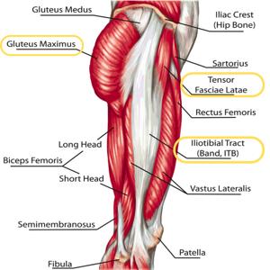
Iliotibial (IT) band syndrome: Irritation of the IT band, a thick tissue that runs from the hip to the knee, common in long-distance runners.
Symptoms of Knee Pain
Knee pain manifests in various ways, depending on the underlying cause. Recognizing these symptoms is crucial for proper diagnosis and treatment. Here are the most common signs to watch for:
Swelling and Stiffness
Inflammation around the knee joint can lead to noticeable swelling and stiffness. This can make it challenging to bend or straighten the knee fully. Swelling may be localized to a specific area or affect the entire knee joint.
Redness and Warmth
In cases of acute inflammation or infection, the knee may appear red and feel warm to the touch. This is often accompanied by increased pain and tenderness.
Instability or Weakness
A sensation of the knee “giving out” or feeling unstable is often associated with ligament injuries or advanced stages of arthritis. This instability can significantly impact your ability to walk, climb stairs, or engage in physical activities.
Pain Patterns
The nature and location of knee pain can provide valuable clues about its underlying cause:
Sharp, localized pain: Often indicative of a meniscus tear or ligament injury.
Dull, aching pain: Commonly associated with osteoarthritis or overuse injuries.
Pain behind the kneecap: Typically seen in cases of patellofemoral pain syndrome or chondromalacia patella.
Pain when climbing stairs: Often experienced by individuals with osteoarthritis or patellar tendonitis.
Show Image
Treatment Options for Knee Pain
The approach to treating knee pain varies depending on the severity and underlying cause of the condition. Here are some effective treatment strategies:
Conservative Treatments
Rest, Ice, Compression, and Elevation (RICE)
For mild injuries or cases of overuse, the RICE method can be highly effective in alleviating pain and reducing swelling:
Rest: Avoid activities that exacerbate the pain and give your knee time to heal.
Ice: Apply cold packs to the affected area for 15-20 minutes at a time, several times a day.
Compression: Use an elastic bandage to provide support and reduce swelling.
Elevation: Keep your leg elevated above heart level when resting to minimize fluid accumulation.
Physical Therapy
Physical therapy plays a crucial role in managing chronic knee pain and recovering from injuries. A tailored exercise program can:
Strengthen the muscles supporting the knee joint, particularly the quadriceps and hamstrings.
Improve flexibility and range of motion.
Enhance overall knee stability and function.
Reduce pressure on the joint through proper biomechanics.
Medications
Various medications can help manage knee pain and inflammation:
Over-the-counter pain relievers: Nonsteroidal anti-inflammatory drugs (NSAIDs) like ibuprofen or naproxen can reduce pain and inflammation.
Acetaminophen: Effective for pain relief without addressing inflammation.
Topical medications: Creams or gels containing NSAIDs or capsaicin can provide localized pain relief.
For more severe cases, prescription medications may be necessary:
Stronger NSAIDs
Corticosteroid injections for rapid inflammation reduction
Hyaluronic acid injections to improve joint lubrication in osteoarthritis cases
Surgical Interventions
When conservative treatments fail to provide adequate relief, surgical options may be considered:
Arthroscopy: A minimally invasive procedure to diagnose and treat various knee conditions.
Partial or total knee replacement: For severe cases of osteoarthritis or significant joint damage.
Ligament reconstruction: To repair or replace damaged ligaments, such as in ACL tears.
Show Image
Preventing Knee Pain
Prevention is key to maintaining healthy knees and avoiding future problems. Here are some essential tips:
Maintain a healthy weight: Excess weight puts additional stress on your knee joints, increasing the risk of osteoarthritis and other knee problems.
Strengthen your leg muscles: Focus on exercises that target your quadriceps, hamstrings, and calf muscles to provide better support for your knees.
Wear proper footwear: Choose shoes that provide adequate support and cushioning, especially during physical activities.
Warm up and cool down: Before and after exercise, perform gentle stretches to improve flexibility and reduce the risk of injury.
Use proper technique: When engaging in sports or physical activities, ensure you’re using correct form to minimize stress on your knees.
Cross-train: Vary your exercise routine to avoid overuse injuries and strengthen different muscle groups.
Listen to your body: If you experience persistent knee pain, don’t ignore it. Seek medical advice to prevent the condition from worsening.
Effectiveness of Treatment Options
To help you understand the potential benefits of various treatment approaches, we’ve compiled two tables comparing their effectiveness:
Knee pain doesn’t have to dictate your life. Whether you’re dealing with a minor sprain or chronic arthritis, there are solutions available to help you regain your mobility and enjoy a pain-free life. Remember, early intervention is key to preventing long-term complications and achieving the best possible outcomes.
By understanding the causes of knee pain, recognizing its symptoms, and exploring various treatment options, you’re taking the first step towards better knee health. Don’t let knee pain hold you back from the activities you love or impact your quality of life.
Ready to Say Goodbye to Knee Pain?
The journey to a pain-free life starts with a single step. Don’t wait any longer to reclaim your mobility and well-being. Whether your goal is to return to your favorite sport, enjoy leisurely walks, or simply move through your day without discomfort, now is the time to act.
Take charge of your knee health by scheduling a consultation with a healthcare provider or physical therapist. They can assess your specific condition, provide an accurate diagnosis, and develop a personalized treatment plan tailored to your needs and goals.
Remember, every journey begins with a single step. Make that step today towards a future free from knee pain. Your knees have supported you throughout your life – now it’s time to give them the care and attention they deserve.
Act now, and open the door to a world of improved mobility, reduced pain, and enhanced quality of life. Your knees – and your future self – will thank you!
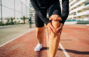
If you’re an athlete or someone who leads an active lifestyle, you’ve likely heard of various sports injuries that can sideline even the most dedicated fitness enthusiasts. One such condition that often flies under the radar until it becomes a significant problem is Iliotibial Band Syndrome (ITBS). This common overuse injury can cause considerable discomfort and hinder your performance, whether you’re a runner, cyclist, or hiker.
In this comprehensive guide, we’ll dive deep into the world of Iliotibial Band Syndrome, exploring its causes, symptoms, and most importantly, how to prevent it. By the end of this article, you’ll have a thorough understanding of ITBS and be equipped with the knowledge to keep your iliotibial band healthy and your activities pain-free.
I. What is Iliotibial Band Syndrome?
Iliotibial Band Syndrome is a common overuse injury that primarily affects the lateral (outer) part of the knee. To understand ITBS, we first need to familiarize ourselves with the iliotibial band itself.
The iliotibial band, often abbreviated as IT band, is a thick band of fibrous tissue that runs along the outside of the thigh, extending from the hip to the shin. This band plays a crucial role in stabilizing the knee during running and other activities that involve repetitive knee flexion and extension.
When the IT band becomes irritated or inflamed, typically due to repetitive friction, it can lead to ITBS. This condition is characterized by pain on the outer part of the knee, which can range from a dull ache to a sharp, burning sensation.
ITBS can significantly impact an individual’s ability to perform physical activities, especially those that involve repetitive knee movements. It’s not just a nuisance; left untreated, it can lead to chronic pain and long-term mobility issues.
II. Who is at Risk for ITBS?
While anyone can develop Iliotibial Band Syndrome, certain groups of people are at a higher risk due to the nature of their activities or physical characteristics. Understanding these risk factors can help you assess your own susceptibility to ITBS and take appropriate preventive measures.
High-Risk Activities
Runners: Distance runners are particularly prone to ITBS due to the repetitive nature of their sport. The constant knee flexion and extension during running can lead to increased friction between the IT band and the lateral femoral epicondyle (a bony prominence on the outer part of the knee).
Cyclists: Cycling, especially long-distance or intense cycling, can also put stress on the IT band. The repetitive pedaling motion and the slightly bent knee position maintained throughout the activity can contribute to the development of ITBS.
Hikers: Hikers, especially those tackling long trails or steep terrains, are at risk due to the combination of repetitive movement and uneven surfaces.
Weight Lifters: Certain weightlifting exercises, particularly those involving deep knee bends like squats, can stress the IT band and potentially lead to ITBS.
Other Risk Factors
Anatomical Issues: Some people may be more susceptible to ITBS due to their physical structure. For example, individuals with leg length discrepancies, excessive pronation (inward rolling of the foot), or bowed legs may have a higher risk.
Improper Training: Rapidly increasing mileage or intensity in running or cycling without proper conditioning can lead to ITBS.
Poor Biomechanics: Inefficient running or cycling form can put extra stress on the IT band.
Inadequate Warm-up: Failing to properly warm up before intense physical activity can increase the risk of developing ITBS.
Worn-out Footwear: Running or exercising in shoes that no longer provide adequate support can contribute to the development of ITBS.
Understanding these risk factors is the first step in preventing ITBS. By identifying which factors apply to you, you can take targeted steps to protect your IT band and maintain your active lifestyle.
III. What are the Symptoms of ITBS?
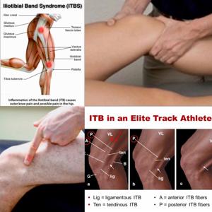
Recognizing the symptoms of Iliotibial Band Syndrome early can help you address the issue before it becomes a chronic problem. While the primary symptom is pain on the outer side of the knee, ITBS can manifest in various ways and may progress if left untreated.
Common Symptoms of ITBS
Lateral Knee Pain: The hallmark symptom of ITBS is pain on the outside of the knee. This pain typically:
Begins as a mild ache and can progress to a sharp, burning sensation
Often starts after a certain time or distance during activity (e.g., after running for 2 miles)
May be more noticeable when going downhill or downstairs
Swelling: Some individuals may experience swelling on the outer part of the knee.
Warmth: The affected area may feel warm to the touch due to inflammation.
Clicking or Popping Sensation: You might feel or hear a clicking or popping sensation when bending the knee, caused by the IT band moving over the lateral femoral epicondyle.
Pain that Worsens with Activity: The discomfort typically intensifies with continued activity and may subside with rest.
Tightness: You might feel tightness along the outer thigh, from the hip to the knee.
Referred Pain: In some cases, the pain may radiate up the thigh or down the calf.
Progression of Symptoms
ITBS symptoms often follow a predictable pattern:
Early Stage: Pain occurs toward the end of a run or workout.
Intermediate Stage: Pain starts earlier in the activity and persists longer after stopping.
Advanced Stage: Pain is present during daily activities and may interfere with sleep.
Show Image Image: Diagram showing the typical location of ITBS pain on the lateral knee
Differentiating ITBS from Other Knee Injuries
Here’s the comparison table for ITBS and other common knee injuries:
Symptom
ITBS
Patellofemoral Pain Syndrome
Meniscus Tear
Pain Location
Outer side of knee
Front of knee, around or behind kneecap
Inside or outside of knee joint
Pain Onset
Gradually during activity
After prolonged sitting or stair climbing
Sudden, often with a popping sensation
Swelling
Minimal to none
Minimal to none
Often present
Range of Motion
Usually not affected
Usually not affected
May be limited
Pain with Squatting
Sometimes
Often
Often
While this table can provide some guidance, it’s crucial to consult a healthcare professional for an accurate diagnosis. Self-diagnosis can lead to improper treatment and prolonged recovery times.
IV. What Causes ITBS?
Understanding the underlying causes of Iliotibial Band Syndrome is crucial for both prevention and treatment. ITBS is primarily an overuse injury, but several factors can contribute to its development.
Primary Causes of ITBS
Repetitive Friction: The most common cause of ITBS is repetitive friction between the iliotibial band and the lateral femoral epicondyle. This friction occurs when the knee flexes and extends, particularly during activities like running or cycling.
Biomechanical Issues:
Overpronation of the foot
Leg length discrepancies
Weak hip abductor muscles
Tight IT band or hip flexors
Training Errors:
Sudden increase in training intensity or duration
Inadequate rest between workouts
Overtraining
Environmental Factors:
Running on banked or uneven surfaces
Worn-out shoes that no longer provide proper support
Normal Function: The IT band helps stabilize the knee during running and walking. It moves back and forth over the lateral femoral epicondyle as the knee flexes and extends.
ITBS Development: When the IT band becomes tight or inflamed, this movement can cause friction and irritation. Over time, this leads to inflammation and pain.
Gait Cycle Impact: ITBS pain is often most noticeable when the knee is flexed at about 30 degrees. This typically occurs during the stance phase of running, particularly when the foot strikes the ground.
Factors Contributing to IT Band Tightness
Several factors can contribute to IT band tightness, increasing the risk of ITBS:
Muscle Imbalances: Weakness in hip abductors (particularly the gluteus medius) can lead to increased stress on the IT band.
Poor Flexibility: Tight muscles in the hips and legs can increase tension on the IT band.
Overtraining: Excessive exercise without proper recovery can lead to muscle fatigue and altered biomechanics.
Improper Form: Poor running or cycling technique can put unnecessary stress on the IT band.
Inadequate Warm-up: Failing to properly warm up before exercise can leave muscles tight and more prone to injury.
Understanding these causes and contributing factors is essential for developing an effective prevention and treatment strategy for ITBS.
V. How is ITBS Diagnosed?
Accurate diagnosis of Iliotibial Band Syndrome is crucial for proper treatment and recovery. While the symptoms of ITBS can be quite distinctive, a healthcare professional should always be consulted to rule out other potential knee injuries.
Diagnostic Process
Medical History: Your doctor will likely begin by asking about your symptoms, when they started, and what activities seem to aggravate them. They’ll also inquire about your exercise habits and any recent changes in your routine.
Physical Examination: This typically involves:
Palpation of the knee and IT band to check for tenderness
Assessment of knee range of motion
Strength testing of the hip and knee muscles
Observation of your gait and running form (if applicable)
Special Tests: Specific tests can help diagnose ITBS:
Noble Compression Test: Applying pressure to the lateral femoral epicondyle while flexing and extending the knee
Ober Test: Assessing IT band tightness
Imaging Tests: While not always necessary, imaging tests may be used to rule out other conditions:
X-rays: To check for bone abnormalities or arthritis
MRI: To visualize soft tissues and rule out other knee injuries
Differential Diagnosis
Condition
Key Symptoms
Diagnostic Tests
ITBS
Lateral knee pain, pain with repetitive knee flexion
Noble Compression Test, Ober Test
Lateral Meniscus Tear
Sharp pain, possible locking or catching sensation
McMurray Test, MRI
Patellofemoral Pain Syndrome
Pain around or behind kneecap, aggravated by stairs
Clarke’s Sign, Patellar Grind Test
Lateral Collateral Ligament Injury
Pain on outer knee, instability
Varus Stress Test
Importance of Professional Diagnosis
While it might be tempting to self-diagnose based on symptoms, seeking professional medical advice is crucial for several reasons:
Accurate Diagnosis: A healthcare professional can differentiate ITBS from other knee conditions that may present similar symptoms.
Underlying Causes: A thorough examination can reveal biomechanical issues or muscle imbalances contributing to your ITBS.
Personalized Treatment Plan: Based on the severity of your condition and your activity level, a professional can develop a tailored treatment plan.
Prevention of Chronic Issues: Early and accurate diagnosis can prevent ITBS from becoming a chronic problem.
Remember, the sooner you seek professional help, the quicker you can begin appropriate treatment and return to your activities.
VI. Treatment Options for ITBS
Once diagnosed with Iliotibial Band Syndrome, a variety of treatment options are available. The appropriate treatment plan will depend on the severity of your condition, your activity level, and your overall health. Most cases of ITBS can be successfully treated with conservative methods, but in rare cases, more aggressive interventions may be necessary.
Conservative Treatments
Rest, Ice, Compression, and Elevation (RICE):
Rest: Avoid activities that exacerbate symptoms
Ice: Apply ice to the affected area for 15-20 minutes every 2-3 hours
Compression: Use a compression bandage to reduce swelling
Elevation: Elevate the leg to minimize sw
Version 1 of 2
Physical Therapy: A physical therapist can design a personalized treatment plan that may include:
Stretching exercises for the IT band, hip flexors, and quadriceps
Strengthening exercises for hip abductors and core muscles
Manual therapy techniques to improve IT band mobility
Gait analysis and retraining to correct biomechanical issues
Foam Rolling: Self-myofascial release using a foam roller can help reduce tension in the IT band and surrounding muscles.
Show Image Image: Demonstration of proper foam rolling technique for the IT band
Medications: Over-the-counter non-steroidal anti-inflammatory drugs (NSAIDs) like ibuprofen can help manage pain and reduce inflammation.
Activity Modification: Temporarily switching to low-impact activities like swimming or cycling (if it doesn’t exacerbate symptoms) can maintain fitness while allowing the IT band to heal.
Orthotics: Custom or over-the-counter orthotics may be recommended to correct biomechanical issues contributing to ITBS.
Kinesiology Taping: Some individuals find relief with specialized taping techniques that support the IT band and knee.
Advanced Treatment Options
If conservative treatments don’t provide sufficient relief after several weeks to months, your healthcare provider might recommend more advanced options:
Corticosteroid Injections: In some cases, a corticosteroid injection into the area of inflammation can provide temporary relief and reduce inflammation.
Platelet-Rich Plasma (PRP) Therapy: This involves injecting a concentration of your own platelets to promote healing in the affected area.
Extracorporeal Shockwave Therapy (ESWT): This non-invasive treatment uses shock waves to stimulate healing in the affected tissues.
Surgical Intervention
Surgery for ITBS is rare and only considered when all conservative treatments have failed over an extended period (usually at least 6 months to a year). Surgical options may include:
IT Band Release: A procedure to release or lengthen the IT band to reduce tension.
Arthroscopic Debridement: Removal of inflamed tissue around the IT band.
It’s important to note that surgery is a last resort and carries its own risks. The success rate of surgery for ITBS varies, and recovery can be lengthy.
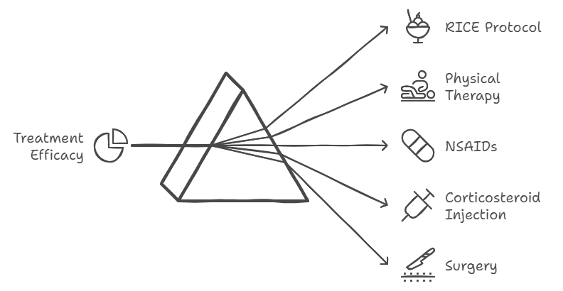
Treatment Efficacy
Remember, the key to successful treatment is early intervention and consistency in following your treatment plan.
VII. Prevention Tips for ITBS
Preventing Iliotibial Band Syndrome is often easier than treating it. By incorporating proper training techniques and maintaining good overall fitness, you can significantly reduce your risk of developing ITBS. Here are some essential prevention strategies:
1. Proper Warm-up and Cool-down Routines
Dynamic Warm-up: Before your workout, engage in dynamic stretching exercises that mimic the movements of your planned activity. This helps prepare your muscles and joints for the upcoming stress.
Static Stretching: After your workout, perform static stretches focusing on the IT band, hip flexors, quadriceps, and hamstrings. Hold each stretch for 30 seconds to a minute.
2. Strengthening Exercises
Focus on exercises that target the hip abductors, glutes, and core muscles. Strong supporting muscles can help maintain proper form during activities and reduce stress on the IT band.
Key exercises include:
Clamshells
Side-lying leg lifts
Glute bridges
Planks and side planks
3. Flexibility Exercises
Maintain flexibility in the IT band and surrounding muscles with regular stretching:
IT band stretches
Quad stretches
Hamstring stretches
Hip flexor stretches
4. Gradual Training Progression
Follow the 10% rule: don’t increase your training volume (mileage, duration, or intensity) by more than 10% per week. This allows your body to adapt to the increased stress gradually.
5. Cross-training
Incorporate low-impact activities into your routine to reduce repetitive stress on the IT band:
Swimming
Cycling (if it doesn’t cause discomfort)
Elliptical machine workouts
Strength training
6. Proper Footwear and Equipment
Replace running shoes every 300-500 miles or when they show signs of wear.
Use shoes appropriate for your foot type and running style.
Consider custom orthotics if you have biomechanical issues.
7. Running Surface and Terrain
Avoid running on banked surfaces for extended periods.
Mix up your running routes to vary the stress on your legs.
Gradually introduce hill training and downhill running.
8. Proper Form and Technique
Maintain good posture during running and cycling.
Avoid overstriding while running.
Keep your knees aligned with your feet during squats and lunges.
9. Listen to Your Body
Pay attention to early warning signs of discomfort.
Don’t push through pain – take rest days when needed.
Gradually return to activity after any injury or extended break.
10. Regular Maintenance
Use a foam roller regularly to maintain IT band flexibility.
Consider regular massage therapy to address muscle tightness.
Incorporate yoga or Pilates for overall flexibility and core strength.
Prevention Checklist
Prevention Strategy
Frequency
Notes
Dynamic Warm-up
Before every workout
5-10 minutes of activity-specific movements
Static Stretching
After every workout
Hold each stretch for 30 seconds to 1 minute
Strength Training
2-3 times per week
Focus on hip abductors, glutes, and core
Foam Rolling
3-4 times per week
Roll each area for 1-2 minutes
Cross-training
1-2 times per week
Incorporate low-impact activities
Footwear Check
Every 300-500 miles
Replace shoes when showing signs of wear
By incorporating these prevention strategies into your routine, you can significantly reduce your risk of developing ITBS and maintain an active, pain-free lifestyle.
VIII. Living with ITBS
While prevention is ideal, if you do develop Iliotibial Band Syndrome, it’s important to know how to manage the condition and maintain your quality of life. Living with ITBS doesn’t mean you have to give up your active lifestyle completely, but it does require some adjustments and ongoing care.
Managing Symptoms and Preventing Flare-ups
Recognize Early Warning Signs: Learn to identify the early symptoms of an ITBS flare-up, such as mild discomfort on the outside of your knee during or after activity.
Implement the RICE Protocol: At the first sign of discomfort, use the Rest, Ice, Compression, and Elevation (RICE) protocol to manage symptoms.
Modify Activities: During a flare-up, switch to low-impact activities that don’t exacerbate your symptoms. This might include swimming, using an elliptical machine, or upper body strength training.
Maintain Flexibility: Continue with your stretching routine, focusing on the IT band, hip flexors, and surrounding muscles.
Use Self-Massage Techniques: Regular use of a foam roller or massage stick can help maintain IT band flexibility and prevent tightness.
Wear Supportive Gear: Consider using a knee support or IT band strap during activities to provide additional support.
Gradual Return to Activity
After an ITBS flare-up, it’s crucial to return to your regular activities gradually:
Start Slow: Begin with low-intensity, short-duration activities and gradually increase as tolerated.
Follow the 10% Rule: Increase your activity level by no more than 10% per week.
Monitor Pain Levels: Use a pain scale of 0-10. If pain exceeds 3/10 during activity, stop and rest.
Incorporate Cross-training: Mix in low-impact activities to maintain fitness while reducing stress on the IT band.
Continue Strengthening Exercises: Maintain your strength training routine, focusing on hip abductors and core muscles.
Long-term Management Strategies
Regular Check-ins: Schedule periodic check-ins with your healthcare provider or physical therapist to assess your progress and adjust your management plan as needed.
Maintain Good Biomechanics: Regularly assess and correct your form during activities. Consider periodic gait analysis if you’re a runner.
Stress Management: Chronic stress can contribute to muscle tension. Incorporate stress-reduction techniques like meditation or yoga into your routine.
Nutrition: Maintain a balanced diet rich in anti-inflammatory foods to support overall joint health.
Sleep: Ensure you’re getting adequate sleep to support recovery and overall health.
Support Resources and Communities
Living with ITBS can be challenging, but you’re not alone. Consider connecting with support resources and communities:
Online Forums: Websites like Reddit’s r/running or r/fitness have communities where you can share experiences and get advice.
Local Running or Cycling Clubs: Many clubs have members who have dealt with ITBS and can offer support and advice.
Physical Therapy Support Groups: Some physical therapy clinics offer support groups for individuals dealing with common sports injuries.
Sports Medicine Clinics: These specialized clinics often offer resources and educational materials for managing conditions like ITBS.
Remember, everyone’s experience with ITBS is unique. What works for one person may not work for another, so it’s important to work closely with your healthcare provider to develop a management plan that’s tailored to your specific needs and goals.
Conclusion
Iliotibial Band Syndrome can be a frustrating condition for active individuals, but with proper understanding, prevention, and management, it doesn’t have to sideline you permanently. Let’s recap the key points we’ve covered:
ITBS is an overuse injury affecting the thick band of tissue that runs from the hip to the knee on the outside of the thigh.
Common symptoms include pain on the outside of the knee, which typically worsens with repetitive activities like running or cycling.
Risk factors include biomechanical issues, training errors, and certain anatomical predispositions.
Diagnosis typically involves a physical examination and may include imaging tests to rule out other conditions.
Treatment options range from conservative measures like rest and physical therapy to more advanced interventions in severe cases.
Prevention strategies include proper warm-up and cool-down routines, strengthening exercises, and gradual training progression.
Living with ITBS involves ongoing management, including recognizing early warning signs and implementing long-term strategies to prevent flare-ups.
Remember, the key to managing ITBS is being proactive. Don’t ignore early signs of discomfort, and don’t be afraid to seek professional help. With patience, consistency, and the right approach, you can overcome ITBS and return to the activities you love.
We encourage you to share your own experiences with ITBS in the comments below. What strategies have worked for you? What challenges have you faced? Your insights could be invaluable to others dealing with this condition.
Stay active, stay healthy, and listen to your body. Here’s to pain-free movement and achieving your fitness goals!
If you’re reading this, chances are you or someone you love has dealt with the frustrating twinge of knee pain. You’re not alone. This common complaint affects people of all ages and walks of life. It’s a topic that doctors and researchers at renowned institutions like the Mayo Clinic study in-depth.
Knee pain can be particularly frustrating because it’s hard to ignore. Every step, every movement can serve as a reminder of your discomfort. This comprehensive guide is all about understanding why your knee might be acting up and what you can do about it. We’ll explore the causes, dive into the types of pain you might experience, and offer practical solutions to help you find relief.
Table of Contents:
Decoding Your Knee Pain: Common Culprits
The Usual Suspects: Injuries and Overuse
Arthritis: When Wear and Tear Takes Its Toll
Beyond the Obvious: Other Potential Causes of Knee Pain
Say What? Types of Knee Pain
When It’s Time to Call the Doc
Treatment Options: From Home Remedies to Medical Interventions
Prevention: Keeping Your Knees Healthy
FAQs About Knee Pain
Conclusion
1. Decoding Your Knee Pain: Common Culprits
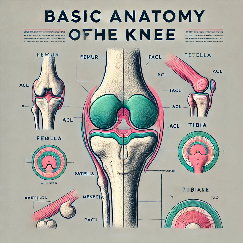
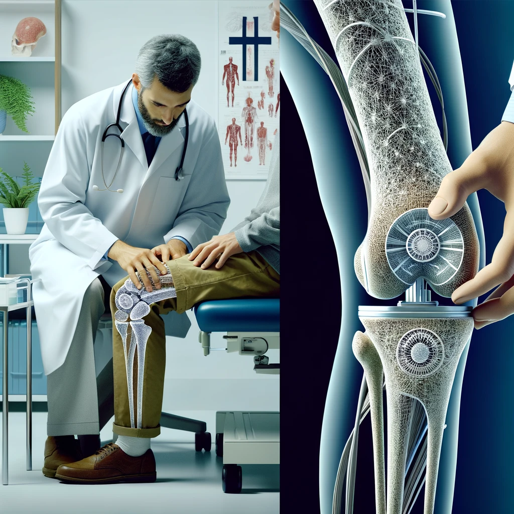
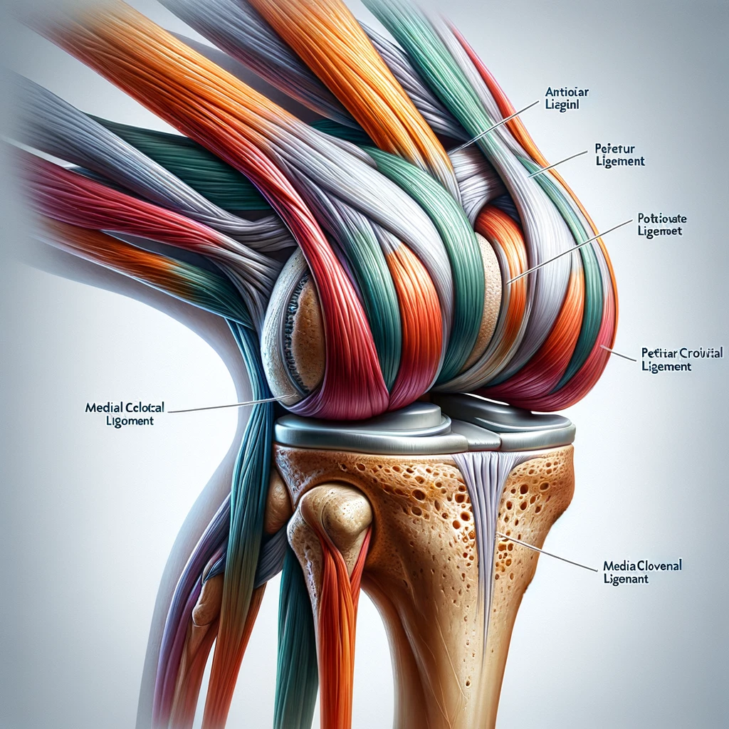
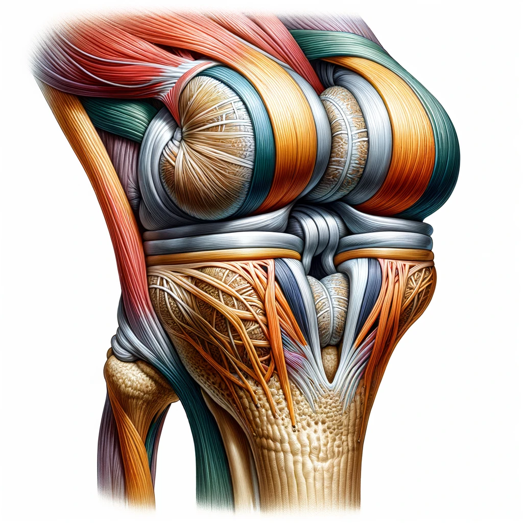
Let’s face it: knee pain is rarely ever welcome. Sometimes, it feels like it appears out of nowhere. Other times, it builds gradually. But why does it happen? Understanding the root cause of your knee pain is the first step towards finding effective relief.
The knee is a complex joint, and pain can originate from various structures within it. The image above illustrates the basic anatomy of the knee, highlighting key components that are often involved in knee pain.
2. The Usual Suspects: Injuries and Overuse
Our knees go through a lot. They bear our weight, help us move, and sometimes, we ask a little too much of them. Think about the impact when you run, jump, or even just walk for extended periods. Over time, this can lead to wear and tear or sudden injuries.
Ligament Troubles
You’ve got these strong bands of tissue, like your anterior cruciate ligament (ACL), that hold your knee joint together. Sudden twists or impacts (common in sports) can lead to painful sprains or tears.
ACL Injuries: These often occur during sports that involve sudden stops or changes in direction, like basketball or soccer.
MCL Injuries: The medial collateral ligament can be damaged by a blow to the outer part of the knee.
PCL Injuries: While less common, posterior cruciate ligament injuries can occur from a direct blow to the front of the knee.
Recovery time for ligament injuries can vary widely. For instance, a minor sprain might heal in a few weeks with proper care, while a severe tear might require surgery and months of rehabilitation.
Meniscus Mishaps
That rubbery cushion in your knee, the meniscus, can get torn. Twisting movements, especially when your knee is under pressure, are often to blame. Meniscus tears are common in sports but can also occur during everyday activities, especially as we age and the meniscus becomes more prone to wear and tear.
Symptoms of a meniscus tear include:
A popping sensation
Swelling
Stiffness
Difficulty fully straightening the knee
Tendon Tantrums
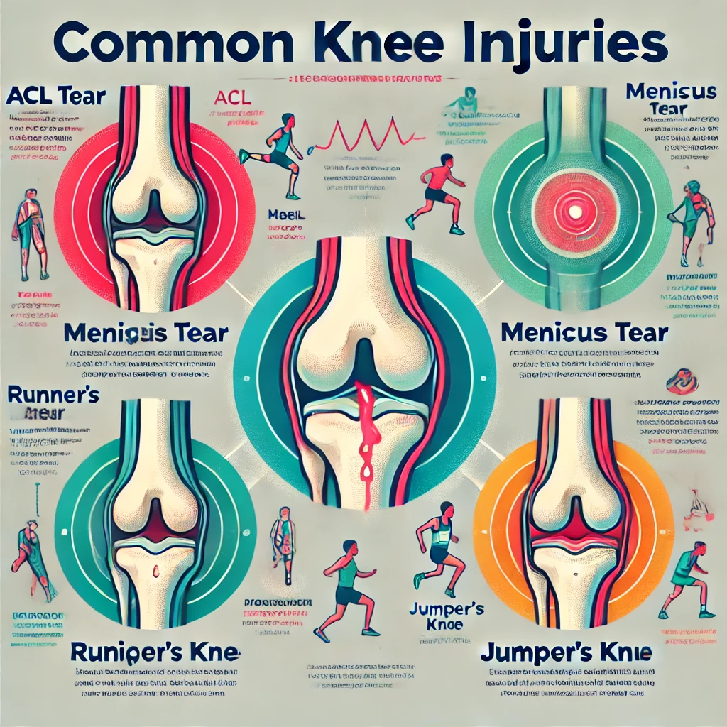
Tendons, which attach muscle to bone, can become inflamed with repetitive activities. This is where conditions like runner’s knee (patellofemoral pain syndrome) and jumper’s knee (patellar tendinitis) come into play.
Runner’s Knee: This condition causes pain around the kneecap, especially when climbing stairs, kneeling, or sitting with bent knees for long periods.
Jumper’s Knee: Common in athletes who do a lot of jumping, this condition causes pain below the kneecap.
These conditions often develop over time due to overuse or improper form during activities. They can be particularly frustrating for athletes or active individuals, as they may require a period of rest and rehabilitation to resolve.
This common knee injuries we’ve discussed. Understanding these injuries can help you identify potential causes of your knee pain and seek appropriate treatment.
3. Arthritis: When Wear and Tear Takes Its Toll
Arthritis is a common cause of knee pain, especially as we age. It occurs when the joint becomes inflamed, leading to pain, stiffness, and sometimes swelling. There are several types of arthritis that can affect the knee:
Osteoarthritis
This is the most common form of arthritis affecting the knee. Think of it like this: remember those car commercials showing shock absorbers wearing down over time? Over the years, the cartilage that cushions our bones can wear away, making movement painful.
In the United States alone, knee osteoarthritis affects around 10 percent of men and 13 percent of women over the age of 60. That’s a significant portion of the population dealing with this type of knee pain.
Symptoms of knee osteoarthritis include:
Pain that increases with activity
Stiffness, especially in the morning or after sitting for long periods
Swelling
Decreased range of motion
A grinding sensation when moving the knee
Rheumatoid Arthritis
Unlike osteoarthritis, rheumatoid arthritis is an autoimmune condition where the body’s immune system attacks the joints. This can lead to inflammation, pain, and eventual joint damage if left untreated.
Rheumatoid arthritis often affects both knees simultaneously and may be accompanied by fatigue, fever, and weight loss.
Post-Traumatic Arthritis
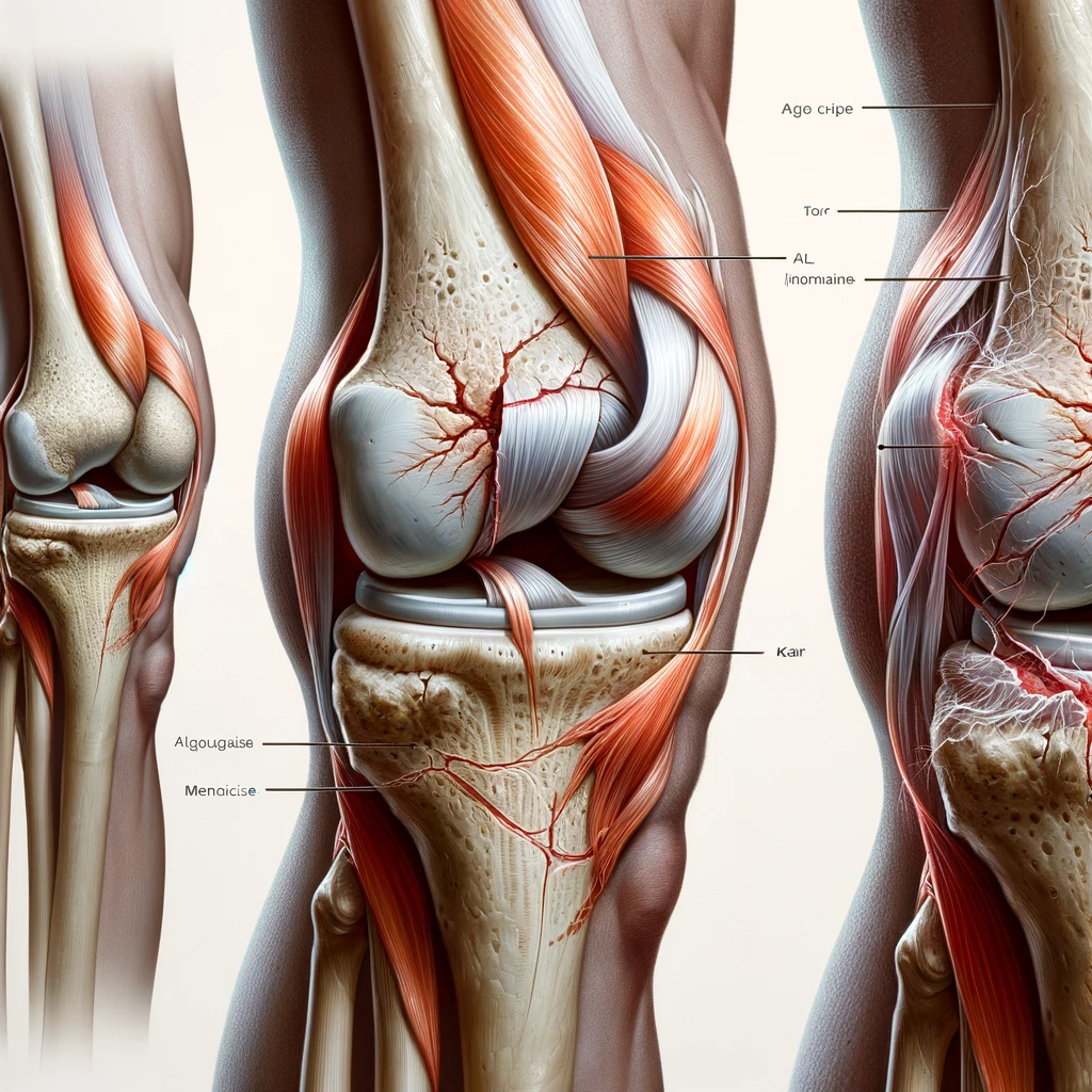
This type of arthritis can develop years after a knee injury, such as a fracture or ligament tear. The initial injury can lead to changes in the knee joint that accelerate the breakdown of cartilage over time.
This image illustrates the difference between a healthy knee joint with smooth cartilage and an arthritic knee with worn, damaged cartilage. In the arthritic knee, you can see how the protective cartilage has deteriorated, potentially leading to bone-on-bone contact and pain.
4. Beyond the Obvious: Other Potential Causes of Knee Pain
While injuries and arthritis are common culprits, sometimes knee pain can be sneaky. It might be a clue that something else is going on. Think about your overall health and any other aches or pains you’ve noticed.
Referred Pain
You might actually have a hip problem or foot issue that’s changing the way you walk, putting extra stress on your knee. It’s like when your car’s tires are out of alignment; the wear and tear show up elsewhere.
For example, flat feet or high arches can alter your gait, potentially leading to knee pain. Similarly, tight hip flexors or weak gluteal muscles can change the mechanics of how you move, putting additional stress on your knees.
Infections
While less common, infections in the knee joint (like septic arthritis) can cause intense pain. This one needs prompt medical attention. Think of it as your body’s way of sounding the alarm.
Symptoms of a knee infection may include:
Severe pain
Swelling
Redness and warmth around the joint
Fever
Fatigue
If you suspect a knee infection, it’s crucial to seek medical attention immediately, as untreated joint infections can lead to permanent damage.
Underlying Conditions
Certain medical conditions can cause joint pain, including knee pain. Some of these include:
Gout: A type of arthritis caused by a buildup of uric acid crystals in the joint.
Lupus: An autoimmune disease that can cause inflammation in various parts of the body, including joints.
Lyme Disease: An infection caused by tick bites that can lead to joint pain and swelling.
Psoriatic Arthritis: A type of inflammatory arthritis that can occur in people with psoriasis.
These conditions often require comprehensive medical management beyond just treating the knee pain.
5. Say What? Types of Knee Pain
Pain relief is possible, but first, we need to determine what type of pain you are experiencing. The type of knee pain can give clues about what’s causing it.
Injury Type
Cause
Description
Pain Type
Example Scenario
Fracture
Direct impact or trauma
Break in one of the knee bones, usually the patella (kneecap)
Sharp, Intense Pain
Falling on a hard surface
Sprain (Entorse)
Sudden twist or wrenching movement
Stretching or tearing of ligaments in the knee
Sharp, Shooting Pain
Twisting the knee while pivoting
Rheumatism
Autoimmune or inflammatory response
Chronic inflammation of the joints, often affecting the knees
Dull, Aching Pain
Persistent knee pain with stiffness
Fall Down Injury
Falling or tripping
Impact injury leading to bruising or damage to knee structures
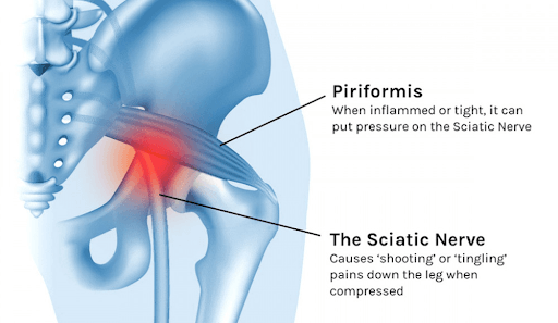
Piriformis syndrome is a common but often misunderstood condition that causes pain, numbness, and tingling in the buttocks and legs. This syndrome occurs when the piriformis muscle, located deep in the buttocks, compresses or irritates the sciatic nerve. Understanding the anatomy, causes, aggravating activities, and effective stretches is crucial to effectively treating this condition.
Anatomy:
The piriformis muscle is a small, pear-shaped muscle located in the buttocks, beneath the gluteal muscles. Its primary function is to assist in the rotation of the hip joint. The sciatic nerve, the largest nerve in the body, runs directly under or sometimes through the piriformis muscle. When the piriformis muscle spasms or becomes tight, it can compress the sciatic nerve, causing pain and discomfort.
Causes:
There are several factors that can contribute to the development of Piriformis syndrome:
1. Muscle imbalance: Weakness or stiffness in the surrounding muscles can lead to compensatory changes in the piriformis muscle.
2. Excessive use or repetitive activities: Activities that involve repetitive motions or prolonged sitting can strain the piriformis muscle.
3. Trauma: Damage to the piriformis muscle or surrounding structures can lead to inflammation and compression of the sciatic nerve.
4. Anatomical variationsSome people may have abnormalities in the anatomy of the piriformis muscle or the sciatic nerve that make them more susceptible to this condition.
Aggravating activities:
Certain activities can aggravate the symptoms of piriformis syndrome:
1. Sitting for long periods of time: Sitting for long periods of time, especially on hard surfaces, can worsen symptoms by putting pressure on the piriformis muscle and sciatic nerve.
2. Running or climbing stairs: Activities that involve repetitive hip movements can strain the piriformis muscle.
3. Lifting heavy objects: Improper lifting techniques or lifting heavy objects can strain the muscles in the lower back and buttocks, including the piriformis muscle.
In order for the inflammation to subside and healing to occur, it is important to stop aggravating the area. Once you have determined what activity is contributing to your symptoms, be cautious about that activity until your body can begin to heal.
Relief:
Performing a specific stretching technique can help relieve piriformis syndrome symptoms by releasing tension in the piriformis muscle and reducing compression on the sciatic nerve. One of the most effective stretches is the contract-relax stretch.
The Best Stretch for Piriformis Syndrome: The “Contract-Relax Stretch”
Starting position: Lie on your back with both knees bent and your feet flat on the floor.
Action: Cross the affected leg over the opposite knee, creating a figure four. Then, gently push the knee of the affected leg away from your body using your hand to resist your leg for 5-10 seconds. Release the contraction and gently pull the knee toward your chest, feeling a gentle stretch in the buttock and outer hip. Hold the stretch for 15-30 seconds, breathing deeply and focusing on relaxing the muscles.
Representatives: Perform 2-3 sets of the stretch, gradually increasing the intensity depending on what you can tolerate.
Please note that there should be NO increase in symptoms after the stretch. If symptoms increase, decrease the intensity of the stretch.
The piriformis stretch is so important that we include it as a foundational stretch in the ACL Strong program. While this stretch can put you on the right path to pain relief, it’s also essential for restoring strength and muscle balance so that pain doesn’t return. ACL Strong can guide you through the exact exercises to improve flexibility, strength, and balance for long-term success. Become a member by taking one of our classes to learn more foundational stretches and strengthening exercises. You can also start with our FREE webinar and get 7 actionable tips right away.
In conclusion, piriformis syndrome can cause significant discomfort and impact daily activities, but with proper understanding and management, symptoms can be effectively relieved. By addressing muscle imbalances, avoiding aggravating activities, and incorporating stretching exercises such as the contract-relax stretch into your routine, you can reduce pain and improve your mobility. If symptoms persist or worsen, consult a healthcare provider for further evaluation and treatment options.
Experiencing knee pain? Don’t struggle in silence! In this comprehensive guide, we’ll decode your symptoms, understand diagnosis, and explore treatment options for a pain-free journey.
Introduction
Knee pain is an exceedingly common complaint, affecting nearly 1 in 4 adults in America every year. Its prevalence spans all ages – from young athletes to the elderly. Persistent knee discomfort can severely impact our mobility and quality of life.
Understanding the source and nature of knee pain provides a pathway to effective diagnosis and treatment. This guide offers clarity by unraveling the location, sensation, severity and associated symptoms that characterize knee pain. We demystify complex diagnostic techniques, illuminate common and uncommon culprits, and chart a course towards evidence-based treatment options for lasting relief.
Bolstered with tips for daily management and resources to empower your health journey, we hope to bring you one step closer to conquering knee pain. You don’t have to struggle alone – knowledge is power, and relief is possible.
1. Demystifying Knee Pain: A Symphony of Symptoms
Knee pain manifests as an intricate web of symptoms, with helpful clues embedded within its location, duration, intensity, triggers and associated manifestations. Let’s decode what your knee is trying to tell you.
a. Recognizing Acute vs Chronic Knee Pain
Acute knee pain is sudden in onset, often from injury or overuse. It usually subsides with rest and basic treatment within days or weeks. Chronic knee discomfort persists for over 3-6 months despite treatment. Understanding duration and precipitating events provides insight.
For example, acute pain after hyper-extending the knee likely indicates a ligament sprain. Conversely, chronic morning knee stiffness hints at inflammatory arthritis. Recognizing acute versus chronic is key for diagnosis.
b. Location Matters: Pinpointing Pain Regions
Knee anatomy is complex – several structures like cartilage, ligaments, tendons and bones comprise the joint. Pinpointing precise regions of knee pain helps diagnose affected structures.
Front of knee pain often indicates patellofemoral syndrome (kneecap issues) or patellar tendonitis (inflammation of the tendon connecting kneecap to shinbone).
Back of knee discomfort could signal injuries in ligaments, cartilage or the popliteal tendon. Cysts or tissue masses can also irritate this region.
Experiencing generalized sides or center knee pain? Consider joint diseases like arthritis or bursitis. Identifying regional pain is Step 1 for your doctor.
c. Intensity & Frequency: A Tale of Throbbing, Aching & Discomfort
Characterizing knee pain sensation and frequency provides more insights. Here are common descriptions:
Throbbing pain often results from inflammation or injury.
A dull ache may indicate arthritis or chronic bursitis.
Sharp, stabbing pain could signal tears, cysts or loose bodies within joint.
Occasional or frequent pain? Note pain-inducing positions or activities too.
Does pain wake you from sleep? Weightbearing activities like walking or climbing stairs can also aggravate certain knee conditions. Clearly communicating pain quality, severity and persistence guides accurate diagnoses.
d. Associated Symptoms: Swelling, Stiffness, Instability & Beyond
Knee pain seldom occurs in isolation – watch for associated red flags:
Joint swelling and warmth indicates inflammation or fluid buildup.
Stiffness upon waking up or sitting hints at arthritis.
Weakness or instability may accompany ligament tears or cartilage loss.
Limping, muscle atrophy and leg numbness warrants evaluation too.
Additionally, pay attention to fevers or unintentional weight loss alongside knee pain – this may indicate infections, autoimmune disease or rarely, bone tumors.
Lateral collateral ligament (LCL) sprain, iliotibial band syndrome
Intensity and Frequency:
Sharp, sudden pain
Injury, ligament tear, fracture
Dull, aching pain
Arthritis, overuse, tendonitis
Pain worsens with activity
Arthritis, tendinitis, bursitis
Night pain
Osteoarthritis, inflammatory arthritis
Associated Symptoms:
Swelling
Inflammation, fluid buildup, injury
Stiffness
Difficulty bending or straightening the knee
Weakness
Feeling unstable or giving way
Clicking or popping sounds
Meniscus tear, loose cartilage
Redness and warmth
Infection, gout
e. When to Seek Immediate Medical Care
While most knee pain responds well to conservative care, prompt medical assessment is key for:
Sudden, severe knee injuries causing immobility
Signs of infection like fever with joint swelling/redness
Unexplained knee pain with trauma indicators like falls
Difficulty bearing weight on leg or knee buckling
Numbness/weakness in leg
Pain unresponsive to home treatment beyond 48-72 hours
Seeking timely care prevents complications like permanent joint damage requiring extensive repair later. Don’t delay if red flags arise!
2. Unmasking the Culprit: Diagnosing the Root Cause
With a myriad potential causes for knee pain, an accurate diagnosis is crucial for effective treatment. A physician deploys an array of tools to unravel root causes, from medical history to advanced imaging. Let’s demystify this complex process.
Knee Pain Diagnoses
Diagnosis
Description
Symptoms
Treatment
Osteoarthritis: Most common type of arthritis, wearing away of cartilage
Dull aching pain, stiffness, worse with activity
Nonsteroidal anti-inflammatory drugs (NSAIDs), physical therapy, weight management, surgery
Meniscus tear: Tear in the cartilage pads
Sharp pain, swelling, locking or catching of the knee
RICE (Rest, Ice, Compression, Elevation), physical therapy, surgery
Ligament sprain: Stretching or tearing of ligaments
a. Medical History & Physical Examination: Uncovering Clues
A detailed medical history unravels duration, location and nature of knee symptoms alongside health conditions, injuries, lifestyle factors and occupational hazards.
For example, an elderly patient with chronic, progressive knee pain that worsens on activity likely has degenerative arthritis. Conversely, acute knee swelling after sports hints at traumatic injuries.
The physical exam assesses injury through range of motion tests, palpation for joint line tenderness, swelling and instability. Gait evaluation and alignment studies are conducted. Preliminary diagnoses take shape through pattern recognition.
Treatment Options for Knee Pain
Treatment
Description
Suitable for
Non-surgical:
RICE: Rest, Ice, Compression, Elevation
Reduces inflammation, swelling, and pain
Most types of knee pain
Pain medication: NSAIDs, acetaminophen
Relieves pain and inflammation
Mild to moderate pain
Physical therapy: Stretches, strengthening exercises, gait training
Improves flexibility, strength, and range of motion
Most types of knee pain
Bracing: Provides support and stability
Ligament sprains, patellofemoral pain syndrome
Injections: Corticosteroids, hyaluronic acid
Reduce inflammation and pain
Tendinitis, bursitis, osteoarthritis
Weight management: Reduces pressure on the joint
Overweight or obese individuals with knee pain
Alternative therapies: Acupuncture, massage, yoga
May provide pain relief and improve mobility
Some types of knee pain
Surgical:
Arthroscopy: Minimally invasive surgery for repairing meniscus tears, cartilage damage, and ligament tears
Meniscus tears, cartilage damage, ligament tears
Joint replacement: Replacing a damaged joint with a prosthetic one
Severe osteoarthritis, severe injuries
b. Imaging Techniques: X-Rays, MRIs & Beyond
Imaging allows visual confirmation of potential problems spotted on exam.
X-rays: Bones and joints. Can detect fractures, arthritis, tumors.
MRI scans: Complex structures like ligaments, cartilage, tendons. Ideal for injury without bone involvement.
CT scans: Detailed bone imaging for injuries, lesions and arthritis.
Tendinitis: Swelling in tendons like patellar/quadriceps due to overuse. Local pain on movement.
Bursitis: Inflamed fluid sacs (bursae) around kneecap/joints from injury/overuse. Restricted mobility.
Meniscal tears: Shock-absorbing cartilage tears causing catching/locking. Sudden onset, often with pivoting motions.
Sports injuries like anterior cruciate ligament (ACL) sprains also manifest with acute trauma. Understanding common knee troubles shapes further evaluation.
e. Less Common Causes: Cysts, Loose Bodies & Tumors
While most causes are chronic wear-and-tear or trauma, we occasionally encounter obscure diagnoses:
Plica syndrome: Irritated synovial folds in the knee joint, eliciting pain.
Loose bodies: Bone or cartilage fragments floating within joint space.
Bone infarcts: Tiny stress fractures with insidious knee discomfort, often in over 50s.
Rarely, infections, jointcrystal deposits or tumors manifest knee troubles without classical triggers. Maintaining an open yet focused diagnostic lens is key.
3. Charting the Path to Recovery: Treatment Options
Once diagnosis crystalizes treatment, we progress to evidence-based modalities spanning rest, physical therapy, injections, medications through surgery when necessary – with prevention always in focus. Let’s explore options.
a. Rest, Ice, Compression & Elevation (RICE)
The venerable RICE protocol alleviates acute sports injuries, sprains or activity-related pain. It halts further damage in the initial inflammatory phase.
Rest: Avoid reinjury from sports, pivoting or straining motions. Use crutches if critical.
Ice: Applying ice packs arrests swelling and relieves throbbing pain via vasoconstriction.
Compression: Wraps, braces or bandages compress the knee, reducing inflammation.
Elevation: Maintains blood flow away from the knee to ease swelling and discomfort.
RICE sets the stage for natural healing while allowing ramp up of mobility via therapy. Potent medicine without pills!
b. Medication Management: Over-the-Counter & Prescription Options
Oral medication in tandem with RICE relieves knee troubles. Analgesics like acetaminophen or NSAIDs (ibuprofen, naproxen) alleviate acute pain, swelling and stiffness.
Seeking stronger options for chronic inflammatory arthritis? Disease-modifying antirheumatic drugs (DMARDs) like methotrexate help minimize joint destruction over time.
For osteoarthritis, supplements like glucosamine/chondroitin provide building blocks to shore up thinning cartilage and lubricate stiff knees.
Topical anti-inflammatories, capsaicin creams or lidocaine patches offer localized relief too. Judicious medication bridges the gap during early rehab.
c. Physical Therapy: Strengthening, Stretching & Regaining Mobility
Physical therapy empowers sustainable healing via targeted exercises. It aims to:
Improve flexibility and range of motion.
Build strength and stability around joints.
Retrain proper gait and movement patterns.
Progress endurance for activities and sports.
Physiotherapy alleviates many knee troubles like osteoarthritis, injuries after ligament tears or surgery and tendinitis. It also prevents future damage through conditioning. Customized exercise is potent medicine!
d. Braces, Tapes and Assistive Devices: Focused Support
Specialized knee braces and supports aid recovery by:
Compressing and stabilizing injury sites.
Offloading pressure on damaged surfaces.
Improving joint alignment and mechanics.
Enhancing proprioception (body awareness).
Medial/lateral supports for arthritis and ACL/PCL braces for ligament tears offer structural assistance. Crutches and canes also temporarily redistribute weight from the knee. Targeted tools supplement therapy.
e. When Conservative Treatment Fails: Surgical Options
Most early knee troubles respond to conservative measures involving RICE, medications, injections or physiotherapy. But when relentless pain or loss of function persists, surgery opens doors to definitive repair.
Key indications for surgery:
Persistent instability from ligament/meniscal tears
Locking/catching with cartilage flaps, loose bodies
Significantly reduced mobility from arthritis or trauma
Failure to improve knee function through nonsurgical options
Advances in arthroscopy and fast-track surgical protocols allow low risk and minimally invasive procedures for commonly torn ACLs or meniscus injuries with faster recovery. Knee replacement surgeries reliably relieve arthritis failing other modalities too. Going under the knife is no longer the end of the road.
4. Living Well with Knee Pain: Practical Tips & Life Hacks
Alongside clinical management, self-care fosters living better with chronic knee troubles through activity modifications, strength and weight optimization. Let’s explore practical coping strategies.
a. Modifying Activity: Finding New Joy In Movement
For knees plagued by osteoarthritis, autoimmune disease or old injuries, some motions spell disaster while others offer comfort. Identifying and avoiding problematic activities allows better daily function.
Pivoting, high impact motions and excessive stair use are notorious knee offenders. Find substitutes – trade basketball for swimming or cycling, or replace stairs with ramps and grab bars through deliberate home adaptations. It’s about rediscovering movement avenues.
b. Losing Weight: Reducing Joint Load
The knee is subjected to almost 3-6 times our body weight with daily activities! Losing even 10-15 pounds makes a tangible difference through reducing force on the knees – easing the burden on joints.
Aim for gradual weight loss through calorie reduction and gentle cardio instead of crash diets and high impact exercising. Aquatic workouts and cycling are gentler complements to walking. Good nutrition and rest keeps energy levels up despite limitations!
c. Muscle Strengthening & Stretching: Building Surrounding Support
Targeted strengthening fosters stability and optimal joint mechanics. Pilates, resistance band workouts and yoga builds hip, core and quadricep control to support knees.
Daily stretching maintains flexibility and limbers up stiff, creaky joints. Gentle warm ups prevent overexertion. When pain flares, ice, rest and over-the-counter anti-inflammatories help recoup. Guided routines prevent future wear and tear.
d. Footwear & Assistive Devices: Offloading Pressure
Cushioned walking shoes with good arch support and shock absorbing soles take literal pressure off knees. Lateral wedging in shoes can stabilize varus arthritic knees.
For severe osteoarthritis, offloader knee braces redistribute up to 30% pressure from diseased cartilage preventing rapid breakdown. Canes and walkers also share the load, assisting stability. Targeted tools offer cheap mobility solutions!
e. Alternative Therapies: Finding Your Comfort Strategy
When all else fails, outside-the-box modalities sometimes deliver – scientifically or via placebo. Many patients benefit from acupuncture, massage, cryotherapy, yoga or tai chi in relieving persistent muscle tightness and knee troubles.
Topical capsaicin creams, menthol gels or CBD balms provide temporary analgesia for some as well. What works for another may fail you – but remain open to potential relief avenues before considering invasive options.
5. Empowering Your Recovery: Resources and Community Support
Despite exhaustive treatment options, living with stubborn knee troubles can feel lonely and frustrating. Connecting with others also seeking diagnosis or struggling with relentless pain provides validation, ideas and hope. Let people give you a leg up!.
a. Patient Advocacy Groups and Online Support Communities
Arthritis support groups and sports injury forums allow sharing challenges and solutions among empathetic ears. Facebook groups like “Knee Pain” or “Arthritis Warriors” connects over 15,000 members in communal griping and laughter – the sense of camaraderie uplifts many.
b. Educational Tools and Medical Apps
Reputable online resources like Arthritis Foundation, AAOS and medical center blogs provide easily digestible education on disease basics, self-care and latest treatment insights without overwhelming medical jargon.
Medical apps like KneeCare facilitate activity logging and appointments alongside providing exercise pointers – portable, searchable knowledge with personalization!
c. Finding the Right Healthcare Provider
Having a caring medical professional in your corner through diagnosis and treatment pitfalls works wonders for motivation and hope.
Seeking a “good match” patient-doctor relationship via referrals and consultations is worth the effort. Once secured, share records freely between interdisciplinary teams – from physiotherapists to rheumatologists and surgeons – to base decisions on collective wisdom.
d. Maintaining Hope and Setting Achievable Goals
Cultivating optimism is crucial when dealing with chronic pain. Set small milestones like swimming five laps or walking for five minutes daily instead of big feats that induce disappointment and despair when missed.
Focusing on things within your control prevents spiraling frustration. Pat yourself on the back for small acts of self care consistency instead of cursing worsening diagnosis or pain that ebbs and flows. Reset goals to what your body currently allows – as abilities evolve, so will your targets.
e. Final Words: You Are Not Alone in this Journey
Through this comprehensive guide on knee troubles, we hope to educate, reassure and empower you, dear reader. Arm yourself with knowledge on symptoms, gear up to navigate precise diagnosis, and judiciously explore treatment modalities catered to your unique needs – while drawing strength from communities should challenges feel overwhelming at any stage.
Relief takes immense patience, but persistence pays off. Stay vigilant, yet gentle with mind and body. We promise there is light at the end of even the darkest pain tunnel. You’ve got this!
Knee pain is a common condition that affects many seniors. It can be caused by a variety of factors, including aging, arthritis, and injury. Knee pain can be debilitating, making it difficult for seniors to perform everyday tasks and enjoy their favorite activities. Fortunately, there are exercises that can help alleviate knee pain and improve mobility.
At our clinic, we recommend a variety of exercises to help seniors manage knee pain. These exercises are designed to strengthen the muscles around the knee joint, improve flexibility, and reduce inflammation. They can be done at home or with the guidance of a physical therapist. It’s important to note that exercise is just one part of a comprehensive treatment plan for knee pain. Seniors should also work with their healthcare provider to address any underlying conditions that may be contributing to their knee pain.
As we age, it’s common to experience aches and pains in various parts of the body. However, knee pain can be particularly challenging, as it can limit mobility and impact overall quality of life. By incorporating targeted exercises into their daily routine, seniors can take an active role in managing their knee pain and improving their health and wellbeing.
Understanding Knee Pain in Seniors
As we age, our body undergoes changes that can lead to various health conditions, including knee pain. Knee pain can be caused by a variety of factors, including injuries, overuse, inflammation, and osteoarthritis. In this section, we will discuss the causes and risk factors of knee pain in seniors, as well as when to consult a doctor.
Causes and Risk Factors
Knee pain can be caused by a variety of factors, including injuries, overuse, inflammation, and osteoarthritis. Injuries such as meniscus tears, ligament sprains, and fractures can cause knee pain. Overuse injuries, such as tendinitis and bursitis, can also lead to knee pain. Inflammation caused by conditions such as rheumatoid arthritis can also cause knee pain.
Osteoarthritis is a common cause of knee pain in seniors. It is a degenerative joint disease that occurs when the cartilage in the knee joint wears away over time. This can lead to pain, stiffness, and swelling in the knee joint.
Aging is also a risk factor for knee pain. As we age, our bones and joints become weaker and more prone to injury and wear and tear. This can lead to conditions such as osteoarthritis and other joint problems.
When to Consult a Doctor
If you are experiencing knee pain, it is important to consult a doctor. A doctor can help you determine the cause of your knee pain and recommend appropriate treatment options. Physical therapy, rehabilitation, and medication are common treatment options for knee pain. In some cases, surgery may be necessary to repair or replace damaged knee joints.
An orthopedic surgeon or physician can provide medical advice and treatment options for knee pain. A physical therapist can also help with rehabilitation and physical therapy exercises to help improve knee function and reduce pain.
In conclusion, knee pain can be caused by a variety of factors, including injuries, overuse, inflammation, and osteoarthritis. If you are experiencing knee pain, it is important to consult a doctor to determine the cause of your pain and recommend appropriate treatment options.
Exercise Program for Knee Pain Relief
If you are experiencing knee pain, incorporating a regular exercise program can help reduce pain, improve flexibility, and increase strength. In this section, we will discuss low-impact exercises, strengthening and flexibility exercises, and exercise safety and techniques.
Low-Impact Exercises
Low-impact exercises are a great way to stay active while minimizing the stress on your knees. Some examples of low-impact exercises include cycling, swimming, water aerobics, and walking. These exercises can help improve cardiovascular health and increase range of motion without putting too much pressure on your knees.
Strengthening and Flexibility Exercises
Strengthening and flexibility exercises can help improve the muscles around your knee joint, providing more support and reducing pain. Some examples of strengthening exercises include squats, lunges, leg raises, and calf raises. For flexibility, try quadriceps, hamstring, and calf stretches. Additionally, exercises like clamshells, leg extensions, and hamstring curls can help target specific muscles like the quadriceps, hamstrings, and hip muscles.
Exercise Safety and Techniques
It’s important to warm up before any exercise program and to use proper technique to prevent injury. Stretching exercises, foam rolling, and myofascial release can help prepare your muscles for exercise. Resistance bands can also be used to add resistance to exercises like squats and lunges.
When starting an exercise program, start slowly and gradually increase the intensity and duration of your workouts. Listen to your body and stop if you experience any pain or discomfort. Always wear proper footwear and use proper technique to prevent injury.
In summary, incorporating low-impact exercises, strengthening and flexibility exercises, and proper exercise safety and techniques can help reduce knee pain and improve overall knee health.
Frequently Asked Questions
What are effective seated knee strengthening exercises for older adults?
Seated knee strengthening exercises are beneficial for seniors with knee pain who may not be able to stand for long periods. Some effective seated knee strengthening exercises include:
Seated leg extension: Lift one leg at a time and extend it straight out in front of you, hold for a few seconds, and lower it back down. Repeat with the other leg.
Seated knee lift: Sit on a chair and lift one knee up towards your chest, hold for a few seconds, and lower it back down. Repeat with the other leg.
Which exercises should be avoided when experiencing knee pain?
High-impact exercises such as running and jumping should be avoided when experiencing knee pain. Exercises that put excessive strain on the knees, such as lunges and deep squats, should also be avoided. Instead, low-impact exercises such as walking, swimming, and cycling are recommended.
How can knee osteoarthritis be managed with exercises for the elderly?
Knee osteoarthritis can be managed with exercises that focus on strengthening the muscles around the knee joint. Some effective exercises for knee osteoarthritis include:
Straight leg raises: Lie on your back and lift one leg straight up towards the ceiling, hold for a few seconds, and lower it back down. Repeat with the other leg.
Wall squats: Stand with your back against a wall and lower yourself down into a squatting position, hold for a few seconds, and stand back up.
Can you recommend knee strengthening exercises suitable for seniors to do at home?
Yes, there are many knee strengthening exercises that seniors can do at home. Some effective knee strengthening exercises for seniors include:
Step-ups: Step up onto a low step with one foot, then step back down. Repeat with the other foot.
Mini-squats: Stand with your feet shoulder-width apart and squat down slightly, hold for a few seconds, and stand back up.
What types of leg exercises are safe for seniors with knee issues?
Low-impact leg exercises such as walking, swimming, and cycling are safe for seniors with knee issues. Strength training exercises that focus on the muscles around the knee joint, such as leg curls and leg presses, can also be beneficial.
What are the best practices for walking with knee pain?
When walking with knee pain, it’s important to wear comfortable, supportive shoes and to walk on flat, even surfaces. Start with short walks and gradually increase the distance over time. Use a walking aid, such as a cane or walker, if necessary. If the pain persists, consult a doctor or physical therapist for further guidance.
As we age, our joints start to wear down, and we become more susceptible to developing arthritis. Knee arthritis, specifically, is a common condition that affects millions of people worldwide. Osteoarthritis and rheumatoid arthritis are the two most common types of knee arthritis, and they both cause joint pain, stiffness, and limited movement.
Fortunately, there are several ways to prevent knee arthritis and maintain healthy joints. First and foremost, it’s essential to stay physically active. Regular exercise helps strengthen the muscles that provide support to the knees, relieve pain and stiffness, and reduce weight gain that puts excess stress on the knees. Additionally, maintaining a healthy weight and eating a balanced diet can also help prevent knee arthritis.
Understanding Arthritis in the Knee
When it comes to preventing arthritis in the knee, it is important to first understand what arthritis is and how it affects the knee joint. Arthritis is a disease that causes inflammation in the joints, leading to pain, stiffness, and swelling. There are different types of arthritis, but the most common type that affects the knee joint is osteoarthritis.
Identifying Types and Symptoms
Osteoarthritis is a degenerative disease that occurs when the cartilage in the knee joint starts to wear away, causing the bones to rub against each other. This can lead to pain, stiffness, and swelling in the knee joint. Rheumatoid arthritis, on the other hand, is an autoimmune disease that can also affect the knee joint, causing inflammation and damage to the cartilage and bone. Symptoms of arthritis in the knee can include pain, swelling, stiffness, and limited range of motion.
Risk Factors and Causes
There are several risk factors that can increase the likelihood of developing arthritis in the knee. These include age, gender (men are more likely to develop knee arthritis than women), injury to the knee joint, obesity or excess weight, genetics, and overuse of the knee joint.
Diagnostic Procedures
If you are experiencing symptoms of arthritis in the knee, it is important to seek medical attention. Your doctor may perform a physical exam to check for swelling, tenderness, and range of motion in the knee joint. They may also order diagnostic tests such as X-rays, MRI, CT scans, or blood tests to help confirm a diagnosis of arthritis in the knee.
In summary, understanding the different types of arthritis, its symptoms, and risk factors can help in preventing arthritis in the knee. Seeking medical attention and undergoing diagnostic procedures can also help in early detection and treatment of knee arthritis.
Prevention and Management Strategies
Arthritis in the knee can be a painful and debilitating condition that can affect your mobility and quality of life. Fortunately, there are several prevention and management strategies that can help you reduce the risk of developing arthritis in your knee or manage the symptoms if you already have the condition.
Lifestyle Modifications
One of the most effective ways to prevent arthritis in the knee is to adopt a healthy lifestyle. Maintaining a healthy weight through regular exercise and a healthy diet is essential as excess weight can put extra pressure on your knee joints. A Mediterranean-style diet rich in fruits, vegetables, whole grains, lean protein, and healthy fats can help reduce inflammation and promote joint health.
Smoking is also a risk factor for developing arthritis, so quitting smoking can also help reduce the risk. Reducing stress levels through relaxation techniques such as deep breathing, meditation, and yoga can also help reduce inflammation and promote joint health.
Medical Interventions
If you are experiencing knee pain or stiffness, it is important to seek medical attention promptly. Your doctor may prescribe medication or injections to help manage the pain and inflammation associated with arthritis.
In severe cases, surgery may be necessary to repair or replace damaged knee joints. Total knee replacement is a common surgical procedure that involves replacing the damaged joint with an artificial one.
Home Remedies and Supportive Care
In addition to medical interventions, there are several home remedies and supportive care strategies that can help manage the symptoms of arthritis in the knee. Rest, ice, compression, and elevation (RICE) can help reduce pain and swelling. Heat therapy can also help reduce pain and stiffness by increasing blood flow to the affected area.
Wearing a knee brace or using a cane or walker can help reduce pressure on the knee joint and improve mobility. Stretching and strengthening exercises can also help improve flexibility, range of motion, and joint health.
In conclusion, there are several prevention and management strategies that can help reduce the risk of developing arthritis in the knee or manage the symptoms if you already have the condition. By adopting a healthy lifestyle, seeking medical attention promptly, and practicing home remedies and supportive care, we can improve our joint health and reduce the impact of arthritis on our lives.
Frequently Asked Questions
What are the early symptoms of knee arthritis to look out for?
The early symptoms of knee arthritis may include pain, stiffness, swelling, and a reduced range of motion in the affected joint. If you experience any of these symptoms, it is best to consult a doctor for a proper diagnosis and treatment plan.
What natural methods are effective in avoiding arthritis?
There are several natural methods that can be effective in avoiding arthritis, such as maintaining a healthy weight, eating a balanced diet rich in anti-inflammatory foods, and getting regular exercise. Additionally, some dietary supplements like glucosamine and chondroitin may also be helpful in preventing arthritis.
What are the primary causes of arthritis and how can it be prevented?
The primary causes of arthritis include age, genetics, and wear and tear on the joints. While some factors like age and genetics cannot be changed, there are steps you can take to prevent arthritis or slow its progression. These include maintaining a healthy weight, getting regular exercise, and avoiding injury to the joints.
How can regular exercise contribute to preventing arthritis?
Regular exercise can help prevent arthritis by strengthening the muscles and bones around the joints, improving flexibility and range of motion, and reducing inflammation in the body. Exercise can also help maintain a healthy weight, which can reduce the stress on the joints.
Which dietary supplements are recommended for arthritis prevention?
Some dietary supplements that may be helpful in preventing arthritis include glucosamine, chondroitin, omega-3 fatty acids, and vitamin D. However, it is important to speak with a doctor before taking any supplements, as they may interact with other medications or have side effects.
What are the latest advancements in treating osteoarthritis of the knee?
There are several new treatments for osteoarthritis of the knee, including platelet-rich plasma (PRP) therapy, stem cell therapy, and hyaluronic acid injections. However, more research is needed to determine the long-term effectiveness of these treatments, and they may not be covered by insurance.
Anterior cruciate ligament (ACL) reconstruction is a common surgical procedure for individuals who have suffered a torn ACL. While outcomes have improved dramatically, knee pain and re-injury continue to be concerns during the recovery process. Recent advances in rehabilitation protocols and biomechanical research are shedding light on optimal recovery strategies.
Knee pain following ACL reconstruction arises due to multiple interrelated biomechanical and physiological factors. The initial trauma of surgery triggers an inflammatory response which can lead to fluid buildup, swelling, and stiffness, resulting in pain with movement. This acute inflammatory phase usually lasts 6-8 weeks. Concurrently, muscular imbalances around the knee joint develop, most notably quadriceps inhibition and weakness. The quadriceps muscles play a crucial role in stabilizing the knee joint. When weakened and inhibited after surgery, abnormal forces are transmitted through the joint, exacerbating pain and dysfunction. Finally, biomechanical alterations including asymmetric gait patterns and improper landing mechanics cause further pain. A careful evaluation of all these contributing trigger points through a full physical therapy examination is key to pinpointing the optimal treatment approach.
Causes of Knee Pain
Knee pain following ACL reconstruction arises due to multiple interrelated biomechanical and physiological factors. The initial trauma of surgery triggers an inflammatory response which can lead to fluid buildup, swelling, and stiffness, resulting in pain with movement.
Inflammation
This acute inflammatory phase usually lasts 6-8 weeks.
A 2022 study found that altered walking biomechanics were present even 2 years after surgery, especially in females (Smith et al). These persistent abnormalities highlight the need for rehabilitation focused on normalizing movement patterns.
Effective knee pain management requires a multifaceted rehabilitation program tailored to the individual’s specific deficits and dysfunctions. The priority initially is controlling inflammation through ice, compression, medication, and activity modification. Once pain and swelling have been reined in, restoring full knee mobility is essential for reducing stiffness and dysfunction. Gentle manual therapy techniques and range of motion exercises facilitate this process. As mobility improves, muscular imbalances must be addressed through targeted stretching and progressive strength training, focused especially on reactivating the quadriceps. Finally, movement pattern retraining helps ingrain proper mechanics. Areas to emphasize include squatting, lunging, walking gait, running, jumping and landing. The goal is to eliminate any asymmetries or compensations that could lead to abnormal forces through the joint. Throughout this process, communication with the care team is key to calibrate the program to avoid flare-ups while still stimulating progress.
Anti-inflammatory medications
Ice, compression, elevation
Restoring range of motion
Eliminating muscle imbalances
Normalizing movement patterns
2: Rehabilitation Phases and Focus Areas
Phase
Timing
Goals
Inflammation & Early ROM
0-2 weeks
– Manage swelling/pain<br>- Regain mobility
Intermediate ROM & Early Strengthening
3-6 weeks
– Improve quad strength<br>- Single leg stability
Advanced Strengthening & Neuromuscular Control
6-12 weeks
– Running mechanics<br>- Multi-plane agility
Return to Sport
4+ months
– Sport-specific training<br>- Confidence in knee function
Muscular Imbalances
Concurrently, muscular imbalances around the knee joint develop, most notably quadriceps inhibition and weakness. The quadriceps muscles play a crucial role in stabilizing the knee joint. When weakened and inhibited after surgery, abnormal forces are transmitted through the joint, exacerbating pain and dysfunction.
Biomechanical Changes
Finally, biomechanical alterations including asymmetric gait patterns and improper landing mechanics cause further pain. A careful evaluation of all these contributing trigger points through a full physical therapy examination is key to pinpointing the optimal treatment approach.
Managing Knee Pain
Rehabilitation Program
Effective knee pain management requires a multifaceted rehabilitation program tailored to the individual’s specific deficits and dysfunctions.
Early Phase
The priority initially is controlling inflammation through ice, compression, medication, and activity modification. Once pain and swelling have been reined in, restoring full knee mobility is essential for reducing stiffness and dysfunction. Gentle manual therapy techniques and range of motion exercises facilitate this process.
Later Phase
As mobility improves, muscular imbalances must be addressed through targeted stretching and progressive strength training, focused especially on reactivating the quadriceps.
Movement Retraining
Finally, movement pattern retraining helps ingrain proper mechanics. Areas to emphasize include squatting, lunging, walking gait, running, jumping and landing. The goal is to eliminate any asymmetries or compensations that could lead to abnormal forces through the joint. Throughout this process, communication with the care team is key to calibrate the program to avoid flare-ups while still stimulating progress.
Optimizing Long-Term Outcomes
Preventing Re-Injury
Due to the high risk of retear after ACL reconstruction, prevention of re-injury is paramount during recovery. Gradually building up strength and then progressing activity in a controlled manner based on specific objective criteria allows tissues to heal while regaining function.
As the graft incorporation stretches beyond 6 months, impact activities can be cautiously integrated. However, pivoting sports may need to wait 9 months or more. Maintaining quadriceps and core strength and allowing the full return of power and agility prior to discharge from therapy ensures the knee can withstand demands of sport. Sticking to activity guidelines, along with smart training periodization can optimize recovery even years after ACL surgery.
Different Types of Knee Ligament Injuries: A Professional Overview
Knee ligament injuries are common among athletes and can result in significant pain and discomfort. Understanding the different types of knee ligament injuries can help individuals recognize the symptoms and seek appropriate treatment. Knee ligaments are strong, fibrous bands that connect the thigh bone to the lower leg bones, providing stability and support to the knee joint.
There are four primary ligaments in the knee: the anterior cruciate ligament (ACL), the posterior cruciate ligament (PCL), the medial collateral ligament (MCL), and the lateral collateral ligament (LCL). Each of these ligaments plays a critical role in the function of the knee joint and can be injured in a variety of ways. For example, ACL injuries are common among athletes who engage in high-impact sports like football and basketball, while MCL injuries are more frequently seen in skiing and other winter sports.
Some common causes of these knee ligament and tendon injuries are sudden stops/twists, blows to the knee, hyperextension, and frequent overuse in sports. Treatment varies depending on severity but may involve RICE (rest, ice, compression, elevation), immobilization brace, physical therapy, medications, or surgery.
details on a few common knee ligament injuries and their typical treatments:
ACL Tear:
Most commonly caused by sudden stops/change in direction, hyperextension, or direct blow
Can be partial or complete tears
Nonsurgical treatment may be used for partial tears or non-athletes: rehab to strengthen muscles around knee, bracing
But most ACL tears, especially complete tears or athletes, require reconstructive surgery using a tissue graft
Caused by direct blow or valgus stress (knee pushed inwards)
Minor sprains can heal with RICE, bracing, rehab
More severe Grade 3 sprains may require surgery to repair or reconstruct ligament
Meniscus Tear:
Often caused by sudden twisting motion
Partial tears may heal with conservative treatment – physical therapy, brace, injections
Surgery usually required for complex, unstable tears or tears with mechanical symptoms (locking, catching, etc.)
Surgery removes damaged meniscus or repairs tears through suturing
PCL Tear:
Most result from direct blow/force to flexed knee (dashboard injuries)
Nonsurgical treatment can be tried especially partial tears – rehab, bracing
Surgical options are PCL repair, reconstruction with tissue graft
The specifics of treatment really depend on factors like location/severity of injury, patient age and activity level. But in general, mild sprains do well with conservative care, while severe ligament tears often require surgery. Let me know if you have any other questions!Copy
knee ligament injuries and their treatments :
Injury
Cause
Symptoms
Nonsurgical Treatment
Surgical Treatment
ACL Tear
Sudden stops/change of direction, hyperextension, direct blows
Knee ligaments are strong, fibrous bands that connect the thigh bone to the lower leg bones, providing stability and support to the knee joint.
The four primary ligaments in the knee are the anterior cruciate ligament (ACL), the posterior cruciate ligament (PCL), the medial collateral ligament (MCL), and the lateral collateral ligament (LCL).
ACL injuries are common among athletes who engage in high-impact sports like football and basketball, while MCL injuries are more frequently seen in skiing and other winter sports.
Understanding Knee Ligament Injuries
As orthopedic specialists, we understand that knee injuries can be debilitating and painful. Knee ligament injuries are a common type of knee injury that can cause knee pain and limit passive range of motion. In this section, we will discuss two of the most common types of knee ligament injuries: Anterior Cruciate Ligament (ACL) Injuries and Meniscal Injuries.
Anterior Cruciate Ligament Injuries
The ACL is one of the four main ligaments in the knee that connects the femur to the tibia. It is responsible for controlling rotation and forward movement of the tibia. ACL injuries are a common type of knee injury, especially among athletes who engage in high-impact sports such as football, soccer, and basketball.
An ACL tear can occur due to a traumatic injury, such as a sudden stop or change in direction, or due to degenerative changes in the knee over time. Symptoms of an ACL tear include knee pain, swelling, and instability. An ACL tear is typically diagnosed through a physical exam, imaging tests such as an MRI, and arthroscopy.
Treatment for an ACL tear may involve nonsurgical options such as physical therapy and bracing, or surgical options such as ACL reconstruction. ACL reconstruction involves replacing the torn ACL with a graft, typically from the patellar tendon or hamstring tendon.
Meniscal Injuries
The menisci are two crescent-shaped pieces of cartilage in the knee that act as shock absorbers and help to distribute weight evenly across the knee joint. Meniscal injuries are a common type of knee injury, especially among athletes who engage in high-impact sports.
Meniscal tears can occur due to a traumatic injury or due to degenerative changes in the knee over time. Symptoms of a meniscal tear include knee pain, swelling, and clicking or locking of the knee joint. A meniscal tear is typically diagnosed through a physical exam, imaging tests such as an MRI, and arthroscopy.
Treatment for a meniscal tear may involve nonsurgical options such as physical therapy and bracing, or surgical options such as meniscal repair or meniscectomy. Meniscal repair involves suturing the torn meniscus back together, while meniscectomy involves removing the torn portion of the meniscus.
In conclusion, knee ligament injuries such as ACL injuries and meniscal injuries can cause knee pain and limit passive range of motion. It is important to seek medical attention if you suspect you have a knee injury. Treatment options vary depending on the severity of the injury and may involve nonsurgical or surgical options.
Treatment and Rehabilitation
When it comes to treating knee ligament injuries, there are a few different options available. Depending on the severity of the injury, surgical procedures may be necessary. In less severe cases, non-surgical treatments such as physical therapy and rehabilitation exercises may be recommended.
Surgical Procedures
Surgical procedures for knee ligament injuries can include knee surgery or knee replacement. Knee surgery may involve arthroscopic lavage or debridement, which is the removal of damaged tissue from the knee joint. In more severe cases, surgical procedures may be necessary to repair or replace damaged ligaments or cartilage.
Cartilage Regeneration Techniques
Cartilage regeneration techniques are used to repair damaged cartilage in the knee joint. Techniques such as marrow stimulation and autologous chondrocyte implantation can be used to regenerate cartilage and promote healing. These techniques involve the use of chondrocytes, which are cells that produce and maintain cartilage tissue.
It is important to note that post-traumatic osteoarthritis can develop after knee ligament injuries, especially if they are not treated properly. To prevent this, it is important to follow a rehabilitation program that includes exercises to strengthen the muscles around the knee joint and improve knee kinematics. Additionally, large animal models such as rabbits, sheep, goats, pigs, and dogs have been used for translational work in this field.
In conclusion, there are various treatments available for knee ligament injuries, ranging from non-surgical options such as physical therapy to surgical procedures such as knee replacement. Cartilage regeneration techniques can also be used to promote healing and prevent post-traumatic osteoarthritis. It is important to follow a rehabilitation program to prevent further damage and promote healing.
Introduction: Knee injuries are an extremely common problem that can happen to athletes, weekend warriors, and even sedentary individuals. The knee joint is complex and vulnerable to various forms of trauma and overuse. Knee problems can develop at any age and affect day-to-day activities. This comprehensive guide covers the most prevalent knee injuries, their typical symptoms and diagnosis, all the treatment options available, as well as the rehabilitation process. Read on to learn how to identify knee issues, seek proper medical care, and safely return to activities after sustaining a knee injury.
Most Common Knee Injuries:
There are several injuries that frequently occur around the knee joint. These include sprains, strains, tears in ligaments or tendons, fractures, dislocations, and inflammatory conditions.
Knee sprains and strains involve overstretching or partial tears of the ligaments and tendons. The most commonly affected ligaments are the ACL, MCL, and LCL. Knee strains can happen to muscles like the quadriceps or hamstrings. These injuries cause pain, swelling, and instability in the knee.
More serious knee injuries involve complete tears of key ligaments like the anterior cruciate ligament (ACL) or posterior cruciate ligament (PCL). Tears can also occur in shock-absorbing structures like the menisci cartilage or patellar tendon. Knee dislocations are when the knee cap slips out of position. Fractures around the knee may occur in the patella, tibia, or femur.
Inflammatory conditions like tendinitis, bursitis, and arthritis can develop over time with overuse or anatomical issues. Patellar tendinitis (jumper’s knee) involves irritation of the tendon below the kneecap. Bursitis affects the small fluid-filled sacs around the knee joint. Arthritis causes progressive cartilage damage and bone spur formation.
Causes and Risk Factors:
Knee injuries can result from sudden trauma or impact to the joint, like a sports tackle or fall. Twisting the knee forcefully can also damage ligaments. Prolonged overuse through repetitive motions, improper training, or imbalance in strength and flexibility increases injury risk. Some anatomical factors that predispose people include being bow-legged or knock-kneed, having flat feet, or a previous knee injury.
Signs and Symptoms:
– Pain – This may be focal or diffuse depending on the injury. Severe initial pain suggests serious trauma. Chronic, progressive pain indicates an overuse injury or degenerative arthritis.
– Swelling – Fluid accumulation indicates inflammation or bleeding inside the joint. Localized swelling affects specific structures.
– Instability – A feeling that the knee will buckle or give out. Significant instability points to ligament tears.
– Locking or catching – Impaired mobility suggesting loose cartilage or tears is blocking joint motion.
– Redness and warmth – The knee appears flushed and inflamed.
– Limited range of motion – The knee lacks full extension or flexion due to pain, swelling, or structural damage.
– Popping or crunching sounds – Results from cartilage and bone rubbing abnormally.
Diagnosis and Testing:
A physician gathers a medical history and performs a physical exam assessing knee range of motion, strength, alignment, swelling, and sites of pain. Imaging tests help evaluate soft tissue and bone structures. X-rays show fractures and arthritis. MRI scans visualize ligaments, tendons, and cartilage in detail. CT scans also assess bony anatomy. Joint fluid analysis under a microscope can identify inflammatory conditions. Assessing walking and alignment helps identify biomechanical issues.
Treatment Options:
– RICE – Rest, ice packs, compression, and elevating the knee reduces swelling and pain.
– Medications – Anti-inflammatories and targeted injections provide pain relief and reduce inflammation.
– Physical therapy – Exercises strengthen muscles, improve range of motion, and promote proprioception.
– Assistive devices – Braces, sleeves, crutches, or canes help immobilize and offload the knee.
– Surgery – Repairing torn ligaments or resurfacing damaged cartilage can restore function.
– Alternative medicine – Treatments like acupuncture, massage, cryotherapy, or prolotherapy may augment conventional care.
Rehabilitation and Recovery:
The initial rehab goals are to control pain and swelling while regaining passive knee mobility. Later, the focus shifts to building muscle strength, endurance, and stability. Sport-specific drills prepare the patient for returning to full activity. Recovery timelines vary based on the injury. Most knee issues require at least 4-6 weeks of rehab. Severe ligament tears may need 3-9 months. Patience during rehab is key to prevent re-injury.
Outlook and Prevention:
With appropriate treatment, most knee injuries can fully recover within 3-12 months depending on severity. However, some may lead to lasting issues like osteoarthritis later in life. Preventive measures include building muscular strength, proprioception, and flexibility through training. Use proper technique when exercising. Wear protective knee pads if necessary. Taping or bracing can also stabilize vulnerable knees during activities.
Conclusion:
Knee injuries are diverse, encompassing everything from minor sprains to severe fractures and ligament tears. Prompt diagnosis and treatment tailored to the specific injury leads to an optimal recovery. A well-structured rehabilitation program further helps restore knee mobility and strength. While knees are susceptible to injury, focusing on prevention and prompt care when problems arise allows most people to return to an active lifestyle.