Author: Mokhtar

  • Korean Stir-Fry Beef |  GF, DF |  BoneCoach™ Recipes – BoneCoach™

    Korean Stir-Fry Beef | GF, DF | BoneCoach™ Recipes – BoneCoach™

    Break the dinner recipe rut with this meal!

    Our Korean Beef Stir-Fry is our favorite recipe for weeknights when both the speed of preparation and the taste of the dish are important.

    This stir-fry dish is marinated with a sweet and savory ginger sauce tender, fragrant and ready to eat in just 15 minutes.

    Moreover, this dish is chock-full protein and bone supporting nutrients such as copper, vitamin C and vitamin K.

    Leftovers reheat nicely and taste just as good as the first time you served them.

    Try our Korean Beef Stir Fry recipe tonight!

    Bone Coach Recipes |  Korean Stir-Fry Beef |  Bone loss Bone Healthy diet Nutrients Osteoporosis

    SERVES: 4

    TOTAL TIME: 15 minutes (+ 30 minutes marinating)

    Ingredients

    1 pound ribeye/loin/flank steak, cut against the grain into 1/2-inch slices

    1 Asian or Bosc pear, peel removed and chopped

    3 cloves of garlic

    1 cm piece of ginger root

    2 tablespoons coconut aminos

    pinch of white pepper (optional)

    1 tbsp toasted sesame oil or avocado oil

    1 shallot, peeled and sliced

    2 tablespoons green onions (30 ml), thinly sliced

    Directions

    1) Place the beef slices in a mixing bowl. In a blender, combine the pear pieces, shallot, garlic, ginger root, coconut aminos and white pepper. Mix until a smooth substance. Pour the marinade mixture over the beef slices. Mix well and let marinate in the refrigerator for at least 30 minutes or up to overnight.

    2) Heat a frying pan over medium heat. Once hot, add the oil, shallots and beef. Also pour in the remaining marinade. Cook the beef, stirring occasionally, until all the marinade juices thicken and begin to caramelize, about 10 minutes.

    3) Serve immediately, garnished with sliced ​​green onions.

    Recipe created by BoneCoach™ Team Dietitian Amanda Natividad-Li, RD & Chef.

    Medical disclaimer

    The information shared above is for informational purposes only and is not intended as medical or nutritional therapy advice; it does not diagnose, treat or cure any disease or condition; it should not be used as a substitute or substitute for medical advice from physicians and trained medical professionals. If you are under the care of a healthcare professional or are currently taking prescription medications, you should discuss any changes in your diet and lifestyle or possible use of nutritional supplements with your doctor. You should not stop taking prescribed medications without first consulting your doctor.

    Source link

  • Many patients with autoimmune diseases struggle with diagnosis, costs, and inattentive care

    Many patients with autoimmune diseases struggle with diagnosis, costs, and inattentive care

    After years of debilitating bouts of fatigue, Beth VanOrden thought she finally had an answer to her problems in 2016 when she was diagnosed with Hashimoto’s disease, an autoimmune disease.

    For her and millions of other Americans, that’s the most common cause of hypothyroidism, a condition in which the thyroid, a butterfly-shaped gland in the neck, doesn’t produce enough of the hormones the body needs to regulate metabolism.

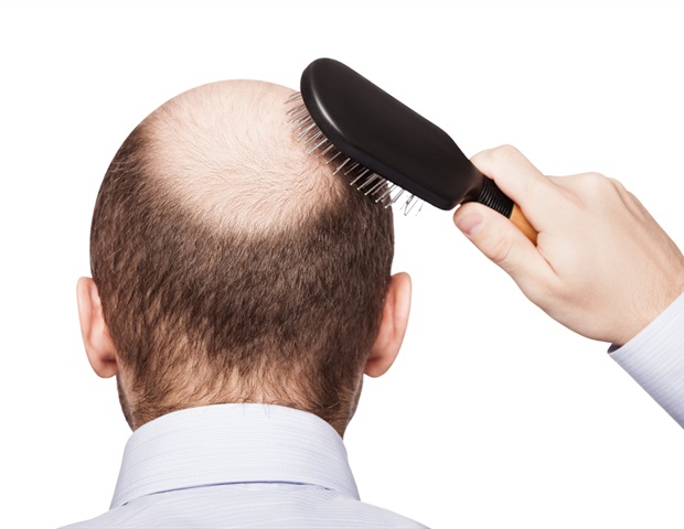
    There is no cure for Hashimoto’s or hypothyroidism. But VanOrden, who lives in Athens, Texas, started taking levothyroxine, a widely prescribed synthetic thyroid hormone used to treat common symptoms such as fatigue, weight gain, hair loss and sensitivity to cold.

    Most patients do well on levothyroxine and their symptoms resolve. But for others, like VanOrden, the drug isn’t as effective.

    For her, that meant traveling from doctor to doctor, test to test, and treatment to treatment, spending about $5,000 a year.

    “I look and act like a pretty energetic person,” said VanOrden, 38, explaining that her symptoms are not visible. “But there’s a hole in my gas tank,” she said. And “stress widens the gap.”

    Autoimmune diseases arise when the immune system mistakenly attacks and damages healthy cells and tissues. Other common examples include rheumatoid arthritis, lupus, celiac disease, and inflammatory bowel disease. There are more than 80 such diseases, affecting an estimated 50 million Americans, disproportionately women. Overall, the cost of treating autoimmune diseases in the US is estimated at over $100 billion per year

    Despite their frequency, finding help for many autoimmune diseases can be frustrating and expensive. Getting diagnosed can be a major hurdle because the set of symptoms is very similar to those of other medical conditions, and there are often no definitive identifying tests, says Sam Lim, clinical director of the Division of Rheumatology at Emory University School of Medicine in Atlanta . . In addition, some patients feel like they have to fight to be believed, even by a doctor. And after a diagnosis, many autoimmune patients rack up big bills while they explore treatment options.

    “They’re often upset. Patients feel rejected,” Elizabeth McAninch, an endocrinologist and thyroid expert at Stanford University, says of some patients who come to her for help.

    Inadequate medical education and a lack of investment in new research are two factors hindering the general understanding of hypothyroidism, according to Antonio Bianco, an endocrinologist at the University of Chicago and leading expert on the condition.

    Some patients become angry when their symptoms don’t respond to standard treatments, either levothyroxine or that drug in combination with another hormone, said Douglas Ross, an endocrinologist at Massachusetts General Hospital in Boston. “We’re going to have to remain open to the possibility that we’re missing something here,” he said.

    Jennifer Ryan, 42, said she has spent “thousands of dollars out of pocket” looking for answers. Doctors did not recommend thyroid hormone medication for the Huntsville, Alabama, resident, who was diagnosed with Hashimoto’s after years of fatigue and weight gain, because her levels appeared normal. She recently changed doctors and is hoping for the best.

    “You don’t walk around in pain all day and there’s nothing to worry about,” Ryan said.

    And health insurers typically deny coverage for new treatments for hypothyroidism, says Brittany Henderson, an endocrinologist and founder of the Charleston Thyroid Center in South Carolina, which treats patients from all fifty states. “Insurance companies want you to use the generics, even though many patients don’t do well with these treatments,” she said.

    Meanwhile, the extent of America’s thyroid problems is reflected in drug sales. Levothyroxine is among the five most prescribed medications in the US each year. Yet research suggests the drug is overprescribed to people with mild hypothyroidism.

    A recent study paid for by AbbVie – maker of Synthroid, a branded version of levothyroxine – found that a database of medical and pharmacy claims found that the prevalence of hypothyroidism, including milder forms, has risen from 9.5% of Americans in 2012 to 11.7%. in 2019.

    The number of people diagnosed will increase as the population ages, McAninch said. Endocrine disruptors — natural or synthetic chemicals that can affect hormones — could explain some of that increase, she said.

    In their search for answers, patients sometimes connect on social media, where they ask questions and describe their thyroid hormone levels, drug regimens and symptoms. Some online platforms provide information that is questionable at best, but overall, social media has increased patients’ understanding of difficult-to-resolve symptoms, Bianco said.

    They also encourage each other.

    VanOrden, who has been active on Reddit, has this advice for other patients: “Don’t give up. Keep standing up for yourself. There is a doctor somewhere who will listen to you.” She has started an alternative treatment — desiccated thyroid medication, an option not approved by the FDA — plus a low dose of the addiction drug naltrexone, although data is limited. She feels better now.

    Research into autoimmune thyroid diseases receives little funding, so the underlying causes of immune dysfunction are not well studied, Henderson said. The medical establishment has not yet fully recognized difficult-to-treat patients with hypothyroidism, but greater recognition of them and their symptoms would help fund research, Bianco said.

    “I would like a very clear, solid acknowledgment that these patients exist,” he said. “These people are real.”


    Kaiser health newsThis article was adapted from khn.org, a national newsroom that produces in-depth journalism on health issues and is one of the core operating programs at KFF – the independent source for health policy research, polling and journalism.

    Source link

  • Tested positive for COVID?  Be careful out there

    Tested positive for COVID? Be careful out there

    Higher concussion rates following COVID-19 infection in high school athletes.

    Bullock GS, Emery CA, Nelson VR, etc. al, Br J Sports Med2023; [epub ahead of print]. two: 10.1136/brjsports-2022-106436.

    Full text freely available

    Take home message

    Athletes who tested positive for a COVID-19 infection were more likely to suffer a concussion within 60 days of recovering from the infection than athletes who did not contract COVID-19.

    Background

    COVID-19 affects the respiratory, cardiovascular and nervous systems. In some cases, COVID-19 can lead to long-term consequences (e.g. impaired cognition). It is currently unknown whether COVID-19 infection affects the risk of sports-related concussions.

    Study aim

    Bullock and colleagues completed a prospective cohort study to compare concussion rates between high school athletes who recently tested positive for COVID-19 and those who did not.

    Methods

    The authors recruited athletes from high schools in six states. A certified athletic trainer recorded all cases of COVID-19 infection or concussion. If an athlete reported COVID-like symptoms or had an elevated temperature during a daily screening, the athletic trainer administered a COVID test. The research team also recorded when an athlete participated in a training or competition. The authors focused on the number of concussions within 60 days after an athlete returned to play following a COVID-19 infection. All data was recorded in the Players Health Rehab system, including demographic information, sports and illness data for all athletes.

    Results

    A total of 72,522 athletes participated in high school sports at the affected school during the 2020-2021 school year. Of these athletes, 430 had COVID-19 infections. Of the athletes with a COVID-19 infection, 32 had a concussion, and 1,241 athletes without COVID-19 had a concussion. An athlete with a history of COVID-19 was approximately 3 times more likely to suffer a concussion within 60 days of returning to play than an athlete without COVID-19.

    Viewpoints

    Overall, the authors found that prior COVID-19 infection increased the risk of concussion during the first 60 days after recovery from COVID-19. Therefore, the effects of a COVID-19 infection may linger after returning to play. Knowing whether this applies to other sports-related injuries would be interesting. Additionally, learning why these athletes are at greater risk for concussion (e.g., deconditioning, persistent symptoms) can help us develop prevention strategies and make informed decisions about when and how to release an athlete to play following a COVID-19 infection.

    Clinical implications

    Doctors should explain to patients with COVID-19 that they may experience lingering effects that predispose them to concussion. We need to help the patient make an informed decision about when to return to play after COVID-19 infection. Once an athlete returns, doctors should monitor these patients for concussions.

    Questions for discussion

    What other measures have you implemented in your clinical practice to monitor athletes after COVID-19 infection? Is a COVID-19 infection something you are currently documenting?

    Written by Kyle Harris
    Reviewed by Jeffrey Driban

    related posts

    COVID recovery may require injury prevention training!
    Use of cardiovascular magnetic resonance (CMR) imaging for return to athletic activities after COVID-19 infection: an expert consensus document on behalf of the American Heart Association Council on Cardiovascular Radiology and Intervention (CVRI) Leadership and endorsed by the Society for Cardiovascular Magnetic Resonance (SCMR)
    Adapted physical activity in subjects and athletes recovering from Covid-19: a position statement of the Società Italiana Scienze Motorie e Sportive
    Cardiopulmonary Considerations for High School Athletes During the COVID-19 Pandemic: Update to the NFHS-AMSSM Guidelines

    Evidence-based assessment of concussion course - 5 EBP CEUs

    Source link

  • Sports managers need sports trainers for a better heat policy

    Sports managers need sports trainers for a better heat policy

    Exercise-induced heat illness: policy adoption and influence on contextual factors reported by athletic administrators

    Scarneo-Miller SE, Adams WM, Coleman KA, Lopez RM. Sports Health. Mar 5, 2023: 19417381231155107. doi: 10.1177/19417381231155107. E-publishing prior to printing. PMID: 36872595.

    https://journals.sagepub.com/doi/10.1177/19417381231155107

    Take home message

    Most high school athletic administrators reported having a written heat illness policy in place, but they were often missing key components. The presence of an athletic trainer helped create a written policy that included more components.

    Background

    Sports administrators play an important role in policy acceptance. Policy measures such as addressing acute heat illness are critical as improper management can lead to poor outcomes. Unfortunately, we know little about the factors that promote and hinder the implementation of heat illness policies.

    Study aim

    The authors surveyed high school athletic administrators in the United States to describe the adoption of exertional heat illness policies and to examine factors that influence the adoption of these policies.

    Methods

    During the 2018-2019 academic school year, the research team emailed nearly 7,000 athletic administrators inviting them to complete a questionnaire asking about 1) demographics, 2) exercise-related illness policies, 3) monitoring and modification of written policy, and 4) enablers and barriers to policy development. The researchers used the precautionary adoption process model to assess an athletics administrator’s willingness to adopt policies. The adoption model is based on 8 phases, from not being aware to maintaining a written policy for more than 6 months.

    Results

    Of 466 athletics administrators (~48 years old, 82% male, 77% worked in the field for more than 15 years), 78% reported having a written policy on the prevention and treatment of exertional heat illness. Only 6% adopted all eleven essential elements of an exertional heat illness policy. Almost half of the managers indicated that they adopted fewer than 5 essential elements.

    Older athletics administrators, those who had previously dealt with heat illness, or those with an athletic trainer on their staff were more likely to have a written policy. Additionally, state mandates and having a medical professional were the most commonly cited facilitators for adopting policies on exertional illness prevention and use of a rectal thermometer. Similarly, the most commonly reported barrier to comprehensive heat illness management was the lack of a full-time athletic trainer (11). Administrators also recognized budget constraints that limited the use of a cold water immersion pool (23%), and the top barriers to using a rectal thermometer were discomfort using the thermometer (32%), parent/guardian resistance (30 %), resistance from parents/guardians (30%). coaches (30%) and liability issues (27%).

    Viewpoints

    Nearly 80% of athletics administrators surveyed reported that they had a written policy on exertional heat illness. Few integrated or were aware of all the necessary components to meet best clinical practices. The authors found that access to athletic training services was associated with better adoption of exercise health policies. This finding is consistent with it being an athletic trainer who would implement such a policy. It’s worth recognizing that only 7% of administrators contacted completed the survey. Therefore, these results may not accurately reflect what is happening in high schools across the country. One possibility is that people more interested in policy or heat illness completed the survey. So these results may show us the best-case scenario for written policies (78%) that include all components (6%) – which is a worrying sign.

    Clinical implications

    Encouraging state mandates and schools to hire athletic trainers can ensure that there are written policies to address heat illness. Clinicians may also want to consider strategies to educate coaches and parents/guardians about the reasons for this policy, such as rectal thermometers and cold water plunge pools.

    Questions for discussion

    Are you having trouble adding rectal temperature to your exercise heat illness protocol? Do you communicate with your athletics administrator regarding the approval, implementation and annual review/practice of your emergency policy?

    related posts

    1. Management of exertional heat stroke still leaves something to be desired
    2. A little more education about heat stroke due to exertion could go a long way
    3. Clinical Pearl: prevention and treatment of exertional heat stroke
    4. Tag us! What do coaches know about heat stroke during exertion and the role of the athletic trainer?
    5. Follow guidelines to prevent exertional heat illness? Let’s reconsider these guidelines

    Written by Jane McDevitt
    Reviewed by Jeffrey Driban

    9 EBP CEU courses

    Source link

  • Activity affected urinary incontinence;  Let’s talk about it!

    Activity affected urinary incontinence; Let’s talk about it!

    Prevalence and normalization of stress urinary incontinence in female strength athletes.

    Mahoney K, Heidel RE, and Olewinski LJ Good luck Cond2023 [epub ahead of print].

    Full text freely available

    Take home message

    Seven in ten female strength training athletes report stress urinary incontinence (SUI) in some aspect of their lives. However, fewer than 2 in 10 athletes talk to their doctor or seek treatment for SUI.

    Background

    Very intensive physical activity can increase the risk of SUI, an involuntary loss of urine due to increased intra-abdominal pressure. Up to 2 in 5 women may experience SUI throughout their lives, which can negatively impact an individual’s quality of life. Pelvic floor training can treat SUI. Although female strength training athletes may be at greater risk for SUI, it remains unclear how many of these athletes experience SUI and seek treatment, as well as how they would prefer to learn about SUI.

    Study aim

    The researchers conducted a cross-sectional study to assess female strength training athletes’ understanding and normalization of SUI, including how often female strength training athletes sought treatment for SUI.

    Methods

    The researchers developed a new study, which was expertly reviewed, to share on various social media platforms. The survey asked respondents about demographic information, risk factors for SUI, experience with SUI, favorite sources of information about SUI, and whether the respondent had sought treatment for SUI. Respondents to the survey were women over the age of 18 who considered powerlifting, weightlifting or strongman as their main activity.

    Results

    Of the 425 respondents, 69% reported experiencing SUI in some aspects of their lives. Of the athletes who experienced SUI, 61% reported that they first experienced SUI after starting their sport. About two-thirds of athletes thought SUI was a normal part of their sport. Only 17% of respondents reported talking to their doctor about SUI, and 9% sought treatment. Nearly 30% of respondents reported seeking advice about SUI from videos and articles on the internet, 23% spoke to friends and 13% spoke to their coach about SUI.

    Viewpoints

    Overall, the results of this study indicated that SUI is common among female strength training athletes, and most consider it a normal part of their sport. However, very few athletes turn to healthcare to address their SUI. It would be interesting to see this study replicated by asking athletes to complete the survey during competitions, as it is unclear whether women with SUI are more likely to complete the online survey. So the online survey may overestimate how many women have SUI, but the answers about engaging the health care system and where they seek advice are still likely to be informative.

    Clinical implications

    Clinicians who work with athletes who are at high risk for developing SUI should educate athletes about effective treatments. It can also be helpful to educate strength and conditioning coaches about SUI and available treatments.

    Questions for discussion

    How do you talk to your athletes about SUI? Have you noticed that other athletes have high SUI rates?

    Written by Kyle Harris
    Reviewed by Jeffrey Driban

    related posts

    Check the lights before you check that urine!
    Throw away your strips for better urine density results
    Altered hydration status may impact concussion assessment
    Cheers! The more you exercise, the more you should drink

    9 EBP CEU courses

    Source link

  • Knee Pain Symptoms and Causes

    Knee Pain Symptoms and Causes

    Knee Pain Symptoms and Causes: A Comprehensive Guide

    Knee pain can be a bothersome issue that affects people of all ages. It may result from a number of factors, such as injuries like torn cartilage or ruptured ligaments, or medical conditions like arthritis, gout, and infections. Understanding the causes and symptoms of knee pain is essential for seeking timely and appropriate treatment to alleviate discomfort and prevent complications.

    In this article, we will explore the various factors that contribute to knee pain and gain insights into different treatment approaches. We will also discuss risk factors and preventive measures to help maintain healthy knees and maintain overall well-being.

    patellofemoral pain syndrome:

    Understanding Patellofemoral Pain Syndrome

    CategoryDetails
    DefinitionPain between the kneecap (patella) and thighbone (femur) due to abnormal biomechanical forces
    Common Causes– Kneecap maltracking/dysfunction<br>- Overuse from sports<br>- Tight quadriceps, IT band, calves <br>- Weak hip and gluteal stabilizers
    Contributing Factors– High Q-angle anatomy<br>- Poor movement patterns<br>- Sudden load increases without adaptation
    Affected Populations– Athletes <br>- Young active adults<br>- Older adults with kneecap arthritis
    Conservative Treatment– Taping/bracing to improve tracking <br>- Targeted stretching & strengthening <br>- Movement pattern retraining
    Surgical Options– Lateral release to loosen tight tissues<br>- Patellar reshaping procedures<br>- Kneecap cartilage resurfacing

    Key components of PFPS involve abnormal kneecap gliding, overloading tissue capacities through sports, and anatomical vulnerabilities. Conservative rehabilitation aims to correct the imbalances, while surgery is occasionally utilized for refractory cases to realign structures or halt arthritis progression.

    Key Takeaways

    • Knee pain can be caused by injuries or medical conditions like arthritis and gout.
    • Timely diagnosis and treatment can alleviate discomfort and prevent complications.
    • Understanding risk factors and preventive measures helps maintain healthy knees.

    Overview

    Knee pain is highly prevalent and affects individuals of all age groups. It can stem from injuries such as torn cartilage or ruptured ligaments or be a result of medical conditions like arthritis, gout, and infections. Fortunately, most minor knee pain cases can be managed through self-care measures, while more severe cases may benefit from physical therapy, knee braces, or even surgical intervention. Our discussion will dive deeper into the complexities of knee pain, exploring muscles, joints, ligaments, tendons, and bones (tibia, patella, and femur) involved in the knee joint.

    3260325e cc5f 426e 80e9 6a1a2a7f263b

    Symptoms

    Recognizing the Signs

    Knee pain can manifest in various ways depending on the underlying issue. Some common symptoms accompanying knee pain are:

    • Swelling and stiffness
    • Redness and a warm sensation
    • Weakness or instability
    • Audible popping or crunching sounds
    • Difficulty fully straightening the knee

    Seeking Medical Attention

    We encourage you to consult your doctor if you experience any of the following:

    • Inability to bear weight on your knee or a feeling of instability
    • Significant swelling of the knee
    • Limited extension or flexing of the knee
    • Visible deformities in your leg or knee
    • Fever, accompanied by redness, pain, and swelling in the knee
    • Severe knee pain associated with an injury

    From Mayo Clinic to Your Inbox

    We’re excited to provide you with the latest research advancements, health tips, current health topics, and advice on managing your health straight to your inbox. Sign up for free and stay informed with information tailored to your needs.

    By signing up, we combine your email and website usage information with other details we have about you, ensuring the most relevant and helpful content is delivered to you. For Mayo Clinic patients, rest assured that your protected health information is treated with utmost care and used according to our privacy practices.

    Remember, you have the option to opt-out of our email communications at any time by simply clicking the unsubscribe link provided in the email.

    Causes of Knee Pain

    Injuries Affecting the Knee

    Knee injuries can involve various structures around the knee joint, such as ligaments, tendons, bursae, bones, and cartilage. Some common knee injuries are:

    • ACL injury: A tear in the anterior cruciate ligament, which connects the shinbone to the thighbone, often occurs in sports that require sudden directional changes like basketball or soccer.
    • Fractures: Knee bones, including the kneecap, may break due to falls or vehicle accidents. Osteoporosis can also lead to knee fractures from minor missteps.
    • Torn meniscus: The meniscus, the cartilage cushioning between the shinbone and thighbone, may tear if the knee is suddenly twisted while bearing weight.
    • Knee bursitis: Injuries may cause inflammation in the knee bursae, the fluid-filled sacs that enable smooth movement between tendons, ligaments, and the knee joint.
    • Patellar tendinitis: This refers to the irritation and inflammation of tendons attaching muscles to bones, particularly the patellar tendon connecting the kneecap and shinbone. It often occurs in runners, skiers, cyclists, and people involved in jumping sports.

    Mechanical Issues

    Mechanical problems causing knee pain include:

    • Loose body: Detached bone or cartilage fragments floating in the joint space can obstruct knee joint movement, akin to a pencil caught in a hinge.
    • Iliotibial band syndrome: Tightening of the iliotibial band, which extends from the hip to the outer knee, can cause rubbing against the thighbone. Distance runners and cyclists are prone to this condition.
    • Dislocated kneecap: The kneecap can slip out of place, typically to the knee’s outside. In some instances, the dislocation remains visible.
    • Hip or foot pain: Compensating for pain in the hips or feet by changing one’s gait can place extra stress on the knee joint, leading to pain.

    Forms of Arthritis

    Various types of arthritis can cause knee pain:

    • Osteoarthritis: This wear-and-tear condition results from the knee cartilage’s deterioration due to use and age.
    • Rheumatoid arthritis: An autoimmune disease potentially affecting any joint, including the knees. This chronic condition’s severity may fluctuate.
    • Gout: Occurs when uric acid crystals accumulate in the joint, typically affecting the big toe but also the knee.
    • Pseudogout: Often mistaken for gout, this condition stems from calcium-containing crystals in the joint fluid. The knee is the most frequently affected joint.
    • Septic arthritis: An infection in the knee joint may lead to swelling, pain, redness, and fever. It can quickly damage the knee cartilage, requiring urgent medical attention.

    Additional Issues

    Patellofemoral pain syndrome (PFPS) refers to discomfort between the kneecap (patella) and underlying thighbone (femur). It involves abnormal forces transmitted through the patellofemoral joint which is the articulation between these two structures. PFPS commonly affects certain populations, especially athletes who overloaded their knees through repetitive overuse from sports.

    The main culprit causing PFPS is maltracking or dysfunction of the kneecap as it glides up and down in a groove on the femur during knee bending. This maltracking indicates a biomechanical imbalance, often from tight muscles or anatomical misalignment pulling the cap sideways. This leads to increased pressures on the back of the patella and the adjacent cartilage, triggering pain.

    PFPS frequently develops in young active adults, particularly females, who have naturally higher “Q angles” – an anatomical measurement of kneecap positioning. It also occurs in some older adults for whom the cartilage under the kneecap has worn away significantly due to arthritis, causing bone-on-bone grinding. These groups appear predisposed towards developing knee pain from the aftermath of abnormal patellofemoral mechanics.

    Besides overuse from sports and vulnerable anatomy, other PFPS triggers involve sudden increases in loading too quickly. The muscles stabilizing the patella require adaptations to control loads safely. When demands exceed abilities, the kneecap tracking suffers. Additionally, poor movement patterns such as unbalanced squatting or jumping/landing with knock knees induces patellofemoral compression. Often tight quadriceps, IT bands and calves paired with weak hips/glutes contributes to dysfunction.

    Several targeted interventions aim at conservatively realigning the patella, strengthening deficient stabilizers, restoring ideal tracking and alleviating compression forces causing PFPS. Using braces, taping, customized exercise corrections and gait pattern retraining are common initial tactics under therapist guidance to calm pain and prevents recurrences. In some cases of refractory PFPS or significant arthritis, surgical options to release or reshape overtightened tissues or resurface damaged articulations provide longer term solutions.

  • Prevention Strategies for Knee Ligament Injuries

    Prevention Strategies for Knee Ligament Injuries

    Prevention Strategies for Knee Ligament Injuries: Professional Tips

    Knee ligament injuries are common among athletes and can be a frustrating setback for anyone who enjoys an active lifestyle. The knee joint is one of the largest and most complex joints in the body, making it vulnerable to various types of injuries. Understanding and identifying knee ligament injuries is crucial to prevent further damage and promote quick recovery.

    v2 32d9h 7v8oi

    The most common knee ligament injuries are the anterior cruciate ligament (ACL), medial collateral ligament (MCL), posterior cruciate ligament (PCL), and lateral collateral ligament (LCL). These injuries can occur due to direct trauma, such as contusions and strains, or from indirect trauma, such as sudden changes in direction. Symptoms of knee ligament injuries include pain, swelling, inflammation, and joint laxity. In this article, we will discuss five prevention strategies for knee ligament injuries to help maintain knee stability and prevent knee instability.

    professional tips for prevention strategies to help avoid knee ligament injuries:

    CategoryTips
    Strength & Flexibility Training– Emphasize hamstring, glute, quad, hip & core exercises<br>- Ensure muscle imbalances are addressed<br>- Use proper form/technique to prevent overstress
    Movement Control & Balance– Practice proper landing mechanics when jumping <br>- Enhance proprioception & body control<br>- Integrate deceleration drills
    Equipment & Environment– Wear supportive & appropriate footwear <br>- Tape or brace vulnerable knees if needed <br>- Ensure safe, even playing surfaces
    Loading & Recovery– Follow loading guidelines for ramping up intensity<br>- Avoid aggressive pivoting until tissues adapted<br>- Schedule adequate rest periods between sessions
    Body Composition & Nutrition– Prevent muscle loss during injury layoffs<br>- Maintain healthy BMI year-round<br>- Support exercise recovery nutritionally
    Mindset & Motivation– Foster positive, confident mentality<br>- Set smaller achievable goal steps <br>- Enlist social support structure

    The most effective ACL/knee ligament injury prevention programs take a comprehensive approach. Optimizing strength, movement mechanics, loading, and nutrition helps create resilience. Confidence in knee stability paired with caution towards unsafe mechanics allows for safer progression. Expert guidance tailoring programs is key for best injury deterrence.

    9c0fad6b da19 4bad 92cb dac1d9705c7d

    Key Takeaways

    • Understanding the anatomy of the knee joint and identifying knee ligament injuries can promote quick recovery.
    • Direct and indirect trauma can cause knee ligament injuries, leading to pain, swelling, inflammation, and joint laxity.
    • Five prevention strategies can help maintain knee stability and prevent knee instability, reducing the risk of knee ligament injuries.

    Understanding and Identifying Knee Ligament Injuries

    Knee ligament injuries are common among athletes and individuals who engage in physical activities that require jumping, pivoting, or sudden changes in direction. In this section, we will discuss the common types and causes of knee ligament injuries, how to identify symptoms and diagnose the injury, treatment and rehabilitation options, and the role of sex and age in knee ligament injuries.

    Common Types and Causes

    The most common types of knee ligament injuries are the anterior cruciate ligament (ACL) and medial collateral ligament (MCL) injuries. The ACL is usually injured during non-contact activities such as landing after a jump or sudden stops and changes in direction. On the other hand, the MCL is usually injured from a direct blow to the outer side of the knee, such as when playing football or hockey.

    Symptoms and Diagnosis

    The symptoms of knee ligament injuries include pain, swelling, bruising, and instability. The diagnosis of knee ligament injuries is usually made through a physical exam and imaging tests such as MRI or X-rays. It is important to seek medical attention if you experience any of these symptoms.

    Treatment and Rehabilitation

    The treatment and rehabilitation of knee ligament injuries depend on the severity of the injury. Treatment options include rest, ice, compression, and elevation (RICE), physical therapy, and surgery. Rehabilitation includes exercises to improve range of motion, strength, and mobility. It is important to follow the guidance of a physical therapist or orthopedic surgeon to ensure proper recovery.

    Role of Sex and Age

    Research shows that females are more prone to knee ligament injuries than males due to differences in anatomy, form, and hormones such as estrogen and relaxin. Additionally, age plays a role in knee ligament injuries, with individuals over 50 years old being at a higher risk due to decreased muscle strength and flexibility.

    Sports and Injuries

    Knee ligament injuries are common in sports that require sudden stops and changes in direction such as soccer, basketball, and football (soccer). Injury prevention programs that include warm-up, neuromuscular training, and proprioception exercises can reduce the frequency of knee ligament injuries by up to 50%. Coaches and athletes should also prioritize proper technique, posture, and fatigue management to prevent knee ligament injuries.

    In conclusion, knee ligament injuries are common among athletes and individuals who engage in physical activities. Understanding the common types and causes of knee ligament injuries, identifying symptoms and diagnosing the injury, and following proper treatment and rehabilitation options can help prevent long-term damage. Coaches, athletes, and individuals should prioritize injury prevention programs and proper technique to reduce the frequency of knee ligament injuries.

    Prevention Strategies for Knee Ligament Injuries

    When it comes to knee ligament injuries, prevention is key. Here are some effective strategies that we recommend:

    Strengthening and Conditioning

    Strengthening and conditioning exercises can help improve muscle strength and endurance, which can help reduce the risk of knee ligament injuries. Exercises that target the hips, ankles, and core can be particularly effective in improving stability and reducing the risk of injury.

    Injury Prevention Programs

    Injury prevention programs can be an effective way to reduce the risk of knee ligament injuries. These programs typically include a combination of warm-up exercises, neuromuscular training, and strength and conditioning exercises. They can be particularly effective for athletes who participate in high-risk sports, such as soccer, basketball, and football.

    Importance of Compliance and Frequency

    Injury prevention strategies are only effective if they are followed consistently and frequently. Compliance with injury prevention programs is essential for reducing the risk of knee ligament injuries. It is important to follow the program as prescribed and to make sure that exercises are performed correctly and with good form.

    In conclusion, preventing knee ligament injuries requires a combination of strategies that target strength, stability, and flexibility. Injury prevention programs that incorporate warm-up exercises, neuromuscular training, and strength and conditioning exercises can be particularly effective. However, compliance with these programs is essential for success. By following these strategies, we can help reduce the risk of knee ligament injuries and keep athletes healthy and active.

  • Rehabilitation Exercises for Knee

    Rehabilitation Exercises for Knee

    Rehabilitation Exercises for Knee Ligaments: Strengthening and Recovery Techniques

    As physical therapists, we often see patients with knee ligament injuries. Knee ligaments are strong bands of tissue that connect the thighbone to the shinbone and provide stability to the knee joint. Injuries to knee ligaments can result in pain, swelling, and decreased range of motion. Rehabilitation exercises for knee ligaments can help improve joint stability, reduce pain, and restore function.

    v2 32db6 rluua

    Understanding Knee Ligament Injuries is important for developing an effective rehabilitation plan. There are four main ligaments in the knee joint: the anterior cruciate ligament (ACL), posterior cruciate ligament (PCL), medial collateral ligament (MCL), and lateral collateral ligament (LCL). ACL and PCL injuries are more common in athletes and can occur during sports that involve sudden stops, changes in direction, or jumping. MCL and LCL injuries can result from a direct blow to the knee or from twisting the knee. Depending on the severity of the injury, surgery may be required to repair or reconstruct the damaged ligament.

    8cbc720a b396 47e0 b03a 307abbe136e3 1

    In this article, we will discuss 6 Rehabilitation Exercises for Knee Ligaments that can help improve joint stability, reduce pain, and restore function. These exercises are designed to strengthen the muscles that support the knee joint and improve biomechanical function. By following a structured rehabilitation program, patients can improve their recovery and return to their daily activities faster.

    Key Takeaways

    • Knee ligament injuries can result in pain, swelling, and decreased range of motion.
    • There are four main ligaments in the knee joint: the anterior cruciate ligament (ACL), posterior cruciate ligament (PCL), medial collateral ligament (MCL), and lateral collateral ligament (LCL).
    • Rehabilitation exercises for knee ligaments can help improve joint stability, reduce pain, and restore function.

    Understanding Knee Ligament Injuries

    Knee ligament injuries are common among athletes and individuals who engage in physical activities. These injuries can be caused by a wide range of factors, including trauma, overuse, and sports-related injuries. The most common types of knee ligament injuries include anterior cruciate ligament (ACL) injuries, medial collateral ligament (MCL) injuries, and posterior cruciate ligament (PCL) injuries.

    Causes and Types of Injuries

    ACL injuries are typically caused by a twisting motion or a direct blow to the knee. MCL injuries, on the other hand, are often caused by a direct blow to the outside of the knee. PCL injuries are usually the result of a direct blow to the front of the knee or a hyperextension injury.

    Ligament injuries can be classified as either partial or complete tears. Partial tears involve damage to a portion of the ligament, while complete tears involve a complete separation of the ligament from the bone. Overuse injuries, such as tendinitis, can also occur in the knee.

    Diagnosis and Assessment

    Diagnosis of knee ligament injuries involves a physical examination, imaging tests, and other diagnostic procedures. Physical examination involves assessing the knee for pain, swelling, and instability. Imaging tests such as magnetic resonance imaging (MRI), radiographs, and ultrasound can also be used to assess the extent of the injury.

    Treatment Options

    Treatment options for knee ligament injuries depend on the severity of the injury. Nonoperative treatment may involve rest, ice, compression, and elevation (RICE), physical therapy, and immobilization. Operative treatment may involve surgery, such as arthroscopy or ACL reconstruction.

    Post-Injury Challenges

    Post-injury challenges may include pain, swelling, and instability. Rehabilitation is essential for restoring full function and reducing the risk of future injuries.

    Understanding the Rehabilitation Process

    Rehabilitation is an essential part of the recovery process for knee ligament injuries. The rehabilitation process involves a series of exercises and other interventions designed to improve muscle strength, proprioception, and joint stability.

    Specific Rehabilitation Exercises

    Specific rehabilitation exercises may include exercises to improve muscle strength, such as knee flexion and extension exercises, as well as exercises to improve proprioception, such as balance training.

    Role of Imaging in Rehabilitation

    Imaging tests such as MRI and radiographs can be used to assess the extent of the injury and monitor progress during rehabilitation.

    Rehabilitation Exercises for Knee Ligament Recovery

    ExerciseDescriptionTarget Area
    Passive Range of MotionGentle movements of knee by therapist/device to improve flexion/extension early post-injury without overstressingRestore mobility; reduce stiffness
    Quad SetsContract quad muscles by pushing knees down into floor/table when in sitting/lying positionActivate quads; prevent atrophy
    Straight Leg RaisesTighten quads to lift straight leg up off floor/bed, keep knee lockedStrengthen quads; knee stabilization
    Hip Abduction/AdductionMove legs outward/inward against resistance or body weightStrengthen hip muscles supporting knee
    Calf RaisesRaise up onto ball/toes of foot, lower with controlStrengthen calves; improve balance/proprioception
    Wall SitsSlide down wall until knees/hips at 90 degree angle, hold positionStrengthen quads; improve endurance
    Terminal Knee ExtensionsSit with resistance band around foot, fully straighten kneeRegain full extension range of motion
    Hamstring CurlsFlex knees to pull heels toward buttress against resistance bandStrengthen hamstrings; decelerate knee extension
    Balance Board/Wobble BoardStand with knee slightly bent on unstable surfaceEnhance balance and knee proprioception
    Mini-SquatsPerform slight squatting motion within pain-free range of motionRegain quad control; restore movement pattern

    The exact exercises and progressions are tailored by physical therapists depending on injury specifics, graft type, and post-surgical timeline. The goal is to restore strength, mobility, proprioception and movement control safely based on healing constraints

    a7918c30 98ad 4e88 b5fa 3668ecb888b6 2

    Preventing Future Injuries

    Preventing future knee ligament injuries involves proper conditioning, warm-up, and cool-down exercises, as well as appropriate protective gear.

    In conclusion, knee ligament injuries are common among athletes and individuals who engage in physical activities. Proper diagnosis, treatment, and rehabilitation are essential for restoring full function and reducing the risk of future injuries.

  • Sports managers need sports trainers for a better heat policy

    Sports managers need sports trainers for a better heat policy


    Exercise-induced heat illness: policy adoption and influence on contextual factors reported by athletic administrators

    Scarneo-Miller SE, Adams WM, Coleman KA, Lopez RM. Sports Health. Mar 5, 2023: 19417381231155107. doi: 10.1177/19417381231155107. E-publishing prior to printing. PMID: 36872595.

    https://journals.sagepub.com/doi/10.1177/19417381231155107

    Take home message

    Most high school athletic administrators reported having a written heat illness policy in place, but they were often missing key components. The presence of an athletic trainer helped create a written policy that included more components.

    Background

    Sports administrators play an important role in policy acceptance. Policy measures such as addressing acute heat illness are critical as improper management can lead to poor outcomes. Unfortunately, we know little about the factors that promote and hinder the implementation of heat illness policies.

    Study aim

    The authors surveyed high school athletic administrators in the United States to describe the adoption of exertional heat illness policies and to examine factors that influence the adoption of these policies.

    Methods

    During the 2018-2019 academic school year, the research team emailed nearly 7,000 athletic administrators inviting them to complete a questionnaire asking about 1) demographics, 2) exercise-related illness policies, 3) monitoring and modification of written policy, and 4) enablers and barriers to policy development. The researchers used the precautionary adoption process model to assess an athletics administrator’s willingness to adopt policies. The adoption model is based on 8 phases, from not being aware to maintaining a written policy for more than 6 months.

    Results

    Of 466 athletics administrators (~48 years old, 82% male, 77% worked in the field for more than 15 years), 78% reported having a written policy on the prevention and treatment of exertional heat illness. Only 6% adopted all eleven essential elements of an exertional heat illness policy. Almost half of the managers indicated that they adopted fewer than 5 essential elements.

    Older athletics administrators, those who had previously dealt with heat illness, or those with an athletic trainer on their staff were more likely to have a written policy. Additionally, state mandates and having a medical professional were the most commonly cited facilitators for adopting policies on exertional illness prevention and use of a rectal thermometer. Similarly, the most commonly reported barrier to comprehensive heat illness management was the lack of a full-time athletic trainer (11). Administrators also recognized budget constraints that limited the use of a cold water immersion pool (23%), and the top barriers to using a rectal thermometer were discomfort using the thermometer (32%), parent/guardian resistance (30 %), resistance from parents/guardians (30%). coaches (30%) and liability issues (27%).

    Viewpoints

    Nearly 80% of athletics administrators surveyed reported that they had a written policy on exertional heat illness. Few integrated or were aware of all the necessary components to meet best clinical practices. The authors found that access to athletic training services was associated with better adoption of exercise health policies. This finding is consistent with it being an athletic trainer who would implement such a policy. It’s worth recognizing that only 7% of administrators contacted completed the survey. Therefore, these results may not accurately reflect what is happening in high schools across the country. One possibility is that people more interested in policy or heat illness completed the survey. So these results may show us the best-case scenario for written policies (78%) that include all components (6%) – which is a worrying sign.

    Clinical implications

    Encouraging state mandates and schools to hire athletic trainers can ensure that there are written policies to address heat illness. Clinicians may also want to consider strategies to educate coaches and parents/guardians about the reasons for this policy, such as rectal thermometers and cold water plunge pools.

    Questions for discussion

    Are you having trouble adding rectal temperature to your exercise heat illness protocol? Do you communicate with your athletics administrator regarding the approval, implementation and annual review/practice of your emergency policy?

    related posts

    1. Management of exertional heat stroke still leaves something to be desired
    2. A little more education about heat stroke due to exertion could go a long way
    3. Clinical Pearl: prevention and treatment of exertional heat stroke
    4. Tag us! What do coaches know about heat stroke during exertion and the role of the athletic trainer?
    5. Follow guidelines to prevent exertional heat illness? Let’s reconsider these guidelines

    Written by Jane McDevitt
    Reviewed by Jeffrey Driban

    9 EBP CEU courses

    Source link

  • Knee Pain After ACL Surgery

    Knee Pain After ACL Surgery

    Managing Knee Pain After ACL Surgery

    Anterior cruciate ligament (ACL) reconstruction is a common surgical procedure for individuals who have suffered a torn ACL. While outcomes have improved dramatically, knee pain and re-injury continue to be concerns during the recovery process. Recent advances in rehabilitation protocols and biomechanical research are shedding light on optimal recovery strategies.

    9566c6af 8e44 4b28 9740 7d191e646e96

    Understanding Knee Pain After ACL Surgery

    Understanding Knee Pain after ACL Surgery

    Knee pain following ACL reconstruction arises due to multiple interrelated biomechanical and physiological factors. The initial trauma of surgery triggers an inflammatory response which can lead to fluid buildup, swelling, and stiffness, resulting in pain with movement. This acute inflammatory phase usually lasts 6-8 weeks. Concurrently, muscular imbalances around the knee joint develop, most notably quadriceps inhibition and weakness. The quadriceps muscles play a crucial role in stabilizing the knee joint. When weakened and inhibited after surgery, abnormal forces are transmitted through the joint, exacerbating pain and dysfunction. Finally, biomechanical alterations including asymmetric gait patterns and improper landing mechanics cause further pain. A careful evaluation of all these contributing trigger points through a full physical therapy examination is key to pinpointing the optimal treatment approach.

    Causes of Knee Pain

    Knee pain following ACL reconstruction arises due to multiple interrelated biomechanical and physiological factors. The initial trauma of surgery triggers an inflammatory response which can lead to fluid buildup, swelling, and stiffness, resulting in pain with movement.

    Inflammation

    This acute inflammatory phase usually lasts 6-8 weeks.

    A 2022 study found that altered walking biomechanics were present even 2 years after surgery, especially in females (Smith et al). These persistent abnormalities highlight the need for rehabilitation focused on normalizing movement patterns.

    1: Common Causes of Knee Pain after ACL Surgery

    CauseContributing Factors
    Inflammation– Surgical trauma<br>- Swelling<br>- Fluid buildup
    Muscle imbalances– Quadriceps inhibition<br>- Hamstring dominance<br>- Calcium deposits
    Biomechanical abnormalities– Gait asymmetries<br>- Improper landing mechanics<br>- Overpronation

    Managing Knee Pain

    Effective knee pain management requires a multifaceted rehabilitation program tailored to the individual’s specific deficits and dysfunctions. The priority initially is controlling inflammation through ice, compression, medication, and activity modification. Once pain and swelling have been reined in, restoring full knee mobility is essential for reducing stiffness and dysfunction. Gentle manual therapy techniques and range of motion exercises facilitate this process. As mobility improves, muscular imbalances must be addressed through targeted stretching and progressive strength training, focused especially on reactivating the quadriceps. Finally, movement pattern retraining helps ingrain proper mechanics. Areas to emphasize include squatting, lunging, walking gait, running, jumping and landing. The goal is to eliminate any asymmetries or compensations that could lead to abnormal forces through the joint. Throughout this process, communication with the care team is key to calibrate the program to avoid flare-ups while still stimulating progress.

    • Anti-inflammatory medications
    • Ice, compression, elevation
    • Restoring range of motion
    • Eliminating muscle imbalances
    • Normalizing movement patterns

    2: Rehabilitation Phases and Focus Areas

    PhaseTimingGoals
    Inflammation & Early ROM0-2 weeks– Manage swelling/pain<br>- Regain mobility
    Intermediate ROM & Early Strengthening3-6 weeks– Improve quad strength<br>- Single leg stability
    Advanced Strengthening & Neuromuscular Control6-12 weeks– Running mechanics<br>- Multi-plane agility
    Return to Sport4+ months– Sport-specific training<br>- Confidence in knee function

    Muscular Imbalances

    Concurrently, muscular imbalances around the knee joint develop, most notably quadriceps inhibition and weakness. The quadriceps muscles play a crucial role in stabilizing the knee joint. When weakened and inhibited after surgery, abnormal forces are transmitted through the joint, exacerbating pain and dysfunction.

    Biomechanical Changes

    Finally, biomechanical alterations including asymmetric gait patterns and improper landing mechanics cause further pain. A careful evaluation of all these contributing trigger points through a full physical therapy examination is key to pinpointing the optimal treatment approach.

    Managing Knee Pain

    Rehabilitation Program

    Effective knee pain management requires a multifaceted rehabilitation program tailored to the individual’s specific deficits and dysfunctions.

    Early Phase

    The priority initially is controlling inflammation through ice, compression, medication, and activity modification. Once pain and swelling have been reined in, restoring full knee mobility is essential for reducing stiffness and dysfunction. Gentle manual therapy techniques and range of motion exercises facilitate this process.

    Later Phase

    As mobility improves, muscular imbalances must be addressed through targeted stretching and progressive strength training, focused especially on reactivating the quadriceps.

    Movement Retraining

    Finally, movement pattern retraining helps ingrain proper mechanics. Areas to emphasize include squatting, lunging, walking gait, running, jumping and landing. The goal is to eliminate any asymmetries or compensations that could lead to abnormal forces through the joint. Throughout this process, communication with the care team is key to calibrate the program to avoid flare-ups while still stimulating progress.

    Optimizing Long-Term Outcomes

    Preventing Re-Injury

    Due to the high risk of retear after ACL reconstruction, prevention of re-injury is paramount during recovery. Gradually building up strength and then progressing activity in a controlled manner based on specific objective criteria allows tissues to heal while regaining function.

    Maintaining Stability

    Furthermore, consistently practicing ideal movement patterns and positioning trains the neuromuscular system to maintain knee stability. For example, focusing on knee alignment over toes during squats and engaging core muscles protects the ACL graft.

    Return to Sports

    As the graft incorporation stretches beyond 6 months, impact activities can be cautiously integrated. However, pivoting sports may need to wait 9 months or more. Maintaining quadriceps and core strength and allowing the full return of power and agility prior to discharge from therapy ensures the knee can withstand demands of sport. Sticking to activity guidelines, along with smart training periodization can optimize recovery even years after ACL surgery.

    Conclusion

    Knee pain and dysfunction after ACL surgery remain ongoing challenges. However, recent research into biomechanics and rehabilitation is improving long-term outcomes. An individualized, progressive recovery program focused on resolving muscle imbalances and normalizing movement is essential to optimizing recovery.