Author: Mokhtar

  • Towards functionally individualised designed footwear recommendation for overuse injury prevention: a scoping review | BMC Sports Science, Medicine and Rehabilitation

    Towards functionally individualised designed footwear recommendation for overuse injury prevention: a scoping review | BMC Sports Science, Medicine and Rehabilitation

    Overview

    Running shoes are often characterised based on their cushioning and motion control functionality. Consequently, we have categorised the literature review results into these sections. We discuss additional FDF that did not fit into the first two sections in a subsequent part, followed by an upper construction segment. In each chapter, we introduce a brief description of the FDF. Next, we present the results of studies, taking covariates into account and analysing BRFs. We further discuss studies that investigated BRFs without considering covariates. Finally, we place our findings in the context of the FDF’s potential to minimise the development of running-related overuse injuries (RRI). We identified 107 articles that met our inclusion criteria (Fig. 4, Supplementary 3 Table 1-12). Most of these articles were published at the start of the twenty-first century and primarily featured data from male runners (Fig. 5). We acknowledge a data gap in running footwear research, which aligns with the female data gap in sport and exercise science [24].

    Fig. 4
    figure 4

    A Scatter plot of the included articles. Articles for each footwear design feature are separated by the number of articles considering covariates (y-axis) and running-related biomechanical risk factors (x-axis). If applicable, covariates for each footwear design feature are reported. According to a recent Delphi study, scatters are scaled to their importance [25]. Larger diameters represent a higher level of importance, and smaller diameters a lower level of importance. White scatters were not reported in the Delphi study and are not scaled

    Fig. 5
    figure 5

    The publication timeline of the included articles, separated by the different footwear design features. Each pie chart represents one study with the fraction of male (dark-grey) and female (light-grey) runners. Pie charts are scaled to the number of runners included in the study. Larger diameters indicate larger sample sizes, and smaller diameters indicate smaller sample sizes
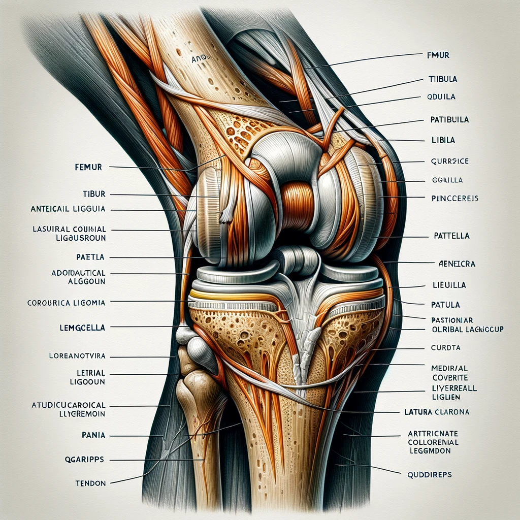
    Cushioning systems

    Cushioned midsoles were one of the first FDF introduced to modern running shoes. They were developed to provide a protective layer, attenuate the shock caused by the collision of the foot with the ground, and reduce local plantar pressure peaks [26]. The cushioning characteristics are modified in the midsole through material and geometry changes.

    Midsole compression stiffness and hardness

    Midsole compression stiffness, also known as hardness, is a fundamental material property that measures the deformation caused by an area load. In the past, midsoles were constructed with uniformly distributed compression stiffness. However, they can now be tailored to individually cushioned midsoles with varying properties at different locations due to the viscoelastic properties of the material [27].

    Twelve of thirty-five articles identified through our literature search considered covariates when analysing the response to differently cushioned midsoles (Fig. 4, Supplementary Table 1). Malisoux et al. considered the runner’s body mass as a covariate [28]. Athletes reported fewer injuries when running in softer midsoles, and lighter runners in hard shoes showed a greater risk of developing an RRI than heavier runners. Three articles investigated the biomechanical response of midsoles with varying hardness during different running speeds. Nigg et al. found that the vertical GRF loading rate increases with speed independent of the cushioning variations, while another study showed unchanged GRF loading rates with footwear of varying cushioning at different speeds, and yet another study showed lower GRF loading rates in harder midsoles with no dependence on running speed [29,30,31]. Running distance or running duration has been considered by five studies [32,33,34,35,36]. None of the studies found significant footwear-by-time/distance interaction effects on vertical GRF loading rates, ground contact times, peak rearfoot eversion angles, and knee flexion angle at initial contact. One article considered the runner’s foot strike pattern as a covariate [37]. Rearfoot strikers reduced the vertical GRF loading rate in a neutrally cushioned shoe, and mid- and forefoot strikers reduced the vertical GRF loading rate in a minimal shoe [37]. We identified one study considering the stiffness of the running surface as a covariate [38]. However, no main and interaction effects were observed in ground contact time and knee flexion angle at touchdown. Another study analysed the effect of surface inclination and midsole cushioning [39]. The authors showed that vertical GRF loading rates are equal when running on different surfaces with either a neutral or a cushioned running shoe. Although studies have examined a variety of covariates, there is much conjecture in the literature regarding their influence on biomechanical measures related to RRI, and no conclusive evidence to suggest that any one covariate is more important than another.

    When considering the effects of midsole hardness on BRFs without considering covariates, five studies found reduced peak rearfoot eversion in harder midsoles than in softer midsoles [40,41,42,43,44]. However, four studies found unchanged peak rearfoot eversion angles when running in soft and hard midsoles [34, 45,46,47]. Four studies reported that different midsole hardness could not systematically affect the rearfoot eversion range of motion [40, 44, 45, 48]. In contrast, one study found a reduction in the rearfoot eversion range of motion in hard midsoles [47], and another study found that the range of motion of the rearfoot was lower when runners were running in softer midsoles [49]. Conflicting findings were also observed for the rearfoot inversion angle at initial ground contact. One study found a reduction in rearfoot inversion when running in soft midsoles [40], and others found reduced inversion angles when running in hard midsoles [40, 48]. Conflicting findings have also been reported for the vertical GRF loading rate. Some studies found an increased vertical GRF loading rate in more cushioned than less cushioned shoes [29, 34]. Other studies found no effects of cushioning [46, 50,51,52], while others found decreased vertical GRF loading rate in cushioned shoes [41]. Only a few studies were identified addressing the effects of different cushioning characteristics on BRFs at more proximal joints. One article’s qualitative data showed that the knee abduction angle during the stance phase was reduced when running in softer than harder midsoles [53]. In contrast, another study found lower peak knee abduction angles when the midsole was manufactured with harder material [47]. A study by Malisoux and colleagues found that both soft and hard midsoles did not change peak hip abduction angles and moments and peak hip internal rotation angles [45]. When considering ground contact time as BRF for PFPS, most studies found no effect of midsole cushioning [29, 36, 38, 45, 49, 51, 54,55,56]. Overall, studies analyzing BRFs without considering covariates, resulted in inconsistent and conflicting findings. Interestingly, the footwear comfort perception reported by participants tends to be higher in regions where softer material is allocated than in those with harder materials [42, 49, 57, 58].

    In summary, the current literature suggests that the midsole hardness can potentially reduce the overall injury risk when adjusted to the runner’s body mass. Reduction in vertical GRF loading rates and subsequent minimizing PF injury risk could be achieved by individualising midsole cushioning to the runner’s foot strike pattern. Specifically, rearfoot strikers might benefit from cushioned shoes, while fore- and midfoot strikers could find minimal shoes advantageous. The lower vertical GRF loading rates observed in neutral shoes compared to cushioned shoes when running downhill suggest that customised midsole cushioning tailored to a runner’s training terrain could benefit runners with a PF history. Based on the limited literature, surface stiffness, running distance, and fatigue might be less important when individualising midsole hardness. Harder midsoles can reduce BRFs associated with MTSS, TSF, AT (rearfoot eversion movement), and ITBS (ground contact times). Indications that different shoe cushioning may alter vertical GRF loading rates are contradictory, and BRFs at more proximal joints have not been well studied.

    Midsole geometry

    Running footwear is often designed with a height gradient from the heel to the forefoot. Running shoes are defined by their heel and forefoot heights, with the difference between the two known as the heel-toe drop. Unlike neutral or motion-control shoes, minimal footwear is typically designed with a lower heel-toe drop. An increase in footwear minimalism generally shifts the foot strike pattern of rearfoot strikers towards a mid- or forefoot strike pattern, and it is further assumed to reduce impact loading parameters [59, 60].

    We identified eighteen articles investigating the effects of geometrical midsole modifications matching our inclusion criteria (Fig. 4, Supplementary Table 2). Out of the eighteen articles, nine accounted for a covariate. The runner’s experience was considered in one article [61]. During a six-month follow-up, it was shown that occasional runners (< 6 months running experience) had reduced injury rates, and recreational runners (≥ 6 months running experience) had increased injury rates when running in footwear with lower heel-toe drop. A subset of this data demonstrated that midsoles with different heel-toe drops were not able to reduce peak rearfoot eversion angle and ground contact time [62]. However, runners who trained for six months in footwear with higher heel-toe drops increased the peak knee abduction angle. On the contrary, runners who trained for six months in footwear with lower heel-toe drops reduced the peak knee abduction angle. Running surface as a covariate was considered by one study [63]. The researchers found smaller knee flexion angles for larger heel-toe drops when running on a treadmill. However, when running overground, the knee flexion angle was not changed when running in shoes with different heel-toe drops. The authors found that increasing the heel-toe drop led to lower vertical GRF loading rates overground, but decreasing the heel-toe drop reduced vertical GRF loading rates during treadmill running. Different running speeds as a covariate were considered by four articles [64,65,66,67]. One study found no changes in the knee flexion angle at initial contact when running at different speeds in midsoles with different heel-toe drop designs [64]. Another study showed that while ground contact time decreased with increasing speed, increasing the heel-toe drop resulted in increased contact time [65]. Other researchers also showed similar results when systematically altering running speed and heel-toe drop [66]. Running speed did not influence the effects of heel-toe drop modifications on vertical GRF loading rates or time spent in rearfoot eversion [67]. The interaction effects of running time and geometrical midsole modifications were investigated in two studies using the same data set [68, 69]. However, neither of the studies reported interaction effects on included BRFs (rearfoot movement, contact time, and knee flexion angle at initial ground contact). Nevertheless, both studies reported longer ground contact times, lower rearfoot eversion range of motion, and greater knee flexion angles at initial contact in thicker than thinner midsoles.

    Concerning the general effects of midsole geometries on BRFs without considering covariates, most of the included studies have addressed the effect of midsole geometry on GRF parameters. An increase in heel-toe drop has been reported to reduce vertical GRF loading rates [70,71,72,73]. Diverse results have been reported for midsole thickness, for which one study found lower vertical GRF loading rates in thicker than thinner midsoles [74], whereas another study could not identify any differences [75]. Three studies showed that geometrical changes at the midsole do not affect rearfoot inversion at touchdown [68,69,70]. Three articles showed that the knee flexion angle at touchdown remains unchanged independent of geometrical midsole configurations [72, 75, 76]. Only one study collected comfort perception data from fifteen male runners [77]. However, no difference in comfort was observed when the heel-toe drop was systematically altered.

    Summarising the results, individualisation of heel-toe drop based on runner experience may reduce the risk of RRI. Although the underlying biomechanical mechanism remains unknown, a gradual transition from shoes with different heel-to-toe drops may allow adequate adaptation of the biological tissues. Running surfaces can affect the response to heel-toe drop alterations by influencing vertical GRF loading rates and knee flexion angles. Runners with a history of PF training on treadmills may benefit from shoes with a lower heel-toe drop, while those with a history of ITBS may benefit from a higher drop. During fatigue, geometric midsole modifications may not affect rearfoot eversion movement or ground contact times. Thinner midsoles with a lower heel-toe drop may reduce ground contact times, peak rearfoot eversion angle and rearfoot eversion duration. Hence, these modifications might be recommended for runners with a risk or a history of PFPS, TSF, or MTSS. Moreover, thicker midsoles with a higher heel-toe drop might shift BRFs related to AT and PF (rearfoot eversion range of motion and vertical GRF loading rate) to potentially less critical BRF magnitudes.

    Motion control features

    Motion control, also called stability, in footwear refers to how the shoe limits pronation (calcaneal eversion) or supination (calcaneal inversion) during the support phase. Much research has been devoted to FDF that purports to control pronation or eversion motion, motivated by the retrospective observations that increased pronation angle is associated with RRI [10, 78,79,80]. Over the initial period of footwear research, various midsole technologies were designed to increase rearfoot stability, including altering the midsole hardness, location of material inserts, flares, arch support systems, and postings. One of the few identified studies utilized a randomized controlled trial with a six-month follow-up. The findings revealed that recreational runners with a motion control shoe developed fewer RRI than runners receiving a standard running shoe [15]. Interestingly, motion-control shoes’ effectiveness in reducing RRI development was more pronounced for runners with pronated feet, indicating some potential for footwear individualisation.

    Postings

    Postings in athletic footwear incorporate elements with higher material densities in the medial rearfoot region and have been reported to limit rearfoot eversion [81]. Unlike wedges, postings are designed without gradual height differences [82].

    Three of seven articles identified through our literature search considered covariates in their analysis (Fig. 4, Supplementary Table 3). The runner’s age was considered by one article [83]. Medial posts effectively reduced the amount of rearfoot eversion in older compared to younger female runners, while vertical GRF loading rates, peak knee abduction moments, and peak knee internal rotation angles remained unchanged. When considering the runners’ fatigue as a covariate, two articles found that rearfoot eversion movement (peak and range of motion) was lower when running in a medially posted than in a neutral running shoe when the runner’s fatigue increased [84, 85].

    When not considering covariates or subgroups of runners, medial postings can reduce peak rearfoot eversion angles and eversion range of motion [86, 87]. Peak knee internal rotation angles are reported to be reduced when running in footwear with medial postings [83, 88]. However, footwear with postings might increase peak hip abduction moments [89]. Diverse results were found for vertical GRF loading rates. One study found lower vertical GRF loading rates in midsoles without medial posts [87], and another found unchanged vertical GRF loading rates in shoes with and without postings [83]. Some runners have perceived the harder posting material without transitions as uncomfortable, potentially resulting in unwanted changes in their biomechanics [88].

    In summary, older female runners with a history of TSF and MTSS might reduce rearfoot eversion in shoes with postings. However, medial posts do not seem to affect the risk of developing PF independent of the runners’ age since changes in vertical GRF loading rates were not observable. Based on the limited literature, posted midsoles may help minimise BRFs (rearfoot eversion movement) associated with AT, MTSS, or TSF as the runners’ fatigue state increases. The limited literature suggests that individualised postings can help runners with a history of AT, MTSS, TSF, or ITBS to reduce biomechanical risk factors. Since postings might increase vertical GRF loading rates, caution needs to be taken by runners with a history of PF.

    Wedges

    Wedges are sloped orthotic inserts, typically with mediolateral elevation, designed to increase foot stability. Mediolateral elevation under different loading conditions can be achieved by incorporating materials with different mechanical properties at distinguished locations of the wedge [90].

    Three out of the ten articles identified in the literature search included a covariate in their analysis (Fig. 4, Supplementary Table 4). One study considered running duration (0–30 min) as a covariate [91]. Independent of the running duration, medially wedged insoles produced lower knee abduction angular impulses than laterally wedged insoles. Another study considered different standing calcaneal angles and injury history as covariates [92]. However, wearing differently wedged insoles showed no effect on female runners’ 3D knee and hip kinematics. Anterior knee pain as a covariate and the response to differently wedged insoles were considered by one article [93]. Independent of knee pain, running in medially wedged insoles reduced maximal rearfoot eversion and range of motion compared to running in footwear without wedges. None of the studies personalised the wedges to the runner’s individual foot anatomy; instead, they used pre-fabricated wedges, which may have confounded these results.

    Seven articles were identified investigating the effect of wedged insoles on BRFs without considering covariates. In a study in which the wedges were customised to individual dynamic barefoot plantar pressure data, all but two subjects reduced peak rearfoot eversion angles compared to footwear without wedges [94]. This finding suggests that wedges bear high potential when individualised to foot pressure mapping. Pre-fabricated medial wedges have proven effective in decreasing maximal rearfoot eversion angles and eversion range of motion [94,95,96,97]. When comparing footwear with and without wedges, non-systematic changes in vertical GRF loading rates and knee abduction angular impulse have been reported [95, 96, 98, 94, 99, 100]. When the mediolateral elevation was systematically altered, no perceived comfort and stability changes were reported [95]. Moreover, neither medially nor laterally wedged insoles were able to relieve runners of patellofemoral pain [99]. One study introduced forefoot wedges with systematic changes in elevation; however, no changes in ground contact times were reported [101].

    In summary, the response to medially wedged insoles is independent for shorter running durations (< 30 min) but may help runners with a history of PFPS to minimise knee abduction angular impulses; however, the effect for longer running durations (> 30 min) remains unknown. The limited literature shows that joint alignments, injury history, and knee pain are less relevant covariates when individualising wedged insoles. Medially wedged insoles might sufficiently limit rearfoot eversion movement and support runners with a history of AT, TSF, and MTSS to reduce reinjury. To attenuate vertical GRF loading rates, runners with a history of PF might refer to other FDF modifications to reduce the overuse injury risk.

    Arch support systems

    Arch support systems help the foot by storing and releasing elastic energy and preventing arch collapse during high loading [102]. Foot arches can be classified as flat/low, normal, or high [103]. Within the three groups, low-arched runners may exhibit greater eversion movement and velocity than high-arched runners [104]. Arch support systems can be integrated into the midsole or achieved through custom-made insoles shaped into the foot arch [105].

    Our review found seven articles, four of which examined the effect of arch support systems on running biomechanics with a covariate (Fig. 4, Supplementary Table 5). Two studies used foot arch height as the covariate, and they found that high-arched runners reduced vertical GRF loading rates in a shoe without arch support, while low-arched runners reduced loading rates in a shoe with arch support. However, both foot arch types experienced reduced rearfoot eversion in a motion control shoe [106]. With a subset of this data, no changes in rearfoot eversion movements for runners with different foot arch types were observed when running in shoes with and without arch support systems during a prolonged run [107]. One article accounted for the runner’s foot strike pattern and found that rearfoot strikers decreased ground contact time in footwear without arch support [108]. In contrast, forefoot strikers reduced contact time in a shoe with arch support [108]. The same study found that forefoot strikers in minimal footwear reduced vertical GRF loading rates, but rearfoot strikers did not. Furthermore, training for three months in footwear with a custom-made arch support system reduced rearfoot eversion [105].

    We identified three articles investigating the effect of arch support systems on BRFs without considering covariates. A study involving female runners found no effect of arch support on vertical GRF loading rates, peak rearfoot eversion angles, and peak femur rotation angles [46]. Another study also found unchanged rearfoot eversion movements (peak eversion angle and rearfoot inversion at initial ground contact) and knee abduction angles when runners with AT symptoms ran in footwear with and without arch support [109]. Although BRFs were unchanged, a 92% relief of AT symptoms was reported when wearing an insole with custom-made arch support. Finally, one study found unchanged ground contact times when running in midsoles with 20 mm and 24 mm high arch support elevations [101].

    The limited literature suggests that arch support systems can potentially reduce BRFs for runners with different arch heights and a history of PF. Runner’s foot strike pattern might be considered when individualising arch support systems. When individualising arch support systems to minimise BRFs associated with PFPS (ground contact time) and PF (vertical GRF loading rate), forefoot strikers might benefit from less arch support than rearfoot strikers. Moreover, customised arch support systems enhance comfort perception without changes in peak knee abduction angles and vertical GRF loading rates. Arch support might reduce rearfoot eversion movements and thus have the potential for individualisation for runners with a history of AT, TSF, and MTSS. BRFs related to ITBS (peak femur rotation angle and peak knee abduction angles) seem to change marginally and unsystematically with arch support.

    Heel flares

    Flares can be described as a projection of the midsole and outsole extending beyond the upper [25]. Flares can be placed medially or laterally along the outline of the midsole and were introduced to alter the rearfoot eversion angle, thus increasing foot stability by changing the ankle joint moment arm [110,111,112].

    After examining all articles, we identified five matching our inclusion criteria (Fig. 4, Supplementary Table 6). None of these articles investigated the effect of a covariate.

    Concerning BRFs, one study altered the medial heel flare from 0° to 15°, and 30°. The 2D video-based analysis indicated higher rearfoot eversion movement in footwear without heel flares [81]. In the same study, runners running in shoes with the most extreme medial heel flare modification had, on average, lower rearfoot eversion range of motion than in shoes with less or without heel flares. These findings were supported by other research showing that footwear with heel flares can reduce the magnitude of rearfoot eversion across the entire stance phase but does not seem to reduce vertical GRF loading rates [110, 112, 113]. On the contrary, one study with only five runners did not show that rearfoot eversion movement (at initial ground contact, peak, and range of motion) changes when running in footwear with different heel flares [111]. From a perception perspective, heel flares can improve perceived foot stability [112].

    None of the articles considered covariates (e.g., foot strike pattern), highlighting future research potential. Although we found diverse results regarding rearfoot eversion movement, midsoles with heel flares might reduce BRFs linked to AT, TSF, or MTSS. Based on the very limited body of literature, midsoles with heel flares are insufficient for reducing vertical GRF loading rates, and individualised heel flares may not target runners with a history of PF.

    Crash pads

    Crash pads are elements incorporated into the posterior-lateral midsole using softer foams, segmented geometries, air pockets, or gel-filled patches. Crash pads in the rearfoot area aim to attenuate the GRF and reduce the GRF’s lever arm to the ankle joint [114].

    After assessing articles for their eligibility, we identified three articles matching our inclusion criteria (Fig. 4, Supplementary Table 7). Out of the three articles, one study considered the fatigue status of female runners as a covariate. As the runners’ fatigue increased, wearing footwear without crash pads increased vertical GRF loading rates compared to the non-fatigue state. However, running in footwear with crash pads maintained consistent vertical GRF loading rates, even as the runners’ fatigue increased. [115]. The same study found no effect of fatigue on the peak free moment amplitude.

    When not considering covariates, two studies found reduced rearfoot inversion angles at touchdown in footwear with smaller compared to larger crash pad dimensions. However, there were no differences in peak rearfoot eversion angles during the stance phase of running and unsystematic changes in vertical GRF loading rates [114, 116]. Crash pad modifications did not affect the peak free moment amplitude, ground contact time, and rearfoot eversion range of motion [114,115,116]. Changes in crash pad dimensions do not seem to influence the runner’s comfort perception [114]. However, they may provide an essential tool for individualisation to tune midsole cushioning properties without increasing stack height which has been shown to increase rearfoot eversion [81].

    Fatigue seems to be a relevant covariate when individualising crash pads to minimise vertical GRF loading rates, thus, might lower the risk of developing PF. However, runners with a history of TSF might need other individualised FDF to lower peak free moment amplitudes. Increasing crash pad height might help runners with plantar fascia complaints by lowering the vertical GRF loading rates. Runners with a history of AT, TSF, or MTSS might benefit from crash pads by reducing rearfoot eversion movement. Surprisingly, although the FDF aimed at attenuating the peak impulse, we have identified only two studies that have analysed vertical GRF loading rate as BRF.

    Other footwear design features

    Rocker

    Rockers in running shoes aim to reduce the strain on the toes, foot, and ankle by altering the midsole’s curvature in the anterior–posterior direction, positioning the apex near the metatarsal heads, and enhancing the midstance-to-push-off transition for a smoother heel-to-toe rolling motion [117].

    Each of the three identified articles considered a covariate in their analysis (Fig. 4, Supplementary Table 8). One study considered running speeds as a covariate. Although running at higher speeds increases the vertical GRF loading rate, no changes in GRF loading rates were observed between shoes with and without rocker [118]. Two studies considered the foot strike pattern and found that a toe spring starting closer to the midfoot reduced pressure in the forefoot compared to a standard rocker placed at 65% of the shoe length [119]. However, runners perceived the traditional rocker as more comfortable. When compared to shoes without rockers, one study found that a rocker shoe reduced ground contact time but did not affect knee flexion angles at initial ground contact [120].

    The number of studies addressing injury-specific BRFs and the effects of rocker designs is limited. Rockers involve different levels of FDF (stack height, cushioning), and therefore it is difficult to assign a specific feature to a specific BRF. More research is needed to understand if certain covariates can cause a specific change in BRFs and how different FDFs that combine a rocker design need to be tuned for individualisation.

    Outsole profile

    A shoe’s outsole interacts with the running surface and requires attributes like traction, waterproofness, durability, and puncture resistance [121]. Material robustness might be related to running shoe comfort, and high traction might increase free moment amplitudes associated with TSF [122].

    After assessing all articles for eligibility, we could not identify any articles matching our predefined inclusion criteria (Fig. 4). Future studies might use wearable sensors or markerless tracking systems to analyse runners wearing shoes with different outsole profiles on natural surfaces.

    Flex grooves

    Flex grooves and zones are included in outsoles and midsoles to enhance flexibility, facilitating metatarsophalangeal joint movement and shock absorption. Their placement is essential for the joint’s variable axis and should be individualised based on foot measurements. Recent 3D measurements indicate significant variation, underscoring the need for personalized flexible zones [123].

    Our literature search identified one article matching our predefined inclusion criteria (Fig. 4, Supplementary Table 9). This article considered running speed as a covariate. In this study, the midsole flexibility was altered by cuts with different orientations at the heel region. Although interaction effects were only marginal when jogging or running in footwear with different groove designs, a 10% lower vertical GRF loading rate was observed in the midsole with grooves compared to the midsoles without grooves at the rearfoot [124]. Interestingly, footwear with greater flexibility is perceived as more comfortable than midsoles with less flexibility [125, 126].

    While there is limited research on the impact of flex grooves on relevant BRFs for common RRI, one identified article found that they can reduce vertical GRF loading rates, suggesting that flex grooves may be customised for runners with PF.

    Longitudinal bending stiffness

    The longitudinal bending stiffness can impact the running economy by optimising energy return and kinematics of the metatarsal joint and force application [127,128,129,130,131]. The bending stiffness can be modified by adding reinforcement materials or changing the geometry of stiff midsole compounds. The optimal bending stiffness depends on factors such as running speed and body weight [128, 132].

    Our literature search identified eleven articles, of which four accounted for a covariate (Fig. 4, Supplementary Table 10). All four articles considered running speed as a covariate. None of these articles found a significant interaction effect on BRFs when running in footwear with different longitudinal bending stiffness at different running speeds [133,134,135,136]. Independent of running speed, studies reported reduced ground contact times when running in shoes with lower bending stiffness, while one article found unchanged ground contact times [136].

    When not considering covariates, three studies found no changes in the GRF braking impulse when running in shoes with different bending stiffness [135, 137, 138]. On the contrary, a reduction in GRF braking impulse in footwear with higher bending stiffness was found in one study [134]. Eight articles found a reduction in the ground contact time [130, 133,134,135, 137,138,139], and two found unchanged ground contact times [134, 140] when running in midsoles with lower bending stiffness. Although studies found lower vertical GRF loading rates [140] and increased comfort perception [135] when athletes ran in more flexible than stiffer midsoles, the relationship between BRFs and injury development when altering the longitudinal bending stiffness has not been sufficiently studied yet, but first studies have evolved showing that bones stress injuries might increase when switching to footwear with carbon fibre plates [18].

    The limited body of literature suggests that fitting longitudinal bending stiffness to the runner’s needs may help with treating PFPS. While reduced bending stiffness can reduce ground contact time, higher stiffness can reduce ground reaction force braking impulse. However, injury prevention and reinjury risk minimisation under the light of different longitudinal bending stiffness has been insufficiently investigated. Furthermore, flexible midsoles with lower longitudinal bending stiffness might reduce vertical GRF loading rates and potentially help runners with a history of PF.

    The upper

    The running shoe upper is comprised of a textile fabric and lacing system that couple the foot and shoe, with reinforcement materials used for stability and breathability. An optimal fit depends on individual foot morphology, while insufficient coupling can negate benefits from other design features. Moreover, excessive pressure can affect comfort by restricting blood supply, making individualisation important [141]. Since foot dimensions differ across sexes, ages, and ethnic origins, individualised upper bears great potential for individualisation [142].

    Upper fabric

    Our systematic literature search identified two articles investigating the effect of different upper modifications (Fig. 4, Supplementary Table 11). None of the articles considered covariates [53, 143].

    The data indicates that a soft-sewed structured fabric reduces knee abduction angles and vertical GRF loading rates compared to a minimalist heat fusion fabric. Furthermore, the ground contact time was reduced when running in minimalist heat fusion fabric.

    The current body of literature is insufficient to give recommendations for upper individualisation concerning the reduction of BRFs. Based on the limited results, upper materials might be individualised to the runner’s preference.

    Lacing

    Five articles have investigated the effect of lacing on the lower extremity joint biomechanics or subjective comfort perception (Fig. 4, Supplementary Table 12).

    One of five studies considered the runner’s experience as a covariate. The researchers found that low-level runners perceived an irregularly (skipping eyelets) laced running shoe as more stable and comfortable than high-level runners who preferred a regular high and tight lacing pattern [144].

    We identified four studies analysing BRFs without accounting for covariates. According to a study, running shoes with traditional lacing and elastic upper material were perceived as more comfortable than footwear without lacing [145]. When running in shoes with various lacings, two studies found no significant difference in the rearfoot eversion angle at initial contact [145, 146]. The same studies found a reduction in the peak rearfoot eversion angle when running in traditionally laced shoes compared to those without traditional lacing. However, another study systematically changed lacing patterns and could not find any differences in the peak rearfoot eversion angle [147]. Different types of lacing patterns, particularly high- and tightly-laced shoes, have been shown to reduce vertical GRF loading rate at the cost of comfort [144, 148].

    Studies analysing BRFs and considering relevant covariates, e.g., foot shape, are required in the future. Notably, no studies have measured the foot-shoe coupling or the relative movement of the foot within the shoe, highlighting the potential for future research to determine individualised fits and their interactions with other FDF. Since peak rearfoot eversion angles and vertical GRF loading rates are reported to be lower when running in tightly and high-laced shoes, runners with a history of MTSS and TSF might target individualised lacing systems.

    Source link

  • Osteoarthritis: Causes, Symptoms, and Treatment

    Osteoarthritis: Causes, Symptoms, and Treatment

    As we age, our bodies go through various changes, and one of the most common conditions that develop is osteoarthritis. Osteoarthritis is a type of arthritis that affects millions of people worldwide, and it occurs when the protective cartilage that cushions the ends of our bones wears down over time. Although it can damage any joint, the disorder most commonly affects joints in our hands, knees, hips, and spine.

    image 99

    Understanding Osteoarthritis is crucial for anyone who may be experiencing joint pain or stiffness. This condition is often referred to as “wear and tear” arthritis, and it occurs when the cartilage within a joint begins to break down, causing the underlying bone to change. These changes usually develop slowly and get worse over time, leading to pain, stiffness, and difficulty moving the joint.

    Key Takeaways

    • Osteoarthritis is a type of arthritis that affects millions of people worldwide, and it occurs when the protective cartilage that cushions the ends of our bones wears down over time.
    • This condition is often referred to as “wear and tear” arthritis, and it occurs when the cartilage within a joint begins to break down, causing the underlying bone to change.
    • Symptoms of osteoarthritis include joint pain, stiffness, and difficulty moving the joint.

    Understanding Osteoarthritis

    Osteoarthritis is a degenerative joint disease that affects millions of people worldwide. It is the most common form of arthritis, and it occurs when the protective cartilage that cushions the ends of the bones wears down over time. This can cause discomfort when moving the joint, and it can lead to stiffness and pain.

    Osteoarthritis is also known as “wear and tear” arthritis or degenerative joint disease. It can damage any joint in the body, but it most commonly affects joints in the hands, knees, hips, and spine. In some cases, osteoarthritis can also affect the neck, shoulders, and ankles.

    The main cause of osteoarthritis is the breakdown of joint cartilage, which can be caused by a combination of factors such as heredity, obesity, injury, and overuse. Cartilage is a tough, flexible tissue that covers the ends of bones where they meet to form a joint. It acts as a cushion and helps to absorb shock when the joint is used.

    When cartilage breaks down, the underlying bone can also change, leading to the development of osteoarthritis. The changes usually develop slowly and get worse over time. As the condition progresses, the joint may become painful and stiff, making it difficult to move.

    There is no cure for osteoarthritis, but there are several treatments available to help manage the symptoms. These include medications, physical therapy, and surgery. In some cases, lifestyle changes such as losing weight or exercising regularly can also help to reduce the symptoms of osteoarthritis.

    In conclusion, osteoarthritis is a common joint disease that affects millions of people worldwide. It is caused by the breakdown of joint cartilage, and it can lead to pain and stiffness in the affected joints. While there is no cure for osteoarthritis, there are several treatments available to help manage the symptoms and improve quality of life.

    Symptoms of Osteoarthritis

    Osteoarthritis is a type of arthritis that affects the joints. It is a degenerative disease that causes the cartilage in the joints to break down, leading to pain, stiffness, and other symptoms. In this section, we will discuss the most common symptoms of osteoarthritis.

    Pain and Stiffness

    The most common symptoms of osteoarthritis are pain and stiffness in the affected joints. The pain may be mild or severe and can be felt during or after movement. Joint stiffness is most noticeable in the morning or after being inactive for a period of time. The affected joint may also feel tender when light pressure is applied to it.

    Decreased Range of Motion

    Osteoarthritis can also cause a decreased range of motion in the affected joint. This means that you may not be able to move the joint as freely as you used to. This can make it difficult to perform daily tasks, such as getting dressed or reaching for objects.

    Swelling and Tenderness

    Swelling and tenderness are also common symptoms of osteoarthritis. The affected joint may become swollen and tender to the touch. This can make it difficult to move the joint and can cause discomfort.

    Bone Spurs

    Osteoarthritis can also cause the growth of bone spurs around the affected joint. These bone spurs can cause additional pain and can make it difficult to move the joint.

    Complications

    Osteoarthritis can lead to complications, such as disability and falls. In some cases, the pain and stiffness caused by osteoarthritis can make it difficult to perform daily tasks, such as walking or climbing stairs. This can lead to disability and a decreased quality of life. Falls are also a common complication of osteoarthritis, as the pain and stiffness can cause a loss of balance.

    Joint Damage

    Osteoarthritis can also cause damage to the joint over time. The cartilage in the affected joint can wear down, leading to bone-on-bone contact. This can cause further pain and damage to the joint.

    Other Symptoms

    Other symptoms of osteoarthritis may include joint inflammation, a cracking or popping sound in the joint, and the formation of cysts around the joint.

    Risk Factors

    There are several risk factors that can increase your likelihood of developing osteoarthritis. These include age, obesity, joint injuries, and genetics.

    In summary, osteoarthritis is a degenerative disease that affects the joints. The most common symptoms of osteoarthritis include pain, stiffness, decreased range of motion, swelling, tenderness, bone spurs, complications, joint damage, and other symptoms. If you are experiencing any of these symptoms, it is important to seek medical attention to receive an accurate diagnosis and appropriate treatment.

    Frequently Asked Questions

    image 99

    What are the risk factors for developing osteoarthritis?

    We can develop osteoarthritis due to various factors such as age, obesity, previous injuries, and genetics. Women are also more likely to develop osteoarthritis than men.

    What are the different types of osteoarthritis?

    There are different types of osteoarthritis that can affect different parts of the body. Some of the common types include knee osteoarthritis, hip osteoarthritis, and hand osteoarthritis.

    What are some common medications used to treat osteoarthritis?

    There are various medications that can help manage osteoarthritis symptoms such as pain and inflammation. Some common medications include acetaminophen, nonsteroidal anti-inflammatory drugs (NSAIDs), and corticosteroids.

    What are some ways to prevent osteoarthritis?

    While we cannot prevent osteoarthritis entirely, we can take steps to reduce our risk of developing it. Maintaining a healthy weight, staying active, and avoiding injuries can help prevent osteoarthritis.

    How does osteoarthritis affect the body?

    Osteoarthritis can cause pain, stiffness, and decreased range of motion in the affected joints. It can also lead to the breakdown of joint cartilage and the development of bone spurs.

    What are some strategies to manage osteoarthritis pain?

    There are various strategies that can help manage osteoarthritis pain such as exercise, physical therapy, and hot/cold therapy. We can also use assistive devices like braces or canes to help reduce joint stress.

    Add Tables

    image 99

    We can use tables to organize and present information about osteoarthritis in a clear and concise manner. Here are some examples of tables that can be useful:

    Table 1: Common Symptoms of Osteoarthritis

    SymptomDescription
    PainPain in the affected joint, especially during or after movement.
    StiffnessStiffness in the affected joint, especially after periods of inactivity.
    SwellingSwelling and tenderness in the affected joint.
    Cracking or popping soundsCracking or popping sounds when moving the affected joint.
    Limited range of motionDifficulty moving the affected joint through its full range of motion.

    Table 2: Risk Factors for Osteoarthritis

    Risk FactorDescription
    AgeThe risk of developing osteoarthritis increases with age.
    GenderWomen are more likely to develop osteoarthritis than men.
    ObesityExcess weight puts extra stress on the joints, increasing the risk of osteoarthritis.
    Joint injuriesPrevious joint injuries or surgeries can increase the risk of osteoarthritis.
    GeneticsOsteoarthritis may run in families.

    Table 3: Treatment Options for Osteoarthritis

    TreatmentDescription
    MedicationsPain relievers, nonsteroidal anti-inflammatory drugs (NSAIDs), and corticosteroids can help manage pain and inflammation.
    Physical therapyExercises and stretches can help improve joint flexibility and strength.
    Assistive devicesBraces, canes, and other devices can help take pressure off the affected joint.
    SurgeryJoint replacement surgery may be necessary in severe cases.

    By using tables, we can easily compare and contrast different symptoms, risk factors, and treatment options for osteoarthritis. This can help us make informed decisions about managing this common joint condition.

  • Knee Anatomy and Functions And the Key Components of Your Knee

    Knee Anatomy and Functions And the Key Components of Your Knee

    As a physical therapist, I have seen many patients with knee injuries and conditions. Understanding the anatomy and function of the knee joint is essential to prevent injuries, manage pain, and recover from surgery. In this article, I will explain the basic structure of the knee, the role of ligaments and tendons, and the muscles responsible for movement.

    v2 2i6y0

    The knee joint is the largest joint in the body and connects the thigh bone (femur) to the shin bone (tibia). The kneecap (patella) is a small bone that sits in front of the knee joint and protects the joint. The knee joint is a hinge joint that allows for flexion and extension of the leg. It also has some rotational movement, which is important for activities such as walking and running.

    The knee joint is supported by several ligaments and tendons. Ligaments are tough bands of tissue that connect bones to bones, while tendons connect muscles to bones. The ligaments and tendons around the knee joint work together to provide stability and support to the joint. In the next section, I will discuss the role of these structures in more detail.

    Key Takeaways

    • The knee joint is the largest joint in the body and allows for flexion, extension, and some rotational movement.
    • The knee joint is supported by ligaments and tendons that provide stability and support to the joint.
    • Understanding the basic structure and function of the knee joint is essential for preventing injuries, managing pain, and recovering from surgery.

    Basic Structure of the Knee

    As the largest joint in the body, the knee is a complex structure that allows us to walk, run, jump, and perform other physical activities. It is a synovial joint, meaning it contains a fluid-filled capsule that lubricates the joint and reduces friction during movement.

    Bones and Joints

    The knee joint is formed by the articulation of three bones: the femur, tibia, and patella. The femur, or thigh bone, is the longest bone in the body and forms the upper part of the knee joint. The tibia, or shin bone, is the larger of the two bones in the lower leg and forms the lower part of the knee joint. The fibula is the smaller bone in the lower leg and is not directly involved in the knee joint.

    The knee joint is actually two joints in one: the tibiofemoral joint and the patellofemoral joint. The tibiofemoral joint is the main joint between the femur and tibia, while the patellofemoral joint is the joint between the patella and the femur.

    Patella

    The patella, or kneecap, is a sesamoid bone that sits in front of the knee joint and helps to protect the knee and improve the leverage of the quadriceps muscle. The patella is unique in that it is not directly attached to any other bone in the body. Instead, it is connected to the quadriceps tendon and the patellar ligament.

    In summary, the knee joint is a complex structure that is formed by the articulation of three bones: the femur, tibia, and patella. The knee joint is actually two joints in one: the tibiofemoral joint and the patellofemoral joint. The patella, or kneecap, is a sesamoid bone that sits in front of the knee joint and helps to protect the knee and improve the leverage of the quadriceps muscle.

    Knee Ligaments and Tendons

    The knee joint is stabilized and supported by a network of ligaments and tendons. These structures work together to provide strength and stability to the knee joint, allowing us to perform various activities such as walking, running, and jumping.

    Collateral Ligaments

    The collateral ligaments are located on the sides of the knee joint. The medial collateral ligament (MCL) is located on the inner side of the knee, while the lateral collateral ligament (LCL) is found on the outer side. These ligaments help to prevent excessive side-to-side movement of the knee joint.

    Cruciate Ligaments

    The cruciate ligaments are located inside the knee joint and cross each other to form an “X” shape. The anterior cruciate ligament (ACL) is located in the front of the knee, while the posterior cruciate ligament (PCL) is located at the back. These ligaments help to prevent excessive forward and backward movement of the knee joint.

    Tendons

    The patellar tendon and quadriceps tendon are two important tendons that are located in the knee joint. The patellar tendon connects the patella (kneecap) to the tibia (shinbone), while the quadriceps tendon connects the quadriceps muscle to the patella. These tendons help to provide stability to the knee joint and allow us to perform various movements such as jumping and climbing stairs.

    In summary, the knee joint is stabilized and supported by a network of ligaments and tendons. The collateral ligaments help to prevent excessive side-to-side movement of the knee joint, while the cruciate ligaments help to prevent excessive forward and backward movement. The patellar and quadriceps tendons provide stability to the knee joint and allow us to perform various movements.

    Muscles and Movement of the Knee

    v2 2i6yj oazrc

    The knee joint is a hinge joint that allows for flexion and extension of the lower leg. The movement of the knee is controlled by a complex system of muscles, tendons, and ligaments that work together to stabilize and move the joint.

    9d23c210 985b 4333 a7fa ab54f1b02ac7

    Anatomical illustration of the human knee joint, showing all the components along with their names. This includes the bones, ligaments, cartilage, and tendons, with clear labels for each part such as the femur, tibia, fibula, patella, meniscus, and various ligaments and tendons.

    Quadriceps Muscles

    The quadriceps muscles are a group of four muscles located on the front of the thigh that work together to extend the knee joint. These muscles include the rectus femoris, vastus lateralis, vastus intermedius, and vastus medialis. The rectus femoris also works to flex the hip joint.

    Hamstring Muscles

    The hamstring muscles are a group of three muscles located on the back of the thigh that work together to flex the knee joint. These muscles include the biceps femoris, semitendinosus, and semimembranosus. The gracilis muscle also works to flex the knee joint.

    Movements of the Knee

    The knee joint allows for a variety of movements, including flexion, extension, lateral rotation, and medial rotation. Flexion is the movement that brings the heel towards the buttocks, while extension is the movement that straightens the leg. Lateral rotation is the movement that turns the lower leg outward, while medial rotation is the movement that turns the lower leg inward.

    The popliteus muscle is a small muscle located at the back of the knee joint that works to unlock the knee joint during flexion. The flexors and extensors of the knee joint work together to stabilize the joint during movement.

    The tibiofemoral joint is the main joint of the knee, while the patellofemoral joint is the joint between the kneecap and the femur. The articularis genus muscle is a small muscle located at the front of the knee joint that works to pull the synovial membrane of the joint upward during extension.

    The gastrocnemius and plantaris muscles are located at the back of the knee joint and work to plantarflex the ankle joint. Instability of the knee joint can lead to pain and difficulty with activities such as running and walking.

    Knee Injuries and Treatment

    v2 2i6yr 6vmiv

    Common Knee Injuries

    The knee is a complex joint that is susceptible to a variety of injuries. Some common knee injuries include:

    • Anterior Cruciate Ligament (ACL) Tear: This is a common injury that occurs when the ACL is stretched or torn. It can happen during sports that involve sudden stops or changes in direction, such as basketball or soccer. Symptoms include pain, swelling, and instability in the knee.
    • Meniscus Tear: The meniscus is a piece of cartilage that helps cushion the knee joint. A tear can occur when the knee is twisted or bent forcefully. Symptoms include pain, swelling, and difficulty moving the knee.
    • Patellar Tendinitis: This is an injury to the tendon that connects the kneecap to the shinbone. It is often caused by overuse and can result in pain and swelling in the front of the knee.

    how to prevent knee injuries and arthritis

    SectionDescription
    1.Maintain a healthy weight to reduce stress on the knee joint.
    2.Wear appropriate shoes for the activity you are doing.
    3.Warm up before exercising to increase blood flow to the muscles.
    4.Use proper technique when exercising to avoid unnecessary stress on the knee joint.
    5.Incorporate exercises that strengthen the muscles around the knee joint.
    6.Avoid activities that put excessive stress on the knee joint, such as jumping or running on hard surfaces.
    7.Take breaks during activities to rest and stretch the knee joint.
    8.Use knee pads or braces for added support during high-impact activities.
    9.Stay hydrated to help keep the joints lubricated.
    10.Consult with a healthcare professional before starting a new exercise program.

    Treatment and Rehabilitation

    Treatment for knee injuries varies depending on the severity and type of injury. Some common treatments include:

    • Rest: Resting the knee and avoiding activities that aggravate the injury can help reduce pain and swelling.
    • Ice: Applying ice to the knee can help reduce pain and swelling. Ice should be applied for 15-20 minutes at a time, several times a day.
    • Physical therapy: Physical therapy can help strengthen the muscles around the knee joint and improve flexibility and range of motion.
    • Medication: Over-the-counter pain relievers, such as ibuprofen or acetaminophen, can help reduce pain and swelling.

    In some cases, surgery may be necessary to repair a knee injury. Rehabilitation after surgery may include physical therapy and exercises to help regain strength and range of motion.

    Overall, it is important to take steps to prevent knee injuries and to seek treatment promptly if an injury does occur. With proper care and treatment, many knee injuries can be successfully treated, allowing individuals to return to their normal activities.

    Frequently Asked Questions

    v2 2i6yz bljme

    What is the knee joint called?

    The knee joint is a synovial joint that connects three bones: the femur, tibia, and patella. It is a complex hinge joint composed of two articulations: the tibiofemoral joint and patellofemoral joint.

    Is the knee a hinge joint?

    Yes, the knee is a hinge joint. It allows for flexion and extension, as well as a small degree of medial and lateral rotation.

    What are the 3 most commonly injured knee structures?

    The three most commonly injured knee structures are the anterior cruciate ligament (ACL), medial collateral ligament (MCL), and meniscus.

    What are the major anatomical features of the knee?

    The major anatomical features of the knee include bones (femur, tibia, and patella), cartilage, ligaments, tendons, and muscles.

    What is the functional anatomy of the knee?

    The knee joint is responsible for weight-bearing and movement, allowing for flexion and extension, as well as a small degree of medial and lateral rotation. The major muscles involved in knee movement include the quadriceps, hamstrings, and calf muscles.

    What are the symptoms of a torn ligament in your knee?

    Symptoms of a torn ligament in your knee include pain, swelling, instability, and difficulty bearing weight. Depending on the severity of the tear, surgery may be necessary to repair the ligament.

  • 2024-2025 Orthopedic Value-Based Healthcare Research Fellowship

    The Fellow will collaborate with department faculty on research projects related to the design, implementation and dissemination of value-based care models, outcome measurement (including the effect of interventions on patient-reported outcomes), costs of care, impact of psychosocial aspects in conditions to the musculoskeletal system, advances in shared decision-making and patient and caregiver engagement, and the adoption of digital health solutions.

    Faculty

    The Fellow will work directly with several leaders in healthcare innovation, including:

    • Kevin Bozic, MD, MBA, professor and chairman of the Department of Surgery and Perioperative Care
    • David Ring, MD, PhD, Associate Dean of Comprehensive Care; Professor of Surgery and Psychiatry
    • Karl Koenig, MD, MS, associate professor and executive director of the Musculoskeletal Institute
    • Prakash Jayakumar, MBBS, PhD, Assistant Professor and Director of Value-Based Healthcare and Outcomes Measurement

    Key Responsibilities and Fellowship Functions

    1. Research: Core research tasks include the development of research protocols, administration of Institutional Review Board (IRB) applications, patient recruitment in the Musculoskeletal Institute’s Lower Extremity Integrated Practice Unit (IPU), data collection and dissemination via abstracts, presentations and manuscripts for multiple publications. The Fellow will serve as a principal investigator in the lower extremity IPU (2.5 days/week) and supervise student and medical student volunteers in patient recruitment and data collection.
    2. Strategy and Operations: Gain valuable insights into the organization’s strategic planning and clinical/operational needs by attending business and management meetings and becoming involved in operational projects as necessary.
    3. Educational opportunities and networking
      1. Immersion program. The Fellow will participate in our quarterly Musculoskeletal Institute Immersion Program in Value-Based Health Care, which accommodates up to two dozen attendees, including a variety of leading stakeholders in orthopedics and beyond from across the country. The program provides comprehensive insight into our value-based model for musculoskeletal care delivery over two half days of presentations and interactive sessions and ample opportunity for networking and in-depth discussions.
      2. Value-based healthcare curriculum. Complete courses related to Value-Based Health Care through an executive education course from UT Austin’s Value Institute for Health and Care.
      3. AAOS Annual Meeting. The department will support the Fellow’s participation in the AAOS Annual Meeting.
      4. Grand rounds. The Fellow may attend Grand Rounds and other clinical conferences.
    4. Clinical: Exposure to clinical patient care and surgical practices by shadowing faculty in clinical activities (operating room, hospital, and outpatient clinic). Although the Fellow will be most integrated with our Musculoskeletal Institute team, opportunities exist for shadowing and research in other surgical specialties within the Department of Surgery and Perioperative Care, based on the Fellow’s interests. He/she will have ample opportunities to interact with orthopedic surgery residents, as well as other residents and students in the department and throughout Dell Medical School.

    A compensation

    Salary: $35,000 plus benefits. Read more about UT Austin’s employment benefits here.

    Eligibility

    Applicant must have been admitted or enrolled in medical school at the time of application.

    Required: strong critical thinking skills; very motivated; can work well independently and in a team; strong written/verbal communication and organizational skills; basic knowledge of statistics.

    Preferred: Demonstrated interest in value-based healthcare; experience in conducting research projects through publication; completion of core clinical internships (e.g., current third-year medical student); experience creating PowerPoint presentations; skills with data analysis; fluent in Spanish.

    Timeline

    The Fellowship runs from July 1, 2024 to June 30, 2025, although there is some flexibility in dates. The deadline for registration is January 15, 2024. Zoom interviews will be conducted with finalists and we plan to fill the position by March 1stst.

    How to apply

    Submit your resume (with USMLE Step 1 and Step 2 scores, if applicable), a one-page cover letter, and a list of three references via our Workday post. If available, include an example of previous work (PowerPoint presentations, published research, data analysis, etc.).

    If you have any questions about the position or the application process, please contact Lauren Uhler, MPH, Associate Director of Research, at This email address is being protected from spambots. You need JavaScript enabled to view it..

    Learn more

    For more information, visit our website https://dellmed.utexas.edu/education/academics/additional-training-programs/orthopaedic-value-based-health-care-fellowship

    Source link

  • You Don’t Look Sick – Living with Rheumatoid Arthritis: DAY 5 HAWAI’I

    I woke up early so I could go to the farmers market. I like looking at the crafts. This time I bought a ring. It is a stainless steel brushed silver ring with very thin rainbow colors along each edge. I had the word ‘resilience’ engraved on it. I like it very much. I also gave my niece a birthday present.

    AVvXsEgeFse6oRwaczJTCOcAV3ZD

    Then I went to the Hawaii Tropical Bioreserve and Garden. It is very beautiful. Jungle and flowers and ocean.

    I bought some vegan gelato before heading to an outdoor area with live music and dancing.

    AVvXsEhYkJAtqMRVo dgjH2AL15uAHS9RKn0CsmJHjMF9svMSlP7bwSoFCb3hSWe7Ha o2191n2A2rJTxVwn 7Y2j2CW0MqezMtFzsyximHputFILAvbC7jT j6ybGB1q7Z04K1ervPCGI81oDa3HaWkdi G2pMAZ Xp8aooL2D9m5tfSZahdWTCb2saYFn14

    When I got back to my hotel and opened my door, the hotel cat ran into my room. He wouldn’t leave. I tried to ignore him, use a cord to play with him and feed him. Nothing worked. It took more than an hour before he could leave. I used a paper bag! He wanted to climb in. After I got him out, he climbed onto a table outside my door and tried to knock over a vase. I told him to stop and then he lay down in front of my door and scratched it.

    It was a long night.

    Source link

  • RA patients in remission should continue taking TNF inhibitors to prevent flare-ups, the study found

    RA patients in remission should continue taking TNF inhibitors to prevent flare-ups, the study found

    shutterstock 390538711 6b3c40fdd32742caa54307db3553cab1

    New research at ACR Convergence 2023, the annual meeting of the American College of Rheumatology (ACR), found that patients with rheumatoid arthritis (RA) in sustained remission who stopped taking TNF inhibitors (TNFi) had significantly more flares and lower Boolean 2.0 remission rates compared to those who continued treatment. Boolean 2.0 is a revised definition for evaluating disease activity in RA, classifying more patients as achieving remission than Boolean 1.0. It is endorsed by the American College of Rheumatology and the European Alliance for Associations in Rheumatology (EULAR) (Abstract #L07).

    As more RA patients achieve durable remission, questions remain about the long-term effectiveness of tapering and stopping TNFi treatment. In the randomized, multicenter, noninferiority ARCTIC REWIND trial, Siri Lillegraven, MD, MPH, PhD at Diakonhjemmet Hospital, Oslo, Norway, and colleagues compared the three-year effect of tapering versus stable treatment in RA patients in sustained remission. It follows a trial last year.

    The current study included 92 patients from Norwegian rheumatology centers who were randomized 1:1 to taper off TNF inhibitors until discontinuation or continuation of treatment. During the three-year study period, all received study visits every four months. Patients restarted treatment at the full dose if they experienced a flare, which was defined as loss of remission plus an increase in disease activity score of 0.6 units or more and two or more swollen joints. In lieu of these criteria, a doctor and a patient might agree that a significant flare had occurred. The study also looked at remission status, medication use and serious side effects or complications.

    Of the original 92 patients, 80 (87%) completed three-year follow-up. At the end of the study, 75% of patients in the tapering group experienced a flare, compared to 15% in the stable group. Most of those who experienced a flare were in remission by their next office visit (81% in the taper group and 67% in the stable group), although the taper group had significantly lower Boolean 2.0 remission rates throughout the study.

    Lillegraven says the researchers were “somewhat surprised by the difference in the proportion of patients in ACR/EULAR Boolean remission in the two groups,” noting that “although most patients in the taper group experienced a flare within the first year and the earlier resume treatment at full dose Boolean 2.0 remission rates were significantly lower in the tapering TNFi group than in the stable group throughout the study period.”

    The risk difference for flares observed in this data [-24% over three years] is quite similar to what was observed in the one-year study. That’s a bit surprising, because we might have expected that more patients receiving stable treatment would develop a flare over time, narrowing the difference between the two groups.”


    Siri Lillegraven, MD, MPH, PhD at Diakonhjemmet Hospital, Oslo, Norway

    Lillegraven notes that the study’s open-label design could influence the evaluation of flares, but says that study staff “were continuously instructed on the importance of recording flares similarly in both groups, a pragmatic approach that will improve clinical care reflects, where patients know which treatment they are receiving. received.”

    Lillegraven says her team has many studies planned to better understand how to personalize treatment for RA patients in remission. This includes factors that can help determine which patients should and should not taper off their treatment.

    “We have begun planning a 10-year follow-up of the study to better understand the long-term outcomes of different treatment strategies in RA remission. We are [also] consider studies to better understand patient preferences regarding medication tapering.”

    Shared decision-making is central to any consideration of tapering, she says.

    “The patient should be informed of the risks and benefits of tapering, and the patient’s overall situation should be taken into account before the decision is made. Although the data do not support tapering off TNFi at a group level, factors such as side effects related to the treatment or the patient having a strong preference for tapering will obviously influence such a decision.”

    Source:

    American College of Rheumatology

    Source link

  • John Booth retires as CEO of Spineology Inc.

    John Booth retires as CEO of Spineology Inc.

    Brian Snider appointed new CEO

    SAINT PAUL, Minn., November 9, 2023–(BUSINESS WIRE)–Spineology Inc. (“Spineology” or the “Company”), the leader in ultra-minimally invasive spine surgery, today announced that John Booth will resign from his position as Chief Executive Officer of Spineology, effective November 10, 2023. The Board of Directors has selected Brian Snider as the next Chief Executive Officer, effective November 13, 2023. Mr. Booth will remain with Spineology through 2024 and serve in an advisory role to enable a smooth transition. Mr. Booth will also resign from the Board of Directors, and the Board of Directors has nominated Mr. Snider as Director.

    Snider joins Spineology with nearly two decades of progressive leadership experience in the medical device industry. Most recently, he served as Executive Vice President of Marketing for Alphatec Spine (NASDAQ: ATEC), as a member of the executive leadership team responsible for growing the company’s market capitalization from $20 million to more than $1.2 billion. During his tenure at ATEC, he was responsible for a variety of marketing and product development disciplines. Most recently he was responsible for the Biologics, Cervical and Thoracolumbar business units. Before joining ATEC, Snider spent nine years at NuVasive, Inc., a leader in innovative products and procedures for minimally disruptive spine surgery. During his tenure at NuVasive, Snider held senior-level marketing roles in the Thoracolumbar business segment, including its flagship procedure, XLIF®. Mr. Snider received his BBA in Marketing and Information Systems from George Washington University and his MBA from the Fuqua School of Business at Duke University.

    “It has been a great privilege to work with a talented and passionate group of employees within the company, who together have significantly advanced the field of ultra-minimally invasive spine surgery over the company’s history,” said John Booth. “I am confident that the company will continue to grow and deliver disruptive solutions under Brian’s leadership as Spineology’s next CEO.”

    Ed Spencer, chairman of the Spineology Board, expressed gratitude for Booth’s leadership. “On behalf of Spineology’s Board of Directors, our employees and shareholders, I express my deep gratitude to John for his success in leading the company, as well as for his service as a member of Spineology’s Board of Directors for more than 20 years .”

    “As we prepared for John’s retirement, the Board unanimously agreed that Brian Snider was well suited to lead Spineology,” Spencer continued. “His years of experience growing evolutionary businesses in the minimally invasive spine market will enable him to immediately contribute to Spineology’s continued success.”

    “I am excited to join the Spineology team,” said Snider. “Spineology is a company with a unique procedural foundation backed by strong clinical data. I am confident that we will achieve growth and surgical advancements in this next phase. I would like to thank the Board of Directors for the opportunity to serve the Spineology team, shareholders and, most importantly, our patients.”

    About Spineology:

    Spineology Inc. is at the forefront of ultra-minimally invasive spine surgery, revolutionizing the way spine surgeons treat and heal back pain. Our patented Mesh technology sets us apart from traditional fusion procedures, allowing surgeons to optimize results while minimizing tissue disruption and improving patient recovery. With a strong commitment to patient-centered care and enabling disruptive technologies, Spineology continues to push the boundaries of what is possible in spine surgery today with the tools of tomorrow.

    Contacts

    Jamison Young
    Finance Director
    651-256-8504
    jyoung@spineology.com

    Source link

  • PLAR: a combined percutaneous and arthroscopic treatment for iliotibial band syndrome description of surgical technique and short-term results: description of surgical technique and short-term results |  BMC Sports sciences, medicine and rehabilitation

    PLAR: a combined percutaneous and arthroscopic treatment for iliotibial band syndrome description of surgical technique and short-term results: description of surgical technique and short-term results | BMC Sports sciences, medicine and rehabilitation

    Study design

    A prospective case series study was conducted between 01/01/2018 and 31/06/2020. All patients provided informed consent to participate in the study, which was conducted in accordance with institutional standards.

    Patient population

    The patients were enrolled consecutively. The inclusion criteria were all adult distance runners diagnosed with iliotibial band syndrome and with a negative response to non-operative treatment after six months. Distance runners were defined as professional or amateur subjects completing medium (1500 m) and long (marathon and ultra-trail runners) distances.

    The exclusion criteria were: (i) incomplete clinical reports; (ii) non-distance runners; (iii) additional injuries that interfere with running; (iv) bilateral involvement, (v) negative local anesthetic infiltration test; and (vi) revision surgeries after previous ITB procedures.

    The patient must meet all inclusion criteria and none of the exclusion criteria. Before inclusion in the study, all patients completed a preoperative protocol regardless of additional tests performed up to that point.

    Preoperative protocol

    Complete medical history and physical examination were recorded in all patients. A local anesthetic infiltration test was performed, which consisted of an ultrasound-guided sub-iliotibial bursa infiltration with 2 ml of 2% mepivacaine, immediately followed by a 5 km race. If the patient’s symptoms were temporarily relieved during the race, the test was considered positive.

    High-field MRI (≥ 1.5 T) was performed in all cases after the patient had exercised in the 72 hours before the scan, increasing the sensitivity of the imaging technique when edema appeared at the level of the LFC or ITB ( Figure 1 ).

    figure 1
    Figure 1

    Preoperative MRI: coronal (right) and axial (left) images showing edema at the ITB.

    Before the surgical indication, a specific rehabilitation program was performed to optimize conservative treatment with techniques not previously used in the patient, including fascia lata stretching exercises, proximal eccentric muscle training, intra-tissue percutaneous electrolysis and at least three focal shock wave exercises. sessions.

    Independent variables and outcome variables

    Demographic data (age, gender and body mass index -BMI-), comorbidities, athletic discipline, time to surgery and postoperative follow-up time were collected in all patients.

    The intraoperative characteristics (time of ischemia, confirmation of ITBS, identification of concomitant lesions and need for drainage) and intraoperative and postoperative complications were also recorded.

    The main variables of the study were the rate and time of return to the previous sports level, which were reported by patients during follow-up visits. Return to the previous sport level was considered a dichotomous outcome and was defined as participation after undergoing the PLAR technique in at least one race of the same distance as before the injury, at or above the pre-injury competitive level. The return to sport percentage was calculated from the number of athletes who returned to sport, from the number of athletes who underwent the PLAR technique, and expressed as a percentage.

    The secondary variables were the clinical evaluation of the patients based on the Activity Rating Scale (ARS), the International Knee Documentation Committee (IKDC) questionnaire and the level of satisfaction. The results of the ARS and IKDC scales were interpreted as follows: excellent = 95–100 for IKDC and 15–16 for ARS; good = 84–94 for IKDC and 13–14 for ARS; and fair = 65–83 for IKDC and 10–12 for ARS. The level of satisfaction was evaluated in all patients with a poll based on the question: did the operation meet your expectations? The possible answers were: completely satisfied, largely satisfied, somewhat satisfied, dissatisfied.

    Surgical procedure

    All procedures were performed by the same surgeon. The ITBS diagnosis was confirmed intraoperatively by observing a collapse of the space between the LFC and the ITB due to a combination of bursitis and hard fibrotic adhesions that prevented the passage of the arthroscopy optic (Fig. 2).

    Fig. 2
    Figure 2

    Intraoperative view. Fibrotic adhesions between the LFC and the ITB.

    Patients were placed supine on a conventional table with arthroscopic support, during which an ischemia cuff was placed around the thigh and standard aseptic preparation was performed. The LFC, fibular head, Gerdy’s tubercle, and anteromedial (AM) and anterolateral (AL) standard portals were identified and marked.

    The procedure began with routine diagnostic arthroscopy through the AL portal. If there was any doubt about additional lesions, an additional AM portal was used to allow tactile examination of the knee structures. Under direct intra-articular view, the superolateral (SL) portal was prepared using a 16G Abbocath spinal needle (Hospira, Lake Forest, IL, USA) as a guide, always passing through the tendon portion of the vastus lateralis muscle or the capsule, taking care not to perforate the quadriceps muscle tissue (Fig. 3). All portals were prepared with a No. 11 scalpel blade.

    Fig. 3
    figure 3

    Intraoperative view. Superolateral portal (SLP) using a 16G Abbocath spinal needle as a guide

    With the knee in 30° flexion, we initially performed debridement and resection of the lateral synovial recess, using a motorized shaver (Fig. 4) and a vaporizer (90 degrees, model 405Q3, Bonss Medical Tech, Taizhou, Jiangsu, China) (Fig. 5). In patients with ITBS, we can observe abnormal anatomy with increased fibrosis in the lateral synovial recess. Therefore, we consider it of utmost importance to perform a wide resection in this area until we obtain a complete view of the iliotibial band externally and the LFC medially, even including the external meniscal wall in the anterior half, and able are to pass the optic from the anterior to the popliteal tendon in the posterior zone, always preserving the meniscal-tibial and meniscal-femoral ligaments. This procedure was performed primarily from the SL portal under visual control from the AL portal, with reversal of the two portals to complete the release.

    Fig. 4
    figure 4

    Intraoperative view. Loosening the fibrous adhesions in the space between the LFC and ITB using a motorized shaver

    Fig. 5
    figure 5

    Intraoperative view. Releasing the fibrous adhesions in the space between the LFC and ITB using a vaporizer

    The second part of the procedure involved the percutaneous lengthening of the ITB under direct vision by arthroscopy. This was done with controlled knee varus at 30° flexion, seeking a balance between extension and maintenance of muscle function. An 18G 3-mm needle scalpel (Nokor needle; Becton Dickinson and Co., Franklin Lakes, NJ, USA) was used to perform controlled micro-tenotomies as a micro-pie crust technique on the ITB. In all cases they were made longitudinally and parallel to the fibers, and in those cases with greater fibrosis of the ITB, the tenotomies were also made transversely in the posterior third (Fig. 6).

    Fig. 6
    figure 6

    Intraoperative view. Micro-tenotomies on the ITB with an 18G 3 mm needle scalpel

    After completion of the procedure, the skin was closed with Prolene (Ethicon, Inc.) 2/0, and a compressive elastic bandage was placed, with semi-rigid support in the external zone, where a bulge typically forms due to fluid extravasation via the microfibers. -tenotomies. Redon drainage (Fresenius Kabi AG, Bad Homburg, Germany) was used for 12 hours in patients with intraoperative identification of a sub-iliotibial bursa associated with significant vascular infiltration, and in all cases we infiltrated a mixture of corticosteroids and local anesthetic (2 ml Celestone Cronodose + 4 ml 2% mepivacaine).

    Postoperative protocol

    All patients were discharged with full weight bearing assisted by two crutches depending on tolerance.

    Rehabilitation started from the first postoperative day. During the first two weeks, full joint range recovery exercises, isometric exercises, and even post-assisted squats were allowed to minimize muscle atrophy. Between weeks 2 and 4, eccentric muscle training (free, weight-bearing and single-foot squats, as well as frontal and lateral lunge exercises) combined with proprioception exercises using a BOSU ball (both sides up) or an unstable platform was allowed. From weeks 4 to 8, plyometric exercises, elliptical taping, and static cycling exercises were increased, and gentle jumping exercises were allowed depending on tolerance. From the 8th week onwards, and depending on the patient’s muscular and proprioceptive status, we allowed running a distance of 1 km every other day, combining walking and running exercises, and added distance or running exercises every two days. speed increases of 10% if tolerance was found. Good. From the 12th week after the operation, recovery was allowed to continue at the athletics club under the supervision of the coach or physiotherapist.

    Follow-up protocol

    A minimum follow-up of 12 months was performed. Postoperative data were collected in all patients at 15 days, 1, 3, 6 and 12 months and at the end of follow-up (medical discharge). Complications and clinical course were assessed at all visits, while sports performance and the ARS and IKDC questionnaires were assessed at 3, 6 and 12 months, without access to a copy of the scale during the intervening period, to avoid the patient himself – monitoring the recovery and influencing the final result. The level of satisfaction was recorded at the last follow-up visit.

    static analysis

    The statistical analysis was performed using the SPSS® version 22.0 package for Mac (IBM, NY, USA). Statistical significance was considered for p ≤ 0.05 and a statistical power of 90%.

    Standard descriptive statistics including measures of central tendency (mean/median) and variance (standard deviation). [SD]/interquartile range [IQR]) were calculated, as well as frequencies and ratios.

    The preoperative and final follow-up functional scores were compared using the Wilcoxon Signed-Rank test.

    A multiple nonparametric analysis comparing the IKDCS and ACS scales preoperatively and at 6 and 12 months was performed using Friedman’s statistical test.

    Source link

  • Various Forms of Knee Arthritis

    What is Knee Arthritis?

    If you’ve been experiencing chronic knee pain and stiffness, you may be wondering if arthritis is to blame. There are various forms of knee arthritis, each with their own causes, symptoms, and treatments. Understanding the differences is key to obtaining an accurate diagnosis and effective relief. This comprehensive guide provides an overview of the most common types of knee arthritis, so you can work with your doctor on the best care plan for your joint health.

    What is Knee Arthritis?

    Arthritis is inflammation affecting the joints. In a healthy knee, the bones are cushioned by smooth cartilage and lubricated by fluid that allows flexible movement. Arthritis damages this cartilage over time, causing pain, swelling, and stiffness.

    Typical blood test findings for different types of knee arthritis:

    Type of Knee ArthritisCommon Blood Test Findings
    OsteoarthritisNormal white blood cell count, ESR, CRP. No presence of rheumatoid factor or anti-CCP antibodies.
    Rheumatoid ArthritisIncreased white blood cell count, elevated ESR and CRP indicating inflammation. Presence of rheumatoid factor and anti-CCP antibodies.
    Post-traumatic ArthritisNormal white blood cell count, ESR, CRP unless complicated by infection. No autoantibodies present.
    GoutIncreased uric acid level. Presence of monosodium urate crystals in synovial fluid. Raised white blood cell count during acute flares.
    PseudogoutNormal serum uric acid. Presence of calcium pyrophosphate crystals in synovial fluid. Elevated white blood cell count during flares.
    Psoriatic ArthritisNormal uric acid level. Possible mild increase in ESR and CRP. No distinct blood markers but associated with psoriasis skin condition.
    Infectious ArthritisSignificantly elevated white blood cell count, ESR, CRP indicating active infection. Positive culture from synovial fluid confirms bacteria or virus.

    Key: ESR = erythrocyte sedimentation rate CRP = C-reactive protein

    Let me know if you need any clarification or have additional questions!

    The knee is a complex joint containing the femur (thigh bone), tibia (shin bone), fibula, and patella (kneecap). Multiple joints and connective tissues provide stability for standing, walking, running, and other activities. Like other joints, the knees are vulnerable to various forms of arthritis.

    General symptoms of knee arthritis include:

    • Joint stiffness, especially in the morning or after sitting
    • Pain and aching during or after movement
    • Swelling around the knee joint
    • Reduced flexibility and range of motion
    • Tenderness when pressure is applied
    • Crackling/popping sounds during movement
    • Feeling that the knee will “give out”

    Osteoarthritis (OA) of the Knee

    Osteoarthritis is the most common type of knee arthritis, affecting over 14 million Americans. It occurs when protective cartilage in the joint gradually wears down over time, allowing painful bone-on-bone friction.

    OA can be primary (idiopathic) with no known cause, or secondary due to injury, obesity, overuse, or other joint stressors. As cartilage erodes, movement becomes stiff and painful. Fluid-filled cysts and bony growths may also develop around the joint.

    Common OA symptoms include:

    • Aching pain during activity that worsens over time
    • Morning joint stiffness lasting under 30 minutes
    • Tenderness, swelling, or inflammation around the knee cap
    • Hard lumps (bone spurs) around the joint
    • Gradual loss of flexibility and range of motion
    • Grating sensation when moving the knee

    Risk factors like age, female gender, genetics, and previous joint injury make OA more likely. Treatment focuses on pain relief, anti-inflammatories, physical therapy, weight loss, braces, and if necessary knee replacement surgery. Lifestyle changes are key to preserving joint function.

    Rheumatoid Arthritis (RA) in the Knee

    Rheumatoid arthritis is an autoimmune disease causing chronic inflammation of the joints and other body tissues. With RA, the immune system attacks the synovial membrane lining the joint. This leads to pain, swelling, and eventual cartilage and bone damage if untreated.

    RA typically begins in smaller upper body joints, but knees and other lower extremity joints can be affected as it progresses. Distinct symptoms of knee RA include:

    • Symmetrical pain in both knees rather than just one
    • Morning stiffness lasting over 30 minutes
    • Systemic symptoms like fatigue and fever along with joint pain
    • More severe pain with movement than at rest
    • Limping, difficulty walking or standing from kneeling
    • Joint deformity over time if inflammation isn’t controlled

    Medications like DMARDs and biologics aim to stop RA progression and preserve joint health. Low-impact exercise and splints can also help reduce knee symptoms.

    Post-Traumatic Arthritis of the Knee

    Post-traumatic arthritis develops after an injury damages structures inside the knee joint. Injuries like anterior cruciate ligament (ACL) tears, meniscus tears, or fractures commonly lead to post-traumatic arthritis over time. The initial injury causes instability and extra wear that degrades cartilage and leads to osteoarthritic changes.

    Symptoms of post-traumatic knee arthritis may include:

    • Pain that increases with activity
    • Recurring swelling and inflammation
    • Reduced knee extension and flexion
    • Tenderness along the joint line
    • Knee buckling or giving way

    X-rays, MRIs, and physical examination of the knee help diagnose post-traumatic arthritis. Treatments like icing, immobilization braces, medications, hyaluronic acid injections, and physical therapy can help manage pain in early stages. But if conservative treatment fails, knee replacement surgery may be necessary.

    Gout and Pseudogout in the Knee

    Gout and pseudogout are inflammatory types of arthritis caused by uric acid crystals and calcium pyrophosphate crystals depositing in joints. This triggers sudden pain, swelling, and stiffness, often in a single joint like the knee.

    Gout arises when excess uric acid in the blood crystallizes. Issues like kidney disease, certain cancers, genetics, diet, and some medications can increase uric acid levels. Pseudogout occurs due to abnormal calcium pyrophosphate crystal formation related to aging, joint injury, or metabolic factors.

    Flare-ups in the knee joint are excruciatingly painful. Other symptoms include:

    • Rapid joint swelling, redness, and heat
    • Extreme tenderness to touch
    • Decreased range of motion
    • Fever and chills if infection occurs
    • Shiny, tense skin over the joint area

    Gout and pseudogout require careful diagnosis and management of underlying causes. Typical treatments include NSAIDs, steroids, colchicine, and dietary changes. Draining fluid from the joint may relieve pressure.

    Psoriatic Arthritis Affecting the Knee

    Up to 30% of people with the autoimmune skin condition psoriasis develop psoriatic arthritis – an inflammatory arthritis distinct from rheumatoid arthritis. The knees are a common location for psoriatic arthritis flare-ups.

    Psoriatic arthritis affects joints asymmetrically, often striking just one knee rather than both sides equally. Symptoms include:

    • Joint pain, swelling, and stiffness
    • Reduced range of motion, difficulty bending the knee
    • Pitted, crumbling nails or nail separation from the nail bed
    • Eye inflammation (uveitis)
    • Fatigue and loss of appetite when flaring
    • Sausage-like swelling of fingers or toes

    Treatment involves NSAIDs, DMARDs, biologics, and other immunosuppressants to relieve knee inflammation and prevent joint damage. Gentle stretching and exercise is also beneficial once flare-ups subside.

    Infectious Arthritis of the Knee

    Infectious arthritis, also called septic arthritis, occurs when bacteria, viruses, or fungi enter the joint space and trigger inflammation. Without prompt antibiotic treatment, infectious arthritis can rapidly destroy knee cartilage and surrounding tissue.

    Infectious knee arthritis may arise from:

    • Bacterial spread from infection elsewhere in the body
    • Penetrating injury introducing pathogens into the joint
    • Surgery complications
    • Joint injections with improperly sterilized equipment

    Distinct symptoms signaling a possible knee joint infection include:

    • Sudden onset of severe knee pain
    • Fever and chills
    • Shaking and weakness
    • Extreme joint swellling, redness, and warmth
    • Inability to walk or bend the knee at all

    Prompt medical attention is crucial to avoid permanent joint damage. Treatment involves strong antibiotics, draining the infected fluid, and sometimes surgery to fully clean out the joint space.

    Managing Knee Arthritis

    Whether you have osteoarthritis, rheumatoid arthritis, or another form, there are many ways to ease knee arthritis symptoms and improve function:

    • Losing weight to reduce joint stress
    • Wearing a knee brace for support and stability
    • Using heat/ice therapy to relieve pain and stiffness
    • Doing gentle knee stretches and low-impact exercises like swimming or cycling
    • Physical therapy to improve flexibility and strength
    • Over-the-counter pain relievers like acetaminophen or NSAIDs
    • Mind-body practices like yoga, tai chi, and meditation to help cope with chronic pain
    • Viscosupplementation injections to replenish knee joint fluid
    • Surgery like arthroscopy, osteotomy, or knee replacement if other therapies fail

    Consulting an orthopedist, rheumatologist, or physical therapist can help determine the safest, most effective treatment plan. Don’t resign yourself to living with constant knee pain – explore the many options available to get you moving comfortably again.

    Conclusion

    Knee arthritis can negatively impact mobility and quality of life. But while there are various types of knee arthritis, there are also a multitude of ways to manage symptoms. Understanding the differences between osteoarthritis, rheumatoid arthritis, and other forms helps you obtain an accurate diagnosis. Work closely with your doctor to find the optimal combination of lifestyle changes, medications, therapies, and possibly surgery to relieve your knee pain and restore function. The more informed you are about your specific type of knee arthritis, the better equipped you’ll be to gain control and get back to healthy, active living.

  • More than 314,000 cells analyzed for precise treatment strategies

    More than 314,000 cells analyzed for precise treatment strategies

    Rheumatoid arthritis (RA) is one of the first autoimmune diseases to be identified and remains incurable. Despite the discovery of several disease-modifying treatments, the response to each treatment remains unpredictable. This indicates a difference in the pathophysiology of RA between patients.

    Study: Deconstruction of the synovium of rheumatoid arthritis defines inflammatory subtypes.  Image credits: Oporty786/Shutterstock.com
    Study: Deconstruction of the synovium of rheumatoid arthritis defines inflammatory subtypes. Image credits: Oporty786/Shutterstock.com

    A new article recently appeared in Nature, reported the examination of synovial tissue from the joints of nearly 80 people with RA, combined with RNA sequencing and surface protein analyses. This allowed the researchers to assemble an atlas of RA synovial changes from more than 314,000 individual cells. This could help develop targeted therapies that recognize the diversity of RA disease processes.

    Background

    RA affects about 1 in 100 people worldwide. The main feature is the painful swelling of synovial joints that ultimately culminates in joint damage and disability. Recognition of the immunological origins of RA has led to the deployment of therapies that target inflammatory cytokines and pathways, including tumor necrosis factor (TNF), IL-6, stimulation of T and B cells together, and the pro-inflammatory JAK -STAT transcription. regulatory process.

    Genetic differences have been identified, as well as diverse clinical features, but these do not fully predict or explain why treatment response varies between patients, nor do they help identify therapeutic targets. The need for a more detailed picture of RA synovial disease activity motivated the current study.

    Multiple effector cells participate in RA activity at the synovial level. Previous research suggests that the synovial cellular profile could predict response to treatment. Furthermore, the presence of common cell state compounds could extend the utility of this study to other autoimmune or inflammatory conditions.

    What does the research show?

    The study was based on 82 synovial tissue samples taken from patients with a spectrum of RA activity from moderate to high. This is measured by the CDAI (clinical disease activity index), which was ten or higher for all participants. The samples came from those who had not yet started treatment, some with a poor response to methotrexate (which stops the proliferation of inflammatory cells), those who responded poorly to anti-TNF agents (to stop pro-inflammatory signaling) and some who had osteoarthritis.

    The scientists were able to divide the RA synovium into six groups based on the cell types that were selectively enriched in each group. Each group is accordingly called a cell type abundance phenotype (CTAP) and is defined by specific cell states.

    While some samples showed very low levels of lymphocytes, others were abundant in T and B cells, indicating clear synovial differences. Each cell state reflects different disease stages and types, as well as varying cytokine profiles, and the risk genes were differentially expressed between groups.

    The researchers created an atlas of RA synovial cell states, consisting of 77 cell states, including 24 T cell clusters, 9 B cell clusters, 14 natural killer (NK) cell clusters, and 15 myeloid clusters. There were also ten stromal cells and five endothelial clusters. This confirmed RA-associated cell states identified in a previous study from more than 5,000 synovial cells.

    For example, the CTAP-TB was enriched in TPH and TFH cells, perhaps because these promote the differentiation of B cells into plasmablasts and ABC cells, as opposed to non-TFH/TPH memory CD4+ T cells that only do the latter. Both TFH and TPH cells are enriched in the synovial tissue of all CTAPs, but extra-follicular activation pathways also appear to be present in CTAP-TB.

    Conversely, the CTAP-TF mainly involves cytotoxic together with naive CD4 and CD8 T cells, with selective NK cells that can share their transcriptional profile promoted by the tissue microenvironment. Fibroblast subsets were differentially enriched in this CTAP versus CTAP-M. The latter also showed enrichment of myeloid cells, perhaps because inflammatory monocytes were recruited to transform into macrophages as a result of exposure to the specific cell types and soluble factors present in each CTAP.

    These cell neighborhoods did not show consistent associations with aggregated RA scores from histology, which are based on the extent and type of inflammatory cell infiltration. This is probably because the former are so diverse. However, the CTAPs each contribute one-fifth of the variance of the histological density and total scores and are associated with inflammation scores.

    Interestingly, the CTAPs showed a close relationship with clinical parameters such as the commonly used autoantibodies against cyclic citrullinated peptide (CCP), reflecting increased lymphocyte infiltration into CCP-positive synovial tissue. CTAP-M was associated with CCP-negative synovial tissue. There was no clear association with the strongest genetic risk predictor, HLADRB1.

    The CTAPs showed distinct cytokine profiles. For example, the T cell neighborhood of CTAP-TB expressed the TFH/TPH highlight genes CXCL13 as expected, while for CTAP-TF the T and NK cell neighborhood was associated with the expression of the genes IFNG And TNF.

    As expected, there was little correlation between disease activity and CTAP or treatment response. This supports the theory that inflammatory phenotypes in different types of RA are reflected in the CTAPs and not in clinical disease activity, as reflected by CDAI and other clinical scores.

    However, CTAPs change over time, usually to CTAP-F, after anti-inflammatory therapies such as rituximab and the anti-IL-6 agent tocilizumab. CTAP-F is a predictor of poor response to treatment.

    What are the implications?

    The CTAP paradigm has the potential to serve as a powerful prototype to classify other types of tissue inflammation.” The subtypes of enriched inflammatory cells in different CTAPs also reveal new research questions about how these interact to produce a range of inflammatory phenotypes in such diseases.

    CTAPs are dynamic and can predict response to treatment, highlighting the clinical utility of classifying synovial phenotypes of rheumatoid arthritis.” It was possible to predict the CTAP using RNA sequencing with different methods. This offers potential therapeutic targets for the future.

    Meanwhile, the spectrum of inflammatory changes in RA explains why treatment responses vary so widely among patients treated with anti-TNF agents. This may imply that specific therapies that target the cells and pathways enriched in each CTAP could induce better responses, and advance drug development and precision medicine.

    Source link