Category: Knee Arthritis

  • Research shows that there is a doubled risk of fractures in patients with acute calcium pyrophosphate crystal arthritis

    Research shows that there is a doubled risk of fractures in patients with acute calcium pyrophosphate crystal arthritis

    Arthritis

    Researchers will present the first-ever study on fractures and calcium pyrophosphate deposition disease at ACR Convergence 2023, the annual meeting of the American College of Rheumatology (ACR). They report a doubled risk of fracture in patients with acute calcium pyrophosphate crystal arthritis compared with those without the disease (Abstract #0235).

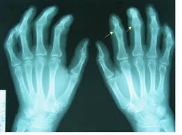
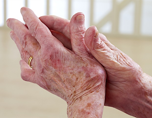
    Calcium pyrophosphate deposition disease (CPPD) occurs when calcium pyrophosphate (CPP) crystals form near cartilage cells, sometimes leading to joint inflammation, pain, and swelling. It is often called pseudogout because of its clinical similarity to gout, yet much less is known about CPPD than about gout and other forms of inflammatory arthritis.

    Rheumatologist Sara Tedeschi, MD, MPH, her colleagues at Brigham and Women’s Hospital in Boston, and fellow at the Medical College of Wisconsin, wanted to expand the knowledge base by investigating whether patients with CPPD disease are at increased risk for fractures. Previous studies had shown a link between low bone density and CPPD. Recent data from experimental models suggest that increased formation of osteoclast (cells that break down old bone) due to loss of function of osteoprotegerin (a protein that normally inhibits bone resorption) may contribute to the pathogenesis of the disease.

    To find out more, Tedeschi and her team conducted a matched cohort study using electronic health records (EHR) from Mass General Brigham’s health care system. The study included more than 1,100 patients who had at least one episode of acute CPP crystal arthritis; the acute inflammatory form of CPPD – between 1991 and 2017. They were compared with more than 3,300 comparison researchers who did not have acute CPP crystal arthritis, although they could have other types of arthritis. The average age in both groups was 73 years, and more than half were women.

    The index date for patients with CPP crystal arthritis was either the first mention of pseudogout in their chart or the first synovial fluid analysis with the finding of CPP crystals. The period from registration of the EPD to the index date was at least 180 days. The index date for the matched comparators was a medical encounter within 30 days of the matched pseudogout patient’s index date.

    The primary outcome of the study was the first fragility fracture (fractures resulting from a fall from standing height or lower) at the humerus, wrist, hip or pelvis. Secondary outcomes were the first fracture at each of these anatomic locations. For patients with more than one fracture, only the earliest fracture was used. Fragility fractures were identified using published algorithms with a positive predictive value of greater than 90%.

    The researchers estimated the incidence rates and incidence ratios for each type of fracture and for fractures at each individual body location. They used Cox models (a statistical technique that can be used to measure time-to-event results on one or more predictors) to estimate adjusted risk ratios for fractures. Patients who had rheumatoid arthritis (RA) or were prescribed corticosteroid or osteoporosis treatment were excluded from the sensitivity analyzes in an attempt to rule out the influence of these diagnoses/medications, which are known to increase the risk of fracture.

    The researchers found that the fracture rate was twice as high in the acute crystal CPP arthritis cohort as in the comparison group, after adjusting for traditional fracture risk factors: 11.2 per 1,000 person-years versus 5.6 per 1,000 person-years. The disparity between the two groups increased over time and the sensitivity analyzes yielded similar findings.

    Tedeschi says the increased risk of fractures wasn’t particularly surprising, but the difference was large. Also surprising, she says, was “that differences in fracture risk were seen, of similar magnitude, after excluding patients who had used corticosteroids in the 90 days before the index date.” [Moreover]Fracture rates varied within the first months of follow-up, indicating a pre-existing difference in bone health between cohorts.”

    Tedeschi notes that the study does not indicate whether patients with acute CPP crystal arthritis had repeat episodes or used corticosteroids after the index date, either of which could influence the findings. She adds that they could not assess falls, which would affect fracture risk and may have differed between CPPD and comparators. She concludes by noting: “The analysis did not assess vertebral fractures as they may be asymptomatic and not captured in diagnosis codes.”

    Yet the findings are clear: patients with acute CPP crystal arthritis have a doubled risk of fragility fractures.

    “At the very least, we hope that physicians will consider assessing bone mineral density in patients with CPPD to determine whether osteoporosis treatment is necessary,” says Tedeschi.

    This research was supported by grants from the NIH’s National Institute of Arthritis and Musculoskeletal and Skin Diseases (NIAMS).

    Source:

    American College of Rheumatology

    Source link

  • Salivary gland abnormalities in primary Sjögren’s become more serious over time, research shows

    Salivary gland abnormalities in primary Sjögren’s become more serious over time, research shows

    shutterstock 312351317 82a9e0224160430890b6b34a2b64af19

    New research at ACR Convergence 2023, the annual meeting of the American College of Rheumatology (ACR), shows that ultrasound-detected salivary gland abnormalities in primary Sjögren’s become more severe over time and that the slowly progressive disease likely begins long before it reaches the is first. detected (summary #1371).

    Sjögren’s disease, also called primary Sjögren’s syndrome, is a systemic autoimmune disease. It is characterized by inflammation of the tear and salivary glands, leading to chronic dry eyes and mouth. Fatigue is common and about a third of patients have complications affecting the lungs, skin, kidneys and joints. Up to 60% of patients may develop systemic symptoms.

    Salivary gland ultrasound (SGUS) is a safe and non-invasive method for diagnosing and monitoring Sjögren’s disease. Still, it’s unclear whether the abnormalities it detects become more notable over time. Valérie Devauchelle-Pensec, M.D., Ph.D., professor of rheumatology in the Department of Clinical Immunology and Rheumatology at the University of Brest Occidentale and Cavale Blanche Hospital in Brest, France, designed a cross-sectional international study to find out.


    I have been caring for patients with Sjögren’s disease for years and I am always surprised that when I see them at the beginning of their disease, their first ultrasound scan of the salivary gland shows severe lesions. I also have many patients with rheumatoid arthritis. In rheumatoid arthritis, the joints are destroyed, but not at the onset of the disease. Sjögren’s seems different. I wondered, ‘When does the disease really start and do the lesions evolve over time or not?’ Many of my colleagues, who are experts in Sjögren’s and ultrasound, agreed to participate [in the study].”


    Valérie Devauchelle-Pensec, MD, Ph.D., professor of rheumatology, department of clinical immunology and rheumatology at the University of Brest Occidentale


    Between May 2019 and February 2022, 247 patients from 11 international centers consecutively participated in the study. Most were women, with an average age of 58 years. Nearly 100% of patients reported dry mouth; 75% had abnormal saliva production and 85% were positive for anti-SSA autoantibodies, a hallmark of Sjögren’s. The median EULAR Sjögren’s disease activity score (ESSDAI) was 3, indicating low disease activity.

    Ultrasound-detected functional abnormalities of the parotid and submandibular gland were classified according to the most recent Outcome Measures in Rheumatology (OMERACT) score, a four-grade semiquantitative scoring system. The patients were then grouped according to the duration of illness from the onset of dry mouth symptoms.

      • Group A: less than five years (47 patients)


      • Group B: five to nine years (69 patients)


      • Group C: 10 to 20 years (78 patients)


      • Group D: More than 20 years (53 patients)


    When the researchers looked at the most serious node for each patient, they found a significant association between disease duration and the OMERACT score. The odds ratio for progression over a five-year interval was 1.23.

    There was no statistical difference between the groups with regard to the various ultrasound parameters, with the exception of the proportion of hyperechoic bands, which are associated with damage in established Sjögren’s patients.

    “We hypothesized that hyperechoic bands represent the slow fibroadipose evolution of the disease,” says Devauchelle-Pensec. “To me, this means that Sjögren’s disease starts long before we find it, so it is important to treat patients early.”

    She adds that the study highlights the importance of adding ultrasound findings to the classification criteria for Sjögren’s syndrome and the need for a better understanding of when the disease begins.

    Source:

    American College of Rheumatology

    Source link

  • New training program expands access to rheumatology care for Native American communities

    New training program expands access to rheumatology care for Native American communities

    Rheumatology

    New research at ACR Convergence 2023, the annual meeting of the American College of Rheumatology (ACR), describes the expansion of a new program to train primary care physicians (PCPs) in the diagnosis and treatment of rheumatoid arthritis (RA) in Native American communities that little or no access to rheumatology care (Abstract #2455).

    Despite the high prevalence of RA and other rheumatologic diseases among Native Americans, many Native American communities lack adequate access to subspecialty care. As a result, the responsibility for treatment has shifted to primary care providers, who often lack confidence in prescribing RA medications or managing the disease. To help offset the critical shortage of rheumatologists, the Rheumatology Access Expansion (RAE) Initiative launched RA ECHO (Extension for Community Healthcare Outcomes) in 2021, a 12-week training program to teach Navajo Nation PCPs how to diagnose and treat RA. The goal was to improve outcomes and reduce entrenched disparities in healthcare. This year, the project expanded to tribes in 15 states.


    We successfully offered the RA ECHO curriculum three times on the Navajo Nation from 2021-2022. For our fourth cohort – Spring 2023 – we have dramatically expanded our target audience and invited healthcare professionals serving Native American communities across the country to participate.”


    Jennifer Mandal, MD, assistant professor at the University of California, San Francisco (UCSF) and director of the RAE Initiative


    Mandal says she and her team worked with an organization called Indian Country ECHO to recruit participants for cohort four.

    “We knew that Indian Country ECHO’s established lines of communication with tribes across the country would allow us to reach a much broader audience for our RA ECHO program. And indeed, when Indian Country ECHO put out a call for interested health care providers the response was overwhelming in attending our program. Over 100 providers responded that they would like to participate, and after the final dates and times were selected, 50 providers registered,” says Mandal.

    Most participants were PCPs, but pharmacists, community health care representatives, and providers from non-primary care settings such as emergency medicine, ophthalmology, and orthopedics also enrolled.

    The Spring 2023 program followed the same format as the first three: 12 weekly interactive classes held virtually, with approximately 30 minutes of high-level didactics on key aspects of RA diagnosis and management, followed by case-based discussion. Participants were encouraged to bring their own anonymized patient cases to class. In addition to the weekly sessions, there were also biweekly virtual ‘office hours’, where participants could interact with a panel of rheumatologists.

    To measure how successful the training actually was, the RAE Initiative team collected data on PCP responses to tests and surveys. Before and after each of the four programs (cohort five is currently underway), participants completed a medical knowledge test about RA and surveys about their confidence in the diagnosis and treatment of RA on a five-point Likert scale. Starting in cohort three, participants were also asked to rate changes in their own clinical behavior, such as how often they performed joint screening exams or blood tests before starting immunosuppressants.

    Pre- and post-intervention scores were available for more than one-third of participants. When data across cohorts were pooled, test scores increased by 26% and PCP confidence increased by more than one point on the Likert scale. Nearly 80% of participants reported that they performed important clinical behaviors related to the diagnosis and treatment of RA “more often” or “much more often” after taking the course.

    While the results are encouraging, Mandal says one limitation is that they did not look directly at patient outcomes.

    “While the RAE Initiative team hopes to eventually measure patient health data, it is critical to recognize that, due to centuries of exploitation, there is widespread mistrust of requests for access to private medical records in the Navajo community. Before seeking out personal health information, we strive to prioritize respectful and considerate handling of sensitive information, while still striving to achieve our educational and empowerment goals.”

    In the meantime, she lists other next steps, including:

      • Creating educational materials for RA patients that are culturally and linguistically tailored to the Navajo community


      • Organizing in-person training for community health representatives in Navajo Nation to increase awareness about joint health and different types of arthritis


      • Creating online training resources for PCPs who want to learn more about common rheumatologic diseases


      • Creating a new ECHO training program for spondyloarthritis


    Mandal hopes that the RA ECHO program can serve as a model for creating similar rheumatology training programs for other communities with limited access to rheumatologists, saying, “We are eager to collaborate with others who are interested in joining this important mission to expand access to rheumatology care.”

    This work was funded by a grant from the Bristol Myers Squibb Foundation.

    Source:

    American College of Rheumatology

    Source link

  • A deep learning system accurately identifies joint space narrowing and erosions on hand X-rays

    A deep learning system accurately identifies joint space narrowing and erosions on hand X-rays

    shutterstock 390538711 6b3c40fdd32742caa54307db3553cab1

    New research at ACR Convergence 2023, the annual meeting of the American College of Rheumatology (ACR), shows that a deep learning system can accurately identify and predict joint space narrowing and erosions in hand x-rays of patients with rheumatoid arthritis (RA) (Abstract #0745) .

    X-rays are the most commonly used imaging technique for detecting and monitoring RA in the hand. Radiologists often use the well-validated Sharp/van der Heidje (SvH) method to evaluate joint space narrowing and erosions by assessing specific locations in each hand and wrist. However, scoring SvH is time-consuming and requires expertise that is not always available. This has led to increased use of deep learning (also called machine learning) to analyze hand X-ray data in RA.

    According to Carol Hitchon, MD, FRCPC, MSc, associate professor at the University of Manitoba and clinical scientist in rheumatology and lead co-author of the study: “Machine learning offers a powerful and complementary approach to traditional RA detection and diagnosis methods. It improves the accuracy, efficiency and objectivity of RA radiograph assessment, while providing the opportunity for early detection of damage and valuable insights into the disease.”

    For the current study, Hitchon and colleagues aimed to develop and validate a deep learning system for the automated detection of joints and prediction of SvH scores on hand radiographs of patients with RA.

    They used a convolutional neural network (CNN)-based algorithm called You Only Look Once (YOLO). CNN is a deep learning neural network commonly used in computer vision and recognition tasks and has been successfully used in medical image classification. YOLO is a type of CNN model specifically designed for real-time object detection in images and videos and known for its speed and efficiency in image processing. Hitchon and colleagues used a recent version of YOLOv516, which they showed to be more than 90% accurate in detecting hand joints.

    The YOLO model was trained to detect joints in 240 training and evaluation pediatric hand radiographs from the Radiologic Society of North America database.

    The researchers boxed and labeled the different joints of interest: proximal interphalangeal, metacarpophalangeal, wrist, distal radius, and distal ulna. The joint detection model was validated with 54 clinician-labeled radiographs from four adult RA patients followed for more than ten years.

    Researchers then applied a vision transformer model (VTM) to predict the erosion and joint space narrowing score of each joint. Hitchon explains that a VTM is a deep learning architecture designed to efficiently process and understand sets of data.


    It works by splitting an image into small chunks, transforming or flattening the chunks into a sequence, creating low-dimensional linear embeddings from the flattened spots, adding the positional embedding, and then running the encoded sequence into a standard transformer encoder for the remaining prediction task. ”


    Carol Hitchon, MD, FRCPC, MSc, Associate Professor, University of Manitoba


    The VTM was validated using more than 2,200 hand radiographs from 381 RA patients to whom the physician assigned SvH scores. Patients were from the Canadian Early Arthritis Cohort, a multicenter Canadian study. These scored radiographs were used as the gold standard for this study.

    The joint detection model was trained to detect the entire wrist, but the researchers had SvH scores for individual wrist joints, so they trained a separate model to detect joint space narrowing and erosion in each joint.

    When they evaluated the accuracy of their models, they found:

      • The joint detection model accurately identified target joints. The F1 score for children was 0.991 and the F1 score for adults was 0.812. (In machine learning, the F1 score is a metric that measures the accuracy of a model).


      • VTM predictions for joint space narrowing and erosion were very accurate. The principal square error, which evaluates the accuracy of predictions, was 0.91 and 0.93, respectively.


      • The multitask models predicted SvH erosion and joint space narrowing scores of individual wrist joints with moderate accuracy (0.6 to 0.91).


    Hitchon says they weren’t surprised by the performance of their model.

    “The AI ​​technologies we applied in this study have been successfully and widely used in other domains, some of which have been commercialized. Compared to the model’s performance in other domains, our performance is relatively low in predicting X-ray scores for some joint types, such as the wrist. [This] may be due to the relatively small sample size in our study or to the complexity of the anatomy of the wrist joint,” she notes.

    Hitchon also says the model’s performance does not match that of human radiologists for joints such as the wrist.

    “The AI ​​models cannot replace human radiologists at this stage, but they will be excellent complementary tools that can improve the overall quality and efficiency of radiograph scoring analysis when used in conjunction with the radiologist’s judgment. [these models] may be applicable to the interpretation of large volumes of radiographs in clinical trials.”

    The study has two major limitations: X-rays were obtained from cohorts composed almost entirely of white women, and the findings may not apply to races and ethnicities traditionally underrepresented in research studies. Hitchon acknowledges that the findings need to be replicated in other groups. The model also lacks the ability to learn and become more accurate with subsequent images, although Hitchon says they are developing a new deep learning framework so that the model continuously learns as new data is available.

    This study received local funding from the Health Science Center Foundation, a hospital charity in Winnipeg, Manitoba, Canada. One of the co-authors, Pingzhao Hu, is supported by the Canada Research Chair Program. The Canadian Early Arthritis Cohort, which provided one set of radiographs, is funded by multiple sources.

    Source:

    American College of Rheumatology

    Source link

  • Research shows that depression increases the risk of disability in patients with rheumatoid arthritis

    Research shows that depression increases the risk of disability in patients with rheumatoid arthritis


    In a review published in Nature Reviews Rheumatologyresearchers discussed the interactions between central and peripheral immunobiological mechanisms associated with rheumatoid arthritis (RA) and major depressive disorder (MDD).

    They further described the role of inflammatory proteins, the effect of peripheral inflammation on different parts of the brain, and the relationship between changes in the brain and inflammation-induced depression.

    Study: Immune mechanisms of depression in rheumatoid arthritis. Image credits: pikselstock/Shutterstock.comStudy: Immune mechanisms of depression in rheumatoid arthritis. Image credits: pikselstock/Shutterstock.com

    Background

    RA is a chronic autoimmune inflammatory disease that negatively affects synovial joints and several other organs. Depression is a common, clinically heterogeneous condition that affects all other patients with RA. There is increasing evidence that RA and depression have overlapping features and can be modulated by each other.

    Data suggest that depression is a risk factor for RA, and that patients diagnosed with RA at a young age are more susceptible to depression. Furthermore, RA patients with depression are observed to exhibit functional progression as well as decreased response to treatment, leading to poor outcomes. However, the precise biological mechanisms underlying this association are not clearly understood.

    Therefore, this review focuses on understanding the link between these two conditions and the underlying mechanisms, while exploring the interplay between the nervous system and the immune system in RA patients.

    Shared cytokines in RA and depression

    Proinflammatory cytokines amplified in RA are also known to be causally linked to depression. Several cytokines have been implicated in RA and depression, including interleukin (IL)-16, IL-18, IL-1, IL-6, and tumor necrosis factor (TNF).

    Peripheral immune signals to the brain

    The peripheral immune system signals the brain through two known pathways: neural and humoral. In the neural pathway, molecules that mediate inflammation can bind and activate receptors on sensory neurons, including those in the dorsal root ganglia (DRG) and the vagus nerve.

    The activated sensory neurons then send the signal back to the cerebral cortex of the brain via the spinal cord. The signal is then passed on to higher brain centers, which modulate the immune system locally and systemically.

    Through the humoral pathway, immune cells release molecules capable of crossing the blood-brain barrier (BBB) ​​and affecting brain cells or activating the endothelial cells of BBB.

    As observed in experimental studies in mice, this pathway leads to the release of chemokines involved in neuronal plasticity, resulting in depression-like behavior and cognitive impairment.

    Immune responses in the brain

    In the brain, existing neural cells and recruited immune cells release various inflammatory proteins that support neuroimmune communication. When cytokines and chemokines are released by neurons, microglia, astrocytes, peripheral immune cells and endothelial cells, they influence neurological and immunological processes.

    For example, during inflammation, the recruitment of peripheral monocytes to the brain is associated with dendritic remodeling and cognitive impairment, potentially leading to depression. Chronic peripheral inflammation in RA induces local microglial activation in the brain, leading to altered microglial expression.

    Although microglia are often associated with inflammatory changes in the brain, recent studies indicate a more complex role for microglia in neurological health.

    Contrary to previous belief, microglia found in the brain, according to studies in mice, originate not only from peripheral blood, but also from meninges and bone marrow in the skull. However, there is a lack of studies examining this aspect in humans.

    Astrocytes also play a role in brain inflammation. Activation of astrocytes by cytokines from microglia has been shown to result in the release of neurotoxic factors that influence neuronal health and behavior.

    Mechanisms linking depression and inflammation

    Immune-related inflammation has been implicated in the pathophysiology of depression. In RA, several pathways are activated, which can lead to inflammation-related behavior.

    These pathways include inflammasome activation, the kynurenine pathway, neuroplasticity, and the pathways of the glutamatergic and serotonergic systems.

    Regional variation in the brain

    Although regional changes in the brain and the underlying mechanisms continue to be studied, mainly using animal models, neuroimaging studies in humans have significantly improved our understanding of inflammation-related changes in the brain.

    Advances in magnetic resonance imaging (MRI) have provided insights into the role of inflammation in depression beyond traditional structural assessments and histology-based studies. Emerging evidence suggests that the brain regions affected by inflammation and depression are the striatum, hippocampus, amygdala, and insula.

    Conclusion

    This review article provides a comprehensive overview of the association between immune mechanisms and depression in patients with rheumatoid arthritis. It highlights the need for further research in this area.

    Furthermore, data from clinical trials suggest that immune modulation may be a promising approach for treating comorbid depression in patients with rheumatoid arthritis, potentially reducing the global burden of this debilitating condition.

    Source link

  • You Don’t Look Sick – Living with Rheumatoid Arthritis: HAWAII DAY 1


    Well, my start to my trip to Hawaii definitely started with a bang. First I got up early and got to the airport well in time. I had a wonderful taxi driver who was very sweet and he took me and my luggage to the airport lobby and arranged a wheelchair for me. I was so early that I was ahead of the people pushing the wheelchairs to your gate. So I had to sit and wait for about half an hour for someone to push me to my gate.

    Getting through TSA was a lesson in patience. Since I was in a wheelchair, I was wheeled to the front of the line, but then had to wait for a female TSA agent to check my trunk. My boot had to be taken off and put through the X-ray machine and then my sock had to be inspected. I offered to take off my sock and they can take it to the x-ray machine, but they refused and instead felt all over my foot. It seemed kind of stupid. That whole process took about another 45 minutes.

    My flight was very nice. I sat next to two very nice women who both wore masks the entire time and we were super helpful when we landed. In Hawaii the plane lands right on the tarmac and you have to walk down a flight of stairs. The two women carried my backpack and medicine bag so I could hold on to the railing.

    Once I had my luggage, I waited in a long line to get the shuttle to the rental car. When I got to the rental car, I went to the Fast Track thinking I would get my rental car, but unfortunately they claimed I didn’t have my fast track for this reservation. So I had to wait in a long line for him to pick up my rental car. Luckily there were some very nice people in line who held my seat while I sat down for a while. It was very hot and I stood for a long time.

    Finally I get to the front of the line and pick up my rental car. I drove away happily and went to the supermarket to get food. I drove just 20 miles further and it was clear something was wrong with the car. Every time I drove over 60 miles per hour there was a chattering sound like someone was banging in the trunk. After stopping a few times to see if I could see what the problem was, I called roadside assistance. That was an incredibly useless call because the guy kept asking me where I was, but I just knew I was on a highway in the middle of nowhere in Hawaii. So I finally turned around and went back to the airport.

    AVvXsEgRy9qBgcR6vJg4f6G3U3pVjAhTWaud7nZq mFSPBLXKiJlAPBJUS E

    Once back at the car rental company at the airport, I saw the same service representative I had spoken to twice. I called him and told him what happened and he quickly brought my new car over.

    When the car came by, I had some really nice people helping me with my bags and groceries. This time the car was great. I arrived at the hotel and the nice lady at the reception helped me with my bags and groceries.

    To give you an idea, I landed at 11:30 AM and arrived at the hotel at 6:00 PM. It took 6.5 hours to get off the plane, pick up my luggage, go to the rental location, get car #1, then a grocery store and drive 20 miles and then drive back and get car #2 and then drive 2 hours to the hotel. It was a lot of standing. Let’s see how my feet feel tomorrow. Oh yeah, I was late for dinner, so I ate snacks.

    See you tomorrow…

    Source link

  • Shift towards personalized treatment approaches for chronic inflammatory diseases

    Shift towards personalized treatment approaches for chronic inflammatory diseases

    shutterstock 390538711 6b3c40fdd32742caa54307db3553cab1

    Chronic inflammatory diseases affect 5% to 7% of the population, regardless of age or gender, from children to the elderly. Chronic inflammatory diseases include a range of conditions. The most common inflammatory diseases include rheumatoid arthritis, Crohn’s disease, psoriasis and multiple sclerosis.

    Few of the current therapies for chronic inflammatory diseases are effective and while some can control the disease, they do not provide a cure. To overcome these problems, researchers and physicians are focusing on several topics to improve patient care in therapeutic antibody treatment.

    The landscape of treating chronic inflammatory diseases has undergone a significant transformation with the advent of targeted therapies using therapeutic antibodies. However, a significant proportion of patients do not respond adequately to treatment or experience a decrease in response over time. This challenge mainly relates to issues such as suboptimal dosing, immunogenicity and variations in pharmacokinetics (how the drugs circulate through the body) in different patients.

    In response to this critical need for more effective treatment, researchers have initiated a strategic shift toward personalized treatment approaches. This includes the development of patient stratification tools and the use of therapeutic drug monitoring (TDM) to adjust dosages based on serum drug concentrations.

    The potential for substantial improvement in patient care is enormous by implementing individualized (TDM-guided) dosing regimens of therapeutic antibodies into routine clinical practice for the treatment of chronic inflammatory diseases. This tailored approach will ultimately lead to more efficient use of these valuable but expensive medicines the right medicine in the right dose for the right patient”.

    However, Europe faces a challenge in the fragmentation of expertise regarding individualized (TDM-guided) treatment optimization. The knowledge and techniques required for effective implementation are concentrated in a limited number of pioneering centers, which makes dissemination to other centers difficult. In particular, the lack of standardization in TDM testing contributes to the complexity.

    Introducing ENOTTA COST promotion

    To address these challenges and promote a more coherent approach, a comprehensive, interdisciplinary pan-European network is being established. ENOTTA COST Action, which stands for “European Network on Optimizing Treatment with Therapeutic Antibodies in chronic inflammatory diseases”,brings together 156 experts from 31 countries, including key scientific disciplines such as physicians, basic researchers, biologists, computer scientists, pharmacometrists, patients, small and medium-sized enterprises (SMEs) and health authorities, to cover all aspects of the challenge.


    ENOTTA advocates personalized use of therapeutic antibodies to become the new standard of care for patients with chronic inflammatory diseases.”


    Prof. Denis Mulleman, chairman of ENOTTA


    This initiative aims to consolidate and structure scientific research in this area and thus promote collaboration and knowledge exchange. The ultimate goal is to facilitate the seamless integration of individualized (TDM-guided) cost-effective dose optimization of therapeutic antibodies into daily clinical practice for the treatment of chronic inflammatory diseases.

    Added value with ENOTTA

    The ENOTTA COST action is groundbreaking and aims to create a distinctive framework, unlike all existing initiatives. This innovative initiative is designed to facilitate networking, sustainable collaboration and expansion of partnerships between participants across Europe.

    ENOTTA stands ready to catalyze progress in this therapeutic area, facilitating the exchange of expertise and the wide dissemination of valuable knowledge for the benefit of patients suffering from long-standing inflammatory diseases. By fostering scientific collaboration between key players in the European research landscape, this initiative will play a crucial role in advancing the field of personalized use of therapeutic antibodies. Furthermore, it will establish critical connections with leading experts on a global scale.

    Taken together, the innovative nature of this action will come from the ability of the entire group to create tools for patient stratification, test harmonization, universal standards and the availability of guidelines and treatment algorithms that are accepted by health insurers, physicians and patients. . These developments will be crucial for implementing individualized (TDM-based) dose optimization of therapeutic antibodies in daily clinical practice. Ultimately, the optimal use of therapeutic antibodies using TDM will alleviate the burden on the healthcare system. Furthermore, the insights gained from this action can also guide future research in other disciplines, such as oncology, metabolic diseases and cardiovascular diseases, using therapeutic antibodies.

    Source:

    European cooperation in science and technology (COST)

    Source link

  • YKL-40 serum levels are predicted by inflammatory state, age, and diagnosis of idiopathic inflammatory myopathies

    YKL-40 serum levels are predicted by inflammatory state, age, and diagnosis of idiopathic inflammatory myopathies


    In this study, YKL-40 serum levels are influenced by factors such as age, inflammation and diagnosis of autoimmune diseases (RA/MII)3. Currently, due to its participation in tissue remodeling and degradation, attempts have been made to use it as a biomarker in pro-inflammatory states and as an indicator of poor prognosis in inflammatory diseases.6.7. However, its usefulness is still controversial because its full biological effects are still unknown. Furthermore, the specific factors that promote its expression, as well as its interaction with the majority of cytokines and molecules involved in the development and establishment of autoimmune inflammatory diseases, are not well established.17.

    YKL-40 serum levels have been reported to increase with age in various cardiovascular, metabolic and systemic inflammatory diseases3. Bojesen et al. found that serum levels of YKL-40 increased exponentially with aging. In subjects with two YKL-40 measurements ten years apart, the average increase in YKL-40 was 1.5 μg/l/year18. Regarding inflammatory diseases, increased serum levels of YKL-40 were reported in anti-neutrophil cytoplasmic antibody (ANCA)-associated vasculitis, which was hypothesized to play a role in promoting chemotaxis, tissue damage and vascular damage.19.

    In RA it has been recognized as a potential candidate autoantigen. Furthermore, in these patients it is produced and secreted by monocytes differentiated into macrophages, articular chondrocytes, synovium, peripheral blood mononuclear cells (PBMCs), and endothelium.17,19,20. It has been proposed that the pathogenic mechanism of YKL-40 in RA initiates through its binding to the HLA-DR4 peptide-binding motif promoting mononuclear cell proliferation, and HLA-DM plays a key role in presenting YKL-40 to CD4+ T cells. Furthermore, antigen-presenting cells (APCs) present YKL-40 at early-stage RA sites, suggesting an association for YKL-40 in the pathogenesis of RA.20,21,22. On the other hand, differentiated DR4+ dendritic cells and macrophages are similar to APCs of synovial joints and have the potential to carry out MHC II presentation of YKL-40 epitopes, resulting in higher levels in synovial and serum.21,22. Although the pathogenic mechanism in RA has been elucidated, agents that promote YKL-40 expression in RA are still lacking. It has been associated with the development of chronic, destructive, relapsing arthritis due to its role in tissue remodeling and breakdown. . It is considered an effective marker in estimating RA disease activities and prognostic value, and may be a therapeutic target19.

    As for IIM, information is even more limited as few studies have been conducted in this regard and thus the role YKL-40 plays in this area has not yet been established. Regarding the pro-inflammatory effect and its relationship with the diagnosis and phenotype of IIM, Noguchi et al. found significantly increased serum in patients with PM/DM compared to the healthy population, as well as age-adjusted serum YKL-40 levels were significantly increased in patients with PM/DM compared to HC. In muscle biopsies, infiltration of YKL-40-positive inflammatory cells (probably macrophages) in the endomysium and perimysium was found. This suggests that cells other than CD8+ and CD4+ T cells can cause inflammation.23.

    Ming-Zhu Gao et al. measured YKL-40 levels in patients with DM/PM and HI and reported significantly higher levels in patients with IIM compared to controls (51.6 vs. 27.8 ng/ml, respectively)6. In a systematic review by Cui et al. reports levels of 84.09 ng/ml in patients with PM and DM versus 27.37 ng/ml in HI24. On the other hand, Carboni et al. analyzed YKL-40 serum levels and its expression in muscle tissue in patients with ASSD. However, serum levels of YKL-40 did not correlate with other clinical, laboratory, disease status, or therapeutic parameters. Furthermore, YKL-40 was expressed by the inflammatory cells of the muscle tissue.13. Our study reinforces these results with serum levels of 187.80 ng/ml in patients with IIM versus 46.82 ng/ml in RA and 57.17 ng/ml in HI, as well as their presence mainly in inflammatory cells. This increase can be explained by inflammation, activation of macrophages, destruction of fibroblasts and existing vascular changes.

    As previously mentioned, several biological effects of YKL-40 are known, such as inflammation and tissue remodeling, as well as its main sources, which, however, highlights its exacerbated expression in inflammatory diseases such as RA or SLE; some researchers have pointed out that this expression may vary depending on the disease type, which could be due to the multi-organ damage in IIM compared to RA, where the damage is mainly directed against the joints19.24. Tang et al. found that YKL-40 concentration was significantly higher in IIM patients with myocardial injury than without myocardial injury25.

    In addition, YKL-40 plays a role in cardiovascular diseases such as early atherosclerosis, essential hypertension and other progressive vascular complications. In IIM patients, as in many other autoimmune diseases, serum levels of this protein have a positive association, particularly with atherosclerosis, and may predict both overall and cardiovascular mortality.5,26,27.

    We wanted to know whether YKL-40 is affected by certain factors in IIM, as mentioned by Tizaoui et al. First, we compared some demographic, laboratory and clinical variables between patients with RA and IIM mentioned in Table 1. Of all After analyzing the variants, we found a significant difference in pDBP (P = 0.024) and pMBP (P = 0.035) which were higher in IIM patients. This may be due to the fact that blood vessels suffer damage in the early stages of the development of inflammatory diseases, altering blood pressure and increasing the risk of cardiovascular damage.28.29. Furthermore, the endothelial changes in IIM progress to microangiopathy, causing blood pressure changes12. Although information on exactly how the process occurs is scarce, it has been suggested that cardiovascular disease and cardiovascular risk increase mortality in IIM patients, but this is becoming controversial because a single center cross-sectional study recently reported that it risk of cardiovascular disease increases. factors in IIM patients are not significant compared to HI, but are significant in IIM if they are related to age, disease duration, duration of therapy and body composition, which could be related to our patients included in our study25.30. Although these evaluated variables were significant between these two groups, serum levels of YKL-40 were the most significant variable (P = 0.010). These were higher in IIM than in RA patients (187.80 ng/ml). vs 46.82 ng/ml, respectively).

    Once we established that IIM exhibits higher serum levels of YKL-40 than RA, we examined whether serum levels of YKL-40 were influenced by age or disease duration. Our results show that only aging has a positive correlation with increased YKL-40, but not with disease duration. The reports conducted by Johansen in 2006 and Schultz 2010 have clearly shown that aging is predictive of the increase of YKL-40 in HI, but in our IIM patients the concentration of this protein is higher due to inflammation and damage to multiple organs.2.3.

    We evaluated the predictive value of inflammatory state, age and diagnosis of IIM on serum levels of YKL-40 and clarified that CRP has predictive value on serum YKL-40 levels in IIM patients.P= 0.038) which corresponds to a cross-sectional survey and systematic review published by Cui andYou at the . In addition, age and the IIM diagnosis ( P=0.008AndP=0.001respectively) were found to be powerful predictors of YKL-40 serum levels24. On the other hand, we confirmed that age and IIM diagnosis have an important influence on YKL-40 concentration, because YKL-40 serum levels are the highest compared to control and RA groups, thus we know that the presence of the disease or its type influences the disease. the YKL-40 concentration. Some reports state that the YKL-40 concentration in HI is stable for many years, but increases with aging or inflammatory conditions. Other researchers reported that normal YKL-40 serum levels may be different among a healthy population. Therefore, they recommend establishing baseline values ​​for each study, because in addition to environmental factors, genetic load is another variable that can influence the expression pattern. of this protein26. We previously mentioned the pathogenic mechanism by which YKL-40 expression is mediated in RA and its possible role in IIM, but the information is still insufficient.

    Regarding the in situ analysis of YKL-40 muscle expression, we observed that YKL-40 is mainly expressed in inflammatory cells rather than in muscle cells. We also observed that YKL-40 expression is associated with higher CPK serum levels and MYOACT score which are often related. to higher inflammation and muscle weakness. This observation is in agreement with the unique previous report of YKL-40 in muscle tissue made by Carboni et al.13It thus supports its role in inflammation, as well as its function as a clinical marker for poor prognosis in inflammatory diseases.

    Taking into account all our findings, we demonstrated the expression of YKL-40 in both HI and in patients with RA and IIM and the possible factors that could influence its expression.

    Source link

  • New exercise test aims to improve the quality of life of people with rheumatoid arthritis

    New exercise test aims to improve the quality of life of people with rheumatoid arthritis

    shutterstock 390538711 6b3c40fdd32742caa54307db3553cab1

    Rheumatoid arthritis is an inflammatory disease that can cause severe pain and swelling of the joints. But a new exercise intervention could help improve physical function and quality of life in people struggling with this debilitating condition.

    In a new trial, researchers from the University of South Australia are working with Arthritis SA to investigate the potential of Blood Flow Restriction (BFR) training to improve the strength and mobility of people with rheumatoid arthritis.

    BFR training is an exercise technique in which people wear pressurized bands – much like blood pressure cuffs – to slow blood flow to the muscles while they exercise. The cuff allows blood flow to the limb but slows its outflow, developing muscle strength without the need for heavy weights.

    In Australia, rheumatoid arthritis is the second most common form of arthritis, affecting more than 450,000 people. More than 18 million people worldwide live with a condition. Women are two to three times more likely to develop rheumatoid arthritis than men.

    Sports scientist Dr. UniSA’s Hunter Bennett says the research hopes to identify interventions that can improve the quality of life for people with rheumatoid arthritis.

    “Rheumatoid arthritis can be a particularly debilitating disease. It is caused by the immune system attacking healthy tissues, leading to pain and swelling, joint destruction and loss of muscle mass and strength,” says Dr. Bennett.

    Although medications can reduce symptoms, they do not address the loss of muscle strength and function.

    The best way to increase strength and combat muscle loss is through resistance training, but this is often problematic for people with rheumatoid arthritis due to pain, fatigue or risk of injury.

    Blood flow restriction training (BFR) offers an alternative. BRF is used in many sports and rehabilitation settings in Australia and is considered a safe and effective method for improving strength and function in many clinical populations, including people with osteoarthritis.

    Because this technique uses very low loads, it is a viable option for people with rheumatoid arthritis. So in our research we look at how BRF can increase people’s strength and hopefully increase their freedom of movement and overall well-being.”

    Dr. Hunter Bennett, exercise scientist, UniSA

    The research team is currently seeking expressions of interest from women and men aged 45 to 75 years diagnosed with rheumatoid arthritis.

    Exercise intervention eases pain for people with rheumatoid arthritis

    Video credit: University of South Australia

    Source:

    University of South Australia

    Source link

  • JAK inhibitors provide effective and convenient treatment options for arthritis patients

    JAK inhibitors provide effective and convenient treatment options for arthritis patients

    Arthritis

    According to a new article in Rheumatology, published by Oxford University Press, JAK inhibitors, which doctors have used to treat patients with arthritis despite concerns about the effectiveness of such drugs, actually work quite well. In a multicenter retrospective study, Japanese researchers found that the drugs resulted in impressive remission rates in patients, most of whom choose to continue such treatment.

    Rheumatoid arthritis is a common autoimmune disease characterized by chronic inflammation of the joint linings and results in progressive joint destruction and other systemic complications. The use of biologic disease-modifying drugs allows patients to enjoy achieving low disease activity and remission. But clinics must administer such medications via subcutaneous or intravenous routes, which is unpleasant for patients, and over time these medications often become less effective.

    Recently, scientists have developed Janus kinase (JAK) inhibitors for the treatment of arthritis. Patients take such medications orally. Previous research has demonstrated the efficacy and safety of JAK inhibitors in randomized controlled trials. However, some researchers have questioned the potential efficacy of JAK inhibitors for widespread use by patients. In practice, doctors usually treat patients with JAK inhibitors, precisely because these patients have other health problems and conventional medications such as methotrexate are less effective for them. Real-world patients have distinctive characteristics compared to those recruited in randomized controlled trials.

    In the current multicenter retrospective study, using data from 622 patients treated at seven major university hospitals in Japan, researchers compared the efficacy and safety of four common JAK inhibitors: tofacitinib, baricitinib, peficitinib, and upadacitinib.

    The researchers here found that about one in three patients achieved remission, and three in four achieved at least low disease activity, with both figures representing impressive efficacy. They noted that more than 80% of patients were still taking the JAK inhibitors after six months.

    They believe that this is especially relevant because with these oral medications there cannot be a failure of immunological secondary treatment, where medications are no longer effective because they cause adverse immune system responses in patients. Failure of secondary immunological treatment is common in patients treating their arthritis with drugs such as methotrexate.

    Source:

    Oxford University Press USA

    Magazine reference:

    Hayashi, S., et al. (2023) Real-world comparative study of the efficacy of Janus kinase inhibitors in patients with rheumatoid arthritis: the ANSWER cohort study. Rheumatology. doi.org/10.1093/rheumatology/kead543.

    Source link